❌

Normal view

Education Alone Is Not Enough: What Nursing Home Staff Really Think About Palliative Care

11 October 2026 at 09:20

Palliative care has long been framed as a specialist discipline reserved for hospices and oncology wards, but a growing body of evidence insists it belongs everywhere people approach the end of life, including nursing homes, where a large share of deaths in ageing societies now occur. A new qualitative study from Sweden, published in BMC Nursing, offers an unusually candid look at what happens inside care teams after they receive structured palliative care education, and the central finding is striking: training changes how staff think, but thinking alone does not change care. The researchers, Camilla Ekeblom, Carina Lundh Hagelin and Jane Österlind of Marie Cederschiöld University in Stockholm, found that the value of an educational intervention depends on an intricate alignment between individual emotions, relational dynamics and organisational realities, and that when any one of these dimensions falls out of step, the benefits of education can quietly dissolve.

The study set out to explore how nursing home staff reason about providing palliative care after participating in an interprofessional educational intervention, abbreviated in the literature as IPEI. Rather than measuring outcomes with questionnaires, the team chose a descriptive qualitative design, interviewing eighteen staff members drawn from five Swedish nursing homes. The participants represented the full spectrum of people who actually deliver and manage care: registered nurses, assistant nurses, a physician, first-line managers and designated palliative care champions. This breadth matters, because palliative care in nursing homes is not delivered by any single profession; it emerges, or fails to emerge, from the daily interplay of people with different training levels, responsibilities and emotional exposures to dying residents.

Data were collected through semi-structured individual interviews conducted between March and April 2024, and analysed using inductive qualitative content analysis, a method that allows themes to rise from the material itself rather than being forced into pre-existing theoretical boxes. From this analysis, three main categories emerged, and together they map the terrain on which palliative care is actually practised. The first concerns managing one’s own emotions, thoughts and attitudes. The second addresses the significance of relationships, communication and interactions in care. The third captures how staff navigate norms, assumptions and organisational structures in everyday palliative care. The categories correspond, respectively, to the individual, relational and organisational dimensions of the work, and the authors argue that it is the interplay among all three, not any single factor, that determines whether good palliative care becomes possible.

The individual dimension is perhaps the most human part of the findings. Staff described the ongoing internal work of confronting their own feelings about death, ageing and suffering, and of examining assumptions they carried about what palliative care even means. A recurring tension in the literature, echoed here, is the tendency to equate palliative care narrowly with end-of-life care, the final days and hours, rather than understanding it as an approach that can and should be integrated into everyday care over a longer trajectory. The interviews suggested that the educational intervention gave participants opportunities for personal as well as professional development, creating space to reflect on attitudes that had previously gone unexamined. In this sense, education functioned less like a knowledge transfer and more like a mirror, prompting staff to notice how their own emotional responses shaped their clinical decisions.

The relational dimension proved equally consequential. Palliative care in a nursing home is saturated with communication: conversations with residents about what matters to them, exchanges with distressed relatives, coordination between assistant nurses who know the residents intimately and nurses and physicians who carry clinical responsibility. The participants’ reasoning placed relationships and interactions at the heart of care provision, indicating that no amount of theoretical knowledge substitutes for the ability to build trust, listen and communicate across professional boundaries. This is where the interprofessional design of the intervention appears to have paid off. By bringing different professions into a shared educational space, the programme created a common language and, implicitly, a shared understanding of what the team was trying to achieve together, something that fragmented, profession-specific training often fails to produce.

The third dimension, the organisational, is where the study’s message becomes most uncomfortable for policymakers and care home leadership. Staff described navigating norms and assumptions embedded in their workplaces, along with structural conditions that could either support or obstruct a palliative approach. Staffing levels, routines, leadership priorities and workplace culture all shape what is practically possible on a shift where residents are dying, families are anxious and time is scarce. The authors’ conclusion is explicit on this point: providing palliative care was not solely a matter of enhancing staff knowledge. Knowledge and the conditions for care are shaped through the interplay between staff attitudes, their reasoning about care, relational dynamics and organisational conditions. When these dimensions align and mutually reinforce one another, favourable conditions for palliative care may be supported; when they do not, even well-trained staff may find their intentions frustrated.

Why does this matter on a societal scale? Nursing homes accommodate older people with complex needs and account for a large proportion of deaths, which makes palliative care a central component of care in these settings rather than an occasional emergency. Yet access to palliative care remains unequal, particularly for older people, and staff in long-term care often lack sufficient training. The demographic arithmetic is unforgiving: as populations age, the number of people who will die in nursing homes rises, and the quality of their final months depends heavily on the competence and confidence of the workforce that cares for them. Studies like this one move the conversation beyond the simplistic assumption that a training course fixes the problem, and toward a systems view in which education is necessary but insufficient on its own.

The study also carries a methodological lesson for how we evaluate educational interventions in care settings. Much of the existing research measures knowledge gains or self-reported confidence before and after training. Those metrics are useful, but they cannot reveal how staff actually reason about applying what they have learned in the messy reality of daily care. By using inductive content analysis of interviews, the Swedish team surfaced the texture of that reasoning: the hesitations, the emotional labour, the negotiations with colleagues and managers, the quiet adjustments between ideal practice and what the shift allows. The findings suggest that interprofessional educational initiatives work best when they combine theoretical knowledge with opportunities for reflection and practical application, allowing staff to translate concepts into their own working context rather than leaving translation to chance.

There are limits worth noting. The study involved eighteen participants in five Swedish nursing homes, interviewed at a single point in time after the intervention, and qualitative findings are not designed to be statistically generalised. The researchers themselves submitted the work for ethical review, receiving approval from the Swedish Ethical Review Authority, and obtained written informed consent from all participants, but the transferability of the results to other health systems will depend on how similar those systems’ nursing homes and educational programmes are to the Swedish context. Still, the three-dimensional framework that emerged, individual, relational and organisational, offers a portable lens that other teams can use to examine their own settings, and it aligns with a broader international consensus that palliative care competence must be built at every level of an organisation simultaneously.

The most durable insight from this research may be its refusal to locate the solution in any single place. Staff who completed the intervention perceived it as an opportunity for growth, and their reasoning showed genuine movement in how they understood palliative care as an ongoing approach rather than a final act. But their accounts also made clear that attitudes, relationships and structures must pull in the same direction for that understanding to reach residents’ bedsides. For nursing homes everywhere, the implication is a practical checklist of a different kind: train your staff together, give them room to reflect, examine the norms your workplace quietly enforces, and make sure the organisation’s daily conditions match what the education teaches. Where those elements align, palliative care stops being an aspiration and becomes, simply, the way care is done.

Subject of Research: Nursing home staff's reasoning about providing palliative care after an interprofessional educational intervention

Article Title: “Nursing home staff’s reasoning about providing palliative care following an educational intervention – a qualitative interview study”

Article References: Ekeblom, C., Lundh Hagelin, C., & Österlind, J. (2026). “Nursing home staff’s reasoning about providing palliative care following an educational intervention – a qualitative interview study”. BMC Nursing, 25(1), Article 956. https://doi.org/10.1186/s12912-026-05480-4

Image Credits: AI Generated

DOI: 10.1186/s12912-026-05480-4

Keywords: palliative care, nursing homes, older people, interprofessional education, qualitative research, nursing education, end-of-life care, care staff attitudes, organisational conditions, Sweden, BMC Nursing, healthcare workforce
Cellular Stress Hubs Help Oral Cancer Spread, Study Finds

11 October 2026 at 09:19

Inside every stressed cell, a quiet emergency response unfolds. When oxygen runs low, toxins accumulate, or nutrients grow scarce, mammalian cells assemble temporary structures known as stress granules, non-membranous aggregates of proteins and RNA that condense in the cytoplasm. In healthy tissue, these assemblies act as a protective pause button, halting protein production so the cell can weather the storm and resume normal function once conditions improve. But a new study suggests that in oral squamous cell carcinoma, one of the most common and disfiguring malignancies of the head and neck, this ancient survival machinery may be hijacked to serve a darker purpose: helping tumors grow and spread.

The research, conducted by a team at Kangwon National University in South Korea and published in Cell Death Discovery, examined how stress granule formation influences tumor progression and metastasis in oral squamous cell carcinoma, or OSCC. The findings point to a specific molecular switch, the phosphorylation of a protein called eIF2α, as a central driver of the process. When that switch is suppressed, the study reports, stress granule formation drops, and with it the growth and metastatic burden of tumors in mouse models. The work adds oral cancer to a growing list of malignancies in which the integrated stress response appears to fuel, rather than restrain, disease.

To understand why this matters, it helps to consider what stress granules actually do. Under stress, cells activate kinases that phosphorylate eIF2α, the alpha subunit of a translation initiation factor. Phosphorylated eIF2α dampens global protein synthesis, and the messenger RNAs that would otherwise be translated are instead sequestered, along with RNA-binding proteins, into dense cytoplasmic droplets. Key markers of these structures include G3BP1, TIAR, and PABP, proteins that researchers use to identify stress granules under the microscope. In normal cells, this translational arrest conserves energy and prevents the accumulation of damaged or misfolded proteins. The system is meant to be reversible: when the stress passes, the granules dissolve and translation restarts.

Cancer cells, however, live in a state of chronic stress. Tumors outgrow their blood supply, creating regions of hypoxia, low oxygen tension that would kill most normal cells. They also face nutrient deprivation, acidic microenvironments, and assault from chemotherapy and radiation. In this context, the stress response can become a liability for the patient rather than a safeguard for the cell. Previous work in other cancers has implicated stress granules in tumor progression and metastasis, partly through their interactions with key signaling pathways, but their role in oral squamous cell carcinoma had remained poorly understood, despite growing scientific interest in the topic.

The Korean team began by comparing human tumor tissue with adjacent normal tissue. They found that markers of stress granules, including G3BP1, eIF2α, TIAR, and PABP, were significantly overexpressed in OSCC tissues compared with their normal counterparts. This observation established that the stress granule machinery is not merely present in oral cancer but is amplified, suggesting that tumor cells may rely on it more heavily than the healthy cells around them. Overexpression of these markers alone does not prove causation, but it set the stage for the functional experiments that followed.

Those experiments turned to mouse models built from OSCC cells that had been preconditioned with hypoxia, deliberately exposing the cells to low oxygen before implantation. The logic was to mimic the stressful conditions tumor cells encounter in a real growing tumor and to ask whether cells that had already mounted a stress response behaved differently afterward. In parallel, the researchers used a compound called ISRIB, an inhibitor of the integrated stress response that acts downstream of eIF2α phosphorylation, in mice subjected to sodium arsenite-induced stress. Sodium arsenite is a classic laboratory trigger of oxidative stress and a reliable inducer of stress granule formation, making it a useful tool for probing the pathway.

The results were striking. In both experimental settings, suppression of eIF2α phosphorylation was associated with reduced stress granule formation, reduced tumor growth, and a reduced metastatic burden. In other words, when the researchers blocked the molecular event that nucleates stress granules, the tumors not only grew more slowly but also spread less aggressively. Conversely, the study found that stress granule formation was elevated during hypoxia-induced stress, underscoring a stress-responsive regulatory mechanism in which the harsh conditions of the tumor microenvironment actively promote the assembly of these granules. Together, the data sketch a coherent chain of causation: hypoxia and other stresses trigger eIF2α phosphorylation, phosphorylation drives stress granule assembly, and the granules support the cellular programs that allow OSCC cells to proliferate and colonize distant sites.

Why would a structure designed to protect cells become an engine of metastasis? The authors and other researchers in the field point to the selective nature of stress granules. While global translation is arrested, certain messenger RNAs are preferentially stored within the granules and protected from degradation. Among the transcripts sheltered this way are often those encoding proteins that promote survival, invasion, and epithelial-to-mesenchymal transition, the process by which stationary epithelial cancer cells acquire the motility needed to invade surrounding tissue and enter the bloodstream. By acting as temporary warehouses for pro-metastatic messages, stress granules may allow tumor cells to stockpile the tools of aggression, releasing them for translation once the acute stress subsides. In this view, the granule is not merely a damage-control structure but a staging ground for the next phase of tumor progression.

The therapeutic implications are considerable. Oral squamous cell carcinoma is frequently diagnosed at advanced stages, and metastasis to cervical lymph nodes is one of the strongest predictors of poor outcome. Existing treatments, surgery, radiation, and chemotherapy, remain limited in their ability to control disseminated disease. If stress granule formation genuinely drives metastatic behavior, then the pathway offers a new target. ISRIB, the compound used in the mouse experiments, has attracted attention in recent years for its ability to blunt the integrated stress response, and the new findings suggest that agents with a similar mechanism could be explored as anti-metastatic therapies in oral cancer. The authors conclude that stress granules promote OSCC progression via eIF2α phosphorylation and might serve as a novel therapeutic target, a formulation that is deliberately cautious but clinically suggestive.

As with any preclinical study, important caveats apply. The evidence comes from human tissue comparisons and mouse models, and translating these findings into patients will require further work to confirm that the same mechanism operates in human tumors in real time, to identify which patient subsets have the highest stress granule activity, and to develop inhibitors with acceptable safety profiles. Suppressing a fundamental cellular stress pathway carries risks, since normal tissues also rely on the integrated stress response during injury and inflammation. Still, the study fills a genuine gap in the literature by connecting a well-characterized cellular stress mechanism to one of the most clinically feared behaviors of oral cancer. It also adds to a broader shift in cancer biology: the recognition that tumors are not just collections of mutated genes but ecosystems shaped by physical and chemical stress, and that the survival programs cells use to endure hardship can be turned against the organism that hosts them. For patients with oral squamous cell carcinoma, whose disease too often spreads before it can be stopped, the humble stress granule may prove to be an unexpected and consequential adversary.

Subject of Research: The role of stress-induced eIF2α phosphorylation and stress granule formation in promoting metastasis of oral squamous cell carcinoma

Article Title: Stress-induced eIF2α phosphorylation promotes tumor metastasis in oral squamous cell carcinoma

Article References: Kim, S., Park, S., Lee, Y., Park, G., & Choi, J. (2026). Stress-induced eIF2α phosphorylation promotes tumor metastasis in oral squamous cell carcinoma. Cell Death Discovery. https://doi.org/10.1038/s41420-026-03412-4

Image Credits: AI Generated

DOI: 10.1038/s41420-026-03412-4

Keywords: stress granules, eIF2α phosphorylation, oral squamous cell carcinoma, metastasis, integrated stress response, ISRIB, hypoxia, G3BP1, tumor progression, translational arrest, cancer biology, therapeutic target

New Scale Reveals How Culture Shapes What We Call Child Abuse

11 October 2026 at 09:09

Where is the line between firm discipline and child abuse? For millions of families in the United States, the answer depends on where they grew up, what their parents did, and how their culture defines good parenting. A new study published in the Journal of Child and Family Studies tackles this thorny question with an ambitious goal: building a scientific instrument that can measure how people from different backgrounds judge parenting practices that are accepted in some communities but condemned in others. The Parenting Practices and Discipline Scale, or PPDS, developed by Kevin Ortiz Diaz, Maureen C. Kenny, and Helia Baez at Florida International University, is one of the first tools designed specifically to capture culturally situated discipline practices that existing measures simply ignore.

The stakes are enormous. Child abuse is recognized as a global health issue, and in the United States it occurs at what researchers describe as epidemic proportions. Federal data show 546,159 victims of child abuse and neglect in 2023 alone, and the vast majority of these cases, roughly 89 percent, are committed by one or both parents. Children from minority groups are often the most affected, with Black or African American children among the most likely to experience abuse, second only to American Indian or Alaska Native children. Yet the definitions of what constitutes abuse vary dramatically across countries and cultures, creating a legal and ethical minefield for immigrant families who may not realize that practices considered normal at home are illegal in their new country.

The problem is particularly acute for Hispanic families, who make up 19 percent of the American population and represent the largest share of recent immigrants. Hispanic culture is far from monolithic, encompassing communities from South and Central America and the Caribbean with distinct traditions, but certain common threads run through the research. The concept of familismo, a cultural orientation and sense of obligation to family, has been linked to greater parental warmth in some studies. At the same time, physical violence as a disciplinary method remains common in many Hispanic cultures, and the chances that such behaviors are carried over by families immigrating to the United States are high. The result is a potential collision between generations of cultural practice and a legal system, anchored by the Child Abuse Prevention and Treatment Act of 1974, that defines abuse in terms of serious physical or emotional harm.

What makes the new study distinctive is its grounding in intersectionality theory, a framework rooted in feminist scholarship that examines how multiple categories of identity, such as race, ethnicity, gender, and nativity, interact to shape experience. Rather than treating demographic characteristics as independent variables, the researchers designed their analyses to test whether these dimensions jointly predict how individuals classify discipline practices as culturally acceptable or abusive. The framework has practical urgency: racial and ethnic disparities exist in both the reporting and substantiation of child maltreatment, and current child protection policies can disproportionately affect women of color who use social services and face heightened surveillance and risk of reports to child protective services.

The scale itself emerged from a careful two-stage piloting process. First, four experienced mental health clinicians specializing in child abuse, with 10 to 20 years of experience each, reviewed a draft pool of 54 items describing parenting behaviors and case vignettes. Then ten first-generation Hispanic immigrants from a range of nationalities, including Puerto Rican, Cuban, Colombian, Mexican, Dominican, and Nicaraguan participants, examined the refined items and identified which behaviors they would associate with parents in their own cultures and whether they would label those behaviors abusive or acceptable. Consensus required agreement from at least 70 percent of the pilot group. Eighteen items survived, ten identified as culturally acceptable behaviors such as yelling, using the chancla, the slipper famously wielded in many Latino households, or letting a child sleep with parents, and eight identified as abusive behaviors such as spanking, leaving a child alone in a car, or forcing a child to bathe with an adult.

The main study recruited 1,477 undergraduates at a large Hispanic-Serving Institution in Miami, ultimately yielding an analytic sample of 1,089 participants after exclusions for failed attention checks, missing data, and groups too small for meaningful analysis. The final sample was 72.2 percent Hispanic, 16.3 percent non-Hispanic Black, and 11.6 percent non-Hispanic White, nearly 70 percent U.S.-born, and predominantly female at 88.8 percent. Participants rated each of the 18 behaviors on a three-point scale: not abusive, inappropriate but not abusive, or abusive. The choice of a college population was deliberate, since emerging adults are on the cusp of parenthood, attitudes at this life stage predict later parenting intentions, and parenting practices tend to be transmitted intergenerationally.

Psychometrically, the study unfolded in two parts. Study 1 established the scale’s initial properties using exploratory factor analysis with principal axis factoring and oblique rotation on a polychoric correlation matrix, an approach suited to the ordinal, non-normally distributed responses. The analysis supported a two-factor structure: Factor 1 captured discipline and psychological aggression, including spanking, yelling, the chancla, cursing, ignoring, comparing, fighting, and religion, while Factor 2 reflected neglect and physical endangerment, such as using drugs in front of a child, refusing treatment for illness, and allowing smoking. The two factors accounted for 29.5 percent of the variance and were moderately correlated. Internal consistency for the full 18-item measure was acceptable at a Cronbach’s alpha of 0.71, though the abusive subscale showed weaker reliability, a limitation the authors acknowledge candidly.

Study 2 then deployed regression models to test how demographic identities shaped perceptions, and the results were revealing. Gender and parental status emerged as the strongest and most consistent predictors: women and parents were significantly more likely to classify culturally acceptable practices as abusive. Among Hispanic participants, those not born in the United States were more likely than U.S.-born Hispanics to view behaviors like spanking as abusive, a finding that ran counter to the researchers’ hypothesis and suggests that acculturation may shift parenting beliefs toward American child protection norms. Across racial and ethnic groups, Hispanic participants were more likely than Black participants to classify culturally acceptable practices as abusive. Notably, for the more overtly harmful behaviors in Factor 2, parental status was the only significant predictor, suggesting broad agreement that neglecting a sick child or encouraging substance use transcends cultural and gendered expectations.

The effect sizes were small, with the models explaining roughly 1 to 6 percent of the variance, a reminder that unmeasured cultural and contextual factors account for most of the variation in how people judge discipline. Still, the patterns carry real implications for prevention. The authors suggest that father-focused education programs may be valuable, since men were less likely to identify physical discipline as harmful and may therefore be at greater risk of normalizing such practices. Culturally adapted interventions have already shown promise, from brief summer parent training programs benefiting Hispanic children to the multinational Triple P Positive Parenting Program, and the PPDS could help practitioners identify which disciplinary practices are perceived as acceptable within specific communities, allowing messaging to be tailored rather than uniform.

The researchers are careful to frame their findings as preliminary. The sample was heavily female and drawn from a single Miami institution in a minority-majority city, socioeconomic status was not assessed, and social desirability bias may have influenced responses to questions about parenting. The abusive subscale needs refinement, and future work should apply confirmatory factor analysis and measurement invariance testing across cultural groups. Yet the core contribution stands: a novel measure that captures culturally situated discipline practices invisible to existing tools like the Alabama Parenting Questionnaire or the Child Abuse Potential Inventory. As parenting practices pass from generation to generation and families cross borders into legal systems with different rules, understanding exactly how culture shapes perceptions of abuse may be the first step toward protecting children without criminalizing the traditions their parents brought with them.

Subject of Research: Development and validation of a scale measuring cultural perceptions of parenting and discipline practices

Article Title: Development and Validation of the Parenting Practices and Discipline Scale in an Ethnically Diverse Sample

Article References: Ortiz Diaz, K., Kenny, M. C., & Baez, H. (2026). Development and Validation of the Parenting Practices and Discipline Scale in an Ethnically Diverse Sample. Journal of Child and Family Studies. https://doi.org/10.1007/s10826-026-03370-6

Image Credits: AI Generated

DOI: 10.1007/s10826-026-03370-6

Keywords: child abuse, parenting practices, discipline, Hispanic families, intersectionality, psychometrics, scale development, cultural perceptions, child welfare, corporal punishment, acculturation, exploratory factor analysis

Cargo Mission and Crew-13 Updates; Station Research, Maintenance Continue

16 September 2026 at 20:40
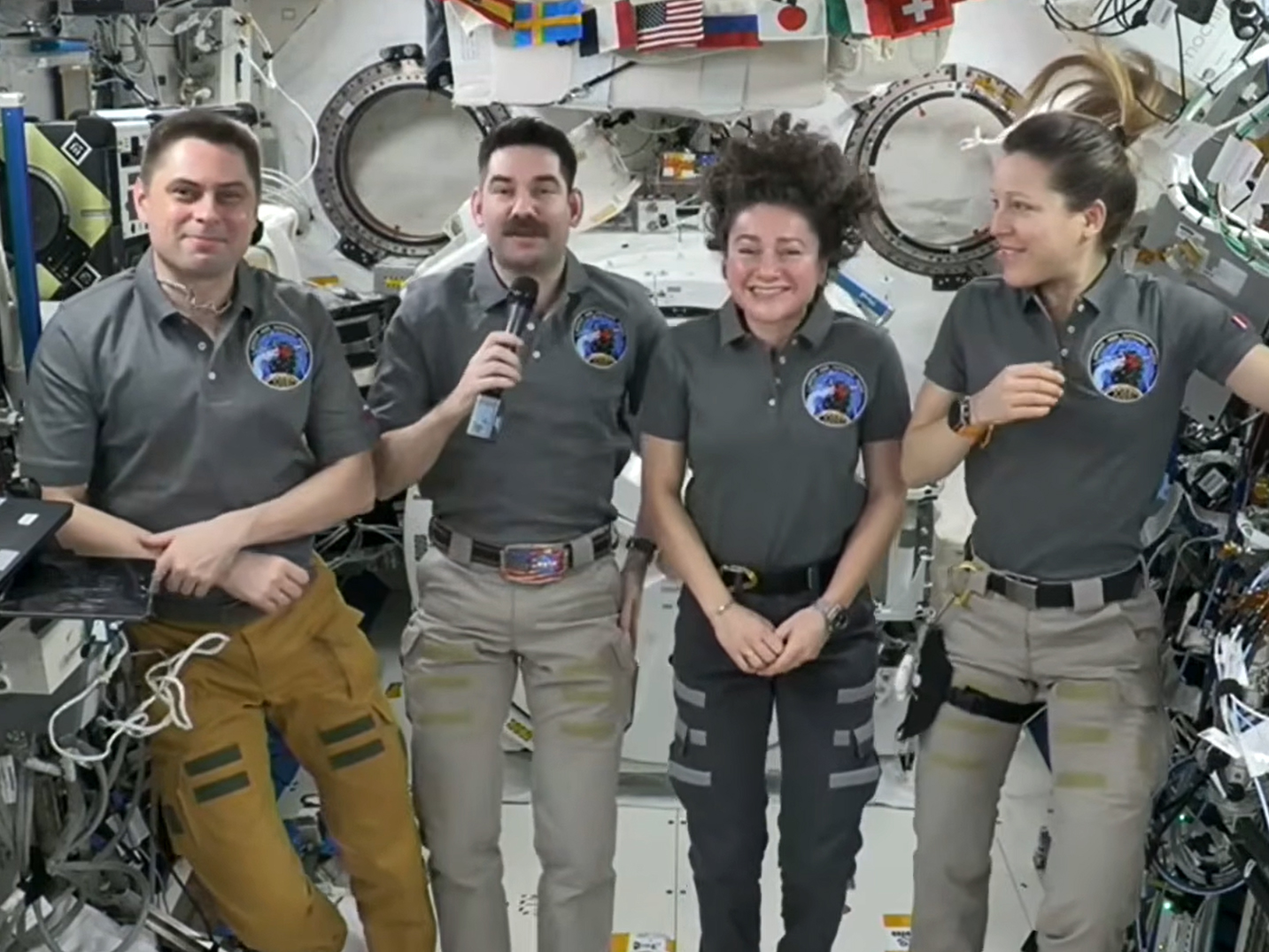
NASA's SpaceX Crew-12 members (from left), Andrey Fedyaev of Roscosmos, Jack Hathaway and Jessica Meir of NASA, and Sophie Adenot of ESA speak to journalists on the ground during a news conference on Wednesday, Sept. 16.
NASA’s SpaceX Crew-12 members (from left), Andrey Fedyaev of Roscosmos, Jack Hathaway and Jessica Meir of NASA, and Sophie Adenot of ESA speak to journalists on the ground during a news conference on Wednesday, Sept. 16.

New supplies are on their way to the International Space Station after launching aboard a Progress 96 cargo spacecraft at 9:33 a.m. EDT on Wednesday. Progress 96 will arrive Saturday for an automated docking to the Poisk module at 9:46 a.m. EDT bringing nearly three tons of food, fuel, and cargo to the Expedition 75 crew members.

NASA and SpaceX are now targeting as soon as early October for the launch of the Crew-13 mission to allow for completion of prelaunch activities, readiness reviews, and coordination of space station operations. The team successfully replaced an oxidizer valve in Dragon’s propulsion system after a leak was detected earlier in processing.

In the meantime, the seven residents aboard the orbiting outpost stayed focused on ongoing microgravity research, cargo operations, and lab maintenance throughout Wednesday.

Station commander Jessica Meir of NASA began her shift documenting her sleep quality, collecting a saliva sample, and downloading biomedical data collected from her smart watch to a computer tablet. Researchers will analyze Meir’s health metrics for the RelaxPro investigation to help reduce stress and improve sleep for astronauts. Next, she transferred cargo in and out of Northrop Grumman’s Cygnus XL cargo spacecraft which has been berthed to the Unity module’s Earth-facing port since April 14.

NASA flight engineer Anil Menon once again soldered components on a circuit board in the Harmony module using lead-free soldering techniques to demonstrate the safe manufacturing and repairing of electronics in microgravity. The space-soldered samples will be returned to Earth and compared to identical ground-soldered samples. Results may lead to reduced reliance on costly resupply missions.

NASA flight engineer Jack Hathaway spent his shift inside the Destiny laboratory module installing new water filter cartridges in the oxygen generation system. The filters remove fluoride ions keeping water pure and clean as it passes through the spacecraft’s systems.

Flight engineer Sophie Adenot of ESA (European Space Agency) worked out on the new European Enhanced Exploration Exercise Device (E4D) installed inside the Columbus laboratory module. Controller software on the E4D tracked her performance helping researchers evaluate its effectiveness for use on future long-duration missions.

The three cosmonauts from Roscosmos, flight engineers Pyotr Dubrov, Anna Kikina, and Andrey Fedyaev, spent their shift on station maintenance. Dubrov tested power systems and cleaned vents in the Zvezda service module. Kikina serviced Zvezda’s Elektron oxygen generator and cleaned fan screens in the Rassvet module. Fedyaev inspected and photographed laptop computers and their power supplies.

Fedyaev and his SpaceX Crew-12 crewmates Meir, Hathaway, and Adenot—who have been aboard the space station since February 14—also participated in a news conference with journalists on the ground. Meir commented on her recent spacewalk when she rode the Canadarm2 robotic arm, “I felt like I was just there levitating above the Earth. I think that was the biggest eureka moment for me.”

Learn more about station activities by following the space station blog, @space_station on X, as well as the ISS Facebook and ISS Instagram accounts.

Get the latest from NASA delivered every week. Subscribe here.

The post Cargo Mission and Crew-13 Updates; Station Research, Maintenance Continue appeared first on NASA Science.

NASA’s Moon Orbiter Spots New, ‘Once-in-Century’ Moon Crater

16 September 2026 at 19:06
6 Min Read

NASA’s Moon Orbiter Spots New, ‘Once-in-Century’ Moon Crater

Black-and-white image of a textured, cratered surface. Two black arrows near the center point inward toward a small, dark, circular feature.
A zoomed-in view of the Moon made from images from NASA’s Lunar Reconnaissance Orbiter Camera. (More information at the bottom of article.)
Credits: NASA Goddard/Intuitive Machines/Robert Wagner

It started as a routine data-quality check. But as Robert Wagner, a scientist with NASA’s Lunar Reconnaissance Orbiter (LRO), scanned a giant Moon map on his computer screen, an unusually large bright spot circled by a dark halo caught his eye, as it implied that surface material in that area had been shaken up.

“I just stopped, dropped everything, and started looking into what that spot was,” said Wagner, an image-processing specialist from Intuitive Machines who works with data from the Lunar Reconnaissance Orbiter Camera (LROC) system.

By comparing before and after images of the Moon, Wagner realized he had discovered the largest, newly formed crater ever found in the solar system, as scientists reported Wednesday in Science Advances. This discovery highlights the value of NASA’s Moon orbiter data in studying a dynamic landscape as the agency advances a sustained human presence and expanded scientific and commercial activity on the Moon.

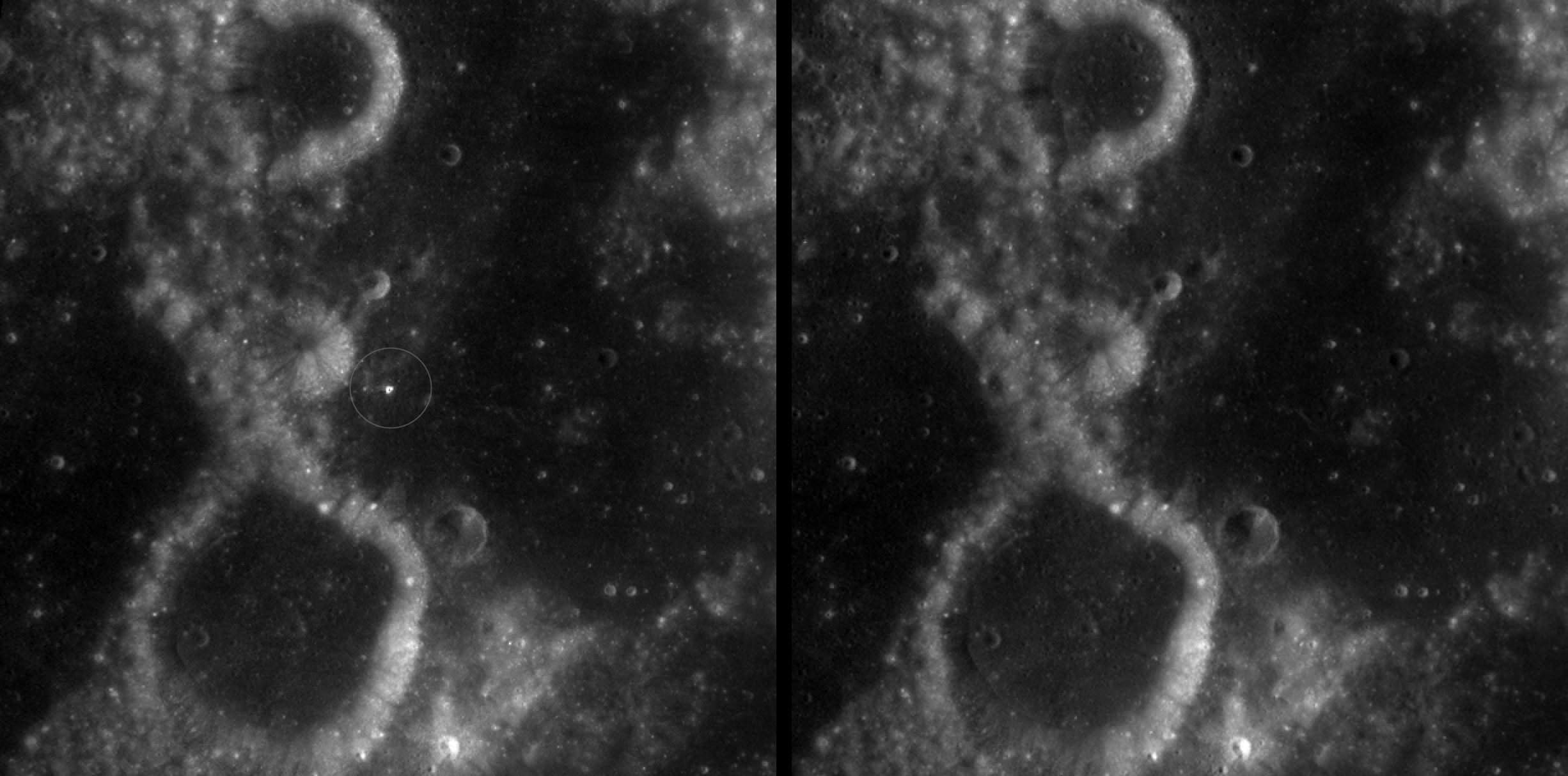
Two nearly identical black-and-white images appear side by side, separated by a vertical black line. Each shows a dark, uneven surface with scattered small circular depressions and several larger round formations with bright, raised rims. In the left image, a thin white circle marks a small, white spot near the center; there is no circled, white spot in the the corresponding area in the right image.
A zoomed-in view of McGetchin crater, circled on the left, next to a “before” image on the right. The panel on the left is made from images captured by NASA’s Lunar Reconnaissance Orbiter Wide-Angle Camera in summer 2025; the right panel was made from images taken in spring 2011. The white spot is not the crater itself, but rather material flung out of the crater, which formed when a rock, possibly this size of a three- to six‑story building, crashed into the Moon between April 11 and May 22, 2024 – a once-in-a-century event, as scientists reported on Sept. 15, 2026, in a pair of papers in Science Advances. The area shown in the images is 38 miles wide.
NASA Goddard/Intuitive Machines/Robert Wagner

Officially named McGetchin after pioneering lunar scientist Tom McGetchin, the crater formed on the Moon’s eastern edge sometime between April 11 and May 22, 2024, after a comet or asteroid the size of a three- to six-story building hit the surface.
 
The crash left a crater, 728 feet wide, that spans the length of two football fields. And at 141 feet deep, the crater could fit three vertically stacked yellow school buses.
 
Scientists estimate that an impact of this magnitude happens on the Moon about once in a century or even longer.

Moon takes some hits

For more than 17 years, LRO has been circling the Moon and using its seven instruments to map the topography, surface composition, temperature, and radiation environment there. The spacecraft’s team has identified at least 1,000 new impact craters throughout the mission and flagged 100,000 more surface changes from an object smashing into the Moon or from the debris that flung out after.

With no atmosphere to slow them or burn them up, space rocks and other objects easily reach the lunar surface. Most are much smaller compared to the object that carved out McGetchin crater. The smallest craters scientists can distinguish from LRO images are about 30 feet wide, the length of a three-story building laid on its side, made by rocks about 43 inches wide, the size of a monster-truck tire. Scientists estimate that impacts of this scale produce about 140 new craters across the Moon each year. But most new ones are made by microscopic projectiles that leave holes too small to identify in orbital images.

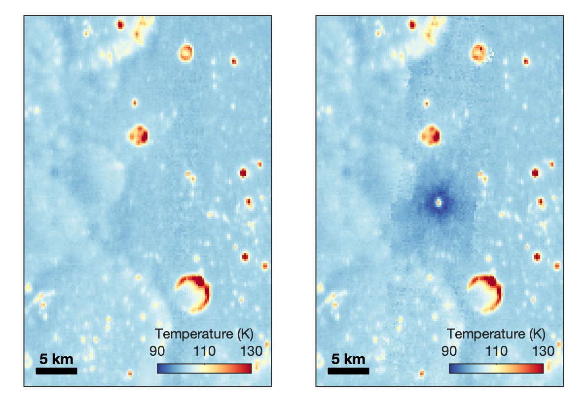
With its numerous instruments, the spacecraft can observe changes to the lunar surface that aren’t apparent in the LROC images alone. After the crater was discovered, scientists working with LRO’s thermal instrument, Diviner, made follow-up observations of the site. They found a 4-mile-wide area around the crater that is about 16 degrees Fahrenheit cooler at night than its surroundings.

Animated temperature map that alternates between two images. A vertical strip through the center blinks on and off, revealing a dark blue circular area surrounding a small white spot near the middle. The pale blue map also contains scattered yellow, orange, and red spots and rings, including a large, curved red-and-yellow feature near the lower center. A 5-kilometer scale bar appears at lower left, and a color key ranges from 90 kelvin in dark blue to 130 kelvin in dark red.
This animated image set shows an area on the eastern limb of the Moon before and after McGetchin crater formed there sometime between April 11 and May 22, 2024. The post-impact image was constructed using data taken by NASA’s Lunar Reconnaissance Orbiter (LRO) thermal instrument, Diviner, between Nov. 15, 2025, and Feb. 18, 2026, after scientists discovered the crater in visible-light images taken by LRO’s Wide-Angle Camera. Pre-impact temperatures are from the Diviner Global High-Resolution Mosaics (GHRM). The crater formed on the Moon after a comet or asteroid, possibly the size of a six-story building, hit the surface in a rare, once-in-a-century collision. The dark blue strip that flashes into view reveals a 4-mile-wide “cold spot” around the crater that is about 16 degrees Fahrenheit cooler at night than its surroundings. This cooling happens because an impact fluffs up regolith around a crater, making it less dense and therefore less able to retain heat. Scientists on the Diviner team reported this finding in a paper published on Sept. 16, 2026, in Science Advances. The red and yellow spots indicate areas typically associated with rocks excavated by long-ago impacts which, though still at cryogenic temperatures like their surroundings, remain slightly warmer at night.
NASA Goddard/UCLA/JHU APL

Reporting in a second paper Wednesday in the same journal, researchers say that the cooling happens because the impact fluffs up the regolith around the crater, making it less dense and therefore less able to retain heat.

The large extent of this “cold spot” is striking, scientists say, because it shows that impacts can modify the Moon’s surface far beyond the crater itself. These physical changes could affect how rover wheels interact with the surface, for instance.

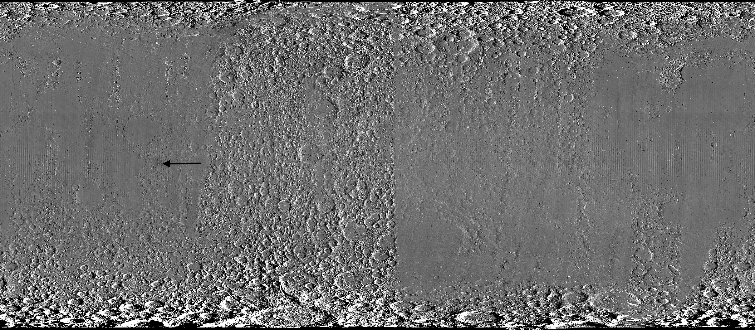
Wide black-and-white image of a rough, uneven surface densely covered with circular depressions and raised areas. The upper and lower edges are especially rugged, while the center is smoother and divided by subtle vertical bands. A black arrow left of center points toward a faint, narrow vertical mark.
A global view of the Moon made by stitching together hundreds of images from the Wide-Angle Camera aboard NASA’s Lunar Reconnaissance Orbiter (LRO). Robert Wagner, image processing specialist for LRO, made this map using two sets of mages taken several years apart. He used a software designed to highlight any changes between the two sets. Anything that stayed the same turned gray; anything different showed up as bright or dark patches. A black arrow just left of center points to McGetchin crater.
NASA Goddard/Intuitive Machines/Robert Wagner

Road to discovery

The LROC system collects images from about 60 miles above the Moon as LRO loops from pole to pole. The system includes two cameras that capture high‑resolution black‑and‑white images and one camera for moderate‑resolution multispectral images. Over the years and thousands of passes, scientists have built maps detailed enough to spot not only new craters, but also landslides, landers, seismic faults, and even hints of lava tubes.

LROC scientists regularly analyze close-up images of small portions of the Moon’s surface taken by the Narrow-Angle Camera. Using these images, they look for changes that are typically less than 30 feet across. But every few years the team searches for large (wider than 150 feet) features by creating global Moon maps and comparing them to older versions.

Square black-and-white image of a dark, uneven surface. At the center is a dark, roughly circular depression surrounded by a bright irregular area, with long streaks of lighter material radiating outward in all directions.
An overhead, close-up view of McGetchin crater that was taken on Dec. 5, 2025, by NASA’s Lunar Reconnaissance Orbiter (LRO) Narrow-Angle Camera (NAC). After the crater was discovered on Oct. 24, 2025, in global images from LRO’s Wide-Angle Camera, which captures broad views with pixels the size of American football fields, scientists turned to the NAC, which takes much sharper views at about 3 feet per pixel. The NAC images, taken as LRO flew over the impact site again, revealed the crater size, shape, and how the surrounding terrain was affected by the impact that carved McGetchin. The image covers an area that’s ¾ mile wide.
NASA Goddard/Intuitive Machines
Square black-and-white image of a dark, gently uneven surface scattered with small circular depressions. Near the center is a small oval crater with a bright raised rim and dark interior. Fine, bright streaks radiate outward from the crater across the surrounding surface.
This view from the side (55° away from straight down) towards the east, covers an area of the Moon that’s about 1.5 miles wide. It was taken by NASA’s Lunar Reconnaissance Orbiter Narrow-Angle Camera on March 3, 2026.
NASA Goddard/Intuitive Machines

That’s what Wagner was doing on Oct. 24, 2025, when he came across McGetchin. Using images from LROC’s Wide-Angle Camera, which captures broad views with pixels the size of football fields, he stacked hundreds of “before” and “after” frames with software designed to highlight change. Anything that stayed the same turned gray while anything different showed up as bright or dark patches.

While the process sounds straightforward, spotting real craters requires a lot of manual work. The software flags every tiny shift in lighting or shadow, generating hundreds of false alarms. For this reason, Wagner typically verifies the software, looking for small fuzzy halos around pixel‑wide bright points, which indicate splashes of regolith around a new crater.

Spanning hundreds of pixels, McGetchin stood out immediately. “It was by far the most obvious impact debris pattern I’ve ever seen in one of these images,” Wagner said.

After his discovery, scientists turned to the Narrow-Angle Camera, which takes much sharper views at about 3 feet per pixel. This camera’s close-up images, taken as LRO flew over the impact site again, revealed the crater size, shape, and how the surrounding terrain was affected. These images also helped researchers estimate the size and force of the rock fragment that formed the new crater, details that are expected to appear in a future paper.

Banner image caption: A zoomed-in view of the Moon, with debris around McGetchin crater visible as a white spot circled by a dark halo just right of center of the image (marked with black arrows). This is the view that Robert Wagner saw on Oct. 24, 2025, during a routine data-quality check. Wagner is an image processing specialist for NASA’s Lunar Reconnaissance Orbiter Camera, a system of three cameras. Using images from the Wide-Angle Camera (WAC), which captures broad views with pixels the size of American football fields, he stacked hundreds of images captured by WAC over the last several years with software designed to highlight change. Anything that stayed the same is gray; anything that changed showed up as bright or dark patches. The stripes in the image are due to slight changes in lighting between the older and newer images. This image composite shows an area of the Moon approximately 900 miles across.

About the Author

Lonnie Shekhtman

Lonnie Shekhtman

Senior Science Writer

Shekhtman helps communicate NASA planetary science to the world through news and feature stories on NASA.gov, videos for NASA+ and YouTube, and by working with the media. She reports on lunar and Mars science and exploration; NASA's search for life; missions to Venus, Titan, and Jupiter's Trojan asteroids; and many other topics related to NASA's exploration of our solar system and beyond.

Keep Exploring

Discover More Topics From NASA

Simplified Summary

Scientists discovered a crater on the Moon that formed sometime between April 11 and May 22, 2024, after a comet or asteroid the size of a three- to six-story building hit the surface.   The crash left a crater, 728 feet wide, that spans the length of two football fields. And at 141 feet deep, the crater could fit three vertically stacked yellow school buses.

The post NASA’s Moon Orbiter Spots New, ‘Once-in-Century’ Moon Crater appeared first on NASA Science.

NASA’s Moon Orbiter Spots New, ‘Once-in-Century’ Moon Crater

16 September 2026 at 19:06
6 Min Read

NASA’s Moon Orbiter Spots New, ‘Once-in-Century’ Moon Crater

Black-and-white image of a textured, cratered surface. Two black arrows near the center point inward toward a small, dark, circular feature.
A zoomed-in view of the Moon made from images from NASA’s Lunar Reconnaissance Orbiter Camera. (More information at the bottom of article.)
Credits:
NASA Goddard/Intuitive Machines/Robert Wagner

It started as a routine data-quality check. But as Robert Wagner, a scientist with NASA’s Lunar Reconnaissance Orbiter (LRO), scanned a giant Moon map on his computer screen, an unusually large bright spot circled by a dark halo caught his eye, as it implied that surface material in that area had been shaken up.

“I just stopped, dropped everything, and started looking into what that spot was,” said Wagner, an image-processing specialist from Intuitive Machines who works with data from the Lunar Reconnaissance Orbiter Camera (LROC) system.

By comparing before and after images of the Moon, Wagner realized he had discovered the largest, newly formed crater ever found in the solar system, as scientists reported Wednesday in Science Advances. This discovery highlights the value of NASA’s Moon orbiter data in studying a dynamic landscape as the agency advances a sustained human presence and expanded scientific and commercial activity on the Moon.

Two nearly identical black-and-white images appear side by side, separated by a vertical black line. Each shows a dark, uneven surface with scattered small circular depressions and several larger round formations with bright, raised rims. In the left image, a thin white circle marks a small, white spot near the center; there is no circled, white spot in the the corresponding area in the right image.
A zoomed-in view of McGetchin crater, circled on the left, next to a “before” image on the right. The panel on the left is made from images captured by NASA’s Lunar Reconnaissance Orbiter Wide-Angle Camera in summer 2025; the right panel was made from images taken in spring 2011. The white spot is not the crater itself, but rather material flung out of the crater, which formed when a rock, possibly this size of a three- to six‑story building, crashed into the Moon between April 11 and May 22, 2024 – a once-in-a-century event, as scientists reported on Sept. 15, 2026, in a pair of papers in Science Advances. The area shown in the images is 38 miles wide.
NASA Goddard/Intuitive Machines/Robert Wagner

Officially named McGetchin after pioneering lunar scientist Tom McGetchin, the crater formed on the Moon’s eastern edge sometime between April 11 and May 22, 2024, after a comet or asteroid the size of a three- to six-story building hit the surface.
 
The crash left a crater, 728 feet wide, that spans the length of two football fields. And at 141 feet deep, the crater could fit three vertically stacked yellow school buses.
 
Scientists estimate that an impact of this magnitude happens on the Moon about once in a century or even longer.

Moon takes some hits

For more than 17 years, LRO has been circling the Moon and using its seven instruments to map the topography, surface composition, temperature, and radiation environment there. The spacecraft’s team has identified at least 1,000 new impact craters throughout the mission and flagged 100,000 more surface changes from an object smashing into the Moon or from the debris that flung out after.

With no atmosphere to slow them or burn them up, space rocks and other objects easily reach the lunar surface. Most are much smaller compared to the object that carved out McGetchin crater. The smallest craters scientists can distinguish from LRO images are about 30 feet wide, the length of a three-story building laid on its side, made by rocks about 43 inches wide, the size of a monster-truck tire. Scientists estimate that impacts of this scale produce about 140 new craters across the Moon each year. But most new ones are made by microscopic projectiles that leave holes too small to identify in orbital images.

With its numerous instruments, the spacecraft can observe changes to the lunar surface that aren’t apparent in the LROC images alone. After the crater was discovered, scientists working with LRO’s thermal instrument, Diviner, made follow-up observations of the site. They found a 4-mile-wide area around the crater that is about 16 degrees Fahrenheit cooler at night than its surroundings.

Animated temperature map that alternates between two images. A vertical strip through the center blinks on and off, revealing a dark blue circular area surrounding a small white spot near the middle. The pale blue map also contains scattered yellow, orange, and red spots and rings, including a large, curved red-and-yellow feature near the lower center. A 5-kilometer scale bar appears at lower left, and a color key ranges from 90 kelvin in dark blue to 130 kelvin in dark red.
This animated image set shows an area on the eastern limb of the Moon before and after McGetchin crater formed there sometime between April 11 and May 22, 2024. The post-impact image was constructed using data taken by NASA’s Lunar Reconnaissance Orbiter (LRO) thermal instrument, Diviner, between Nov. 15, 2025, and Feb. 18, 2026, after scientists discovered the crater in visible-light images taken by LRO’s Wide-Angle Camera. Pre-impact temperatures are from the Diviner Global High-Resolution Mosaics (GHRM). The crater formed on the Moon after a comet or asteroid, possibly the size of a six-story building, hit the surface in a rare, once-in-a-century collision. The dark blue strip that flashes into view reveals a 4-mile-wide “cold spot” around the crater that is about 16 degrees Fahrenheit cooler at night than its surroundings. This cooling happens because an impact fluffs up regolith around a crater, making it less dense and therefore less able to retain heat. Scientists on the Diviner team reported this finding in a paper published on Sept. 16, 2026, in Science Advances. The red and yellow spots indicate areas typically associated with rocks excavated by long-ago impacts which, though still at cryogenic temperatures like their surroundings, remain slightly warmer at night.
NASA Goddard/UCLA/JHU APL

Reporting in a second paper Wednesday in the same journal, researchers say that the cooling happens because the impact fluffs up the regolith around the crater, making it less dense and therefore less able to retain heat.

The large extent of this “cold spot” is striking, scientists say, because it shows that impacts can modify the Moon’s surface far beyond the crater itself. These physical changes could affect how rover wheels interact with the surface, for instance.

Wide black-and-white image of a rough, uneven surface densely covered with circular depressions and raised areas. The upper and lower edges are especially rugged, while the center is smoother and divided by subtle vertical bands. A black arrow left of center points toward a faint, narrow vertical mark.
A global view of the Moon made by stitching together hundreds of images from the Wide-Angle Camera aboard NASA’s Lunar Reconnaissance Orbiter (LRO). Robert Wagner, image processing specialist for LRO, made this map using two sets of mages taken several years apart. He used a software designed to highlight any changes between the two sets. Anything that stayed the same turned gray; anything different showed up as bright or dark patches. A black arrow just left of center points to McGetchin crater.
NASA Goddard/Intuitive Machines/Robert Wagner

Road to discovery

The LROC system collects images from about 60 miles above the Moon as LRO loops from pole to pole. The system includes two cameras that capture high‑resolution black‑and‑white images and one camera for moderate‑resolution multispectral images. Over the years and thousands of passes, scientists have built maps detailed enough to spot not only new craters, but also landslides, landers, seismic faults, and even hints of lava tubes.

LROC scientists regularly analyze close-up images of small portions of the Moon’s surface taken by the Narrow-Angle Camera. Using these images, they look for changes that are typically less than 30 feet across. But every few years the team searches for large (wider than 150 feet) features by creating global Moon maps and comparing them to older versions.

Square black-and-white image of a dark, uneven surface. At the center is a dark, roughly circular depression surrounded by a bright irregular area, with long streaks of lighter material radiating outward in all directions.
An overhead, close-up view of McGetchin crater that was taken on Dec. 5, 2025, by NASA’s Lunar Reconnaissance Orbiter (LRO) Narrow-Angle Camera (NAC). After the crater was discovered on Oct. 24, 2025, in global images from LRO’s Wide-Angle Camera, which captures broad views with pixels the size of American football fields, scientists turned to the NAC, which takes much sharper views at about 3 feet per pixel. The NAC images, taken as LRO flew over the impact site again, revealed the crater size, shape, and how the surrounding terrain was affected by the impact that carved McGetchin. The image covers an area that’s ¾ mile wide.
NASA Goddard/Intuitive Machines
Square black-and-white image of a dark, gently uneven surface scattered with small circular depressions. Near the center is a small oval crater with a bright raised rim and dark interior. Fine, bright streaks radiate outward from the crater across the surrounding surface.
This view from the side (55° away from straight down) towards the east, covers an area of the Moon that’s about 1.5 miles wide. It was taken by NASA’s Lunar Reconnaissance Orbiter Narrow-Angle Camera on March 3, 2026.
NASA Goddard/Intuitive Machines

That’s what Wagner was doing on Oct. 24, 2025, when he came across McGetchin. Using images from LROC’s Wide-Angle Camera, which captures broad views with pixels the size of football fields, he stacked hundreds of “before” and “after” frames with software designed to highlight change. Anything that stayed the same turned gray while anything different showed up as bright or dark patches.

While the process sounds straightforward, spotting real craters requires a lot of manual work. The software flags every tiny shift in lighting or shadow, generating hundreds of false alarms. For this reason, Wagner typically verifies the software, looking for small fuzzy halos around pixel‑wide bright points, which indicate splashes of regolith around a new crater.

Spanning hundreds of pixels, McGetchin stood out immediately. “It was by far the most obvious impact debris pattern I’ve ever seen in one of these images,” Wagner said.

After his discovery, scientists turned to the Narrow-Angle Camera, which takes much sharper views at about 3 feet per pixel. This camera’s close-up images, taken as LRO flew over the impact site again, revealed the crater size, shape, and how the surrounding terrain was affected. These images also helped researchers estimate the size and force of the rock fragment that formed the new crater, details that are expected to appear in a future paper.

Banner image caption: A zoomed-in view of the Moon, with debris around McGetchin crater visible as a white spot circled by a dark halo just right of center of the image (marked with black arrows). This is the view that Robert Wagner saw on Oct. 24, 2025, during a routine data-quality check. Wagner is an image processing specialist for NASA’s Lunar Reconnaissance Orbiter Camera, a system of three cameras. Using images from the Wide-Angle Camera (WAC), which captures broad views with pixels the size of American football fields, he stacked hundreds of images captured by WAC over the last several years with software designed to highlight change. Anything that stayed the same is gray; anything that changed showed up as bright or dark patches. The stripes in the image are due to slight changes in lighting between the older and newer images. This image composite shows an area of the Moon approximately 900 miles across.

About the Author

Lonnie Shekhtman

Lonnie Shekhtman

Senior Science Writer

Shekhtman helps communicate NASA planetary science to the world through news and feature stories on NASA.gov, videos for NASA+ and YouTube, and by working with the media. She reports on lunar and Mars science and exploration; NASA’s search for life; missions to Venus, Titan, and Jupiter’s Trojan asteroids; and many other topics related to NASA’s exploration of our solar system and beyond.

Keep Exploring

Discover More Topics From NASA

Simplified Summary

Scientists discovered a crater on the Moon that formed sometime between April 11 and May 22, 2024, after a comet or asteroid the size of a three- to six-story building hit the surface.
 
The crash left a crater, 728 feet wide, that spans the length of two football fields. And at 141 feet deep, the crater could fit three vertically stacked yellow school buses.

New Crops, Health Research Proposals to Help NASA Advance Exploration

14 September 2026 at 20:00

NASA has selected 12 new investigations to help support human explorers in space. Six will help advance the ability to produce food off-planet, while another six advance the understanding of how unique conditions associated with space exploration can impact the health and physiology of astronauts.  

The six food-focused investigations, supported by the agency’s Space Crops grants, advance NASA’s goal of harnessing the power of plants to enhance quality of life for humans in space, as well as to enable lunar and Mars exploration. Four of the space crops proposals focus on studies of edible plants and how their growth, physiology, and associated microbial communities are affected by environmental conditions in space or on other planetary bodies.

Another proposal involves understanding mechanisms by which certain plant species, called resurrection plants, can withstand extreme, dry conditions. The last space crops proposal builds on previous studies on the International Space Station to uncover functions of algal genes, segments of DNA controlling algae survival, whose expression is modified by spaceflight. Such studies can help scientists integrate biological-based solutions into space life-support systems.

The second set of six proposals fall under the agency’s Biological and Physical Sciences Division’s focus on precision health, which seeks to unravel the mechanisms by which space exploration affects health and well-being. One proposal studies how lunar dust exposure impacts respiratory, immune, and inflammation responses.  A second proposal identifies repurposed drugs that can mitigate the effects space travel has on health and physiology.

Two more of the research proposals look at how radiation combined with other stressors affects age-relevant characteristics seen in various organ and physiological systems. The final two proposals assess molecular and cellular changes induced by stressors in space that can alter gene expression or protein function. The research will identify ways to mitigate health issues and develop early-warning detection systems to notify crew of potential concerns.

The principal investigators who conduct the space biology studies come from 10 institutions across eight states. Nine of the awards are for investigators who are receiving funding from the Space Biology Program for the first time.

Selected Space Crops Investigations

  • Jannell Bazurto, University of Minnesota
    Cultivating the Cosmos: Investigating Plant-Microbe Dynamics for Sustainable Space Farming
  • Miranda Haus, Michigan State University
    Investigating Legume Crop Rotations and Microbial Interactions in Regolith Simulants
  • Andrew Settles, NASA Ames Research Center
    Genetic Mitigation of Spaceflight Stressors on Production of Chlamydomonas
  • Christopher Taylor, Ohio State University
    Hydroponic Microbes in a Low Carbon, High-Radiation Environment; Identifying the Genetic Determinants of Water- and Plant-Associated Microbial Survival in Extraterrestrial Environments
  • Robert VanBuren, Michigan State University
    Leveraging the Natural Resilience of Resurrection Plants for Space Bioregenerative Systems
  • Shuyang Zhen, Texas A&M AgriLife Research
    Environmental Optimization and Cultivar Selection to Enhance Dwarf Tomato Growth and Stress Tolerance for Space Agriculture

Selected Precision Health Investigations

  • Archana Dhasarathy, University of North Dakota
    Epigenetic Memory as an Adaptive Mechanism to Microgravity and Radiation Exposure
  • Andrij Holian, University of Montana
    Contribution of Aging to Particle-Induced Chronic Inflammation
  • Mert Gur, University of Pittsburgh
    Probing Pathogenic Mutations Under Spaceflight-Relevant Stressors Via All-Atom Molecular Dynamics Simulations
  • Grace O’Connell, University of California
    Persistent Spine Health Impacts of Microgravity and Radiation Following Recovery
  • Satoshi Okawa, University of Pittsburgh
    Investigating Space Flight-Induced Brain-Heart Axis Aging and Identifying Repurposed Drugs to Mitigate Its Effects
  • Sanghee Yun, Ph.D., The Children’s Hospital of Philadelphia
    Spaceflight-Relevant Hypercapnia Effects on Space Radiation- and Social Isolation-Induced Biological Aging: A Systems Biology Analysis of Multiple Aging Hallmarks

When fully implemented, more than $7 million in grant money will be awarded throughout fiscal years 2026-2030. These awards were made in response to research proposals submitted to ROSES-2024: Program Element E.9: Space Biology Research Studies (NNH24ZDA001N-SBR).

To learn more about NASA’s biological and physical research in space, visit: https://science.nasa.gov/biological-physical/

Biological & Physical Sciences Division

  • NASA’s Biological and Physical Sciences Division pioneers scientific discovery and enables exploration by using space environments to conduct investigations not possible on Earth. Studying biological and physical phenomenon under extreme conditions allows researchers to advance the fundamental scientific knowledge required to go farther and stay longer in space, while also benefitting life on Earth.

3D imaging techniques reveal hidden damage in soybean seeds

11 September 2026 at 11:50

Scientists in Brazil have demonstrated that a combination of three non-destructive imaging techniques, visible-spectrum (RGB) photography, X-ray radiography, and three-dimensional magnetic resonance imaging (MRI), can detect and characterize hidden damage in soybean seeds that conventional quality tests frequently miss. The study, published in Smart Agricultural Technology, comes at a critical moment: Brazil is the world’s largest soybean producer and exporter, with an estimated harvest of 166 million tons for the 2024/2025 growing season, and seed quality is increasingly under threat from the very climate extremes that help drive the country’s agricultural productivity.

The research, led by Rafael Mateus Alves and colleagues at institutions including the Seed Technology Center of Embrapa Soybean and the University of São Paulo, was funded by the São Paulo Research Foundation (FAPESP). Its central premise is deceptively simple: because seed quality is largely determined in the field, before harvest even begins, researchers and seed companies need tools that can look inside a seed without killing it. Destructive assays such as the tetrazolium viability test, which stains metabolically active tissue red, are the industry standard, but they end the life of the sample. That is a particular problem for breeding programs, where each seed represents a unique genetic combination that must survive analysis if it is to be planted.

The seed itself is a small miracle of engineering. A protective coat, derived from the ovule integuments and marked by a scar called the hilum, regulates water uptake and shields the embryo within. The embryo contains the plumule, which gives rise to the plant’s first true leaves; the apical meristem, precursor of the aerial plant; and the hypocotyl–radicle axis, which breaks through the soil and forms the primary root. Flanking this axis are the cotyledons, storage organs packed with macromolecules that are metabolized and shipped to the growing axis during germination. Damage to any of these structures, from insects, fungi, drought, heat, or rough handling during harvest, can silently reduce a seed lot’s vigor, the capacity for rapid, uniform germination that underpins successful crop establishment.

To interrogate this architecture non-invasively, the team assembled ten representative seeds from production fields, commercial lots, and storage samples, each embodying a distinct damage category: stink bug injury, purple seed stain caused by fungi, green seeds from incomplete maturation, wrinkled seeds from heat and drought stress, weathering-related coat ruptures, severe structural failure, mechanical harvest damage, and internal galleries bored by the cigarette beetle (Lasioderma serricorne) during storage. Every seed was imaged sequentially with each of the three modalities and then subjected to the tetrazolium test as a qualitative reference standard.

The technical pipeline was meticulously standardized. For visible-light imaging, seeds were mounted on acetate sheets and photographed with a Leica DMLB microscope equipped with a DFC 310FX digital camera, oriented in four standardized positions with the hilum consistently to the right. X-ray radiographs were acquired on a Faxitron MX-20 DC-12 system operating at 34 kilovolts, with a nine-second exposure and an object-to-source distance of 11.4 centimeters; uniform contrast settings were applied to all images to make damaged tissues directly comparable. The MRI work used a horizontal 2 Tesla superconducting magnet coupled to a Bruker Avance III spectrometer operating at 85.24 megahertz for proton nuclei. Before scanning, seeds were conditioned to raise their moisture content from roughly 8–10 percent (wet basis) by incubation on moistened germination paper for three hours at 25 °C, a step that ensured sufficient hydrogen signal for imaging. A three-dimensional FLASH pulse sequence, with a 3-millisecond echo time and 75-millisecond repetition time, collected data on a 128×128×128 matrix over a 12.8-millimeter field of view, yielding isotropic voxels of 100 micrometers per side. Datasets were zero-padded to 256³ before Fourier transformation and rendered in three dimensions using ImageJ with the 3Dscript plugin.

The resulting image galleries are striking. On seeds punctured by stink bugs, the dominant pest of Brazilian soybean fields, RGB images revealed darkened patches on the cotyledons and near the plumule, the visible legacy of insect feeding that lacerates tissue and injects yeasts that degrade it from within. X-ray images flagged zones of reduced radiation absorption in the same darkened regions, while MRI cross-sections exposed the depth of tissue destruction with unusual clarity, precisely because degraded tissue no longer absorbs water and therefore produces no magnetic resonance signal. The tetrazolium test confirmed that these zones lacked respiratory activity, meaning they were metabolically dead.

Not every symptom tells the same story, however, and the multimodal approach proved especially powerful at distinguishing superficial from structural damage. Seeds with purple seed stain, caused by Cercospora fungi that secrete the toxin cercosporin, showed internal structures that appeared entirely normal in both X-ray and MRI. Yet the tetrazolium test produced an unusually intense red coloration at the stained coat, indicating that fungal activity had compromised the seed coat’s ability to regulate water uptake, allowing the staining solution to penetrate more deeply into the embryo. Similarly, green seeds, which retain chlorophyll because heat and drought interrupted maturation, showed no structural anomalies under X-ray or MRI, but their membranes proved unusually permeable to the tetrazolium solution, hinting at the physiological deterioration typical of immature tissue.

Where MRI truly distinguished itself was in detecting damage invisible to every other method. One seed that appeared flawless in visible light and radiographically normal in X-ray images revealed, in its three-dimensional MRI reconstruction, internal ruptures running through the central cotyledons, the likely consequence of rapid, uneven drying that forces differential cellular expansion and eventual tissue collapse. In mechanically damaged seeds, the X-ray images showed a crack in the hypocotyl–radicle axis that the camera could not see, but MRI went further, revealing the full extent of the fissure and the displacement of the axis relative to the cotyledons. The team also documented a dramatic structural failure nicknamed the “popcorn effect,” in which alternating wetting and rapid drying cycles tear the seed coat and force the cotyledons apart, a phenomenon increasingly reported in Brazilian seed lots as late-season weather grows more erratic.

To move beyond qualitative observation, the researchers extracted grayscale histograms from each X-ray image, normalizing pixel frequencies across the full 0–255 intensity range. Two statistical descriptors, mean gray level and Shannon entropy, were then compared across damage categories using one-way ANOVA followed by Tukey’s honest significance test. The analysis yielded clear separation: undamaged seeds showed the narrowest distributions with peaks at high grayscale values, while beetle-infested seeds and wrinkled seeds displayed the broadest distributions with heavy weight in the lower gray ranges, reflecting hollowed-out tissues and sparse, deformed structures. Mean gray level differed significantly across categories, peaking in undamaged seeds and bottoming out in wrinkled ones, while Shannon entropy, a measure of histogram complexity, was highest in beetle-damaged seeds and lowest in healthy ones. Because each damage category produced a characteristic radiographic fingerprint, the authors suggest these histogram metrics could support automated, quantitative screening in commercial seed laboratories.

Each modality, the authors stress, occupies a distinct niche. RGB imaging is fast, cheap, and ideal for surface screening but blind to anything beneath the coat. X-ray radiography, already a mainstay of seed testing labs, reveals internal voids, malformations, and cavities rapidly and non-destructively, though it cannot report on metabolic viability and demands careful standardization. MRI, with its superb soft-tissue contrast and genuine three-dimensional resolution, excels at detecting subtle physiological and structural alterations, latent mechanical injury, hydration-induced ruptures, and moisture redistribution, but its cost and acquisition time currently confine it to research settings. The tetrazolium test remains valuable as a reference but is destructive and subject to interpreter bias.

The authors argue the techniques should be viewed as complementary rather than competing, and they sketch a future in which RGB and X-ray screening handle high-throughput commercial quality control while MRI resolves ambiguous or high-stakes cases, such as breeding lines and seed physiology studies. Advances in imaging hardware, automation, and artificial intelligence-based analysis, they note, could eventually fuse these modalities into scalable phenotyping workflows. For now, the message is clear: in an era when a single season of heat, drought, and hungry insects can undermine an entire seed lot, the ability to see damage in all three dimensions, without sacrificing the seed itself, may prove as transformative for agriculture as it has been for medicine.

Subject of Research: Non-destructive multimodal imaging (RGB, X-ray, and MRI) for detecting abiotic- and biotic-stress-induced damage in soybean seeds

Article Title: When damage goes deep: a multi-technique approach including 3D for soybean seed damage assessment

Article References: Alves, R. M., Teixeira, J. M., Marassi, A. G., Henning, F. A., Umburanas, R. C., Gomes-Junior, F. G., & Tannús, A. (2026). When damage goes deep: a multi-technique approach including 3D for soybean seed damage assessment. Smart Agricultural Technology, 15, Article 102523. https://doi.org/10.1016/j.atech.2026.102523

Image Credits: AI Generated

DOI: 10.1016/j.atech.2026.102523

Keywords: soybean seeds, seed quality, MRI, X-ray imaging, RGB imaging, seed vigor, multimodal imaging, stink bug damage, tetrazolium test, non-destructive testing, seed coat rupture, cigarette beetle

New Electrochemical Method Boosts Ocean Alkalinity to Capture Carbon

11 September 2026 at 11:26

The ocean has quietly absorbed billions of tons of humanity’s carbon emissions over the past two centuries, but scientists and companies racing to supercharge that natural service have long been hampered by a stubborn and costly engineering problem. Now researchers at Stony Brook University, working with the Research Foundation for the State University of New York, have unveiled a method for electrochemical ocean alkalinity enhancement that they say can dramatically lower the cost of ocean-based carbon dioxide removal by eliminating one of the most expensive steps in the entire process: the pretreatment of seawater before it enters an electrochemical reactor. The innovation, now available for licensing through SUNY TechConnect, promises high-quality carbon offsets at a substantially reduced cost while simultaneously helping to counteract the acidification that threatens marine ecosystems worldwide.

At the heart of the challenge is a delicate chemical balancing act. As atmospheric carbon dioxide dissolves into seawater, it forms carbonic acid, which lowers the ocean’s pH and erodes the carbonate chemistry that corals, shellfish and other calcifying organisms depend upon. Ocean carbon dioxide capture exploits a simple principle: if the ocean becomes more alkaline, it can absorb more CO2 from the air and lock it away safely as bicarbonate, one of the most stable and abundant forms of inorganic carbon on Earth. This process, known as ocean alkalinity enhancement, effectively turns the surface ocean into a vast, distributed carbon sink. But adding alkalinity to seawater is not trivial, and doing it in a way that is energy-efficient, scalable and safe has become one of the central engineering puzzles of the emerging carbon removal industry.

One of the most promising approaches relies on electrochemistry, specifically a technique called bipolar membrane electrodialysis. In this system, ion-selective membranes are stacked between two end electrodes, and the arrangement allows operators to split ordinary saltwater into its constituent acid and base. When brine containing sodium chloride flows through the stack, the process generates hydrochloric acid on one side and sodium hydroxide, a strong base, on the other. The elegance of the scheme lies in its product handling: if the hydrochloric acid is kept on land, where it can be stored, neutralized or put to industrial use, and only the sodium hydroxide along with the treated seawater is returned to the ocean, the receiving waters gain a net infusion of alkalinity. That added alkalinity shifts the carbonate equilibrium, allowing the ocean to draw down carbon dioxide from the atmosphere and convert it into dissolved bicarbonate that remains stable for millennia.

This electrochemical pathway has attracted serious commercial interest precisely because it produces what carbon markets call negative emissions, meaning carbon dioxide is durably removed from the atmosphere rather than merely avoided. Companies pursuing net-zero targets increasingly rely on such carbon removal credits to offset their residual emissions, and ocean alkalinity enhancement is widely regarded as one of the largest-capacity avenues available, given the sheer scale of the world’s oceans. Yet the technology has been held back by a deceptively mundane problem: what flows into the reactor matters just as much as what flows out.

Seawater, and many other brine streams that might feed these systems, contains divalent cations, most notably calcium and magnesium ions, alongside the sodium and chloride that the process is designed to split. When these divalent ions encounter the high-pH conditions created at the base-producing side of a bipolar membrane stack, they precipitate as solid calcium and magnesium compounds that coat the membranes in a crust known as scale. The consequences are cascading. Scaled membranes lose their ion-selective efficiency, forcing the system to push harder and consume more electricity to achieve the same output. Over time, the deposits shorten membrane lifetime, increasing maintenance frequency and replacement costs. The conventional answer has been water softening pretreatment, in which the incoming seawater is stripped of calcium and magnesium before it ever reaches the electrodialysis unit. While effective, such pretreatment adds significant capital and operating expense, undermining the economic case for the entire carbon removal operation and eroding the value proposition of the resulting carbon credits.

The Stony Brook researchers behind the new method have essentially designed around this bottleneck. Their technology enables electrochemical enhancement of ocean alkalinity without any pretreatment of the incoming seawater at all. By rethinking how the electrochemical system handles brine streams laden with divalent cations, the researchers have created a process that tolerates raw seawater directly, sidestepping the water softening infrastructure that previous designs demanded. According to the announcement from the Research Foundation for SUNY, the result is a system that produces negative emission carbon offsets of very high quality at a substantially reduced cost, simply because the most expensive upstream step has been engineered out of existence. The advantages claimed for the approach are straightforward and commercially significant: it is cheaper, more energy efficient, and requires no expensive water softening pretreatment.

The implications ripple across both the carbon markets and the broader climate technology landscape. Carbon offset buyers, including corporations striving to meet net-zero commitments, place a premium on removal credits that are durable, verifiable and cost-competitive. Ocean alkalinity enhancement already offers exceptional permanence, since bicarbonate dissolved in seawater represents one of the most long-lived forms of carbon storage known. By cutting costs at the front end, the Stony Brook method could help push the price of these high-quality ocean-based offsets toward levels that make them viable at gigatonne scale, a threshold that analysts consider essential if carbon removal is to play a meaningful role in stabilizing the global climate. Cheaper alkalinity generation also strengthens the second major application of the technology: mitigating ocean acidification directly, without necessarily framing it as a carbon market product at all.

Ocean acidification itself is a mounting crisis. Since the industrial revolution, the average surface ocean pH has dropped measurably as the sea has absorbed roughly a quarter to a third of anthropogenic CO2 emissions. The consequences have been documented across coral reefs, oyster hatcheries, pteropods and other calcifying organisms whose shells and skeletons become harder to build in increasingly acidic waters. A technology that adds alkalinity to coastal waters and open ocean regions simultaneously addresses this chemical stress and harvests the carbon removal benefit, a dual function that distinguishes ocean alkalinity enhancement from many other carbon removal approaches. In regions where fisheries, aquaculture and reef ecosystems are economically vital, localized deployment of alkalinity-enhancing electrochemical systems could offer both environmental resilience and revenue through carbon credit sales.

The scientific pedigree of the work reflects the growing maturity of the carbon removal field. Stony Brook University, a flagship research institution within the SUNY system, has become an active hub for climate and energy innovation, and the new method is protected under intellectual property identified in the licensing announcement. The Research Foundation for the State University of New York, the nation’s largest research foundation supporting the nation’s largest public university system, is actively seeking development partners, commercial partners and licensees to translate the laboratory advance into deployed systems. The foundation describes a broad portfolio of SUNY-led research spanning artificial intelligence for the public good, quantum technologies, next-generation semiconductors, biotechnology and medicine, and energy and climate solutions, with system-wide research expenditures approaching 1.5 billion dollars in fiscal year 2025.

For the carbon removal industry, the timing of the announcement is notable. As voluntary and compliance carbon markets mature, buyers are demanding higher fidelity in the offsets they purchase, favoring durable removal over avoidance-based credits. Electrochemical ocean alkalinity enhancement, with its well-understood chemistry and measurable inputs and outputs, is well positioned to meet rigorous monitoring, reporting and verification standards. The obstacle has always been cost, and the pretreatment bottleneck has been a substantial contributor. If the Stony Brook design performs as described at industrial scale, it could remove one of the largest cost wedges standing between laboratory demonstration and commercial deployment, accelerating a technology class that many climate analysts view as indispensable to achieving deep decarbonization.

The broader lesson of the advance may be as important as the technology itself. In the race to scale carbon removal, progress often comes not from exotic new chemistry but from eliminating the hidden costs and fragilities that make promising concepts uneconomic in practice. Water softening pretreatment is exactly the kind of unglamorous engineering detail that determines whether a climate technology remains a paper exercise or becomes infrastructure. By designing a bipolar membrane electrodialysis system that thrives on untreated seawater, the Stony Brook researchers have addressed one of those decisive details. The ocean, meanwhile, stands ready. Every unit of alkalinity added is an invitation for the sea to take up more carbon dioxide and, in doing so, to heal a little of the acidification that a warming world has inflicted upon it. What remains now is the work of scaling, partnering and commercializing, and the SUNY licensing announcement signals that the researchers and their institution intend to see this chemistry leave the laboratory and reach the sea.

Subject of Research: Electrochemical ocean alkalinity enhancement for carbon dioxide removal and ocean acidification mitigation

Article Title: Method for electrochemical ocean alkalinity enhancement

Article References: Method for electrochemical ocean alkalinity enhancement. Research Foundation for the State University of New York, via EurekAlert! News by Subject: Tech & Engineering. Available at: SUNY TechConnect (https://suny.technologypublisher.com/) Original publication

Image Credits: AI Generated

DOI: Not provided

Keywords: ocean alkalinity enhancement, carbon dioxide removal, bipolar membrane electrodialysis, ocean acidification mitigation, carbon offsets, Stony Brook University, negative emissions, seawater pretreatment, net-zero goals, electrochemistry, Research Foundation for SUNY

OpenAI anuncia novidades para o ChatGPT Voice, Work, Codex e a Biblioteca

11 September 2026 at 01:23

A OpenAI anunciou recentemente uma série de novidades para o ChatGPT, incluindo uma atualização muito bem-vinda para usuários do ChatGPT Voice.

Agora, o assistente pode usar os modelos GPT-5.6 ou GPT-6 Astra quando precisa pesquisar ou raciocinar sobre questões mais difíceis — e o usuário pode escolher qual usar com um seletor.

Além disso, a empresa também simplificou os limites de uso diários do GPT-Live:

  • Go: até 3 horas com o GPT-Live-1 mini, substituindo o acesso ao GPT-Live-1;
  • Plus: até 3 horas com o GPT-Live-1;
  • Pro (US$100/mês): até 15 horas com o GPT-Live-1;
  • Pro (US$200/mês): uso ilimitado do GPT-Live-1;
  • Plus e Pro: não mudam mais para o GPT-Live mini depois de atingir o limite de uso do Voice.

Com essas mudanças, os planos Plus e Pro não mudam mais para o GPT-Live-1 mini depois de atingir o limite de uso do Voice. Além disso, os níveis separados de inteligência do Voice (Instant, Medium e High) foram descontinuados.

Novidades para o ChatGPT Work e Codex

A OpenAI anunciou também que a Deep Research está agora disponível no ChatGPT Work e no Codex para os planos Plus, Pro, Business, Enterprise e Edu. Basta ao usuário digitar @Deep Research ou pedir explicitamente para usá-la no comando.

Outra novidade chegando aos dois recursos é o plugin Data, que ajuda a analisar dados de negócios conectados. É possível fazer uma pergunta sobre um negócio, investigar mudanças ou até mesmo criar um dashboard interativo sobre o conteúdo analisado.

Now everyone can put data to work.

We’re introducing a new Data agent in ChatGPT Work so you can turn your company’s data into answers, interactive dashboards, and action—just by asking.

Just add the Data Plugin in ChatGPT Work, connect to the data sources and context you… pic.twitter.com/R7N3I2NFCN

— ChatGPT (@ChatGPT) September 10, 2026

Biblioteca

Agora também é possível compartilhar arquivos e pastas no ChatGPT pela Biblioteca. É possível compartilhar com pessoas específicas ou com integrantes do workspace, com opções de gerenciamento de acesso e uma visualização dos conteúdos compartilhados.

Por fim, a Biblioteca também suporta agora o Box, o Dropbox e o SharePoint, que se juntam ao Google Drive para permitir que o usuário pesquise com facilidade por arquivos e pastas disponíveis nessas plataformas e os usem como contexto em conversas.

❌