❌

Normal view

Cargo Mission and Crew-13 Updates; Station Research, Maintenance Continue

16 September 2026 at 20:40
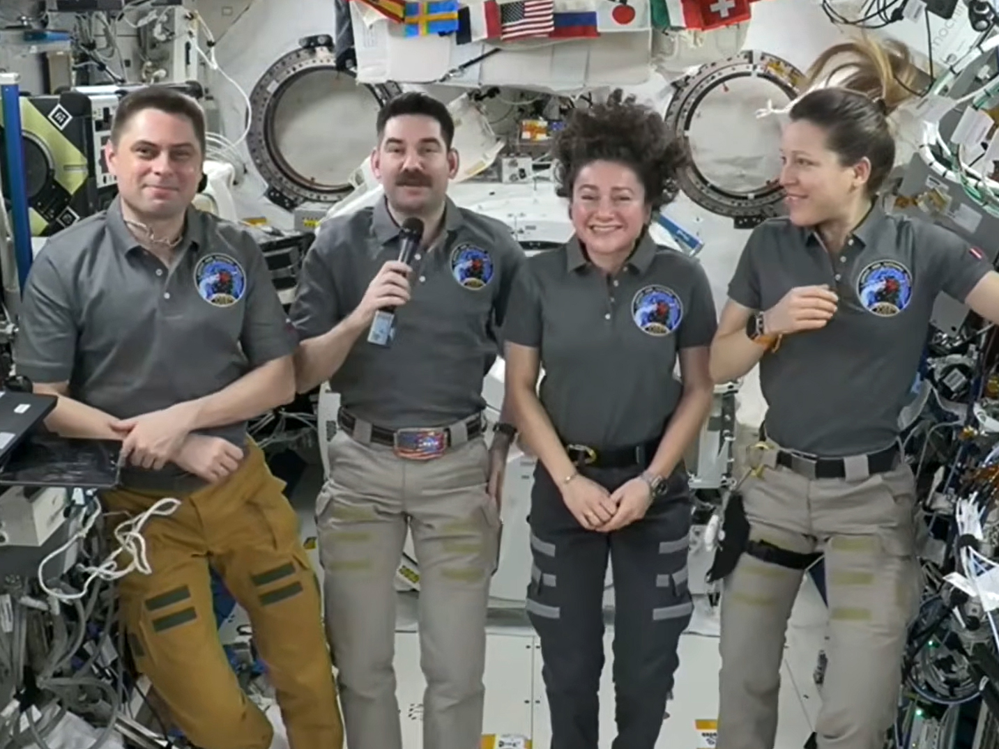
NASA's SpaceX Crew-12 members (from left), Andrey Fedyaev of Roscosmos, Jack Hathaway and Jessica Meir of NASA, and Sophie Adenot of ESA speak to journalists on the ground during a news conference on Wednesday, Sept. 16.
NASA’s SpaceX Crew-12 members (from left), Andrey Fedyaev of Roscosmos, Jack Hathaway and Jessica Meir of NASA, and Sophie Adenot of ESA speak to journalists on the ground during a news conference on Wednesday, Sept. 16.

New supplies are on their way to the International Space Station after launching aboard a Progress 96 cargo spacecraft at 9:33 a.m. EDT on Wednesday. Progress 96 will arrive Saturday for an automated docking to the Poisk module at 9:46 a.m. EDT bringing nearly three tons of food, fuel, and cargo to the Expedition 75 crew members.

NASA and SpaceX are now targeting as soon as early October for the launch of the Crew-13 mission to allow for completion of prelaunch activities, readiness reviews, and coordination of space station operations. The team successfully replaced an oxidizer valve in Dragon’s propulsion system after a leak was detected earlier in processing.

In the meantime, the seven residents aboard the orbiting outpost stayed focused on ongoing microgravity research, cargo operations, and lab maintenance throughout Wednesday.

Station commander Jessica Meir of NASA began her shift documenting her sleep quality, collecting a saliva sample, and downloading biomedical data collected from her smart watch to a computer tablet. Researchers will analyze Meir’s health metrics for the RelaxPro investigation to help reduce stress and improve sleep for astronauts. Next, she transferred cargo in and out of Northrop Grumman’s Cygnus XL cargo spacecraft which has been berthed to the Unity module’s Earth-facing port since April 14.

NASA flight engineer Anil Menon once again soldered components on a circuit board in the Harmony module using lead-free soldering techniques to demonstrate the safe manufacturing and repairing of electronics in microgravity. The space-soldered samples will be returned to Earth and compared to identical ground-soldered samples. Results may lead to reduced reliance on costly resupply missions.

NASA flight engineer Jack Hathaway spent his shift inside the Destiny laboratory module installing new water filter cartridges in the oxygen generation system. The filters remove fluoride ions keeping water pure and clean as it passes through the spacecraft’s systems.

Flight engineer Sophie Adenot of ESA (European Space Agency) worked out on the new European Enhanced Exploration Exercise Device (E4D) installed inside the Columbus laboratory module. Controller software on the E4D tracked her performance helping researchers evaluate its effectiveness for use on future long-duration missions.

The three cosmonauts from Roscosmos, flight engineers Pyotr Dubrov, Anna Kikina, and Andrey Fedyaev, spent their shift on station maintenance. Dubrov tested power systems and cleaned vents in the Zvezda service module. Kikina serviced Zvezda’s Elektron oxygen generator and cleaned fan screens in the Rassvet module. Fedyaev inspected and photographed laptop computers and their power supplies.

Fedyaev and his SpaceX Crew-12 crewmates Meir, Hathaway, and Adenot—who have been aboard the space station since February 14—also participated in a news conference with journalists on the ground. Meir commented on her recent spacewalk when she rode the Canadarm2 robotic arm, “I felt like I was just there levitating above the Earth. I think that was the biggest eureka moment for me.”

Learn more about station activities by following the space station blog, @space_station on X, as well as the ISS Facebook and ISS Instagram accounts.

Get the latest from NASA delivered every week. Subscribe here.

The post Cargo Mission and Crew-13 Updates; Station Research, Maintenance Continue appeared first on NASA Science.

NASA’s Moon Orbiter Spots New, ‘Once-in-Century’ Moon Crater

16 September 2026 at 19:06
6 Min Read

NASA’s Moon Orbiter Spots New, ‘Once-in-Century’ Moon Crater

Black-and-white image of a textured, cratered surface. Two black arrows near the center point inward toward a small, dark, circular feature.
A zoomed-in view of the Moon made from images from NASA’s Lunar Reconnaissance Orbiter Camera. (More information at the bottom of article.)
Credits: NASA Goddard/Intuitive Machines/Robert Wagner

It started as a routine data-quality check. But as Robert Wagner, a scientist with NASA’s Lunar Reconnaissance Orbiter (LRO), scanned a giant Moon map on his computer screen, an unusually large bright spot circled by a dark halo caught his eye, as it implied that surface material in that area had been shaken up.

“I just stopped, dropped everything, and started looking into what that spot was,” said Wagner, an image-processing specialist from Intuitive Machines who works with data from the Lunar Reconnaissance Orbiter Camera (LROC) system.

By comparing before and after images of the Moon, Wagner realized he had discovered the largest, newly formed crater ever found in the solar system, as scientists reported Wednesday in Science Advances. This discovery highlights the value of NASA’s Moon orbiter data in studying a dynamic landscape as the agency advances a sustained human presence and expanded scientific and commercial activity on the Moon.

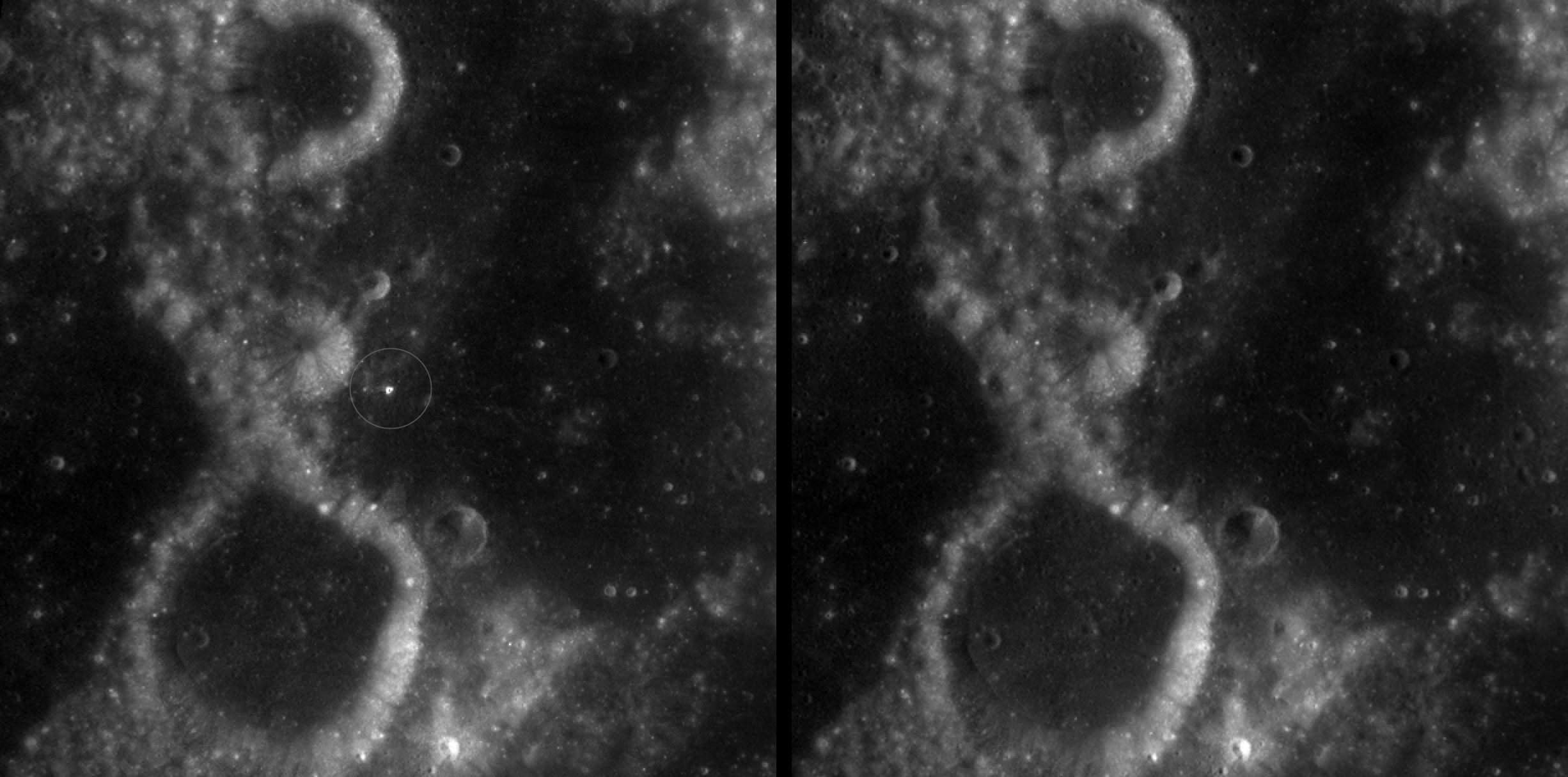
Two nearly identical black-and-white images appear side by side, separated by a vertical black line. Each shows a dark, uneven surface with scattered small circular depressions and several larger round formations with bright, raised rims. In the left image, a thin white circle marks a small, white spot near the center; there is no circled, white spot in the the corresponding area in the right image.
A zoomed-in view of McGetchin crater, circled on the left, next to a “before” image on the right. The panel on the left is made from images captured by NASA’s Lunar Reconnaissance Orbiter Wide-Angle Camera in summer 2025; the right panel was made from images taken in spring 2011. The white spot is not the crater itself, but rather material flung out of the crater, which formed when a rock, possibly this size of a three- to six‑story building, crashed into the Moon between April 11 and May 22, 2024 – a once-in-a-century event, as scientists reported on Sept. 15, 2026, in a pair of papers in Science Advances. The area shown in the images is 38 miles wide.
NASA Goddard/Intuitive Machines/Robert Wagner

Officially named McGetchin after pioneering lunar scientist Tom McGetchin, the crater formed on the Moon’s eastern edge sometime between April 11 and May 22, 2024, after a comet or asteroid the size of a three- to six-story building hit the surface.
 
The crash left a crater, 728 feet wide, that spans the length of two football fields. And at 141 feet deep, the crater could fit three vertically stacked yellow school buses.
 
Scientists estimate that an impact of this magnitude happens on the Moon about once in a century or even longer.

Moon takes some hits

For more than 17 years, LRO has been circling the Moon and using its seven instruments to map the topography, surface composition, temperature, and radiation environment there. The spacecraft’s team has identified at least 1,000 new impact craters throughout the mission and flagged 100,000 more surface changes from an object smashing into the Moon or from the debris that flung out after.

With no atmosphere to slow them or burn them up, space rocks and other objects easily reach the lunar surface. Most are much smaller compared to the object that carved out McGetchin crater. The smallest craters scientists can distinguish from LRO images are about 30 feet wide, the length of a three-story building laid on its side, made by rocks about 43 inches wide, the size of a monster-truck tire. Scientists estimate that impacts of this scale produce about 140 new craters across the Moon each year. But most new ones are made by microscopic projectiles that leave holes too small to identify in orbital images.

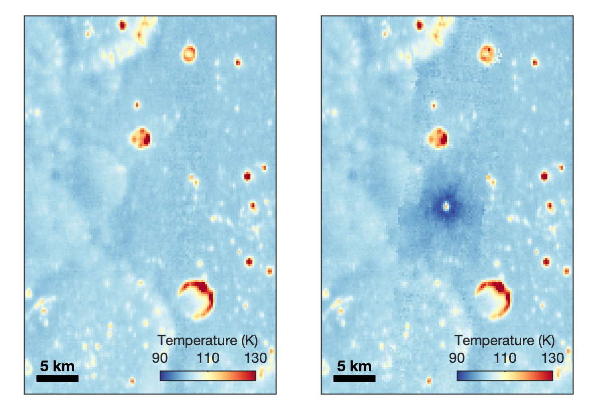
With its numerous instruments, the spacecraft can observe changes to the lunar surface that aren’t apparent in the LROC images alone. After the crater was discovered, scientists working with LRO’s thermal instrument, Diviner, made follow-up observations of the site. They found a 4-mile-wide area around the crater that is about 16 degrees Fahrenheit cooler at night than its surroundings.

Animated temperature map that alternates between two images. A vertical strip through the center blinks on and off, revealing a dark blue circular area surrounding a small white spot near the middle. The pale blue map also contains scattered yellow, orange, and red spots and rings, including a large, curved red-and-yellow feature near the lower center. A 5-kilometer scale bar appears at lower left, and a color key ranges from 90 kelvin in dark blue to 130 kelvin in dark red.
This animated image set shows an area on the eastern limb of the Moon before and after McGetchin crater formed there sometime between April 11 and May 22, 2024. The post-impact image was constructed using data taken by NASA’s Lunar Reconnaissance Orbiter (LRO) thermal instrument, Diviner, between Nov. 15, 2025, and Feb. 18, 2026, after scientists discovered the crater in visible-light images taken by LRO’s Wide-Angle Camera. Pre-impact temperatures are from the Diviner Global High-Resolution Mosaics (GHRM). The crater formed on the Moon after a comet or asteroid, possibly the size of a six-story building, hit the surface in a rare, once-in-a-century collision. The dark blue strip that flashes into view reveals a 4-mile-wide “cold spot” around the crater that is about 16 degrees Fahrenheit cooler at night than its surroundings. This cooling happens because an impact fluffs up regolith around a crater, making it less dense and therefore less able to retain heat. Scientists on the Diviner team reported this finding in a paper published on Sept. 16, 2026, in Science Advances. The red and yellow spots indicate areas typically associated with rocks excavated by long-ago impacts which, though still at cryogenic temperatures like their surroundings, remain slightly warmer at night.
NASA Goddard/UCLA/JHU APL

Reporting in a second paper Wednesday in the same journal, researchers say that the cooling happens because the impact fluffs up the regolith around the crater, making it less dense and therefore less able to retain heat.

The large extent of this “cold spot” is striking, scientists say, because it shows that impacts can modify the Moon’s surface far beyond the crater itself. These physical changes could affect how rover wheels interact with the surface, for instance.

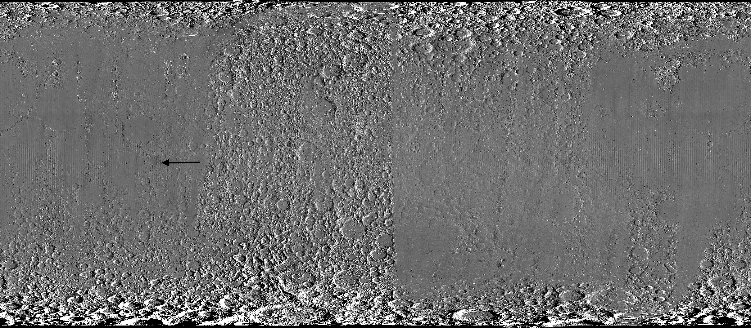
Wide black-and-white image of a rough, uneven surface densely covered with circular depressions and raised areas. The upper and lower edges are especially rugged, while the center is smoother and divided by subtle vertical bands. A black arrow left of center points toward a faint, narrow vertical mark.
A global view of the Moon made by stitching together hundreds of images from the Wide-Angle Camera aboard NASA’s Lunar Reconnaissance Orbiter (LRO). Robert Wagner, image processing specialist for LRO, made this map using two sets of mages taken several years apart. He used a software designed to highlight any changes between the two sets. Anything that stayed the same turned gray; anything different showed up as bright or dark patches. A black arrow just left of center points to McGetchin crater.
NASA Goddard/Intuitive Machines/Robert Wagner

Road to discovery

The LROC system collects images from about 60 miles above the Moon as LRO loops from pole to pole. The system includes two cameras that capture high‑resolution black‑and‑white images and one camera for moderate‑resolution multispectral images. Over the years and thousands of passes, scientists have built maps detailed enough to spot not only new craters, but also landslides, landers, seismic faults, and even hints of lava tubes.

LROC scientists regularly analyze close-up images of small portions of the Moon’s surface taken by the Narrow-Angle Camera. Using these images, they look for changes that are typically less than 30 feet across. But every few years the team searches for large (wider than 150 feet) features by creating global Moon maps and comparing them to older versions.

Square black-and-white image of a dark, uneven surface. At the center is a dark, roughly circular depression surrounded by a bright irregular area, with long streaks of lighter material radiating outward in all directions.
An overhead, close-up view of McGetchin crater that was taken on Dec. 5, 2025, by NASA’s Lunar Reconnaissance Orbiter (LRO) Narrow-Angle Camera (NAC). After the crater was discovered on Oct. 24, 2025, in global images from LRO’s Wide-Angle Camera, which captures broad views with pixels the size of American football fields, scientists turned to the NAC, which takes much sharper views at about 3 feet per pixel. The NAC images, taken as LRO flew over the impact site again, revealed the crater size, shape, and how the surrounding terrain was affected by the impact that carved McGetchin. The image covers an area that’s ¾ mile wide.
NASA Goddard/Intuitive Machines
Square black-and-white image of a dark, gently uneven surface scattered with small circular depressions. Near the center is a small oval crater with a bright raised rim and dark interior. Fine, bright streaks radiate outward from the crater across the surrounding surface.
This view from the side (55° away from straight down) towards the east, covers an area of the Moon that’s about 1.5 miles wide. It was taken by NASA’s Lunar Reconnaissance Orbiter Narrow-Angle Camera on March 3, 2026.
NASA Goddard/Intuitive Machines

That’s what Wagner was doing on Oct. 24, 2025, when he came across McGetchin. Using images from LROC’s Wide-Angle Camera, which captures broad views with pixels the size of football fields, he stacked hundreds of “before” and “after” frames with software designed to highlight change. Anything that stayed the same turned gray while anything different showed up as bright or dark patches.

While the process sounds straightforward, spotting real craters requires a lot of manual work. The software flags every tiny shift in lighting or shadow, generating hundreds of false alarms. For this reason, Wagner typically verifies the software, looking for small fuzzy halos around pixel‑wide bright points, which indicate splashes of regolith around a new crater.

Spanning hundreds of pixels, McGetchin stood out immediately. “It was by far the most obvious impact debris pattern I’ve ever seen in one of these images,” Wagner said.

After his discovery, scientists turned to the Narrow-Angle Camera, which takes much sharper views at about 3 feet per pixel. This camera’s close-up images, taken as LRO flew over the impact site again, revealed the crater size, shape, and how the surrounding terrain was affected. These images also helped researchers estimate the size and force of the rock fragment that formed the new crater, details that are expected to appear in a future paper.

Banner image caption: A zoomed-in view of the Moon, with debris around McGetchin crater visible as a white spot circled by a dark halo just right of center of the image (marked with black arrows). This is the view that Robert Wagner saw on Oct. 24, 2025, during a routine data-quality check. Wagner is an image processing specialist for NASA’s Lunar Reconnaissance Orbiter Camera, a system of three cameras. Using images from the Wide-Angle Camera (WAC), which captures broad views with pixels the size of American football fields, he stacked hundreds of images captured by WAC over the last several years with software designed to highlight change. Anything that stayed the same is gray; anything that changed showed up as bright or dark patches. The stripes in the image are due to slight changes in lighting between the older and newer images. This image composite shows an area of the Moon approximately 900 miles across.

About the Author

Lonnie Shekhtman

Lonnie Shekhtman

Senior Science Writer

Shekhtman helps communicate NASA planetary science to the world through news and feature stories on NASA.gov, videos for NASA+ and YouTube, and by working with the media. She reports on lunar and Mars science and exploration; NASA's search for life; missions to Venus, Titan, and Jupiter's Trojan asteroids; and many other topics related to NASA's exploration of our solar system and beyond.

Keep Exploring

Discover More Topics From NASA

Simplified Summary

Scientists discovered a crater on the Moon that formed sometime between April 11 and May 22, 2024, after a comet or asteroid the size of a three- to six-story building hit the surface.   The crash left a crater, 728 feet wide, that spans the length of two football fields. And at 141 feet deep, the crater could fit three vertically stacked yellow school buses.

The post NASA’s Moon Orbiter Spots New, ‘Once-in-Century’ Moon Crater appeared first on NASA Science.

NASA’s Moon Orbiter Spots New, ‘Once-in-Century’ Moon Crater

16 September 2026 at 19:06
6 Min Read

NASA’s Moon Orbiter Spots New, ‘Once-in-Century’ Moon Crater

Black-and-white image of a textured, cratered surface. Two black arrows near the center point inward toward a small, dark, circular feature.
A zoomed-in view of the Moon made from images from NASA’s Lunar Reconnaissance Orbiter Camera. (More information at the bottom of article.)
Credits:
NASA Goddard/Intuitive Machines/Robert Wagner

It started as a routine data-quality check. But as Robert Wagner, a scientist with NASA’s Lunar Reconnaissance Orbiter (LRO), scanned a giant Moon map on his computer screen, an unusually large bright spot circled by a dark halo caught his eye, as it implied that surface material in that area had been shaken up.

“I just stopped, dropped everything, and started looking into what that spot was,” said Wagner, an image-processing specialist from Intuitive Machines who works with data from the Lunar Reconnaissance Orbiter Camera (LROC) system.

By comparing before and after images of the Moon, Wagner realized he had discovered the largest, newly formed crater ever found in the solar system, as scientists reported Wednesday in Science Advances. This discovery highlights the value of NASA’s Moon orbiter data in studying a dynamic landscape as the agency advances a sustained human presence and expanded scientific and commercial activity on the Moon.

Two nearly identical black-and-white images appear side by side, separated by a vertical black line. Each shows a dark, uneven surface with scattered small circular depressions and several larger round formations with bright, raised rims. In the left image, a thin white circle marks a small, white spot near the center; there is no circled, white spot in the the corresponding area in the right image.
A zoomed-in view of McGetchin crater, circled on the left, next to a “before” image on the right. The panel on the left is made from images captured by NASA’s Lunar Reconnaissance Orbiter Wide-Angle Camera in summer 2025; the right panel was made from images taken in spring 2011. The white spot is not the crater itself, but rather material flung out of the crater, which formed when a rock, possibly this size of a three- to six‑story building, crashed into the Moon between April 11 and May 22, 2024 – a once-in-a-century event, as scientists reported on Sept. 15, 2026, in a pair of papers in Science Advances. The area shown in the images is 38 miles wide.
NASA Goddard/Intuitive Machines/Robert Wagner

Officially named McGetchin after pioneering lunar scientist Tom McGetchin, the crater formed on the Moon’s eastern edge sometime between April 11 and May 22, 2024, after a comet or asteroid the size of a three- to six-story building hit the surface.
 
The crash left a crater, 728 feet wide, that spans the length of two football fields. And at 141 feet deep, the crater could fit three vertically stacked yellow school buses.
 
Scientists estimate that an impact of this magnitude happens on the Moon about once in a century or even longer.

Moon takes some hits

For more than 17 years, LRO has been circling the Moon and using its seven instruments to map the topography, surface composition, temperature, and radiation environment there. The spacecraft’s team has identified at least 1,000 new impact craters throughout the mission and flagged 100,000 more surface changes from an object smashing into the Moon or from the debris that flung out after.

With no atmosphere to slow them or burn them up, space rocks and other objects easily reach the lunar surface. Most are much smaller compared to the object that carved out McGetchin crater. The smallest craters scientists can distinguish from LRO images are about 30 feet wide, the length of a three-story building laid on its side, made by rocks about 43 inches wide, the size of a monster-truck tire. Scientists estimate that impacts of this scale produce about 140 new craters across the Moon each year. But most new ones are made by microscopic projectiles that leave holes too small to identify in orbital images.

With its numerous instruments, the spacecraft can observe changes to the lunar surface that aren’t apparent in the LROC images alone. After the crater was discovered, scientists working with LRO’s thermal instrument, Diviner, made follow-up observations of the site. They found a 4-mile-wide area around the crater that is about 16 degrees Fahrenheit cooler at night than its surroundings.

Animated temperature map that alternates between two images. A vertical strip through the center blinks on and off, revealing a dark blue circular area surrounding a small white spot near the middle. The pale blue map also contains scattered yellow, orange, and red spots and rings, including a large, curved red-and-yellow feature near the lower center. A 5-kilometer scale bar appears at lower left, and a color key ranges from 90 kelvin in dark blue to 130 kelvin in dark red.
This animated image set shows an area on the eastern limb of the Moon before and after McGetchin crater formed there sometime between April 11 and May 22, 2024. The post-impact image was constructed using data taken by NASA’s Lunar Reconnaissance Orbiter (LRO) thermal instrument, Diviner, between Nov. 15, 2025, and Feb. 18, 2026, after scientists discovered the crater in visible-light images taken by LRO’s Wide-Angle Camera. Pre-impact temperatures are from the Diviner Global High-Resolution Mosaics (GHRM). The crater formed on the Moon after a comet or asteroid, possibly the size of a six-story building, hit the surface in a rare, once-in-a-century collision. The dark blue strip that flashes into view reveals a 4-mile-wide “cold spot” around the crater that is about 16 degrees Fahrenheit cooler at night than its surroundings. This cooling happens because an impact fluffs up regolith around a crater, making it less dense and therefore less able to retain heat. Scientists on the Diviner team reported this finding in a paper published on Sept. 16, 2026, in Science Advances. The red and yellow spots indicate areas typically associated with rocks excavated by long-ago impacts which, though still at cryogenic temperatures like their surroundings, remain slightly warmer at night.
NASA Goddard/UCLA/JHU APL

Reporting in a second paper Wednesday in the same journal, researchers say that the cooling happens because the impact fluffs up the regolith around the crater, making it less dense and therefore less able to retain heat.

The large extent of this “cold spot” is striking, scientists say, because it shows that impacts can modify the Moon’s surface far beyond the crater itself. These physical changes could affect how rover wheels interact with the surface, for instance.

Wide black-and-white image of a rough, uneven surface densely covered with circular depressions and raised areas. The upper and lower edges are especially rugged, while the center is smoother and divided by subtle vertical bands. A black arrow left of center points toward a faint, narrow vertical mark.
A global view of the Moon made by stitching together hundreds of images from the Wide-Angle Camera aboard NASA’s Lunar Reconnaissance Orbiter (LRO). Robert Wagner, image processing specialist for LRO, made this map using two sets of mages taken several years apart. He used a software designed to highlight any changes between the two sets. Anything that stayed the same turned gray; anything different showed up as bright or dark patches. A black arrow just left of center points to McGetchin crater.
NASA Goddard/Intuitive Machines/Robert Wagner

Road to discovery

The LROC system collects images from about 60 miles above the Moon as LRO loops from pole to pole. The system includes two cameras that capture high‑resolution black‑and‑white images and one camera for moderate‑resolution multispectral images. Over the years and thousands of passes, scientists have built maps detailed enough to spot not only new craters, but also landslides, landers, seismic faults, and even hints of lava tubes.

LROC scientists regularly analyze close-up images of small portions of the Moon’s surface taken by the Narrow-Angle Camera. Using these images, they look for changes that are typically less than 30 feet across. But every few years the team searches for large (wider than 150 feet) features by creating global Moon maps and comparing them to older versions.

Square black-and-white image of a dark, uneven surface. At the center is a dark, roughly circular depression surrounded by a bright irregular area, with long streaks of lighter material radiating outward in all directions.
An overhead, close-up view of McGetchin crater that was taken on Dec. 5, 2025, by NASA’s Lunar Reconnaissance Orbiter (LRO) Narrow-Angle Camera (NAC). After the crater was discovered on Oct. 24, 2025, in global images from LRO’s Wide-Angle Camera, which captures broad views with pixels the size of American football fields, scientists turned to the NAC, which takes much sharper views at about 3 feet per pixel. The NAC images, taken as LRO flew over the impact site again, revealed the crater size, shape, and how the surrounding terrain was affected by the impact that carved McGetchin. The image covers an area that’s ¾ mile wide.
NASA Goddard/Intuitive Machines
Square black-and-white image of a dark, gently uneven surface scattered with small circular depressions. Near the center is a small oval crater with a bright raised rim and dark interior. Fine, bright streaks radiate outward from the crater across the surrounding surface.
This view from the side (55° away from straight down) towards the east, covers an area of the Moon that’s about 1.5 miles wide. It was taken by NASA’s Lunar Reconnaissance Orbiter Narrow-Angle Camera on March 3, 2026.
NASA Goddard/Intuitive Machines

That’s what Wagner was doing on Oct. 24, 2025, when he came across McGetchin. Using images from LROC’s Wide-Angle Camera, which captures broad views with pixels the size of football fields, he stacked hundreds of “before” and “after” frames with software designed to highlight change. Anything that stayed the same turned gray while anything different showed up as bright or dark patches.

While the process sounds straightforward, spotting real craters requires a lot of manual work. The software flags every tiny shift in lighting or shadow, generating hundreds of false alarms. For this reason, Wagner typically verifies the software, looking for small fuzzy halos around pixel‑wide bright points, which indicate splashes of regolith around a new crater.

Spanning hundreds of pixels, McGetchin stood out immediately. “It was by far the most obvious impact debris pattern I’ve ever seen in one of these images,” Wagner said.

After his discovery, scientists turned to the Narrow-Angle Camera, which takes much sharper views at about 3 feet per pixel. This camera’s close-up images, taken as LRO flew over the impact site again, revealed the crater size, shape, and how the surrounding terrain was affected. These images also helped researchers estimate the size and force of the rock fragment that formed the new crater, details that are expected to appear in a future paper.

Banner image caption: A zoomed-in view of the Moon, with debris around McGetchin crater visible as a white spot circled by a dark halo just right of center of the image (marked with black arrows). This is the view that Robert Wagner saw on Oct. 24, 2025, during a routine data-quality check. Wagner is an image processing specialist for NASA’s Lunar Reconnaissance Orbiter Camera, a system of three cameras. Using images from the Wide-Angle Camera (WAC), which captures broad views with pixels the size of American football fields, he stacked hundreds of images captured by WAC over the last several years with software designed to highlight change. Anything that stayed the same is gray; anything that changed showed up as bright or dark patches. The stripes in the image are due to slight changes in lighting between the older and newer images. This image composite shows an area of the Moon approximately 900 miles across.

About the Author

Lonnie Shekhtman

Lonnie Shekhtman

Senior Science Writer

Shekhtman helps communicate NASA planetary science to the world through news and feature stories on NASA.gov, videos for NASA+ and YouTube, and by working with the media. She reports on lunar and Mars science and exploration; NASA’s search for life; missions to Venus, Titan, and Jupiter’s Trojan asteroids; and many other topics related to NASA’s exploration of our solar system and beyond.

Keep Exploring

Discover More Topics From NASA

Simplified Summary

Scientists discovered a crater on the Moon that formed sometime between April 11 and May 22, 2024, after a comet or asteroid the size of a three- to six-story building hit the surface.
 
The crash left a crater, 728 feet wide, that spans the length of two football fields. And at 141 feet deep, the crater could fit three vertically stacked yellow school buses.

New Crops, Health Research Proposals to Help NASA Advance Exploration

14 September 2026 at 20:00

NASA has selected 12 new investigations to help support human explorers in space. Six will help advance the ability to produce food off-planet, while another six advance the understanding of how unique conditions associated with space exploration can impact the health and physiology of astronauts.  

The six food-focused investigations, supported by the agency’s Space Crops grants, advance NASA’s goal of harnessing the power of plants to enhance quality of life for humans in space, as well as to enable lunar and Mars exploration. Four of the space crops proposals focus on studies of edible plants and how their growth, physiology, and associated microbial communities are affected by environmental conditions in space or on other planetary bodies.

Another proposal involves understanding mechanisms by which certain plant species, called resurrection plants, can withstand extreme, dry conditions. The last space crops proposal builds on previous studies on the International Space Station to uncover functions of algal genes, segments of DNA controlling algae survival, whose expression is modified by spaceflight. Such studies can help scientists integrate biological-based solutions into space life-support systems.

The second set of six proposals fall under the agency’s Biological and Physical Sciences Division’s focus on precision health, which seeks to unravel the mechanisms by which space exploration affects health and well-being. One proposal studies how lunar dust exposure impacts respiratory, immune, and inflammation responses.  A second proposal identifies repurposed drugs that can mitigate the effects space travel has on health and physiology.

Two more of the research proposals look at how radiation combined with other stressors affects age-relevant characteristics seen in various organ and physiological systems. The final two proposals assess molecular and cellular changes induced by stressors in space that can alter gene expression or protein function. The research will identify ways to mitigate health issues and develop early-warning detection systems to notify crew of potential concerns.

The principal investigators who conduct the space biology studies come from 10 institutions across eight states. Nine of the awards are for investigators who are receiving funding from the Space Biology Program for the first time.

Selected Space Crops Investigations

  • Jannell Bazurto, University of Minnesota
    Cultivating the Cosmos: Investigating Plant-Microbe Dynamics for Sustainable Space Farming
  • Miranda Haus, Michigan State University
    Investigating Legume Crop Rotations and Microbial Interactions in Regolith Simulants
  • Andrew Settles, NASA Ames Research Center
    Genetic Mitigation of Spaceflight Stressors on Production of Chlamydomonas
  • Christopher Taylor, Ohio State University
    Hydroponic Microbes in a Low Carbon, High-Radiation Environment; Identifying the Genetic Determinants of Water- and Plant-Associated Microbial Survival in Extraterrestrial Environments
  • Robert VanBuren, Michigan State University
    Leveraging the Natural Resilience of Resurrection Plants for Space Bioregenerative Systems
  • Shuyang Zhen, Texas A&M AgriLife Research
    Environmental Optimization and Cultivar Selection to Enhance Dwarf Tomato Growth and Stress Tolerance for Space Agriculture

Selected Precision Health Investigations

  • Archana Dhasarathy, University of North Dakota
    Epigenetic Memory as an Adaptive Mechanism to Microgravity and Radiation Exposure
  • Andrij Holian, University of Montana
    Contribution of Aging to Particle-Induced Chronic Inflammation
  • Mert Gur, University of Pittsburgh
    Probing Pathogenic Mutations Under Spaceflight-Relevant Stressors Via All-Atom Molecular Dynamics Simulations
  • Grace O’Connell, University of California
    Persistent Spine Health Impacts of Microgravity and Radiation Following Recovery
  • Satoshi Okawa, University of Pittsburgh
    Investigating Space Flight-Induced Brain-Heart Axis Aging and Identifying Repurposed Drugs to Mitigate Its Effects
  • Sanghee Yun, Ph.D., The Children’s Hospital of Philadelphia
    Spaceflight-Relevant Hypercapnia Effects on Space Radiation- and Social Isolation-Induced Biological Aging: A Systems Biology Analysis of Multiple Aging Hallmarks

When fully implemented, more than $7 million in grant money will be awarded throughout fiscal years 2026-2030. These awards were made in response to research proposals submitted to ROSES-2024: Program Element E.9: Space Biology Research Studies (NNH24ZDA001N-SBR).

To learn more about NASA’s biological and physical research in space, visit: https://science.nasa.gov/biological-physical/

Biological & Physical Sciences Division

  • NASA’s Biological and Physical Sciences Division pioneers scientific discovery and enables exploration by using space environments to conduct investigations not possible on Earth. Studying biological and physical phenomenon under extreme conditions allows researchers to advance the fundamental scientific knowledge required to go farther and stay longer in space, while also benefitting life on Earth.

3D imaging techniques reveal hidden damage in soybean seeds

11 September 2026 at 11:50

Scientists in Brazil have demonstrated that a combination of three non-destructive imaging techniques, visible-spectrum (RGB) photography, X-ray radiography, and three-dimensional magnetic resonance imaging (MRI), can detect and characterize hidden damage in soybean seeds that conventional quality tests frequently miss. The study, published in Smart Agricultural Technology, comes at a critical moment: Brazil is the world’s largest soybean producer and exporter, with an estimated harvest of 166 million tons for the 2024/2025 growing season, and seed quality is increasingly under threat from the very climate extremes that help drive the country’s agricultural productivity.

The research, led by Rafael Mateus Alves and colleagues at institutions including the Seed Technology Center of Embrapa Soybean and the University of São Paulo, was funded by the São Paulo Research Foundation (FAPESP). Its central premise is deceptively simple: because seed quality is largely determined in the field, before harvest even begins, researchers and seed companies need tools that can look inside a seed without killing it. Destructive assays such as the tetrazolium viability test, which stains metabolically active tissue red, are the industry standard, but they end the life of the sample. That is a particular problem for breeding programs, where each seed represents a unique genetic combination that must survive analysis if it is to be planted.

The seed itself is a small miracle of engineering. A protective coat, derived from the ovule integuments and marked by a scar called the hilum, regulates water uptake and shields the embryo within. The embryo contains the plumule, which gives rise to the plant’s first true leaves; the apical meristem, precursor of the aerial plant; and the hypocotyl–radicle axis, which breaks through the soil and forms the primary root. Flanking this axis are the cotyledons, storage organs packed with macromolecules that are metabolized and shipped to the growing axis during germination. Damage to any of these structures, from insects, fungi, drought, heat, or rough handling during harvest, can silently reduce a seed lot’s vigor, the capacity for rapid, uniform germination that underpins successful crop establishment.

To interrogate this architecture non-invasively, the team assembled ten representative seeds from production fields, commercial lots, and storage samples, each embodying a distinct damage category: stink bug injury, purple seed stain caused by fungi, green seeds from incomplete maturation, wrinkled seeds from heat and drought stress, weathering-related coat ruptures, severe structural failure, mechanical harvest damage, and internal galleries bored by the cigarette beetle (Lasioderma serricorne) during storage. Every seed was imaged sequentially with each of the three modalities and then subjected to the tetrazolium test as a qualitative reference standard.

The technical pipeline was meticulously standardized. For visible-light imaging, seeds were mounted on acetate sheets and photographed with a Leica DMLB microscope equipped with a DFC 310FX digital camera, oriented in four standardized positions with the hilum consistently to the right. X-ray radiographs were acquired on a Faxitron MX-20 DC-12 system operating at 34 kilovolts, with a nine-second exposure and an object-to-source distance of 11.4 centimeters; uniform contrast settings were applied to all images to make damaged tissues directly comparable. The MRI work used a horizontal 2 Tesla superconducting magnet coupled to a Bruker Avance III spectrometer operating at 85.24 megahertz for proton nuclei. Before scanning, seeds were conditioned to raise their moisture content from roughly 8–10 percent (wet basis) by incubation on moistened germination paper for three hours at 25 °C, a step that ensured sufficient hydrogen signal for imaging. A three-dimensional FLASH pulse sequence, with a 3-millisecond echo time and 75-millisecond repetition time, collected data on a 128×128×128 matrix over a 12.8-millimeter field of view, yielding isotropic voxels of 100 micrometers per side. Datasets were zero-padded to 256³ before Fourier transformation and rendered in three dimensions using ImageJ with the 3Dscript plugin.

The resulting image galleries are striking. On seeds punctured by stink bugs, the dominant pest of Brazilian soybean fields, RGB images revealed darkened patches on the cotyledons and near the plumule, the visible legacy of insect feeding that lacerates tissue and injects yeasts that degrade it from within. X-ray images flagged zones of reduced radiation absorption in the same darkened regions, while MRI cross-sections exposed the depth of tissue destruction with unusual clarity, precisely because degraded tissue no longer absorbs water and therefore produces no magnetic resonance signal. The tetrazolium test confirmed that these zones lacked respiratory activity, meaning they were metabolically dead.

Not every symptom tells the same story, however, and the multimodal approach proved especially powerful at distinguishing superficial from structural damage. Seeds with purple seed stain, caused by Cercospora fungi that secrete the toxin cercosporin, showed internal structures that appeared entirely normal in both X-ray and MRI. Yet the tetrazolium test produced an unusually intense red coloration at the stained coat, indicating that fungal activity had compromised the seed coat’s ability to regulate water uptake, allowing the staining solution to penetrate more deeply into the embryo. Similarly, green seeds, which retain chlorophyll because heat and drought interrupted maturation, showed no structural anomalies under X-ray or MRI, but their membranes proved unusually permeable to the tetrazolium solution, hinting at the physiological deterioration typical of immature tissue.

Where MRI truly distinguished itself was in detecting damage invisible to every other method. One seed that appeared flawless in visible light and radiographically normal in X-ray images revealed, in its three-dimensional MRI reconstruction, internal ruptures running through the central cotyledons, the likely consequence of rapid, uneven drying that forces differential cellular expansion and eventual tissue collapse. In mechanically damaged seeds, the X-ray images showed a crack in the hypocotyl–radicle axis that the camera could not see, but MRI went further, revealing the full extent of the fissure and the displacement of the axis relative to the cotyledons. The team also documented a dramatic structural failure nicknamed the “popcorn effect,” in which alternating wetting and rapid drying cycles tear the seed coat and force the cotyledons apart, a phenomenon increasingly reported in Brazilian seed lots as late-season weather grows more erratic.

To move beyond qualitative observation, the researchers extracted grayscale histograms from each X-ray image, normalizing pixel frequencies across the full 0–255 intensity range. Two statistical descriptors, mean gray level and Shannon entropy, were then compared across damage categories using one-way ANOVA followed by Tukey’s honest significance test. The analysis yielded clear separation: undamaged seeds showed the narrowest distributions with peaks at high grayscale values, while beetle-infested seeds and wrinkled seeds displayed the broadest distributions with heavy weight in the lower gray ranges, reflecting hollowed-out tissues and sparse, deformed structures. Mean gray level differed significantly across categories, peaking in undamaged seeds and bottoming out in wrinkled ones, while Shannon entropy, a measure of histogram complexity, was highest in beetle-damaged seeds and lowest in healthy ones. Because each damage category produced a characteristic radiographic fingerprint, the authors suggest these histogram metrics could support automated, quantitative screening in commercial seed laboratories.

Each modality, the authors stress, occupies a distinct niche. RGB imaging is fast, cheap, and ideal for surface screening but blind to anything beneath the coat. X-ray radiography, already a mainstay of seed testing labs, reveals internal voids, malformations, and cavities rapidly and non-destructively, though it cannot report on metabolic viability and demands careful standardization. MRI, with its superb soft-tissue contrast and genuine three-dimensional resolution, excels at detecting subtle physiological and structural alterations, latent mechanical injury, hydration-induced ruptures, and moisture redistribution, but its cost and acquisition time currently confine it to research settings. The tetrazolium test remains valuable as a reference but is destructive and subject to interpreter bias.

The authors argue the techniques should be viewed as complementary rather than competing, and they sketch a future in which RGB and X-ray screening handle high-throughput commercial quality control while MRI resolves ambiguous or high-stakes cases, such as breeding lines and seed physiology studies. Advances in imaging hardware, automation, and artificial intelligence-based analysis, they note, could eventually fuse these modalities into scalable phenotyping workflows. For now, the message is clear: in an era when a single season of heat, drought, and hungry insects can undermine an entire seed lot, the ability to see damage in all three dimensions, without sacrificing the seed itself, may prove as transformative for agriculture as it has been for medicine.

Subject of Research: Non-destructive multimodal imaging (RGB, X-ray, and MRI) for detecting abiotic- and biotic-stress-induced damage in soybean seeds

Article Title: When damage goes deep: a multi-technique approach including 3D for soybean seed damage assessment

Article References: Alves, R. M., Teixeira, J. M., Marassi, A. G., Henning, F. A., Umburanas, R. C., Gomes-Junior, F. G., & Tannús, A. (2026). When damage goes deep: a multi-technique approach including 3D for soybean seed damage assessment. Smart Agricultural Technology, 15, Article 102523. https://doi.org/10.1016/j.atech.2026.102523

Image Credits: AI Generated

DOI: 10.1016/j.atech.2026.102523

Keywords: soybean seeds, seed quality, MRI, X-ray imaging, RGB imaging, seed vigor, multimodal imaging, stink bug damage, tetrazolium test, non-destructive testing, seed coat rupture, cigarette beetle

New Electrochemical Method Boosts Ocean Alkalinity to Capture Carbon

11 September 2026 at 11:26

The ocean has quietly absorbed billions of tons of humanity’s carbon emissions over the past two centuries, but scientists and companies racing to supercharge that natural service have long been hampered by a stubborn and costly engineering problem. Now researchers at Stony Brook University, working with the Research Foundation for the State University of New York, have unveiled a method for electrochemical ocean alkalinity enhancement that they say can dramatically lower the cost of ocean-based carbon dioxide removal by eliminating one of the most expensive steps in the entire process: the pretreatment of seawater before it enters an electrochemical reactor. The innovation, now available for licensing through SUNY TechConnect, promises high-quality carbon offsets at a substantially reduced cost while simultaneously helping to counteract the acidification that threatens marine ecosystems worldwide.

At the heart of the challenge is a delicate chemical balancing act. As atmospheric carbon dioxide dissolves into seawater, it forms carbonic acid, which lowers the ocean’s pH and erodes the carbonate chemistry that corals, shellfish and other calcifying organisms depend upon. Ocean carbon dioxide capture exploits a simple principle: if the ocean becomes more alkaline, it can absorb more CO2 from the air and lock it away safely as bicarbonate, one of the most stable and abundant forms of inorganic carbon on Earth. This process, known as ocean alkalinity enhancement, effectively turns the surface ocean into a vast, distributed carbon sink. But adding alkalinity to seawater is not trivial, and doing it in a way that is energy-efficient, scalable and safe has become one of the central engineering puzzles of the emerging carbon removal industry.

One of the most promising approaches relies on electrochemistry, specifically a technique called bipolar membrane electrodialysis. In this system, ion-selective membranes are stacked between two end electrodes, and the arrangement allows operators to split ordinary saltwater into its constituent acid and base. When brine containing sodium chloride flows through the stack, the process generates hydrochloric acid on one side and sodium hydroxide, a strong base, on the other. The elegance of the scheme lies in its product handling: if the hydrochloric acid is kept on land, where it can be stored, neutralized or put to industrial use, and only the sodium hydroxide along with the treated seawater is returned to the ocean, the receiving waters gain a net infusion of alkalinity. That added alkalinity shifts the carbonate equilibrium, allowing the ocean to draw down carbon dioxide from the atmosphere and convert it into dissolved bicarbonate that remains stable for millennia.

This electrochemical pathway has attracted serious commercial interest precisely because it produces what carbon markets call negative emissions, meaning carbon dioxide is durably removed from the atmosphere rather than merely avoided. Companies pursuing net-zero targets increasingly rely on such carbon removal credits to offset their residual emissions, and ocean alkalinity enhancement is widely regarded as one of the largest-capacity avenues available, given the sheer scale of the world’s oceans. Yet the technology has been held back by a deceptively mundane problem: what flows into the reactor matters just as much as what flows out.

Seawater, and many other brine streams that might feed these systems, contains divalent cations, most notably calcium and magnesium ions, alongside the sodium and chloride that the process is designed to split. When these divalent ions encounter the high-pH conditions created at the base-producing side of a bipolar membrane stack, they precipitate as solid calcium and magnesium compounds that coat the membranes in a crust known as scale. The consequences are cascading. Scaled membranes lose their ion-selective efficiency, forcing the system to push harder and consume more electricity to achieve the same output. Over time, the deposits shorten membrane lifetime, increasing maintenance frequency and replacement costs. The conventional answer has been water softening pretreatment, in which the incoming seawater is stripped of calcium and magnesium before it ever reaches the electrodialysis unit. While effective, such pretreatment adds significant capital and operating expense, undermining the economic case for the entire carbon removal operation and eroding the value proposition of the resulting carbon credits.

The Stony Brook researchers behind the new method have essentially designed around this bottleneck. Their technology enables electrochemical enhancement of ocean alkalinity without any pretreatment of the incoming seawater at all. By rethinking how the electrochemical system handles brine streams laden with divalent cations, the researchers have created a process that tolerates raw seawater directly, sidestepping the water softening infrastructure that previous designs demanded. According to the announcement from the Research Foundation for SUNY, the result is a system that produces negative emission carbon offsets of very high quality at a substantially reduced cost, simply because the most expensive upstream step has been engineered out of existence. The advantages claimed for the approach are straightforward and commercially significant: it is cheaper, more energy efficient, and requires no expensive water softening pretreatment.

The implications ripple across both the carbon markets and the broader climate technology landscape. Carbon offset buyers, including corporations striving to meet net-zero commitments, place a premium on removal credits that are durable, verifiable and cost-competitive. Ocean alkalinity enhancement already offers exceptional permanence, since bicarbonate dissolved in seawater represents one of the most long-lived forms of carbon storage known. By cutting costs at the front end, the Stony Brook method could help push the price of these high-quality ocean-based offsets toward levels that make them viable at gigatonne scale, a threshold that analysts consider essential if carbon removal is to play a meaningful role in stabilizing the global climate. Cheaper alkalinity generation also strengthens the second major application of the technology: mitigating ocean acidification directly, without necessarily framing it as a carbon market product at all.

Ocean acidification itself is a mounting crisis. Since the industrial revolution, the average surface ocean pH has dropped measurably as the sea has absorbed roughly a quarter to a third of anthropogenic CO2 emissions. The consequences have been documented across coral reefs, oyster hatcheries, pteropods and other calcifying organisms whose shells and skeletons become harder to build in increasingly acidic waters. A technology that adds alkalinity to coastal waters and open ocean regions simultaneously addresses this chemical stress and harvests the carbon removal benefit, a dual function that distinguishes ocean alkalinity enhancement from many other carbon removal approaches. In regions where fisheries, aquaculture and reef ecosystems are economically vital, localized deployment of alkalinity-enhancing electrochemical systems could offer both environmental resilience and revenue through carbon credit sales.

The scientific pedigree of the work reflects the growing maturity of the carbon removal field. Stony Brook University, a flagship research institution within the SUNY system, has become an active hub for climate and energy innovation, and the new method is protected under intellectual property identified in the licensing announcement. The Research Foundation for the State University of New York, the nation’s largest research foundation supporting the nation’s largest public university system, is actively seeking development partners, commercial partners and licensees to translate the laboratory advance into deployed systems. The foundation describes a broad portfolio of SUNY-led research spanning artificial intelligence for the public good, quantum technologies, next-generation semiconductors, biotechnology and medicine, and energy and climate solutions, with system-wide research expenditures approaching 1.5 billion dollars in fiscal year 2025.

For the carbon removal industry, the timing of the announcement is notable. As voluntary and compliance carbon markets mature, buyers are demanding higher fidelity in the offsets they purchase, favoring durable removal over avoidance-based credits. Electrochemical ocean alkalinity enhancement, with its well-understood chemistry and measurable inputs and outputs, is well positioned to meet rigorous monitoring, reporting and verification standards. The obstacle has always been cost, and the pretreatment bottleneck has been a substantial contributor. If the Stony Brook design performs as described at industrial scale, it could remove one of the largest cost wedges standing between laboratory demonstration and commercial deployment, accelerating a technology class that many climate analysts view as indispensable to achieving deep decarbonization.

The broader lesson of the advance may be as important as the technology itself. In the race to scale carbon removal, progress often comes not from exotic new chemistry but from eliminating the hidden costs and fragilities that make promising concepts uneconomic in practice. Water softening pretreatment is exactly the kind of unglamorous engineering detail that determines whether a climate technology remains a paper exercise or becomes infrastructure. By designing a bipolar membrane electrodialysis system that thrives on untreated seawater, the Stony Brook researchers have addressed one of those decisive details. The ocean, meanwhile, stands ready. Every unit of alkalinity added is an invitation for the sea to take up more carbon dioxide and, in doing so, to heal a little of the acidification that a warming world has inflicted upon it. What remains now is the work of scaling, partnering and commercializing, and the SUNY licensing announcement signals that the researchers and their institution intend to see this chemistry leave the laboratory and reach the sea.

Subject of Research: Electrochemical ocean alkalinity enhancement for carbon dioxide removal and ocean acidification mitigation

Article Title: Method for electrochemical ocean alkalinity enhancement

Article References: Method for electrochemical ocean alkalinity enhancement. Research Foundation for the State University of New York, via EurekAlert! News by Subject: Tech & Engineering. Available at: SUNY TechConnect (https://suny.technologypublisher.com/) Original publication

Image Credits: AI Generated

DOI: Not provided

Keywords: ocean alkalinity enhancement, carbon dioxide removal, bipolar membrane electrodialysis, ocean acidification mitigation, carbon offsets, Stony Brook University, negative emissions, seawater pretreatment, net-zero goals, electrochemistry, Research Foundation for SUNY

OpenAI anuncia novidades para o ChatGPT Voice, Work, Codex e a Biblioteca

11 September 2026 at 01:23

A OpenAI anunciou recentemente uma série de novidades para o ChatGPT, incluindo uma atualização muito bem-vinda para usuários do ChatGPT Voice.

Agora, o assistente pode usar os modelos GPT-5.6 ou GPT-6 Astra quando precisa pesquisar ou raciocinar sobre questões mais difíceis — e o usuário pode escolher qual usar com um seletor.

Além disso, a empresa também simplificou os limites de uso diários do GPT-Live:

  • Go: até 3 horas com o GPT-Live-1 mini, substituindo o acesso ao GPT-Live-1;
  • Plus: até 3 horas com o GPT-Live-1;
  • Pro (US$100/mês): até 15 horas com o GPT-Live-1;
  • Pro (US$200/mês): uso ilimitado do GPT-Live-1;
  • Plus e Pro: não mudam mais para o GPT-Live mini depois de atingir o limite de uso do Voice.

Com essas mudanças, os planos Plus e Pro não mudam mais para o GPT-Live-1 mini depois de atingir o limite de uso do Voice. Além disso, os níveis separados de inteligência do Voice (Instant, Medium e High) foram descontinuados.

Novidades para o ChatGPT Work e Codex

A OpenAI anunciou também que a Deep Research está agora disponível no ChatGPT Work e no Codex para os planos Plus, Pro, Business, Enterprise e Edu. Basta ao usuário digitar @Deep Research ou pedir explicitamente para usá-la no comando.

Outra novidade chegando aos dois recursos é o plugin Data, que ajuda a analisar dados de negócios conectados. É possível fazer uma pergunta sobre um negócio, investigar mudanças ou até mesmo criar um dashboard interativo sobre o conteúdo analisado.

Now everyone can put data to work.

We’re introducing a new Data agent in ChatGPT Work so you can turn your company’s data into answers, interactive dashboards, and action—just by asking.

Just add the Data Plugin in ChatGPT Work, connect to the data sources and context you… pic.twitter.com/R7N3I2NFCN

— ChatGPT (@ChatGPT) September 10, 2026

Biblioteca

Agora também é possível compartilhar arquivos e pastas no ChatGPT pela Biblioteca. É possível compartilhar com pessoas específicas ou com integrantes do workspace, com opções de gerenciamento de acesso e uma visualização dos conteúdos compartilhados.

Por fim, a Biblioteca também suporta agora o Box, o Dropbox e o SharePoint, que se juntam ao Google Drive para permitir que o usuário pesquise com facilidade por arquivos e pastas disponíveis nessas plataformas e os usem como contexto em conversas.

New EAMA highlights showcase 30-year cohort generation research findings

10 September 2026 at 13:53

Thirty years after a small group of European professors of geriatric medicine first warned that the continent’s fastest-growing population was being cared for by physicians who had never been formally trained to care for them, the program they created to fix the problem is marking its anniversary with a scientific milestone of its own. A new special article published in European Geriatric Medicine documents the research output of the most recent graduating cohort of the European Academy for Medicine of Ageing, or EAMA, and in doing so offers a rare longitudinal measure of how a structured, pan-European leadership program in geriatric medicine translates into scholarly productivity, professional advancement and, ultimately, better care for older people.

The new paper, titled “‘Highlights of latest EAMA’: generation research outputs of the 30-year anniversary cohort,” was authored by Thomas Münzer of the Geriatric Clinic St. Gallen in Switzerland, Hannes B. Stähelin of Basel, and Barbara van Munster of the University of Groningen Medical School in the Netherlands, writing on behalf of the EAMA Board. It arrives at a moment when the demographic arithmetic of Europe has made the question of geriatric capacity more urgent than ever: the proportion of people over 65 continues to climb, multimorbidity, frailty and polypharmacy have become the daily reality of hospital medicine, and health systems across the continent still struggle to recruit and retain physicians trained specifically in the medicine of ageing. The article’s premise is simple but rarely examined with this degree of rigor — if you invest systematically in the education of mid-career geriatricians, what do you get back?

To understand the significance of the anniversary cohort’s research output, one has to go back to the early 1990s, when the group of European Professors in Medical Gerontology published a stark assessment in the journal Age and Ageing. Writing in 1994, Stähelin and colleagues documented that medical gerontology was taught unevenly, and in many countries not at all, in European medical schools. Geriatric medicine, despite being one of the few specialties whose patient population was guaranteed to expand every single year, was frequently a subordinate unit within internal medicine, staffed by physicians who had acquired their expertise on the job rather than through dedicated training. The professors’ conclusion was that Europe needed not merely more geriatricians but a new cadre of academic leaders — clinicians who could teach, publish, shape curricula and advocate for the specialty within their national health systems.

That diagnosis led, in 1995, to the founding of the European Academy for Medicine of Ageing, an intensive postgraduate program deliberately modeled on leadership academies rather than conventional continuing medical education. EAMA’s structure has remained remarkably stable across its three decades: a small, competitively selected cohort of physicians, typically already experienced clinicians, is brought together for a series of residential courses spread over roughly two years, interspersed with mentored projects that require participants to produce original scholarly work. The curriculum spans the full breadth of the medicine of ageing — from the biology of ageing and the physiology of frailty, through geriatric syndromes such as falls, delirium, incontinence and sarcopenia, to the organizational science of acute geriatric units, nursing home medicine, rehabilitation and end-of-life care. Crucially, faculty are drawn from across Europe, exposing participants to radically different national models of geriatric care and building a professional network that persists long after the formal courses end.

The “reloaded” reform of the program, described in a 2018 paper by Roller-Wirnsberger and colleagues in European Geriatric Medicine, formalized what three decades of experience had taught the academy’s faculty. That paper set out explicit competencies and standards for a European leadership program in geriatric medicine, defining what a graduate of EAMA should be able to do: not just diagnose and treat, but design services, lead multidisciplinary teams, mentor juniors, conduct and critique research, and represent the specialty at institutional and governmental levels. The competency framework aligned the academy with modern medical-education theory, in which learning objectives are specified in advance and assessed rather than assumed. It is against this framework that the anniversary cohort’s performance, as documented in the new article, can be read as an evaluation of the program itself.

The central finding of the new special article is that the latest cohort of EAMA graduates has generated a substantial body of research output — the “generation research outputs” of the title — spanning peer-reviewed publications, presentations and ongoing projects in geriatric medicine. Each participant in the program is expected to develop a scholarly project under faculty mentorship, moving from a research question through protocol design, data collection and analysis to presentation and, ideally, publication. For the 30-year anniversary cohort, the academy compiled and highlighted these outputs, creating what the authors describe as the “Highlights of latest EAMA” — a curated snapshot of what a single generation of emerging European geriatric leaders produced during and immediately after their training.

Why does this matter beyond the individual careers involved? The technical answer lies in how research capacity compounds. A single published paper changes one evidence base marginally; a cohort of trained researchers who continue publishing changes the trajectory of an entire specialty. Geriatric medicine has historically been under-represented in high-impact clinical research relative to the burden of disease it manages. Conditions such as delirium, frailty and multimorbidity were for decades excluded from major trials or treated as confounders rather than outcomes, precisely because the specialty lacked enough investigator-trained clinicians to insist on their inclusion. Programs like EAMA attack that bottleneck at its root: every graduate who learns to design a study, secure a mentor and navigate the publication pipeline becomes a node through which clinical questions from the bedside can flow into the literature and back.

The pedagogical machinery behind that outcome is worth examining, because it explains why the anniversary cohort’s productivity is treated by the authors as more than a curiosity. EAMA’s residential courses are built around case-based, small-group learning in which participants bring real clinical and organizational problems from their home institutions and work them through with international faculty. The mentored projects between courses impose deadlines and accountability — the two ingredients that, as anyone who has supervised doctoral students knows, most reliably convert good intentions into submitted manuscripts. The academy also functions as a peer-review incubator: draft papers are critiqued by senior European geriatricians before submission, giving participants exposure to the standards of international journals at a stage of their careers when feedback is most formative.

The anniversary article also carries a personal and institutional poignancy. Hannes B. Stähelin, one of the co-authors, is identified in the publication as deceased; he was among the founding professors whose 1994 analysis launched the academy, and his presence on the author line of a paper published in 2026 spans the full thirty-year arc of the program. The continuity from the GEPMG position paper through the 2002 retrospective review of the academy’s first years, published in Gerontology, through the 2018 competency framework, and now to this documentation of cohort research output, constitutes something rare in medical education: a thirty-year, multi-generation audit trail of a single intervention in specialty training, documented by the people who built it.

For health systems, the implications are concrete. Europe’s national models of geriatric care differ enormously — from the fully established departments of the United Kingdom and the Netherlands to countries where geriatrics is still fighting for recognition as an independent specialty. EAMA’s cohorts deliberately mix these national traditions, so that a geriatrician from a country without an acute geriatric unit model trains alongside colleagues who run such units daily. The research outputs of the anniversary cohort, the authors suggest, function as a form of technology transfer: projects conceived in one system are published in international journals and become adaptable blueprints for colleagues elsewhere. In an era when the European Union explicitly tracks health-workforce capacity, an academy that reliably produces clinicians who can lead services and generate evidence is a strategic asset, not a professional courtesy.

The article is also, implicitly, an argument about how educational interventions should be evaluated. Medical education research is notoriously dominated by satisfaction surveys and self-reported confidence measures, which correlate weakly with actual performance change. Research output — publications generated, projects completed, academic positions attained — is a harder, more objective endpoint, and the EAMA Board’s decision to compile and publish the anniversary cohort’s outputs signals a commitment to accountability by measurable results. It is a methodology other postgraduate leadership programs in medicine, from palliative care to rural health, could plausibly adopt: define the competencies, train against them, and then report what the trainees actually produced.

The publication timeline of the article itself — received in December 2025, revised in April 2026, accepted in June and published on 6 July 2026 in European Geriatric Medicine, the journal of the European Geriatric Medicine Society — places it squarely in the anniversary year it commemorates. As Europe’s population ages at a pace with no historical precedent, the question the founding professors asked in 1994 — who will teach the teachers, and who will lead the specialty? — has an answer that can now be pointed to in print: thirty years of EAMA cohorts, and a newest generation whose scholarly work is documented, highlighted and, the academy clearly hopes, replicated by the cohorts to come.

Subject of Research: Research outputs and scholarly productivity of the 30-year anniversary cohort of the European Academy for Medicine of Ageing (EAMA), a pan-European postgraduate leadership program in geriatric medicine

Article Title: “Highlights of latest EAMA”: generation research outputs of the 30-year anniversary cohort

Article References: Münzer, T., Stähelin, H. B., van Munster, B., & the European Academy for Medicine of Ageing (EAMA) Board (2026). “Highlights of latest EAMA”: generation research outputs of the 30-year anniversary cohort. European Geriatric Medicine. https://doi.org/10.1007/s41999-026-01536-5

Image Credits: AI Generated

DOI: 10.1007/s41999-026-01536-5

Keywords: European Academy for Medicine of Ageing, EAMA, geriatric medicine, medical education, research output, leadership program, ageing, Europe, geriatric care, anniversary cohort, lifelong learning

Virtual screening uncovers tepotinib as LY75 inhibitor against ovarian cancer

10 September 2026 at 13:14

In a finding that could reshape the search for new treatments against one of the deadliest gynecologic malignancies, researchers in Shanghai have identified the approved lung cancer drug tepotinib as a potent inhibitor of ovarian cancer growth, acting through an unexpected molecular target known as LY75. The study, published in the Journal of Ovarian Research, combines large-scale computational screening, structural biology, and preclinical testing to make a case for repurposing an existing medicine against a disease that urgently needs new options.

Ovarian cancer remains one of the most lethal cancers affecting women, largely because it is often diagnosed at an advanced stage and because resistance to platinum-based chemotherapy and PARP inhibitors eventually develops in most patients. High-grade serous ovarian cancer, the most common and aggressive subtype, has proven particularly stubborn, and survival rates have improved only marginally over recent decades. Against this backdrop, the idea of finding new uses for drugs that have already passed safety testing in humans, a strategy known as drug repurposing, has gained considerable traction. The new study demonstrates how modern computational tools can accelerate that process dramatically.

The research team, led by Yang Xiao, Wei Xia, and Yanan Song of Pudong Gongli Hospital and the Shanghai University of Medicine and Health Sciences, began by focusing on LY75, also known as CD205 or DEC205, a C-type lectin receptor best known for its role in antigen uptake by dendritic cells. Mining data from The Cancer Genome Atlas and the Human Protein Atlas, the investigators found that LY75 was significantly upregulated in ovarian cancer tissues compared with healthy tissue. More importantly, elevated LY75 expression correlated with shorter progression-free survival, marking the protein as both a potential biomarker of aggressive disease and an attractive therapeutic target.

With the target in hand, the team turned to structure-based virtual screening. Rather than testing thousands of compounds in the laboratory, they used the published crystal structure of the LY75 protein, deposited in the Protein Data Bank under the identifier 8K8H, as a template to computationally dock molecules from the TargetMol compound library. Docking predicts how well a small molecule fits into a binding pocket on a protein, but the researchers went considerably further. They applied MM/GBSA calculations, a method that estimates binding free energy by combining molecular mechanics forces with implicit solvent models, to rescore candidate poses. They also employed protein-ligand interaction fingerprints, or PLIF analysis, to compare the binding patterns of candidates against known interaction motifs, and ran ADMET predictions to filter out compounds likely to fail on absorption, metabolism, or toxicity grounds before any experiment was performed.

From this computational funnel, tepotinib emerged as the leading candidate. Tepotinib is an orally available small molecule approved in several countries for the treatment of non-small cell lung cancer harboring MET exon 14 skipping mutations, where it acts as a MET kinase inhibitor. Its appearance as a strong LY75 binder raised an obvious question: was any anti-cancer effect simply a consequence of MET inhibition? The team anticipated this concern and designed their study around it, measuring c-MET expression in their cell models, examining MET–LY75 co-expression patterns, and running parallel pharmacological controls with capmatinib, another selective MET inhibitor, to disentangle the two mechanisms.

The laboratory experiments delivered striking results. Tepotinib preferentially inhibited ovarian cancer cell lines with high LY75 expression, achieving half-maximal inhibitory concentrations, or IC₅₀ values, of 16.31 micromolar in SKOV3 cells and 18.91 micromolar in OVCAR-8 cells. In contrast, the drug showed markedly weaker activity against HO8910 cells, which express low levels of LY75, a dose-response pattern consistent with LY75 serving as the drug’s relevant target rather than an incidental one. To confirm a direct physical interaction, the researchers turned to surface plasmon resonance, a label-free optical technique that measures real-time binding between molecules immobilized on a sensor surface. The experiments confirmed that tepotinib binds LY75 directly, with a kinetic dissociation constant of 2.52 micromolar and a steady-state K_D of 3.74 micromolar, values indicating a specific and measurable interaction.

Perhaps the most intriguing mechanistic finding concerns what tepotinib does to the LY75 protein once bound. Treatment with the drug induced apoptosis in the sensitive cell lines, suppressed their migratory capacity, a process closely tied to metastatic spread, and downregulated LY75 protein levels. When the team probed how this downregulation occurred, they found that tepotinib accelerated LY75 degradation through a pathway that does not depend on the ubiquitin-proteasome system, the cell’s standard machinery for tagging unwanted proteins for destruction. Cycloheximide chase experiments, which block new protein synthesis and allow the decay rate of existing proteins to be measured, supported this conclusion. The identity of the alternative degradation route remains an open question, but the observation suggests tepotinib may engage lysosomal or autophagic pathways, a hypothesis that will require further work to confirm.

The in vivo evidence proved even more compelling. In mouse xenograft models implanted with SKOV3 ovarian cancer cells, oral administration of tepotinib markedly suppressed tumor growth, achieving a tumor inhibition rate of 79.6 percent. Analysis of the excised tumors showed reduced LY75 expression in the treated animals, consistent with the drug engaging its target in living tissue. All animal procedures were approved by the Institutional Animal Care and Use Committee of Shanghai Health Medical College and conducted in accordance with the ARRIVE guidelines for reporting animal research.

Crucially, the MET controls strengthened rather than weakened the case for LY75 as the operative target. Capmatinib, a structurally distinct MET inhibitor, failed to reproduce the full anti-tumor activity of tepotinib in the ovarian cancer models, and analyses of c-MET expression and MET–LY75 co-expression in patient datasets suggested that MET inhibition alone could not account for the magnitude of benefit observed. The authors therefore conclude that tepotinib’s effect in ovarian cancer reflects a genuine dual pharmacology, with LY75 binding and subsequent LY75 protein downregulation contributing substantially to its activity.

The implications of the study extend in several directions. First, it elevates LY75 from an immunological curiosity to a candidate therapeutic target in ovarian cancer, a protein whose abundance in tumors and association with poor prognosis make it a marker worth tracking clinically. LY75 has already attracted attention in oncology as a target for antibody-drug conjugates, given its presence on the surface of certain tumor cells and its efficient internalization, and the new findings add a small-molecule dimension to that conversation. Second, the work offers a template for computational drug repurposing: crystal-structure-guided docking, energy-based rescoring, interaction fingerprinting, and ADMET filtering followed by rigorous biochemical and in vivo validation. The approach identifies candidates in silico in a fraction of the time and cost of conventional screening campaigns.

Third, and most immediately, the results argue for clinical exploration of tepotinib in ovarian cancer, particularly in patients whose tumors express high levels of LY75. Because the drug is already approved and its safety profile in humans is documented, the path from laboratory finding to clinical trial is potentially shorter than for a de novo compound. The micromolar potencies observed in cell culture are modest by the standards of modern targeted therapy, and patients would likely require careful dosing studies, biomarker-based selection, and possibly drug combinations to translate the xenograft results into human benefit. Questions also remain about whether the ubiquitin-proteasome-independent degradation mechanism operates identically in human tumors and about the precise structural features of the tepotinib–LY75 interaction that could be optimized in next-generation analogs.

The study is not without limitations, as its authors acknowledge. The work relies on cell lines and xenografts rather than patient-derived models, and the correlation between LY75 expression and drug sensitivity, while suggestive, has been established across only a handful of cell lines. Prospective validation in patient-derived xenografts and organoids, ideally stratified by LY75 expression, would sharpen the biomarker hypothesis considerably. Nonetheless, the convergence of computational prediction, biochemical confirmation, mechanistic insight, and animal efficacy data makes this one of the more complete preclinical repurposing cases published for ovarian cancer in recent memory.

For a disease in which the therapeutic arsenal has expanded slowly and resistance is nearly universal, the prospect that a drug already sitting on pharmacy shelves could be redirected against a newly validated molecular target is the kind of story that resonates far beyond the laboratory. If follow-up studies and early-phase trials bear out these findings, tepotinib’s second act may prove more consequential than its first, and LY75 may take its place among the actionable targets of precision oncology.

Subject of Research: Identification of tepotinib as a LY75-targeting small-molecule inhibitor with anti-tumor activity in ovarian cancer through structure-based virtual screening and drug repurposing

Article Title: Discovery of tepotinib as a novel LY75-targeting small-molecule inhibitor with anti-tumor activity in ovarian cancer via virtual screening

Article References: Xiao, Y., Han, Y., Kong, W., Cheng, J., Xia, W., & Song, Y. (2026). Discovery of tepotinib as a novel LY75-targeting small-molecule inhibitor with anti-tumor activity in ovarian cancer via virtual screening. Journal of Ovarian Research. https://doi.org/10.1186/s13048-026-02230-7

Image Credits: AI Generated

DOI: 10.1186/s13048-026-02230-7

Keywords: Ovarian cancer, LY75, Tepotinib, Virtual screening, Drug repurposing, Molecular targeted therapy, Surface plasmon resonance, Tumor biomarkers

❌