❌

Normal view

Inbreeding Hits Small Males in Threatened Songbird

21 June 2026 at 11:49

Inbreeding depression, a genetic phenomenon where offspring result from the mating of related individuals, has long been understood to compromise individual fitness and elevate the risks of population decline or extinction. However, the depth and nuances of these effects remain largely unexplored in many non-model species, especially those facing dire conservation challenges. A groundbreaking new study investigates this complex relationship in a threatened songbird species, the aquatic warbler (Acrocephalus paludicola), unveiling how inbreeding depression manifests in a phenotype-dependent manner, particularly emphasizing body size as a critical factor in male fitness outcomes.

The aquatic warbler, a habitat specialist bird known for its recent precipitous population decline, poses a unique opportunity to study the genetic intricacies and their ecological consequences. Unlike widely studied model species, this bird’s depleted genetic diversity coupled with habitat specificity creates an ideal system to understand how inbreeding depression impacts survival and reproduction under real-world environmental pressures. Employing cutting-edge genomic tools such as single nucleotide polymorphisms (SNPs), researchers could quantify inbreeding at an unprecedented resolution, offering new insights into the genetic health and viability of this endangered population.

One of the pivotal breakthroughs of this research lies in demonstrating that inbreeding depression is not a uniform or straightforward process but is intricately moderated by the physical and ecological context of the individual. For male aquatic warblers, the genomic markers revealed a pronounced negative effect of inbreeding on breeding success—but only among males characterized by smaller body size, denoted by tarsus length, an established morphological proxy. This finding highlights how phenotypic traits can mediate the severity of inbreeding effects, suggesting that smaller-bodied males experience disproportionately harsh fitness reductions as a consequence of their genetic relatedness.

Equally compelling is the study’s exploration of wing length as another morphological trait potentially influencing inbreeding depression. While the results here were more subtle, a notable weak interaction was found between the degree of inbreeding and shorter wing length in males, correlating with decreased survival probabilities. This interaction suggests that even relatively minor morphological differences may buffer or exacerbate the impacts of genetic relatedness on survival outcomes, adding another layer of complexity to how we understand inbreeding depression in natural populations.

Importantly, the research also reveals a stark contrast between sexes. Female aquatic warblers showed scant evidence of inbreeding depression affecting key fitness components such as clutch size, hatching success, nestling survival, or the number of fledged fledglings. Moreover, no interactions emerged between female fitness measures and phenotypic or environmental variables examined. This asymmetry in inbreeding effects underscores sex-specific pathways in evolutionary and ecological responses to genetic challenges, warranting further inquiry into the mechanistic underpinnings driving such differences.

Prior studies investigating inbreeding depression often relied on relatively simplistic models that did not incorporate potential interactions with individual phenotype or environmental heterogeneity. This omission might explain why many previous estimates of inbreeding load—reflecting the cumulative fitness consequences of deleterious recessive alleles—have been inconsistent or highly uncertain. By integrating detailed phenotypic data with high-resolution genomic analyses, this study advances the field by showing that accounting for individual variation unveils clearer, biologically meaningful patterns of inbreeding depression.

The implications for conservation biology are profound. Small-bodied males, disproportionately burdened by inbreeding loads, might represent a critical demographic subgroup whose reproductive success most strongly influences population trajectories. This insight challenges conventional approaches that treat all individuals as homogenous units within populations, advocating instead for conservation strategies that are finely tuned to the nuanced genetic and phenotypic architectures of endangered species.

The environmental factors considered—timing of breeding and brood size—proved less influential in mediating inbreeding depression compared to phenotypic variables, at least in this particular system. While breeding timing and brood size have been previously hypothesized to influence fitness by affecting resource allocation or offspring survival probabilities, their interaction with genomic inbreeding proved minimal. This result refines our understanding of the complex ecological determinants of fitness and suggests that underlying genetic and phenotypic factors can overshadow the modifying effects of certain environmental variables.

Methodologically, the use of SNP-based genomic inbreeding coefficients represents a significant step forward from traditional pedigree-based measures. Genetic markers enable more precise estimates of genome-wide relatedness and inbreeding, which is especially crucial for populations like the aquatic warbler, where pedigree data are incomplete or unavailable due to logistical challenges in tracking individuals across generations. This technological advance paves the way for more accurate assessments of inbreeding consequences in conservation genetics.

Another noteworthy contribution is the nuanced interpretation of inbreeding load estimates. The study found that models omitting phenotypic interactions yielded highly uncertain inbreeding load figures for both survival and breeding success in males, whereas incorporating body size interactions produced more robust and meaningful estimates. This methodological insight calls for revisiting traditional models that treat inbreeding load as a fixed entity, promoting dynamic frameworks that integrate individual heterogeneity.

The study also contributes to evolutionary theory by demonstrating how inbreeding depression may exert selective pressure that maintains or increases body size in male aquatic warblers. Since smaller males suffer more pronounced fitness detriments due to inbreeding, natural selection may favor larger males, which effectively buffer against the genetic disadvantages. This dynamic adds a rich dimension to sexual selection and life-history evolution, where morphology and genetic fitness interplay to shape population structure.

In broader ecological contexts, understanding conditional inbreeding depression helps elucidate population resilience amid environmental change. As habitats degrade or fragment, and populations become increasingly isolated, the compounding effects of genetic bottlenecks and reduced phenotypic diversity could spell catastrophic declines if certain phenotypic groups disproportionately suffer. This research underscores the urgent need to consider phenotype-genotype-environment interactions in any assessment of vulnerable species.

Moreover, the findings raise compelling questions about the management of breeding programs aiming to mitigate inbreeding effects. Conservation initiatives often emphasize genetic diversity restoration in general terms, yet this study advocates for precision strategies that recognize differential vulnerabilities among phenotypic classes. For the aquatic warbler and similar threatened species, such targeted approaches could optimize reproductive success and survival rates, substantially enhancing recovery prospects.

Overall, this study reshapes scientific paradigms surrounding inbreeding depression by foregrounding the conditionality of its effects. It insists that blanket statements about the genetic risks of inbreeding are oversimplifications. Instead, fitness consequences emerge as a complex tapestry woven from genomic architecture, individual morphology, and ecological context, challenging researchers and conservationists alike to embrace multidimensional models.

Future research inspired by these findings will likely delve deeper into the mechanistic bases for the observed interactions, exploring, for instance, how developmental pathways link body size and genetic load expressions. Additionally, cross-species comparisons incorporating habitat specialists versus generalists may reveal whether this pattern of phenotype-dependent inbreeding depression is widely applicable or highly species-specific.

In conclusion, this landmark study presents a compelling case that it is indeed “hard to be small” for male aquatic warblers facing the genetic drawbacks of inbreeding. Such a phenotype-dependent dimension to inbreeding depression not only enhances scientific understanding but also provides invaluable practical guidance for conserving one of Europe’s most threatened songbirds. As the world grapples with biodiversity loss and climate challenges, integrating genomic, phenotypic, and ecological data remains paramount in crafting effective conservation blueprints.


Subject of Research: Inbreeding depression and its phenotypic and environmental conditionality in a threatened songbird species, the aquatic warbler (Acrocephalus paludicola).

Article Title: It is hard to be small: Inbreeding depression on male breeding success depends on body size in a threatened songbird.

Article References:
Kubacka, J., Arantes, L.S., Herdegen-Radwan, M. et al. It is hard to be small: Inbreeding depression on male breeding success depends on body size in a threatened songbird. Heredity (2026). https://doi.org/10.1038/s41437-026-00859-0

Image Credits: AI Generated

DOI: 21 June 2026

Scientists Just Repeated a Nobel Prize-Winning Experiment in a Creature Older Than Jellyfish

Comb Jellyfish Phylum CtenophoreA century after one of developmental biology’s most influential experiments, researchers have revisited the concept of the embryonic “organizer” in one of the oldest animal lineages alive today. A tiny cluster of cells in an embryo can act like a construction manager for an entire body. It helps determine which end becomes the head, which [...]

This Deadly Disease Was Wiping Out Humans 5,500 Years Ago

20 June 2026 at 15:26
Pathogenic Yersinia pestis Microorganism Plague Disease BacteriaA new study suggests plague was already a deadly threat 5,500 years ago, striking small hunter-gatherer communities long before cities and agriculture emerged. For centuries, plague has been remembered as the disease that devastated medieval Europe, killing millions and reshaping societies. But new research suggests its deadly history stretches much further back than previously thought. [...]

Thinking of Gifting a DNA Test for Father’s Day? This Researcher Warns it Could Lead to Surprising Discoveries

20 June 2026 at 14:45


Have you ever considered buying a DNA kit for a family member as a special gift, such as Father’s Day? On the surface, it may seem like a good idea, as it allows loved ones or friends to peer into their past and family history; however, psychologists have recently warned against it, as it might have profound psychological implications.

In a new book titled The Psychology of Genealogy, Dr. Susan Moore, a retired Australian university researcher in social and developmental psychology, examines the complex emotions associated with DNA testing and uses real-life case studies to illustrate its psychological impact.

“Should you give DNA kits as gifts? It can be fun, [but] it can be risky,” Moore says in a statement. 

Moore cautions that the psychological impact of DNA testing may be far greater than many people anticipate. These tests can reveal unexpected family secrets, uncover cases of misattributed paternity, disclose donor conception, and even link individuals to hundreds of previously unknown half-siblings or relatives they may not want to be connected with.

“Do first think carefully about how your intended recipient might cope with unexpected outcomes,” Moore says.

“Hobbyist family historians are uncovering joyful and inspiring stories of the past as well as sad and unsettling secrets of their ancestry,” Moore explains. “Records and DNA matches provide boosts, challenges, and even threats to one’s sense of self, and this is occurring at unprecedented levels.”

Unexpected DNA results can deeply impact a person’s sense of self and identity, along with family belonging and dynamics—a phenomenon psychologists call “identity disruption.” Moore’s book highlights cases involving revelations of misattributed parentage, late-discovered adoption, and donor conception uncovered through genetic testing, all of which caused more physical harm than good. 

“At the personal level, these may be findings that cause you great distress, that destabilize your sense of who you are, that initially at least, you would have preferred not to know. Once known, however, they cannot be unknown,” says Moore.

DNA testing has also raised privacy concerns in recent years. A 2023 23andMe data breach exposed millions of users’ genetic information, while financial challenges that DNA companies have faced raise questions about what happens to genetic data if these companies fail.

Surprising statistics show that, presently, over 30 million people have tested their DNA through genealogical services like 23andMe and AncestryDNA. 

Still, for many people, even with data breaches and larger companies buying up DNA data sets, it’s still enticing to buy, as their curiosity seems to outweigh the risks.

“How exciting it can be to discover the identity of an ancestor, solve a family mystery, prove or disprove family ‘legends’ or discover previously unknown living relatives,” Moore says. 

“Indeed, genealogists regularly report elation and joy as they unearth lost relatives, discover the identity of a ‘missing’ ancestor, or otherwise break down ‘brick walls’ (genealogical puzzles that seem to resist solutions due to missing, inaccurate, or hard-to-access records).”

“Curiosity is a great motivator,” Moore adds.

Chrissy Newton is a PR professional and the founder of VOCAB Communications. She currently appears on The Discovery Channel and Max and hosts the Rebelliously Curious podcast, which can be found on YouTube and on all audio podcast streaming platforms. Follow her on X: @ChrissyNewton, Instagram: @BeingChrissyNewton, and chrissynewton.com. To contact Chrissy with a story, please email chrissy @ thedebrief.org.

Trigger-Day hCG Impacts DNA Methylation, Neurodevelopment

20 June 2026 at 14:14

In the rapidly advancing field of assisted reproductive technologies (ART), subtle biochemical cues during treatment cycles can have profound and lasting impacts on offspring health. A groundbreaking study by Jiang, Wei, Liu, and colleagues, set to appear in Nature Communications in 2026, reveals intricate links between human chorionic gonadotropin (hCG) exposure on the trigger day of ART protocols and enduring epigenetic modifications that influence neurodevelopmental trajectories in children conceived via these methods. This research shines a spotlight on the complex interplay between hormonal interventions during ART and the epigenetic landscape shaping long-term neurodevelopmental outcomes.

At the core of this investigation lies hCG, a glycoprotein hormone traditionally recognized for its critical role in early pregnancy maintenance. It is clinically administered as a trigger to induce final oocyte maturation and subsequent ovulation in ART cycles. Despite its widespread use, the broader biological consequences of hCG administration timing and dosage, particularly regarding the epigenome of resulting embryos and offspring, remain poorly understood. Jiang and colleagues address this knowledge gap by systematically evaluating how hCG exposure on trigger day affects DNA methylation patterns—a key epigenetic mechanism regulating gene expression—in offspring conceived via ART.

Utilizing a large cohort of ART offspring and sophisticated epigenomic profiling techniques, the researchers uncovered that variations in trigger-day hCG levels corresponded to distinct DNA methylation signatures detectable in peripheral tissues of the children years post-birth. These methylation changes were not random but localized to genes with established roles in neurodevelopment and synaptic function. Notably, altered methylation was observed in loci involved in neuronal proliferation, differentiation, and synaptic plasticity, processes fundamental to cognitive development and neurological health.

The implications of these findings are considerable given the rising prevalence of ART-conceived individuals globally. Epigenetic modifications such as DNA methylation are dynamic yet heritable through cell division, and perturbations during critical windows of embryonic development can have cascading effects on gene regulation. Jiang et al.’s data suggest that the timing and hormonal milieu created by hCG administration on trigger day may inadvertently shape the epigenetic program of the embryo with consequences extending into postnatal brain development.

To unravel the potential neurodevelopmental consequences, the team correlated their epigenetic data with standardized neuropsychological assessments performed on the ART offspring. They reported significant associations between specific methylation patterns and measures of cognitive function, executive processing, and social behavior, domains frequently implicated in neurodevelopmental disorders. This links the biochemical intervention in ART directly to behavioral phenotypes observed in the children, underscoring the functional relevance of epigenetic imprinting driven by hCG exposure.

Mechanistically, the study posits that hCG triggers a cascade of intracellular signaling events including modulation of DNA methyltransferase (DNMT) activity and chromatin remodelers during oocyte maturation and early embryo development. These processes may alter the establishment or maintenance of methylation marks at neurodevelopmentally critical genes. Interestingly, variations in the hCG dose and timing created distinct epigenomic landscapes, emphasizing the fine balance required in ART hormone protocols to avoid unintended epigenetic dysregulation.

This research not only advances understanding of ART-associated neurodevelopmental risks but also offers actionable insights for clinical practice. Optimizing hCG administration parameters could mitigate adverse epigenetic and developmental outcomes, enhancing the safety profile of ART. The notion that a single hormonal manipulation at a specific time point can induce persistent epigenomic and neurodevelopmental alterations demands a reevaluation of ART protocols incorporating epigenetic endpoints.

The findings may also have broader implications beyond ART, revealing fundamental principles of how transient hormonal milieus during early development can imprint long-lasting changes on gene regulation and phenotype. This could inform developmental biology, neuroepigenetics, and endocrine research, potentially identifying biomarkers predictive of neurodevelopmental risk across diverse prenatal exposures.

Furthermore, the study leverages cutting-edge epigenomic technologies such as whole-genome bisulfite sequencing and single-cell methylation analysis, coupled with rigorous neurobehavioral phenotyping in a longitudinal cohort framework. This integrative, multi-omics approach sets a new standard for assessing the safety and long-term effects of reproductive interventions, blending molecular precision with clinical relevance.

Ethically, these insights prompt a dialogue about the need for comprehensive monitoring of ART offspring, integrating epigenetic screening and neurodevelopmental surveillance to detect and potentially intervene in early deviations. The epigenetic alterations detected by Jiang et al. are reversible in principle, raising the prospect of targeted epigenetic therapies or nutritional interventions to restore typical developmental trajectories.

Moreover, the study encourages further exploration into paternal contributions and environmental factors interacting with trigger-day hCG effects, recognizing that ART outcomes emerge from a complex interplay of genetics, epigenetics, and external exposures. Identifying modifiers of hCG-induced epigenetic programming could refine personalized ART approaches tailored to individual risk profiles.

Overall, this landmark research by Jiang and colleagues opens a new frontier in reproductive medicine, illustrating that the endocrinology of ART cycles extends beyond immediate gamete maturation to sculpt the epigenomic and neurodevelopmental fate of future generations. It reinforces the imperative for precision medicine in reproductive technologies and highlights the power of epigenetics in mediating early-life environmental influences on human development.

As ART continues to bring hope to millions struggling with infertility, understanding and controlling the subtle molecular ripples of treatment interventions like hCG administration becomes paramount. Jiang et al.’s study sets a vital precedent, demonstrating that optimizing hormone triggers is not merely a matter of improving pregnancy rates but safeguarding the neurological health and well-being of ART-conceived individuals throughout their lives.

Subject of Research: Human chorionic gonadotropin (hCG) effects on DNA methylation and neurodevelopment in offspring conceived via assisted reproductive technologies (ART).

Article Title: Trigger-day hCG effects on DNA methylation and neurodevelopment in ART offspring.

Article References:
Jiang, Y., Wei, X., Liu, X. et al. Trigger-day hCG effects on DNA methylation and neurodevelopment in ART offspring. Nat Commun (2026). https://doi.org/10.1038/s41467-026-74712-7

Image Credits: AI Generated

Epigenetic Control in Atherosclerosis: New Therapies

19 June 2026 at 19:21

Atherosclerosis remains the foremost cause of cardiovascular disease-related morbidity and mortality globally, posing a substantial challenge to healthcare systems. Although traditional risk factors such as diet, lifestyle, and genetics have been extensively studied, recent advances have illuminated the pivotal role played by epigenetic mechanisms in shaping the onset and progression of atherosclerotic disease. Epigenetics, the reversible modulation of gene expression without alterations to the DNA sequence, encompasses DNA methylation, histone modifications, and non-coding RNAs, all of which intricately orchestrate cellular behavior within vascular tissues.

Emerging research reveals that DNA methylation patterns, both globally across the genome and in gene-specific loci, critically regulate cellular proliferation and inflammatory responses integral to atherogenesis. Hypomethylation or hypermethylation at particular sites can drastically affect the transcriptional activity of genes responsible for maintaining vascular homeostasis. This phenomenon underscores a dynamic regulatory system where environmental cues and pathological stimuli can induce lasting epigenomic changes that propagate vascular dysfunction and inflammation.

Histone post-translational modifications add another sophisticated layer to this epigenetic landscape. Primarily involving acetylation and methylation of histone proteins, these modifications influence chromatin structure and, consequently, gene accessibility. Within atherosclerotic plaques, histone modifications have been shown to govern networks of genes related to lipid metabolism, cellular plasticity, and inflammatory signaling. Such modifications enable vascular cells to adapt to inflammatory and metabolic stress by modulating transcriptional programs essential for plaque development and instability.

Non-coding RNAs have further expanded the complexity of epigenetic regulation within the vascular milieu. Long non-coding RNAs (lncRNAs) shape the transcriptomic profiles of specific cell types and exert control over the localization and activity of epigenetic enzymes. This targeting ensures that epigenetic remodeling is not only precise but also tailored to cellular needs in the context of vascular remodeling. Concurrently, microRNAs (miRNAs), small regulatory RNAs, predominantly mediate post-transcriptional gene silencing but have been identified to participate in non-canonical functions that influence vascular cell phenotype and immune responses.

The integration of multi-omics approaches, combining epigenomics, transcriptomics, and proteomics, has revolutionized our understanding of the cell-specific epigenetic contributions in atherosclerosis. These technological advancements enable the dissection of complex intercellular interactions within plaques and facilitate the mapping of genetically associated loci to discrete epigenetic modifications. This multidimensional insight is vital to unraveling the heterogeneity of atherosclerotic lesions and their diverse clinical manifestations.

In vascular endothelial cells, epigenetic alterations modulate barrier function, inflammation, and response to shear stress, all of which are critical in the initial stages of atherogenesis. Endothelial dysfunction triggered by epigenetic dysregulation sets the stage for immune cell recruitment and plaque formation. Meanwhile, vascular smooth muscle cells (VSMCs) exhibit remarkable plasticity through epigenetic reprogramming that drives their phenotypic switch from a contractile to a synthetic state, facilitating extracellular matrix remodeling and plaque stability or vulnerability.

Immune cells within the atherosclerotic microenvironment are also subject to profound epigenetic modulation. Macrophages, T cells, and other leukocytes display distinct epigenetic signatures that influence their activation states and inflammatory profiles. Epigenetic memory within these immune populations can perpetuate chronic vascular inflammation, underscoring the need for targeted interventions that modulate immune cell epigenomes to mitigate atherosclerotic progression.

Preclinical studies have leveraged these mechanistic insights to explore epigenetic drug candidates capable of attenuating plaque burden and vascular inflammation. Histone deacetylase inhibitors, DNA methylation modulators, and RNA-based therapeutics have demonstrated efficacy in experimental models, highlighting their potential to recalibrate aberrant gene expression patterns driving disease. However, challenges remain in achieving cell-type specificity, minimizing off-target effects, and ensuring the durability and safety of such interventions in humans.

A critical hurdle for translational success lies in the delivery mechanisms for epigenetic therapies. Nanoparticle-based systems and ligand-targeted delivery platforms are under investigation to enhance precision targeting of vascular and immune cells within plaques. These delivery technologies aim to maximize therapeutic benefit while circumventing systemic toxicity, thereby advancing the clinical applicability of epigenetic modulators.

Long-term safety is another paramount consideration as epigenetic drugs inherently affect gene regulation, potentially leading to unforeseen consequences. Rigorous preclinical evaluation and carefully designed clinical trials are imperative to balance efficacy with safety, ensuring that epigenetic therapies can be integrated into cardiovascular medicine without triggering deleterious off-target effects.

The prospect of leveraging epigenetic regulation heralds a transformative shift in cardiovascular therapeutics, enabling precision medicine tailored to an individual’s unique epigenomic landscape. By elucidating the dynamic and reversible nature of epigenetic modifications in vascular cells and immune populations, researchers are charting new pathways to mitigate atherosclerosis beyond conventional lipid-lowering and anti-inflammatory strategies.

Continued exploration into the interplay between genetic predisposition and environmental influences mediated by epigenetic mechanisms promises to deepen our understanding of atherosclerosis pathobiology. This holistic view integrates molecular, cellular, and systemic insights, empowering the development of sophisticated therapeutics capable of interrupting the disease process at its regulatory roots.

In summary, the frontier of atherosclerosis research lies at the intersection of epigenetics and vascular biology. The potential to manipulate gene expression programs with high specificity and reversibility offers an unprecedented opportunity to combat the global burden of cardiovascular disease. As the field advances, collaboration across disciplines and innovative technological approaches will be essential to realize the therapeutic promise of epigenetic modulation in clinical practice.

With ongoing efforts focused on unraveling cell-specific epigenetic mechanisms, refining drug delivery technologies, and ensuring long-term treatment safety, the era of epigenetics-driven precision cardiology is on the horizon. This paradigm shift not only holds the key to diminishing the impact of atherosclerosis but also exemplifies the broader potential of epigenetic therapies across complex diseases.


Subject of Research: Epigenetic regulation mechanisms in atherosclerosis and their potential for therapeutic intervention.

Article Title: Epigenetic regulation in atherosclerosis and its therapeutic potential

Article References:
Santovito, D., Atzler, D. & Weber, C. Epigenetic regulation in atherosclerosis and its therapeutic potential. Nat Rev Cardiol (2026). https://doi.org/10.1038/s41569-026-01313-8

Image Credits: AI Generated

Beyond DNA: Scientists Discover Inheritance That Breaks the Rules of Genetics

19 June 2026 at 16:01
Genetic Information DNA MethylationScientists found that some inherited traits can bypass the traditional rules of genetics, revealing a surprising new layer of inheritance beyond DNA. For more than a century, Gregor Mendel’s laws of inheritance have served as the foundation of genetics. But new research suggests that inheritance can be more complicated than the DNA sequences passed from [...]

Could Humans Regrow Limbs? Scientists Are Closer Than Ever to Unlocking a Hidden Power of Regeneration

19 June 2026 at 14:48

Researchers at Texas A&M University report the successful regrowth of skeletal and connective tissues, in what they call a “critical step” toward one day enabling major tissue regeneration in humans.

Throughout time, the regenerative capabilities that, in some cases, can even restore entire lost limbs, have been viewed as an almost miraculous ability that only certain animals, such as salamanders, some reptiles, and other creatures, possess.

However, regenerative science is now unlocking the secrets of this capability, meaning that one day, even humans might be able to tap into one of nature’s most mysterious superpowers.

Awakening Humanity’s Dormant Regenerative Superpower

Current state-of-the-art research at the Texas A&M College of Veterinary Medicine and Biomedical Sciences is revealing a remarkable possibility: that regenerative abilities may not be absent in mammals after all—they may simply be dormant.

This remarkable possibility was detailed in a recent study published in Nature Communications, which details research led by Dr. Ken Muneoka with the school’s Department of Veterinary Physiology & Pharmacology (VTPP), who says he has devoted most of his career to unraveling this possibility.

Now, Muneoka and his team report a two-step treatment they developed, which successfully triggered the regeneration of bone, joint structures, ligaments, and other tissues following amputation—a significant advancement toward unlocking more powerful regenerative capabilities in humans.

“Why some animals can regenerate and others, particularly humans, can’t is a big question that has been asked since Aristotle,” Muneoka says.

Fibrolasts to the Fore

In their research, the team worked with cells that drive the healing processes and scar formation after wounds occur, which are known as fibroblasts.

In species like salamanders, similar cells go to work when injury or limb loss occurs, which begin to form into structures biologists call blastemas. These formative materials are the primary drivers behind the regrowth of tissues in such creatures.

However, unlike salamanders, mammalian cells need a bit more encouragement before they will begin to follow such regenerative pathways.

To help with this process, Muneoka and his team developed a new sequential treatment based on a pair of primary growth factors. Applying fibroblast growth factor 2 (FGF2) in an area where a wound had already healed stimulated the formation of blastema-like formations in mammal cells. Then, after several days, a bone morphogenetic protein 2 (BMP2) was applied, which similarly prompted bone cells to begin rebuilding their lost tissues.

“You first shift the cells away from scarring, and then you provide the signals that tell them what to build,” Muneoka said of this two-step process.

While the regenerated structures were not perfect replicas of the original anatomy, what Muneoka and his team achieved is still nothing short of remarkable: the team says they were able to successfully restore all major components that are typically lost when amputation occurs, including bone, tendons, ligaments, and joints.

“The Capacity is Not Absent”

Also of key significance is the fact that the team’s findings suggest that such hidden powers of regeneration might be accessible without the need for introducing external stem cells.

“You don’t have to actually get stem cells and put them back in,” Muneoka said. “They’re already there—you just need to learn how to get them to behave the way you want.”

Dr. Larry Suva, the recent study’s co-author, said the results challenge long-held assumptions about the limits of mammalian healing.

“The cells that we thought to be unprogrammable, in fact are,” Suva said. “The capacity is not absent—it’s just obscured.”

The Future of Regenerative Science

The team’s work is breaking new ground in the promising field of regenerative science, although what they have achieved so far is still in the earliest stages of what they believe may ultimately be possible.

Soon, such approaches may yield applications that could help significantly reduce scarring and may greatly help with tissue repair. Also, since BMP2 has already undergone the approval process for medical uses, and FGF2 is undergoing evaluation in clinical trials, Muneoka, Suva, and the Texas A&M team believe the path toward future medical applications may be more accessible than entirely new therapies.

“Regenerative failure in mammals can be rescued,” Muneoka said.

“Now we have a model to begin figuring out how,” he added.

The team’s recent study, “Digit regeneration in mice is stimulated by sequential treatment with FGF2 and BMP2,” was published in Nature Communications.

Micah Hanks is the Editor-in-Chief and Co-Founder of The Debrief. A longtime reporter on science, defense, and technology with a focus on space and astronomy, he can be reached at micah@thedebrief.org. Follow him on X @MicahHanks, and at micahhanks.com.

A Surprising Discovery Challenges What Scientists Thought DNA Methylation Was For

DNA Genetics Breakthrough ConceptDNA carries the instructions for life, but additional molecular systems help determine how those instructions are used. New research in a sea anemone reveals an unexpected role for one of these systems. DNA is often described as the blueprint of life, but a blueprint is only useful if someone knows how to read it. Across [...]

This New DNA Test Solves Rare Disease Mysteries That Standard Genetics Misses

19 June 2026 at 12:19
Blood DNA Forming Inside Test TubeA new long-read DNA test improves rare disease diagnosis, replaces multiple existing tests, and could become the preferred global standard for genetic diagnostics. A new DNA test is providing a far more detailed view of the genome than current standard diagnostic methods, helping doctors identify rare genetic disorders more often. The test can replace up [...]

Insights from 173,303 Pakistan Genome Analyses

18 June 2026 at 00:08

A groundbreaking analysis of 173,303 exomes and genomes from the Pakistan Genome Resource (PGR) has unveiled compelling insights into the genetic architecture of human diseases and the therapeutic potential of loss-of-function (LoF) variants. This expansive dataset offers unprecedented resolution into homozygous loss-of-function (homLoF) mutations across diverse genes, providing critical human biological data that challenge and refine preclinical models. The ramifications for drug discovery are profound, underscoring the value of population-based genomic studies in elucidating gene function and safety profiles of potential therapeutics.

The PGR study corroborated previously established phenotypic associations for numerous homLoF variants. For instance, homozygous LoF mutations in LDLR were linked to hypercholesterolemia, while variants in LPL were associated with elevated triglyceride levels. Similarly, ANGPTL3 and APOB homLoF carriers exhibited notably reduced cholesterol and triglyceride profiles, whereas variants in ADCY3, POMC, MC4R, and MRAP2 were implicated in obesity phenotypes. These observations not only validate the penetrance of key metabolic genes but also affirm the rigorous phenotyping framework employed by the PGR.

Delving into therapeutically relevant homLoF variants, the study sheds light on the nuanced interplay between genotype and phenotype in neurodegeneration. LRRK2, a well-known protein kinase whose gain-of-function variants increase Parkinson’s disease risk, was examined in the context of loss-of-function. Notably, two PGR participants harboring homozygous LRRK2 pLoF variants exhibited early-stage kidney disease, a phenotype absent in heterozygous carriers. This human evidence mirrors renal dysfunction observed in LRRK2 knockout rodent models and preclinical trials with LRRK2 inhibitors in primates, suggesting a need for vigilant renal monitoring in therapeutic contexts targeting LRRK2.

In the realm of metabolic disease, a participant with a homLoF variant in SLC2A4—the gene encoding GLUT4—demonstrated type 2 diabetes, aligning with experimental models where GLUT4 deficiency impairs glucose homeostasis. Contrastingly, homLoF variants in DENND1B, previously linked to obesity and implicated in MC4R signaling pathways, did not confer obesity phenotypes in the cohort. This could indicate a lack of functional penetrance of DENND1B in human energy balance, differing substantially from prior murine models. Moreover, carriers of homLoF mutations in POMC, MC4R, and MRAP2 exhibited elevated BMI, reinforcing their contributory role to obesity.

Cardiovascular research insights emerged with RXFP1, a gene encoding a receptor extensively studied for its role in fertility, fibrosis, and cardiac function. Despite the gene’s pivotal role in murine reproductive and cardiovascular models—where its absence causes pronounced phenotypes—16 PGR individuals with RXFP1 homLoF variants spanned ages 30 to 73 years with no consistent deficits. Subtle associations with reduced waist-to-hip ratio and myocardial infarction risk were noted, and echocardiographic evaluations revealed mild variations in cardiac function among some homozygotes. Such findings suggest that the translational relevance of RXFP1 as a therapeutic target may be overestimated from rodent data, urging a recalibration of expectations for RXFP1 agonist therapies.

The hepatoprotective potential of CIDEB inhibition also gained support from PGR findings. Fourteen individuals homozygous for CIDEB loss-of-function variants displayed no liver disease, and burden analyses correlated CIDEB deficiency with decreased levels of liver enzymes ALT and AST and a reduced risk of non-alcoholic fatty liver disease. This human genetic evidence bolsters the rationale for targeting CIDEB therapeutically with siRNA or other modalities, highlighting an encouraging safety profile for chronic intervention.

Surprising evolutionary biology insights were uncovered in the reproductive gene PRDM9, a histone methyltransferase essential for meiotic recombination hotspot determination. While Prdm9 knockout mice exhibit infertility, PGR identified multiple human homLoF variant carriers of PRDM9 with proven fertility and successful reproduction. This stark species divergence reveals that PRDM9’s indispensability in meiosis is not conserved in humans, emphasizing the complexity of translating mouse genetic models to human biology.

Sensory gene analyses revealed functionally impactful homLoF mutations in TRPM8, an ion channel activated by cold stimuli. RBG cohort phenotyping demonstrated that carriers exhibited delayed cold-induced pain sensitivity and increased tolerance to cold-related discomfort, aligning with murine knockout models. Intriguingly, genome-wide association studies link TRPM8 variants to migraine susceptibility, positing TRPM8 inhibition as a promising therapeutic avenue. The PGR data indicate that complete loss of TRPM8 is generally well-tolerated, mitigating safety concerns for pharmacological antagonism targeting migraine prevention.

Collectively, these results highlight the critical importance of integrating large-scale human genomic data with functional assays to delineate gene-disease relationships and therapeutic potential. The PGR exemplifies how comprehensive population-based sequencing can validate, refine, or refute biological assumptions derived from animal models, fostering a more precise paradigm for drug target validation. By profiling rare homLoF variants and linking them with detailed phenotypic data, the study provides invaluable resources for developing safer, more effective genetic-driven therapies.

This study also accentuates the value of consanguineous populations, such as the Pakistani cohort, which enrich for homozygous rare variants and enable the detection of recessive phenotypes difficult to observe in outbred populations. Such datasets maximize the discovery power for identifying natural human knockouts, thereby informing on both the efficacy and potential deleterious effects of gene inactivation.

Beyond specific gene findings, the broader implication is a caution against uncritical extrapolation from model organisms to human physiology. While murine models remain indispensable, discrepancies evidenced in PRDM9 and RXFP1 underscore that human genetic data are indispensable for accurate target validation and safety assessment. This alignment between genotype, phenotype, and therapeutic action forms the cornerstone of precision medicine.

As the PGR and similar initiatives continue to expand, the integration of genomics with phenotypic and clinical data will catalyze a new era of therapeutic development truly grounded in human biology. The promise of harnessing human loss-of-function variants lies not only in uncovering disease mechanisms but also in guiding drug discovery toward targets with validated human safety and efficacy profiles, ultimately accelerating the path from bench to bedside.

This landmark study published in Nature offers a crucial template for leveraging population genomics in drug development, emphasizing the necessity of human genetic evidence to underpin the next generation of therapies. The insights gleaned provide a roadmap for more rational, genetics-informed clinical decision-making, heralding a future where personalized medicine and pharmacogenomics converge to transform healthcare.


Subject of Research: Genomic analysis of homozygous loss-of-function variants in the Pakistan Genome Resource and their implications for human disease and drug target validation.

Article Title: Analysis of 173,303 exomes and genomes in the Pakistan Genome Resource.

Article References:
Koch, C., Khalid, S., Khan, M.Z. et al. Analysis of 173,303 exomes and genomes in the Pakistan Genome Resource. Nature (2026). https://doi.org/10.1038/s41586-026-10667-5

Image Credits: AI Generated

DOI: https://doi.org/10.1038/s41586-026-10667-5

Researchers Capture the First Atomic-Level Images of a Critical Human DNA Repair Enzyme

17 June 2026 at 21:46
SMUG1 and APE1Researchers have captured the first atomic-level views of human SMUG1, a key enzyme involved in repairing damaged DNA. Every cell in your body is constantly battling damage to its DNA. Now, scientists have captured the first atomic level images of a key human repair enzyme in action, revealing how cells identify and remove some of [...]

Scientists Just Discovered a Cellular Survival System That Was Never Supposed To Exist

17 June 2026 at 21:11
Human Cell Division Genetics DNAA surprising backup pathway allows cells to make a crucial amino acid when their primary machinery fails. For decades, biologists believed cells had only one way to access a molecule they cannot live without. New research suggests they were wrong. Scientists at Montana State University have uncovered a previously unknown cellular survival pathway that allows [...]

“We’ve Never Seen Anything Like This”: Discovery of a Hidden Biological Clock Reveals the “Master Key” to Growth and Development

16 June 2026 at 14:54


A hidden biological clock has been discovered by genetic researchers, revealing how proteins act as a timing mechanism that controls living organisms’ growth into adulthood.

Cold Spring Harbor Laboratory (CSHL) researchers in New York revealed their findings in a recent paper published in the Proceedings of the National Academy of Sciences, identifying how a feedback circuit between the proteins MYRF-1 and LIN-42 functions as the worm genome’s master developmental clock.

Their research focused on the worm species Caenorhabditis elegans, revealing how these proteins set the timing of pulses that guide the worm through its development.

A Missing Biological Clock

“Development requires that cells change identity in the correct order while the organism continues to grow. How multicellular animals generate and synchronize this timing information across tissues has remained unclear,” the authors write.

The search for this missing biological clock was the core of the CSHL team’s research. Genetic and developmental problems are often expressed as a failure of developmental processes to initiate, which could be tied to problems with internal timing sequences that organize such behaviors. This marks the first time researchers have discovered a non-repeating biological clock.

“This is the central clock for all cells in the worm,” explained co-author Christopher Hammell. “It’s responsible for coordinating a finite series of sequential pulses of gene expression that must occur only once, and in order, for proper developmental progression. It’s like a ratchet. It turns genes on and off multiple times during development, but ultimately, it’s only going in one direction.”

Exploring the Genetic Issues

CSHL researchers paired the modern AI tool AlphaFold with more traditional genetic research techniques, such as protein sequencing, molecular experiments, and DNA sequencing, to identify MYRF-1’s role in the worm’s development.

The team found that protein signals both initiate and terminate developmental stages, keeping growth on track. MYRF-1 pulses fire first, then activate LIN-42, which determines the strength and duration of the timing pulses. This biological clock activates stage-specific regulatory RNAs and synchronizes developmental checkpoints, creating a rhythmic gene-regulation metronome that enforces the irreversible nature of growth and molting. 

In tests to confirm how this biological clock operates, researchers discovered that blocking MYRF-1 expression disrupts the worms’ development cycle.

Understanding the Rhythm of Growth

“We’ve never seen anything like this before,” Hammell says. “MYRF-1 is part of this master regulatory clock for all cells, but it’s also acting as a key maker and the master key for each stage of growth. Without the right key for each stage, development hits a wall and can’t progress.”

The researchers say that understanding how these proteins interact to create cellular clocks that guide development will help researchers better understand cell growth, progression, and differentiation.

“The MYRF-1/LIN-42 circuit runs in all cells,” Hammell says. “And every one of these independent cellular clocks appears to be in sync when you watch normal development. But are they communicating with each other? We’ve never thought deeply about that question before.”

A better understanding of these clocks could offer potential routes to cures for genetic and developmental issues by allowing medical science to jump-start processes that fail to initiate naturally.

Fundamentally, by restoring synchronization of these growth, development, and differentiation processes, scientists and medical practitioners could see better future success in alleviating certain genetic conditions.

The team’s recent paper, “A Molecular Timer Couples Organism-Wide Temporal Identity to Developmental Checkpoints,” appeared in Proceedings of the National Academy of Sciences on May 6, 2026.

Ryan Whalen covers science and technology for The Debrief. He holds an MA in History and a Master of Library and Information Science with a certificate in Data Science. He can be contacted at ryan@thedebrief.org, and follow him on Twitter @mdntwvlf.

DNA Breakthrough: Silver Nanoparticles Cut and Join DNA Segments with Incredible Efficiency

14 June 2026 at 19:19


A new ultra-efficient DNA splicing method developed by a Japanese research team using silver nanoparticles offers a dramatic improvement to how scientists stitch genetic blueprints together, with implications for future developments in genetics.

In animal models for drug research, crop breeding, and disease treatment, scientists cut and recombine DNA segments, traditionally relying on “sticky ends,” the overhanging bits left at the ends of the sequences to create a stable join.

Now, researchers at Japan’s Nagoya University and Gifu University have announced a much more precise silver nanoparticle-based solution for DNA cutting and joining in a recent paper published in Nucleic Acids Research, enabling incredible precision.

DNA Splicing Challenges

The sticky ends required in traditional splicing techniques come with significant drawbacks. In these methods, restriction enzymes cut DNA, which is then reattached using T4 DNA ligase. Unfortunately, restriction enzymes lack precision, often producing sticky ends that are not long enough to create an optimal join.

Professor Hiroshi Abe and Assistant Professor Masahito Inagaki at Nagoya University, in collaboration with Professor Natsuhisa Oka at Gifu University, led the effort to improve the efficiency of DNA splicing. The team focused on cutting DNA segments using chemical reactions rather than the restriction enzymes typically employed, drawing inspiration from decades-old research.

Between 1990 and 1992, researchers reported successfully cutting 3′-thiol-modified DNA at specific locations using silver ions.

“Although it had been known for a long time, it was never put to practical use, and we recognized that it could be the key to our goal of creating sticky ends of any length and sequence at any position we choose,” Hiroshi told The Debrief.

Rethinking Old Work

However, this method was impractical because nonspecific binding of silver ions resulted in only a 14% recovery rate.’ The researchers’ breakthrough came from substituting silver nanoparticles for silver ions, aiming to improve DNA recovery by removing them via centrifugation after the reaction was over.

“The turning point was to turn this problem of ‘how to remove the silver’ to our advantage,” Hiroshi explained. “We reasoned that if the silver were in the form of nanoparticles, it could be removed easily by centrifugation after the reaction, greatly improving recovery.”

Another problem quickly arose, as cleaving efficiency required temperatures and durations that were hazardous to the DNA itself. At 70°C, the DNA achieved only 50% efficiency, whereas it took 2 hours at 95°C to reach something approaching 100% efficiency.

Polyethylene glycol (PEG), a water-soluble polymer, provided the solution the researchers required. Coating the silver nanoparticles with PEG increased efficiency at a mere 37°C for two hours, from 36% to 92%. 

“In the end, we optimized the conditions to a practical level and, under ambient temperatures, achieved PEG-modified cleaving efficiency above 91% at 50°C within just one to two hours,” Inagaki said.

A Powerful DNA Editing Breakthrough

The new silver nanoparticle technique improves DNA assembly efficiency by two to five times. Crucially, their work is not theoretical: the team successfully introduced a DNA fragment into live cells, confirming expression of a green fluorescent protein gene.

“We have shown that two DNA fragments can be joined. Now, we need to confirm whether multiple fragments can be joined at the same time—a key step for building genome-scale DNA,” Ingaki said.

The researchers will continue to pursue this new splicing method, both to explore its potential for advanced applications and to consider bringing the technique into routine use.

“To make it a routine, everyday tool, the remaining tasks are to verify reproducibility across a wide range of sequences, to scale up to longer DNA, and to develop standardized, kit-format protocols that anyone can follow,” Hiroshi said. “We see these as engineering and optimization challenges rather than fundamental barriers, and we believe they can be overcome.”

“Our next goal is to extend this technique to longer DNA and, ultimately, to genome-scale DNA assembly,” Hiroshi concluded. “Specifically, we plan to apply it to the construction of mRNA libraries and the synthesis of long-chain DNA encoding therapeutic proteins, while also working on automation and standardization, so that it can grow into a foundational technology actually used in the fields of synthetic biology and nucleic acid medicine.”

The paper, “Silver Nanoparticle Induced Site-Specific Strand Cleavage of Chemically Modified Oligonucleotides for Long-Chain DNA Assembly,” appeared in Nucleic Acids Research on June 10, 2026.

Ryan Whalen covers science and technology for The Debrief. He holds an MA in History and a Master of Library and Information Science with a certificate in Data Science. He can be contacted at ryan@thedebrief.org, and follow him on Twitter @mdntwvlf.

Scientists Discover a Biological Clock Unlike Anything Seen Before

13 June 2026 at 20:53
Developmental Clock Graphic RepresentationA newly discovered genetic clock acts as the body’s developmental timekeeper, coordinating the bursts of gene activity needed for growth. Scientists found that when this clock breaks down, development comes to a halt. Think about a train sitting at a station. Passengers have boarded, conductors are checking tickets, and everything is ready to go. But [...]

The first complex cells had genes from a complex mix of species

11 June 2026 at 13:44

We tend to view ourselves and the complex cells that build us as a distinct branch of the tree of life from the compact, seemingly featureless cells of bacteria and archaea. But we've found that our genome is actually a hybrid, a mish-mash of genes from bacteria and archaea, along with some that have evolved in our own lineage.

Scientists gradually settled on a simple explanation for this: the first complex cells were the product of a fusion between archaeal cells and bacteria, with the bacteria ultimately evolving into the mitochondria, a chemical-power-generating structure that still retains a bit of its own genome. Over time, many of the other bacterial genes were transferred to the nucleus of what was becoming what we now call a eukaryote, intermingling with the archaeal genes there.

But a new study has taken a careful look at some of the genes shared by all eukaryotes and comes to the conclusion that the reality is a little more complicated and that there were several waves of gene transfers from bacteria. The big picture of a merger between bacteria and archaea is still right, but it was only part of a picture where gene transfers among species were commonplace.

Read full article

Comments

© MARK GARLICK/SCIENCE PHOTO LIBRARY

Caffeine may dial down genes crucial for brain development, teen finds

PHOENIX, Ariz. — When someone says they’re “addicted to caffeine,” it might not seem that alarming. After all, many people rely on a morning coffee or an afternoon energy drink to get through the day. But Sophia Zeng, 16, says we might need to reconsider our attitudes about one of the most common drugs in the world.

Her research hints that caffeine might impact genes involved in brain development. And it may affect kids and adults in different ways.

A photo of Sopha Zeng, a smiling young woman with a short haircut and cute glasses
After noticing that she and her mother responded differently to caffeine, Sophia Zeng did a series of experiments to explore the effects of this common drug on developing brains.K.G. Carpenter

The American Academy of Pediatrics advises that kids avoid caffeine. Yet many kids and teens consume caffeine in some form every day. Sophia is one of those caffeine drinkers. She first got curious about the effects of this drug when she noticed differences in how she and her mom reacted to caffeinated tea.

Sophia’s mom is “really a tea lover,” she says. “After drinking tea, she would feel more focused.” But drinking tea didn’t always leave Sophia feeling good. “If I stopped drinking it, I’d receive headaches,” she says. To get rid of those headaches, she needed more caffeine. That got Sophia wondering: Why does caffeine make some people feel happy and helpful, while leaving others with increased stress and headaches?

Sophia’s work earned this sophomore from Tsinghua International School in Beijing, China, a spot at the 2026 Regeneron International Science & Engineering Fair, or ISEF. This is a program run by the Society for Science (which also publishes this magazine). Sophia was among 1,725 students from 65 nations or territories who competed in 2026. Participants shared nearly $7 million in prizes.

A three-stage approach

Dye-ing for caffeine

a series of images showing how rat neurons responded to various levels of caffeine exposure
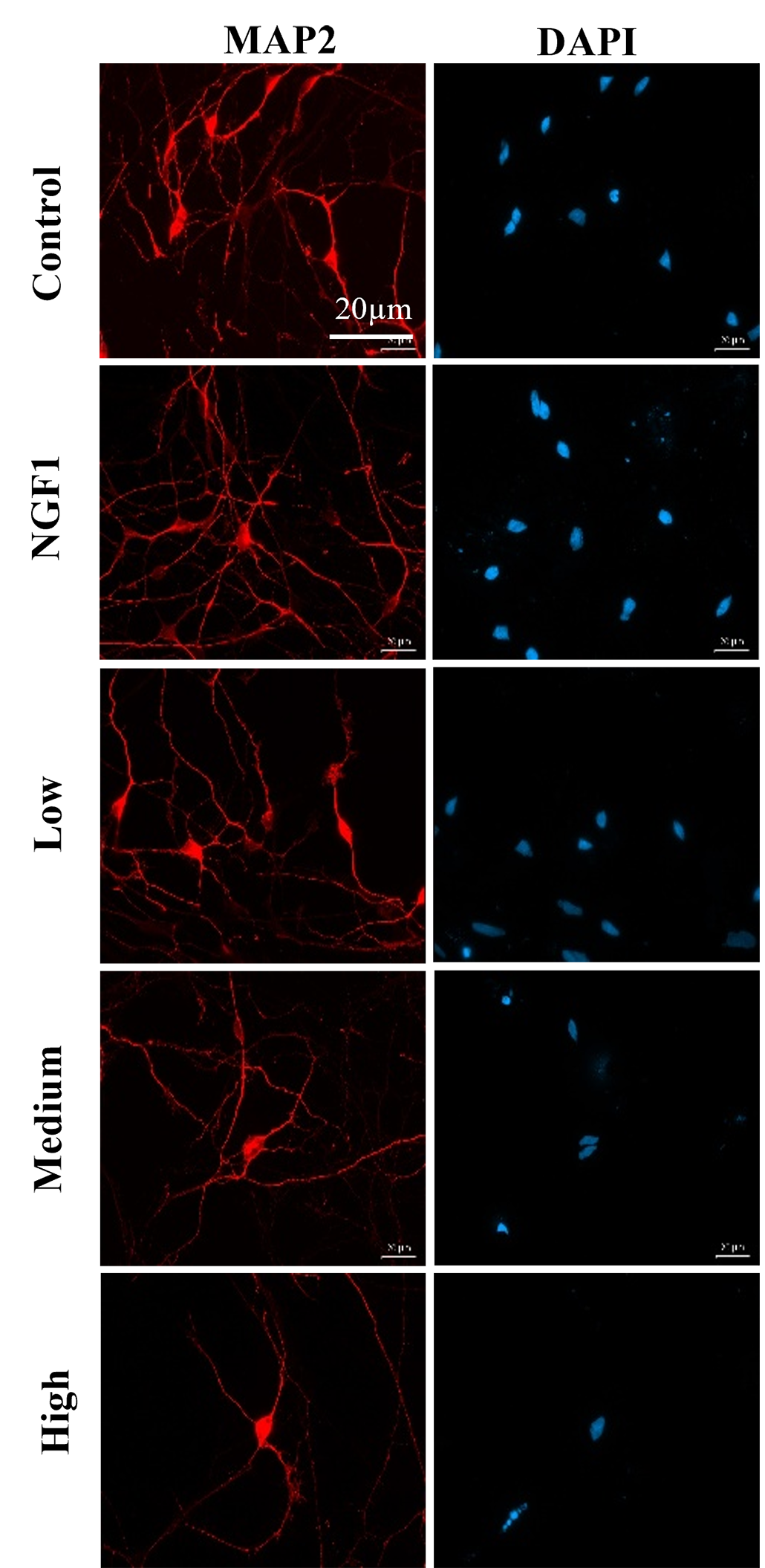
These images show a decrease in rat neuron complexity with increasing caffeine exposure. The “control” group was exposed to no caffeine. “NGF” neurons were exposed to nerve growth factor, which is expected to promote neuron growth. Other neurons were exposed to low, medium and high levels of caffeine. MAP2 is a protein on the surfaces of cells. Red fluorescent dye binds to those proteins to reveal neurons’ shapes and growth. DAPI is a fluorescent dye that highlights the nuclei of cells. Together, these colorful tags reveal that with increasing levels of caffeine, fewer neurons grow and fewer connections form between neurons. S. Zeng

The first thing Sophia did was look at past data on caffeine. She analyzed published studies with the goal of identifying new patterns. This approach is called bioinformatics.

Sophia focused on studies that had looked at how caffeine affected the brains of adult mice. Her analysis suggested that caffeine could alter which genes in a mouse’s DNA were active, or expressed.

Next, Sophia took her work to a lab. She grew rat brain cells in petri dishes for six days, then exposed the cells to different levels of caffeine. Two days later, she compared the neurons under a microscope.

To make the neurons show up clearly, Sophia used fluorescent dyes that bind to the cells. Dyes can highlight important features, such as how the cells are forming connections with one another. Those neuron-to-neuron connections are called synapses.

Dishes exposed to more caffeine contained fewer neurons, Sophia found. What’s more, those neurons showed fewer connections. Caffeine seemed to make neuronal networks simpler and scarcer.

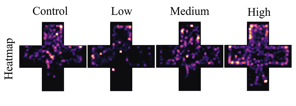
In another set of experiments, Sophia studied young zebrafish that had been exposed to different levels of caffeine. She looked at how far the fish swam and where. More caffeinated fish swam more, she says. They also made erratic choices, spending time in regions of their tank that calm fish typically avoided. Both of these findings hinted that the fish that got more caffeine were more anxious.

But Sophia didn’t stop there. She looked at what changes in the fishes’ brains might have led to this behavior. Sophia measured the activity of four genes involved in brain development. All four were less active in the caffeine-exposed fish, she found.

Takeaways for caffeine drinkers

Interestingly, some genes in the young zebrafish were turned down, or less active, in a way not seen in the neurons from adult rats. So, caffeine may affect the brain differently at different ages, Sophia says. But together, the mouse brain data, the rat neuron experiment and the fish studies all point to the same pattern. Caffeine seems to affect gene activity in neurons. And that in turn may impair brain development and behavior. 

a heat map showing how movement patterns of zebrafish shifted with caffeine exposure
Past caffeine exposure changed the movement patterns of zebrafish. As caffeine exposure increased, fish moved more erratically throughout the four zones of their aquarium. (The bright dots on the heatmaps show where the fish swam.) This evidence points to behavior changes and increased anxiety linked to caffeine.S. Zeng

“I feel that we really don’t talk about caffeine, even though we drink it,” Sophia says. It’s in so many everyday drinks, she adds. Between soda, tea and other beverages, young people may be consuming a lot more of it than they realize.

Sophia suggests greater skepticism about using this drug. People tend to think about caffeine as a helpful tool for focus, she notes, and dismiss potential downsides. But her findings highlight that caffeine comes with trade-offs — and those trade-offs may harm young brains more than we know.

❌