An equation is a mathematical statement that’s written linearly — in a straight line. It has two parts separated by an equal sign (=). And as that sign would suggest, what appears on either side of it has the exact same value.
Math statements weren’t always written this way. Even today, grade-school kids learn to write these statements vertically — with numbers above and below a straight line. That line corresponds to the equal sign. For instance:
One might be tempted to think that equations as we know them are as old as math. But no. Math goes back millennia. The symbols used in equations are relatively recent.
Centuries ago, “math used to be written out” in words, notes British mathematician Robin Wilson. “They call it rhetorical algebra.” A math historian, he authored the 2025 book, Sum Stories: Equations and Their Origins. People used to use regular words to express additions, subtractions and other relationships between numbers and the things they were counting.
Robert Recorde, a 16th-century Welsh mathematician, made equations possible by inventing the “equal to” symbol.Basher Eyre/Wikimedia Commons (CC BY-SA 2.0)
For instance, someone might have written: “Ship one brought two hundred fish to port. Fifty were flounder, seventy-five were bream, twenty-seven were sharks and forty-eight were anchovies.” Now it might be shortened to 50 flounder + 75 bream + 27 sharks + 48 anchovies = 200 fish.
But that shortened version couldn’t have been written before 1557. That’s when the equal sign made its world debut in The Whetstone of Witte. (This math book’s name literally means sharpening your wits). Its author was Welsh mathematician Robert Recorde.
Among many things in this tome, Recorde introduces that equal symbol so he can avoid having to tediously repeat the words “is equal to.” Why choose two horizontal lines of the same length? Because, he argued, no two things can be more equal. (Of course, he said it a bit differently, as he was writing it in a very early version of modern English.)
That equal sign was a relative latecomer among our familiar math symbols. People started using the plus (+) and minus (-) signs around the late 1300s and early 1400s.
The first known definition — and use of “equal to” symbol (14x + 15 = 71) — appears in the 1557 book, The Whetstone of Witte (written in Early Modern English). It includes the phrase: “[T]o avoid the tedious repetition of … ‘is equal to’ I will set … a pair of … [twin] lines of one length, thus: =====, because no 2 things can be more equal.”R. Recorde/Wikimedia Commons (Public Domain)
And the equation at the start of this piece would have made no sense before 1631. That’s when William Oughtred, a 17th-century English math expert, introduced the “x” mark for multiplication (in his book Clavis Mathematicae).
About that equal sign
“The equal sign is quite interesting and is slightly different from an operator like plus, minus, divide or multiply,” says Clemency Montelle. She’s a math historian at the University of Canterbury, New Zealand. The equal sign, she notes, “is not saying calculate anything. But it is indicating a relationship: that one thing on one side is identical [to the other side].”
And that’s true, she notes, “even if they don’t look the same.”
In 4+1 = 5, the two digits on the left-hand side don’t look identical to the digit on the right. Still, they have the exact same value. Montelle finds this quite a “powerful” idea.
“Equations are a way of expressing what you’re doing accurately, precisely and efficiently,” Wilson adds. The “=” signifies a balance — like on a scale — between the quantities on the left and right sides.
The power of balance
Knowing that there’s a balance between the mathematical expressions on either side of an equal sign allows us to solve for something in that expression that may be unknown. This is the basis of algebra, an especially useful branch of math.
Suppose we know that Daniel and Alice picked 9 apples from their backyard tree. We know that Daniel has 5 apples. Given this information, we can calculate how many Alice has. It can be written as this hybrid word problem:
By subtracting 5 from each side, we can calculate that the unknown quantity — the number of apples Alice has — must be 4.
Here the unknown quantity was apples. But it could be anything. Mathematicians generally refer to such an unknown with a letter. Often it’s an “x”.
Earlier in history, some cultures used other approaches. The ancient Indians, for instance, used word abbreviations for various colors to represent some unknown values. Ancient Mesopotamians used different words that meant such things as length, width and area to denote some unknowns.
What is an equation? We learn how its equal sign signals that the two sides of this mathematical expression are like a scale in balance.House of Math/YouTube
Being able to calculate an unknown number makes equations widely useful.
Before equations existed, ancient astronomers painstakingly developed tables of numbers. Think of multiplication tables, with the numbers to be multiplied in the first row and column and the products of those multiplications filling in the rest of the table. This allows anyone to read out the answer for any product quickly.
Similarly, astronomers used tables to track a planet’s motion, observe phases of the moon and predict eclipses. If they wanted to follow a planet’s motion, they’d go down the table to the day and look across to read the number. Such a table could span weeks, months or even years. Creating them took painstaking amounts of mathematical effort.
Equations offer a compact substitute for such tables. Each equation describes the relationship between features of interest, such as a planet’s motion over time. To use it, you input the known values into the equation. For instance, this might be the date and the planet’s position. Then by performing the operations in that equation, you can calculate some unknown value, such as the distance traveled by the planet.
“Equations might seem rather dry,” says Montelle. “But they reveal a hidden structure or numerical pattern that we would otherwise have to write out one by one by one.”
They help engineers design and construct safer buildings. They can let airline pilots calculate how much fuel they’ll need if they know the distance they’re traveling and average speed they’ll be flying. And suppose you have a cake recipe that calls for 2.75 cups of flour. You could use an equation to scale up or down the ingredients needed to make one that’s two and a half times as big or just a mini-cake one-quarter the normal size.
Raindrops may be doing untold damage to cars, bridges, monuments and other outdoor structures. Not all rain, new data show. Just droplets that have first picked up an electric charge. One way they can get that charge: sliding off other surfaces, such as leaves, glass or buildings.
Rain droplets can then transfer their charge to materials that have the right electrical properties. That includes metals. This eventually corrodes, or eats away, at the metals. And even metals treated with protective coatings can corrode, the new data show.
Researchers shared these findings in the September 10 Nature.
Scientists knew water droplets could wear down structures by pounding on them. Rains can also contain small amounts of corrosive compounds, such as acids. But it wasn’t clear whether electrified drops would cause damage, says physicist Zhongyuan Ni. He works at the Max Planck Institute for Polymer Research. It’s in Mainz, Germany.
In early experiments, Ni found no damage to a copper surface after it was pelted with hundreds of water droplets. But one day, he says, “I just let the experiment [keep] running.” He kind of forgot about it as he went “to drink coffee or something.” When he came back, the copper looked different. A co-worker suggested it might have been damaged in some way.
This spurred Ni’s team to investigate further.
Drip, drip, drip
His team continuously released drops of water onto Teflon-coated copper. Copper is more resistant to corrosion than many other metals. And Teflon is an especially strong, protective coating. If the drops started eating into this protected surface, they reasoned, charged raindrops could probably do it to other metals, too.
The researchers gave some drops an electric charge. They did this by having the water slide along leaves or construction materials. The team also tipped uncharged water drops directly onto the Teflon-coated copper.
When viewed under a microscope, all copper surfaces hit with electrically charged drops appeared damaged. The charged water had eaten through the Teflon and begun corroding the copper below. Uncharged water droplets triggered no such damage.
Similar effects appeared on samples of copper and gold covered in polystyrene glass — another protective coating. Clearly, charged water could wear through more than Teflon.
It’s electric
As water droplets slide down common surfaces — such as leaves and glass — they can pick up an electric charge. In a test setup (left), when thousands of these drops landed on Teflon-coated copper, they caused damage that could be seen under a microscope (bottom row; scale bar represents 100 micrometers).
Z. Ni et al./Nature 2026
The new findings could pave the way for better anticorrosion coatings, says Stefan Weber. A physicist at the University of Stuttgart in Germany, he did not take part in the study.
Water drops can become charged in clouds or by sliding along your windshield. But they should not scare you, Ni says. Their effects are small. Still, they could add up when cars, ships, monuments and machinery are exposed to the elements for years.
Small charges shouldn’t be ignored, Ni says. Even if they’re tiny, “sometimes they will play an important role.”
Hydrogels are polymer-based materials that can absorb and release lots of water. These materials are composed of threadlike molecules tangled in a complex mesh. That netting easily snags water, or H2O, molecules.
Some hydrogels are made of natural polymers. Jell-O is a good example. It contains a natural polymer called gelatin. Other hydrogels are made with synthetic polymers — and can be put to work in many different ways.
Some hydrogels can suck moisture out of the air and release it for drinking water. Others can collect H2O from dirty water while leaving pollutants behind. Hydrogels can even be added to soil. When someone waters their crops, those hydrogels soak up the moisture and release it slowly. That keeps water from flowing or evaporating off the soil.
Hydrogels are especially useful for making stuff that must touch living tissue. That’s because hydrogels and living tissue are both mostly water. Hydrogels have been used to make contact lenses and medical implants. Some engineers have also used hydrogels to 3-D print human tissues for transplants. The bioinks used to print these tissues mix live cells with a hydrogel ooze that protects the cells.
Want to know more? We’ve got some stories to get you started:
A new hydrogel could help pull drinking water from the airThe salty gel absorbs more water from the air than similar gels, even in desert climates. This could provide clean water for drinking or farming. (11/3/2023) Readability: 6.8
A new solar-powered gel purifies water in a flashThe unusual, fruit-inspired structure of this material provides quick filtration that could satisfy people’s daily water needs. (4/8/2023) Readability: 6.9
If you’ve got a green thumb, try this experiment from Science Buddies and test how well hydrogels help conserve water in farming. Prefer to take a spa day? Science Buddies has another hydrogel experiment for you — testing the moisturizing power of hydrogel face masks.
For years, scientists have been calculating how much matter should exist in our universe. But more than half of it didn’t appear to be showing up in telescope images. Now an explanation for why is emerging. The “missing” matter is just too diffuse — or spread out — to see. Some of it may not even be made of complete atoms.
“The missing matter exists as a thin gas that’s very spread out,” says Haochen Wang. Wang works at the Massachusetts Institute of Technology in Cambridge. A PhD student in astrophysics, he led the new study. Think of the missing matter, he says, as faint clouds. They’re huge, stretching far beyond the edges of galaxies.
This missing matter is considered ordinary. By that, scientists mean it’s made of everyday particles called baryons. Those include the protons and neutrons that make up stars, planets and people. Ordinary matter is quite different from another type of unseen stuff called “dark matter.” That’s a mysterious substance that doesn’t emit, reflect or absorb light at all. (Scientists only know it’s there because its gravity pulls on other things.)
In some spots, Wang says, the unseen ordinary matter is so spread out that there’s less than one proton or electron in a space about the size of a washing machine. That, he explains, is why finding it has proven “such a hard puzzle to solve.”
Radioing in the solution
To find this diffuse matter, Wang’s group focused on fast radio bursts, or FRBs. Each of these very intense bursts of radio waves lasts about one-thousandth of a second. They flare up somewhere in the far-off universe, then vanish.
As an FRB races toward Earth, it passes through the gas floating between galaxies. That gas, the missing matter, stretches out an FRB’s radio light, Wang explains.
This separates a distant FRB’s light into distinct wavelengths. It’s a bit similar to the way a prism splits white light into a rainbow. When radio waves travel through gas, those with longer wavelengths move more slowly than shorter-wavelength ones. And the difference in their speed grows ever larger as FRBs travel through more matter.
Overall, that change in speed still “is a really small effect,” says astrophysicist Liam Connor. He works at Harvard University in Cambridge, Mass. But “over billions of years of traveling through this … matter, it becomes a detectable effect here on Earth.”
Telescopes pick up a stretched-out signal, where short-wavelength radio waves arrive first, and longer wavelengths trail behind. “The more missing matter the signal goes through,” Connor says, “the more stretched it gets.”
The new findings appear in the July Physical Review Letters.
The upper map shows where fast radio bursts have been spotted across the sky. The lower map shows how galaxies are spread across the sky. The new study found a significant correlation between the two maps. The comparison turned up formerly missing “ordinary” matter in our universe. H. Wang/CHIME/FRB and DESI data
Builds on other work
Connor is part of a different team that’s been searching for the missing matter. Last year, his group measured how much each FRB reaching Earth had stretched. The scientists did this by comparing FRB data to a computer model.
This allowed the team to calculate how much gas the radio waves had passed through. It also pointed to roughly where that gas was. A 2025 study in Nature Astronomy estimated how much missing matter sits between, around and inside galaxies.
Also last year, another team directly detected one specific filament of hot gas. It linked several galaxy clusters and is thought to contain some of the missing ordinary matter. The team, led by Konstantinos Migkas at Leiden University in the Netherlands, used X-rays rather than FRBs to find this gas.
Now Wang’s team has directly measured how far gas typically stretches around galaxies. Unlike Connor’s group, they did this without leaning on a computer model to fill in the picture. They say it’s the first definitive detection of the missing ordinary matter.
Vikram Ravi is hesitant to call it the first. An astronomer, Ravi works at the California Institute of Technology in Pasadena. Connor’s 2025 work, he says, at least hinted the gas was out there — even if no one had directly measured its reach. Ravi also points out that some of the new study’s numbers could be off, if the FRBs’ distances weren’t measured precisely enough.
Still, he considers the latest work “a great signpost of what is to come.”
This image shows a model of how diffuse gas (colored blue, green and yellow) is spread around galaxies (white dots on the image). A new study reports that this gas extends much farther out from galaxies than earlier research had suggested.Illustris/TNG Collaboration
What the new findings show
Wang’s group studied 2,870 FRBs. All had been detected by the CHIME radio telescope in British Columbia, Canada. The team compared these bursts to the known locations of nearly 6 million galaxies. The galaxies had been mapped by the Dark Energy Spectroscopic Instrument, or DESI. It sits atop Manzanita Shrub Mountain in Arizona.
To identify where the missing gas is, the researchers lined up each stretched-out FRB across the galaxy map.
The gas doesn’t cling tightly to galaxies the way gravity alone might suggest. Instead, the diffuse matter puffs outward in thin clouds. Some clouds extend roughly 4 million light-years beyond a galaxy’s edge. That’s much farther than most computer models had predicted.
“Just like fountains that can spout water, there must be activities in galaxies that can push gas out to a large distance, despite gravity’s pull,” Wang says. What might they be? Perhaps jets, he says, that black holes blast out as they’re feeding. Or maybe they’re supernovas, explosions from dying stars. Whatever the sources, they’re exerting more force than scientists had suspected.
Victoria Kaspi is a physicist at McGill University in Montreal, Canada, who worked with Wang on the new study. She thinks their approach to mapping diffuse matter could change how astronomers study the universe. FRBs, she says, “are amazingly effective” in probing how matter is distributed across the cosmos.
Why it matters
Knowing where ordinary matter hides is important. Astronomers map it to understand how galaxies form, evolve and interact with their environment. They also use these matter maps in their search for other unknown phenomena, such as dark energy and dark matter (which is believed to make up the vast majority of our universe).
If the “normal” matter map is wrong, these bigger puzzles get even harder to solve.
The new study is “an exciting and impressive work,” says Connor. “I think it’s a sign of good things to come for the application of FRBs to mapping the cosmic baryons.”
New radio telescopes are also being built. They should be able to pinpoint FRBs far more precisely. In the coming years, these telescopes could boost the number of recorded FRBs 100 times or more.
Ravi’s team, among others, is already preparing to use larger FRB catalogs in hopes of making even sharper maps of cosmic gas.
“The more FRBs we find,” Wang says, “the more accurate our answers on the missing matter will become. That includes the powerful galactic forces shaping it.”
Blood Falls is a five-story waterfall that flows out of Taylor Glacier in Antarctica. The water comes from a salty lake beneath the glacier that’s rich in iron. When the water reaches the glacier’s surface, the iron interacts with oxygen in the air. This reaction turns the water red. It’s the same reaction that creates reddish rust on a nail.
Scientists suspect the lake was filled by ocean water 5 million years ago. But it’s been completely cut off from other water sources for at least a million years. Over time, the water became saltier as the glacier formed above. Now, the lake is more than twice as salty as the ocean. The water can’t freeze because salt prevents water molecules from locking into the crystal structure of ice.
Because the lake is also cut off from the atmosphere, scientists call it a biological time capsule. Microbes have lived here for millions of years with no light or oxygen. Instead, these extremophiles get their energy from molecules called sulfates. This respiration strategy is similar to microbes that live in deep-sea thermal vents.
But the Blood Falls microbes are special. They extract oxygen from the sulfates and leave behind other molecules. When these leftovers interact with iron in the water, they turn back into sulfates. This means these microbes have a practically unlimited energy source.
Scientists suspect planets like Mars and Jupiter’s icy moons could be home to salty underground lakes like this one. How these microbes make a living might be a clue for how life could work on other planets, too.
In a sentence
Blood Falls is located in East Antarctica, more than 3,300 kilometers (2,000 miles) south of New Zealand.
California’s Death Valley is one of the driest, hottest and harshest places on Earth. In summer, thermometers there often climb above 49° Celsius (120.2° Fahrenheit). Such extreme heat can make it hard for desert dwellers to survive. But one shrub has found a curious way not only to survive Death Valley, but also to thrive in its blistering heat.
The Arizona honeysweet essentially sweats.
“This plant has evolved a way to survive temperatures that would kill most other plants and animals,” says botanist Seung Yon Rhee. This plant scientist at Michigan State University (MSU), in East Lansing, worked on the new study. Even on scorching days, this plant grows fast. It can add about one-third to its weight every day.
This “in itself is an amazing biological phenomenon,” says Joanna Feehan. She’s another MSU plant biologist who worked on the study. The team shared its new findings August 3 in bioRxiv. Studies posted there have not yet been peer reviewed by other scientists.
Here’s a closeup of leaves on the Arizona honeysweet (Tidestromia oblongifolia). In high heat, structures in the leaves morph in shape, allowing this plant to boost its growth.Karine Prado
In-leaf air conditioning?
The ability of the Arizona honeysweet (Tidestromia oblongifolia) to endure extreme heat has long fascinated scientists. It performs photosynthesis best at a blazing 47 °C (117 °F). That’s the highest peak-performance temperature for any plant, says Karine Prado, a plant biologist at MSU. “These plants wait [for] the hottest month just to grow fast,” she told Science News.
But to do that, its leaves have to keep from baking into a crisp. This year, Rhee says, her team set out to ask a simple question: “How can a plant survive in one of the hottest places on Earth?”
Scientists knew this honeysweet can cool itself by releasing water vapor. Vapor exits through stomata — tiny pores on its leaves and stems. Called transpiration, this process is like sweating, Feehan says. It was never clear, though, how big a role this method of cooling played in helping the plant survive Death Valley’s heat.
Karine Prado examines T. oblongifolia in Death Valley, Calif.Jennifer Johnson
To find out, the MSU team collected seeds from 223 wild honeysweets growing in and near Death Valley. Then they grew more than 1,200 seedlings in the lab at a comfortable 30 °C (86 °F). Over several more days, the researchers gradually turned up the heat. They maxed out at 60 °C (140 °F) for six to eight hours daily. (Scientists believe that 60 °C is the hottest temperature at which complex life, such as plants or animals, can survive.)
At each step, the team measured how hot the plants got. Once the number of surviving and non-surviving plants did not change, the experiment ended.
Some plants survived repeated exposures to high temperatures. And they didn’t simply tolerate the heat. They actively cooled their leaves through transpiration. Some leaves were able to stay roughly 13 degrees C (23 degrees F) cooler than the surrounding air!
When the team experimentally closed the leaves’ stomata, shutting down this cooling, the plants didn’t live as long.
Clearly, Rhee concludes, “This cooling makes the difference between life and death.”
This is “direct evidence that leaf cooling contributes to survival under extreme heat,” says Wataru Yamori. He is a plant biologist at the University of Tokyo in Japan who did not take part in this work.
However, not all the honeysweets survived.
Some died after only a few days. Others remained healthy for weeks. These differences may trace to their DNA, Rhee suspects. It’s like “how some of us have brown eyes whereas others have blue eyes,” she says. Some of the plants, she explains, carry versions of genes that appear to more effectively help them cool off and survive extreme heat.
The new study did not pinpoint which genes bring better cooling relief, notes Rubén Rellán Álvarez. He studies plants at North Carolina State University in Raleigh and did not take part in the new study. Identifying those genes could help uncover “the secrets of this extreme adaptation,” he says.
That could have out-of-this world payoffs, says Rhee. To grow plants on the moon or Mars, she says “we’ll need to learn from the toughest plants on our own planet first.” The plants in Death Valley — where filmmakers have shot scenes set on desert planets in Star Wars and other movies — might just be the perfect teachers.
One limit in this study was water availability, Yamori says. The experiment was done without water restrictions. Opening the stomata can cool the leaf, but it also increases water loss. If stomata are closed, not only is the plant’s intake of carbon dioxide halted, but its cooling through transpiration is also turned off.
So the plant must balance cooling and CO2 intake as it conserves water, says Feehan. Further work, she says, will need to explore how it does this in nature.
Editor’s note: This story was updated on Oct. 19, 2026, to remove an incorrect mention that Karine Prado was part of the team that collected honeysweet seeds.
Jonathan Growcott’s “happy place” is bouncing across the African savanna in a 4×4 vehicle. As a large-carnivore biologist, he tracks predators in the wild. It’s tough work; these animals can be hard to find.
Common methods often involve driving for hours in search of footprints or droppings. Some researchers set up camera traps in hopes of snapping photos of passing animals.
Growcott felt these still missed something. Maybe scientists could do more than just look for signs of critters. Perhaps they could listen for them, too.
“Species like lions, leopards and hyenas produce some of the most recognizable sounds in the animal kingdom,” notes Growcott. What’s more: “These sounds travel many kilometers [miles].”
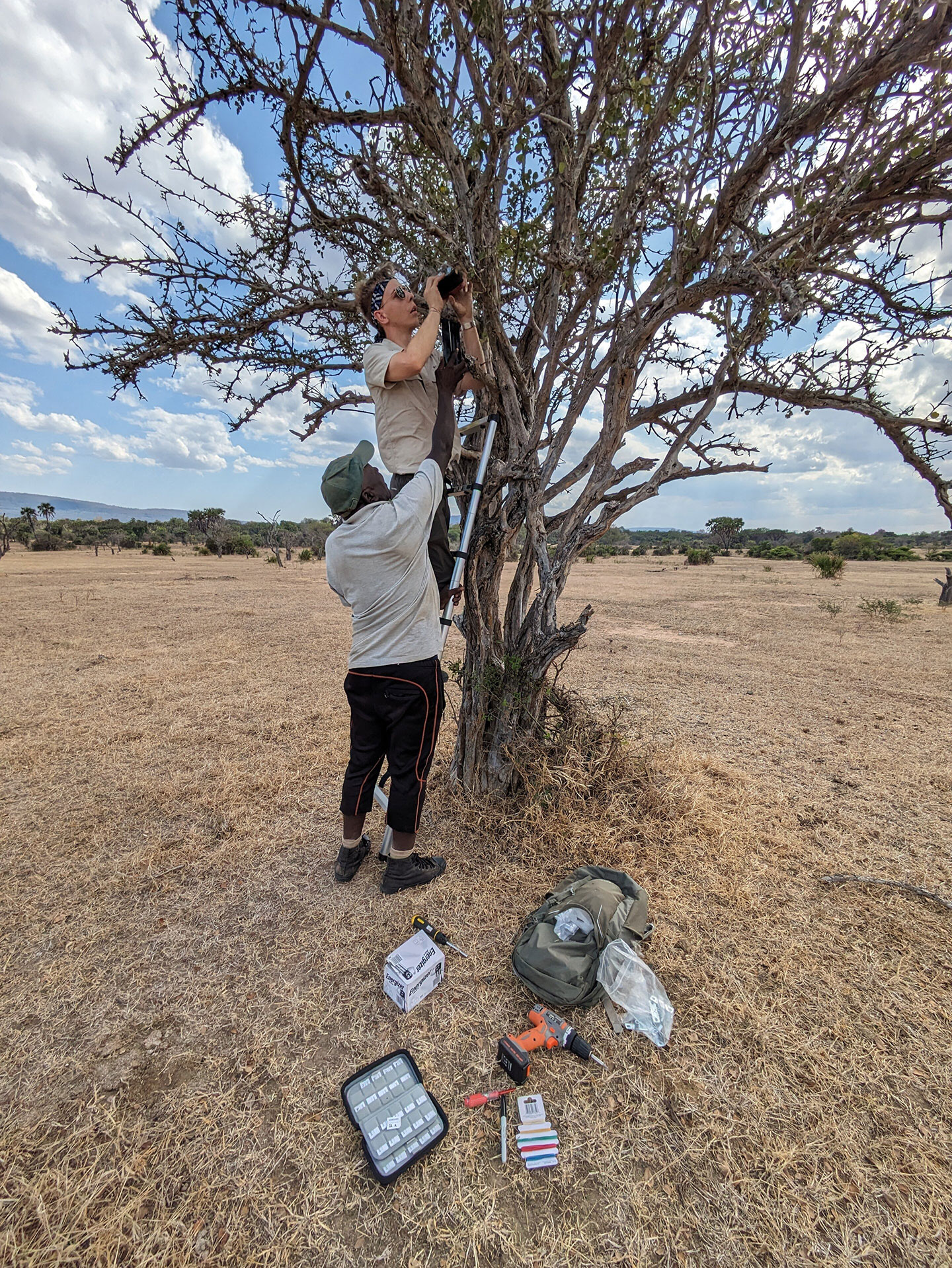
Jonathan Growcott and colleagues set up 50 pairs of camera traps and microphones in East Africa in Nyerere National Park. Combining these two technologies allowed the team to pair photographs with unique roars to identify individual leopards.Charlotte Searle
And these critters are far from the only sound-makers. Birds, mammals and insects are, too. So are reptiles, amphibians, fish — even crustaceans. Some call out to find mates or defend territories. Others use calls to scare off predators. Sounds help many creatures sense their environment and check in with each other. This cacophony of chirps, squeaks, rattles and roars can aid scientists, too — if they know what to listen for.
Across the globe, more and more researchers are studying how living things make, transmit and receive sounds. Audio recorders can be strapped to a tree or set on the ocean floor. Once placed, the devices capture these bioacoustic data without disturbing wildlife. This can make them especially useful in conservation efforts.
By eavesdropping on neighborhood chatter, researchers can find out which species are out there and what they’re up to. They can estimate the size of populations to keep tabs on how critters are doing. Knowing where animals are can also help limit their conflict with people. For all these reasons, bioacoustics can be a powerful tool in the fight to save endangered species.
Hear me roar!
Growcott ran the first large-scale bioacoustics survey of African carnivores. At the time, he was a PhD student at the University of Exeter in England. He and his team worked in East Africa at the Nyerere National Park in Tanzania. They deployed 50 acoustic recorders across 450 square kilometers (175 square miles).
He focused on leopards first. These solitary cats are secretive and nocturnal. Such habits make monitoring their numbers challenging. So Growcott worked to identify the animals by sound.
Leopards
Leopards produce deep “sawing” roars that resemble the sound of cutting wood. Jonathan Growcott and colleagues found that each leopard’s roar was unique. So “you can identify leopards not only by their spots, but also by their roars,” Growcott reports.
J. Growcott et al. (2024)
He set up acoustic recorders next to camera traps. This allowed him to match individual leopards to their roars. With these data, a computer model was able to distinguish between leopards based on their roars with 93 percent accuracy. This revealed that each leopard’s roar is unique enough to identify it.
Could the same be true for other big cats?
When Growcott turned to lions, he noticed patterns. Some of their roars were loud, long and complex. Others sounded shorter and simpler. He wondered whether lions used different types of roars.
Using artificial intelligence (AI), he analyzed those roars. It showed lions produce two distinct types. There’s the familiar, full-throated roar you may have heard at a zoo or in a movie. Then there’s a shorter, lower-pitched one. He called this previously unknown one an “intermediary” roar.
But what Growcott really wanted was to identify individual lions by their roars. For this, he created an AI tool that he calls “Shazam for lions.” It analyzes just the full-throated roars — because they contain the most sound information. AI, he found, could identify a specific lion by its roar with more than 95 percent accuracy. That’s better than human experts can do!
Lions
Lions emit different types of calls within roaring bouts. Full-throated roars are loudest and have the most detailed vocalization. They can be used to identify individual lions. These big cats also produce a previously unclassified vocalization, dubbed the intermediary roar, new research reveals.
J. Growcott et al. (2024)
Linking cats to their roars could help researchers count and track the big cats. That, in turn, could help them gauge the size of their populations while also monitoring the range of individual animals. Conservationists and others need such data to protect wildlife and people.
This work highlights how much we have to learn about even well-known sounds, such as a lion’s roar, Growcott notes.
“At some point, we may better understand why animals make the sounds they do,” he adds. “Trying to work out what animals mean when they make noises is something that has fascinated people for a long time. I think it will continue to fascinate us for a long time to come.”’
Sounds of the forest
Sound is especially useful when animals are rare or hard to see.
The last 74 Cao-vit gibbons on Earth live in a remote part of Southeast Asia. They’re in a patch of mountainous rainforest in northern Vietnam. Their species was thought to be extinct until 2002. That’s when scientists discovered this small population. These critically endangered apes spend their lives in treetops. That makes them almost impossible to observe by eye.
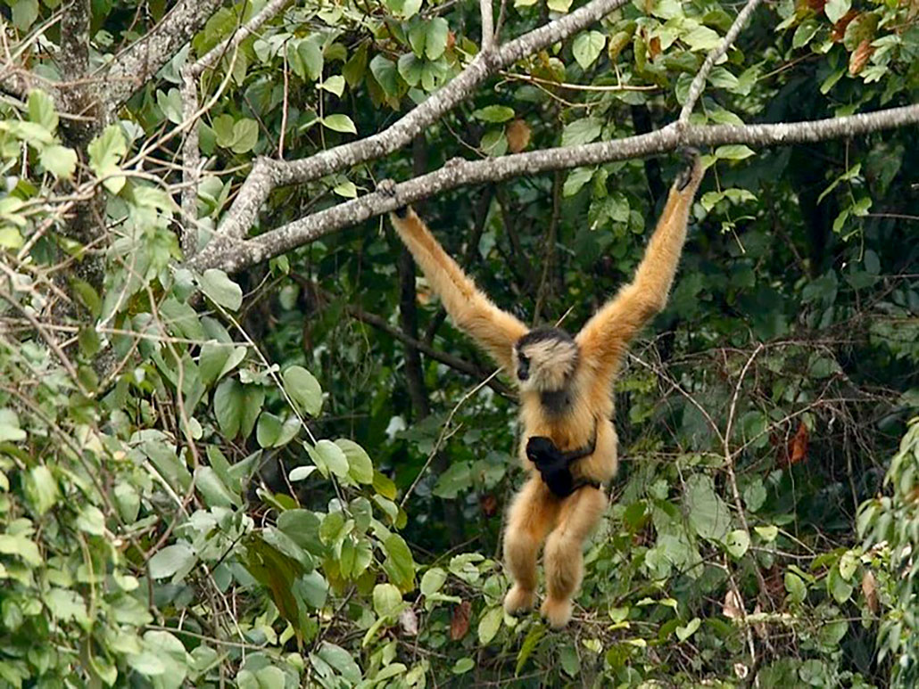
Cao-vit gibbons spend their lives high in treetops within only one tiny patch of a remote Vietnam jungle. This female gibbon is seen carrying a one-month-old baby.Bioacoustics Research Group
Fortunately, Cao-vit gibbons like to make themselves heard. Mated pairs sing loud, melodic duets. They’re music to Angela Dassow’s ears.
“It is a privilege to hear one of the rarest primates on the planet giving their morning duet calls,” she says. These “are kind of eerie,” she notes, “but also beautiful to wake up to.”
Cao-vit gibbons
Fauna & Flora, a conservation group, recorded Cao-vit gibbon calls in northern Vietnam. This clip features two male gibbons singing over each other and then a female calling at the end.Fauna & Flora Vietnam
Dassow is a biologist at Carthage College in Kenosha, Wisc. For the past five years, she’s studied Cao-vit gibbons with partners in Vietnam. To locate the animals, the team uses specially designed acoustic recorders. Dassow places around 20 of them throughout a region. That way, a single gibbon call reaches different recorders at slightly different times. By comparing those times, computers can pinpoint where the sound came from.
“Our recording units don’t just tell us if an animal is present,” says Dassow. “They give us a GPS location of where that animal is, without putting a radio collar on it.”
Dassow tracks gibbon movements by those calls and monitors their population. This soundscape also tells her about the social lives of these apes. Her team shares what it learns with local rangers, who use the info to help protect gibbons and manage their forest home.
Angela Dassow and colleagues filmed a documentary about their efforts to reduce conflict between people and tigers in Nepal. This clip shows the impact that their acoustic tiger-detection system has on the local community.
Dassow is using bioacoustics in another Asian forest to lessen conflicts between people and Bengal tigers.
Over the past 10 years, conservation programs have increased the number of these tigers in Nepal. That’s great news for this endangered species. But it also has led to more encounters between people and these top predators. This is especially true in rural areas, where the lives of people depend on the forest around them.
To help people coexist with the big cats, Dassow developed an acoustic alarm system. It alerts villagers to the presence of approaching tigers.
For this, she had to get a bit creative. “Tigers tend to be quiet. Very rarely do you hear them vocalize,” Dassow notes. “But there are a lot of chital deer in the area, and they are a prey source for tigers. As you might expect, these deer are excellent at detecting when a tiger is near.”
Chital deer are important prey of Bengal tigers in India and Nepal. They make a distinctive alarm call when they spot a tiger or other predator. It sounds like the bark of a small dog or a sharp coughing.Sovietonion321/Wikimedia Commons (CC BY 4.0)
When chital (CHEE-tul) deer notice a tiger, they start barking out an alarm call. Recorders in the forest detect these and alert local villagers. This system warns people to nearing tigers in real time. Villagers can then take extra precautions. They might avoid areas where a tiger has been spotted or travel in groups when moving through the forest.
Researchers have long listened to sounds made by aquatic animals. Those sounds may now help people share the ocean safely with them.
Ana Širović is a marine ecologist at the Norwegian University of Science and Technology in Trondheim. She’s part of the international team behind Whale Safe. It’s a powerful tool for preventing ships from colliding with endangered whales.
Ship collisions often kill large whales. Research shows that reducing a ship’s speed is one way to limit this. But ships need to know when and where to slow down to avoid whale strikes.
San Francisco Bay is near several major shipping ports. It’s also an important feeding ground and migratory route for whales. Scientists estimate that more than 80 blue, humpback and fin whales are killed each year in ship collisions along the U.S. west coast.Bekah Lane/The Marine Mammal Center
That’s where Whale Safe comes in. It maps in nearly real time the locations of large cargo ships as well as blue, fin and humpback whales. To do this, Whale Safe uses data from three sources. The first is a system that monitors whale calls. The second is a mobile app through which community scientists record whale sightings. The third is a model that monitors certain ocean conditions.
What boings below
For her research, Ana Širović leaves acoustic recorders on the ocean floor for long periods. Sometimes, they pick up sounds she cannot identify. This is her favorite recording of an unknown sound. Širović recorded these “mystery boings” at a reef about 80 meters (260 feet) off the coast of southern California.
Data from all these sources are combined to rate how likely it is that whales are near. Ships can use these ratings to understand where they should slow down.
Whale Safe was first used in California’s Santa Barbara Channel. This is near several major shipping ports. It’s also an important feeding ground and migratory route for large whales. Whale Safe has since expanded to San Francisco Bay. The second busiest port in California, it’s another hot spot for whale strikes.
Each year, collisions with large ships kill dozens of whales in these regions. Even a small reduction in speed saves lives. A shipping vessel that slows down from 15 knots to 10 knots cuts in half its risk of striking a whale.
Part of what makes her job fun, Širović says, is listening to parts of the ocean no human has ever heard before. When she leaves acoustic recorders on the sea floor, they often pick up unexpected or unknown sounds.
“The ocean is a vast portion of the planet, but there is still so much of it that’s not explored,” she says. “I enjoy learning new information and figuring out what to do with it. My job is part adventure, part puzzle solving and part new discoveries.”
Scientists from Woods Hole Oceanographic Institution in Massachusetts and The Norwegian University of Science and Technology in northern Europe deploy an acoustic buoy in the Santa Barbara Channel. The buoy contains an underwater microphone and a tiny computer to detect the calls of whales. Benioff Ocean Science Laboratory
Sound science
Acoustic recordings can capture a lot of data. Analyzing these used to be slow and difficult. Researchers had to listen carefully for certain sounds.
Today’s acoustic recorders can be left unattended for weeks, months or even years. They can collect and store huge amounts of data — sometimes, more than someone could listen to in their entire lifetime! What’s a researcher to do?
Seeing sound
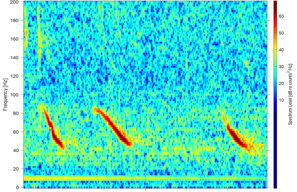
Scientists often transform audio files into images called spectrograms. This spectrogram shows blue whale calls. Horizontal axis shows time; vertical axis shows frequency. Low frequencies (low-pitched sounds) are at the bottom, high frequencies at the top. Colors represent energy or volume at a given time and frequency. Reds and oranges indicate a more intense signal; blues and greens indicate silence.J. H. Rasmussen
Over the past decade, AI technology has evolved and transformed how scientists analyze recorded sounds. Now, AI algorithms can do much of the work. They can sort through recordings to identify sounds. And they process data more quickly and accurately than humans can.
Jeppe Have Rasmussen is a biologist at the University of Copenhagen in Denmark. He specializes in applying AI to animal bioacoustics. AI algorithms are usually not trained to analyze sounds themselves, he points out. Instead, the sound data must first be transformed into some visual form.
“AI is really good at detecting and classifying objects in images,” he says. “So, we convert the recorded sound file into a spectrogram.” This is more like an image, Rasmussen explains. It shows sound frequency on a vertical axis; time is shown on the horizontal axis.
AI models, he says, can then be trained on these spectrograms to identify the calls of specific species.
Rasmussen is using this approach to learn more about endangered species.
Saving the Hawaiian monk seals
Colleen Reichmuth
Hawaiian monk seals are one of the most endangered marine mammals in the world. They live in the warm, subtropical waters around the Hawaiian Islands and spend about two-thirds of their time at sea.
“There are only 1,600 Hawaiian monk seals left in the entire world,” he says. “If we can track where they are, we can better determine where to focus on protecting them. Listening for their underwater calls is a very good way.”
Recently, a team of researchers identified six types of underwater calls made by these seals. They include moans, croaks and whoops. Rasmussen is now using AI to automatically detect these calls. His team hopes to figure out where the monk seals spend their time. Then, conservationists can prioritize those sites for protection.
Seal sounds
Hawaiian monk seals make a host of different sounds underwater. These include croaks, groans, growls, moans, rumbles and whoops. Scientists are using AI to monitor the ocean for these calls. This helps them decide where to focus efforts to protect these endangered marine mammals.
J.M. Sills et al. Endangered Species Research 2021
Rasmussen also is exploring other ways to use AI in bioacoustics. For instance, it can help evaluate the welfare of captive animals. In one recent study, his tools could tell whether a pig sound came from a stressed animal or a happy one.
“The combination of bioacoustics and AI is one window we can use to look into the minds of animals,” he says.
Such techniques, Rasmussen says, will help science “discover that there is much more information hidden in animal communication.”
Bees may be best known for buzzing through the air. But they are surprisingly good swimmers, too. The insects can paddle through the water using the bottoms of their wings. And a new study shows that waterborne bees prefer to swim toward dark areas. This strategy might save them from drowning.
Zach Huang and others studied this behavior in honeybees and mason bees. Huang is a bee researcher at Michigan State University in East Lansing. The team shared its findings February 10 in Communications Biology.
AI today can write our class essays, plan our meals and draft our emails. But not everyone agrees on whether this type of help is good or bad. Recent headlines have warned that artificial intelligence will not just make us lazy and stupid; they’ve suggested AI could lead to the downfall of civilization. But the truth may lie somewhere in the middle of those best- and worst-case scenarios, says Trent Cash. He’s a behavioral scientist at University of Waterloo in Canada.
AI will most likely weaken our skills when it simply does the work for us. “If we offload a specific skill to AI,” Cash says, “we’re probably not going to retain that skill particularly well.” Staying engaged while using AI — by learning from its feedback or examples — may, however, enhance our skills.
Fears of AI whittling away human intelligence like some sort of digital pocketknife are likely overblown, Cash says. He dismisses the idea that AI will cause people to completely forget how to think.
“Humans are resilient,” he points out. “I don’t think that [AI is] going to suddenly turn our brains to mush.” But staying sharp will take effort on our part.
Take one 2025 study of doctors who’d been trained to spot growths — called polyps — on patients’ colons. Doctors got an AI tool’s help spotting polyps for three months. Later, when the doctors returned to working alone, they didn’t detect polyps as well. This suggests the doctors “kind of forgot what to look for,” Cash says.
The same pattern plays out with other skills, such as learning a new math concept. One 2025 study looked at high-school students using an AI tool like ChatGPT to solve practice problems. The AI seemed to help. But take the tool away, and those students did worse than ones who never used it.
Something similar can happen with reading comprehension. Consider people who used an AI tool to solve practice SAT questions. Those people had a hard time coming up with correct answers when the AI was taken away, a new study finds. After losing their AI helper, people were also more likely to skip questions.
More than half of U.S. teens use chatbots for homework. Treating AI like a tutor may help them pick up new skills. But turning to AI to simply churn out answers is unlikely to result in real learning. Thai Liang Lim/iStock/Getty Images Plus
That was “particularly concerning,” the study’s authors conclude, because persistence is key for learning. These researchers shared the findings at the 2026 Conference on Language Modeling in San Francisco, Calif., on October 8.
All these results suggest people can struggle once they let AI take over their tasks.
For Cash, the lesson is “use it or lose it” — where “it” is your own brainpower. Don’t let AI do all your thinking for you, he says. “If you’re not engaging with the cognitive work, you’re not going to learn the skill.”
AI can help when it keeps people thinking
Using AI doesn’t have to mean your skills will crumble. You can put in the effort needed to learn even as you’re getting digital help, says Alp Sungu. The key is using AI tools that offer guidance or examples, rather than spitting out answers.
Sungu is an economist at the University of Pennsylvania in Philadelphia. He worked on the study of high-school students using AI for math. In that study, his team designed an AI tool with some limits. It worked like ChatGPT but didn’t give away answers. Instead, it offered hints and encouragement. That approach helped students learn as well as those who just studied with books.
Using AI as a tutor is different from using it as an answer machine, Sungu says. Students still had to put thought into their studying. The struggle of working through a new concept is where real learning happens, he says. “When you’re solving a problem, you need to get your hands dirty.”
For those willing to put in some effort, AI can be a good teacher, says Benjamin Lira Luttges. This behavioral scientist also works at the University of Pennsylvania.
An AI tool can help people learn how to edit cover letters, his research has shown. After practicing with either an AI tool or human feedback, people edited a poorly written cover letter without any help. Then, human judges picked which letters would likely land a job interview. Letters from both groups were equally likely to get picked, Lira Luttges’ group reports. The team shared these results on arxiv.org in February. (Studies posted there have not yet been vetted by other scientists.)
The AI tool in that study had taught by rewriting an entire cover letter itself. That may sound like its students weren’t doing much thinking. But it’s not what the team found. People didn’t just accept the AI-revised draft. They sat with it for a while, tinkering with the language. “They were still working,” Lira Luttges says.
AI can help us learn new skills when it offers guidance rather than simply spitting out answers.Namthip Muanthongthae/Moment/Getty Images Plus; adapted by L. Steenblik Hwang
This hints that AI can teach by example — “if you have people who are motivated to learn,” he says. In short, AI systems that keep people’s brains engaged might be able to sharpen some of our skills.
Still, we should be careful in choosing what thinking we outsource to computers, experts say. If there’s a skill you value, don’t give it over to AI, Cash advises. “We know skills come from practice and are maintained through practice.”
Lira Luttges agrees. People should think about what skills they want to protect, he says. That might be coming up with the perfect way to phrase a sentence. Or it may be devising a tasty dish using items in your pantry.
“You shouldn’t delegate the things that you enjoy and that give you meaning,” he says. “Outsource the crap, not the craft.”
Each year, about this time, the Karolinska Institute and Royal Swedish Academy of Sciences — both in Stockholm, Sweden — announce three world-changing achievements. People who pioneered the work in these fields will receive the most coveted science awards on Earth: the Nobels.
The first of the 2026 prizes was awarded for finding a light switch for the brain. The second goes for developing a literally super-cool way to study elusive particles bombarding Earth from outside our solar system. And the last prize highlights the importance of being able to preferentially make certain “handed” molecules.
All three achievements opened up major new windows on — and opportunities in — biology, physics and chemistry.
Let’s start with the human brain. It hosts nearly 90 billion neurons. They talk amongst each other through some 100 trillion connections. This brain chatter allows our minds to learn, adapt and interact with the world around us.
The 2026 Nobel Prize in physiology or medicine went to Karl Deisseroth, Peter Hegemann and Georg Nagel for developing ways to control nerve cells with light. The technique, called optogenetics, opened new ways to study the brain by turning individual neurons on and off. #nobel#optogenetics#sciencenewsNikk Ogasa Getty Images/Alexander Pytskiy/JONATHAN NACKSTRAND/JOHN SAEKI,LAURENCE CHU/VALENTINA BRESCHI,JONATHAN WALTER,SYLVIE HUSSON,OLIVIER THIBAULT
To find out how, brain scientists needed a special tool. They long “dreamed” of something that might let them “turn specific neurons on or off with great precision,” notes Abdel El Manira. He spoke on Oct. 5, describing the prize for this tool.
The path to the prize started in an unlikely place: the “eye spot” of a single-celled green alga.
In the early 2000s, biophysicists in Germany discovered a unique protein in that alga’s “eye” spot. The two researchers were Peter Hegemann (who now works at Humboldt University in Berlin, Germany) and Georg Nagel (at the Julius-Maximilians-Universität Würzburg in Germany). The protein that caught their interest had a long name: channelrhodopsin (CHAA-nel-roh-DOP-sin).
When light hits it, the protein produces an electrical impulse. This triggered the alga to move. Later research would show light caused that protein to open like a pore. That let positively charged ions flow into the cell. They are what caused the electrical impulse.
Hegemann and Nagel “had just discovered the switch neuroscientists had long dreamed of,” says El Manira.
Building on the that work, Karl Deisseroth tried using that protein to see if it could turn on brain cells. A neuroscientist, he works at Stanford University in California.
He was part of a team that inserted the gene to make channelrhodopsin into rat nerve cells. These were growing in dishes in the lab. Shining light on the cells triggered an immediate reaction.
This team also put the gene into nerve cells within the brains of living mice. The researchers inserted optical fibers at the same spots, right through the rodents’ skulls. By shining blue light through the fibers, the researchers could now control the movements of the rodents’ whiskers.
Researchers later went on to test the links between other brain circuits and behaviors.
In short order, optogenetics “transformed neuroscience,” El Manira says. It offered a window on the workings of healthy brains, he says. It helped show how brain circuits can be disrupted, too, he adds. Those findings, he notes, could help researchers treat conditions such as blindness, depression and dementia.
In fact, we didn’t have to wait long for news in one of those areas. Just two days after this Nobel was announced, other researchers described a light therapy for retinitis pigmentosa (Reh-tih-NY-tis Pig-men-TOW-sah). This blinding disease slowly kills off light-sensing cells in the eye’s retina. Optogenetics, the new report showed, helped improve light sensitivity in seven of 10 treated people.
Neutrinos have no electric charge. They can pass through objects without interacting. In fact, billions of these ghostly particles pass through each of us every second.
Fusion reactions in our sun form most of them. But some had been thought to come from much farther away — in high-energy particle collisions too far off to observe.
Detecting such neutrinos from well beyond our solar system had long been a goal of scientists. Because neutrinos are not deflected by magnetic fields and rarely interact with matter, detecting their path could point back to their distant source. Scientists hoped to one day figure out where and how the universe accelerated these super-small particles to super-high energies.
In the 1980s, physicist Francis Halzen of the University of Wisconsin–Madison had an insight: The South Pole should be an ideal place to look for the flashes of light created by the rare events when neutrinos collide with atomic nuclei. Ice deep within the Antarctic ice sheet could make a large, dark, transparent stage for such a neutrino light show.
Francis Halzen has won the 2026 Nobel Prize in physics for his vision and scientific leadership of the IceCube Neutrino Observatory. IceCube gave us an entirely new way to see the universe. #nobelprize#physics#sciencenews Nikk Ogasa Getty Images/MARCO BERTORELLO/NSF/Martin Wolf & Sven Lidstrom, IceCube
In part, Halzen got this year’s physics Nobel “for his decisive contributions to [IceCube].” notes physicist Mark Pearce. But Halzen also won for “the discovery of high-energy neutrinos of astrophysical origin,” he adds. Pearce, who chairs the Nobel committee for physics, spoke during the Oct. 6 award announcement by the Royal Swedish Academy of Sciences.
IceCube had been a gamble. “Nobody knew whether a kilometer-field detector was actually large enough to discover neutrinos beyond our atmosphere from the universe,” Halzen said in a phone call during the announcement ceremony.
In fact, he noted, “It only took us two years to detect them.”
This year isn’t the first time a Nobel has gone for work on neutrinos. Two physicists received the prize in 2015 for demonstrating that neutrinos have mass.
In nature, the building blocks of life exhibit a striking pattern. Many can exist in two mirror-image structures, much like your right and left hand. Such molecules are said to be chiral (KY-rul). However, in nature, such chiral molecules overwhelmingly occur in just one form. Amino acids, for instance, are nearly always left-handed.
Known as homochirality, this dominance of one type of handedness had long puzzled scientists.
When chemists first attempted to make chiral molecules in the lab, they ended up with roughly 50-50 mixes of right- and left-handed molecules. Why?
“It is this puzzle that this year’s laureates have solved,” said Heiner Linke. He spoke during the Oct. 7 announcement of the chemistry prize by the Royal Swedish Academy of Sciences.
Two scientists will split this Nobel prize. Henri Kagan works at the Université Paris-Saclay in France. Kenso Soai works at Tokyo University of Science in Japan.
Henri Kagan and Kenso Soai won the 2026 Nobel Prize in chemistry for their work on controlling the production of mirror-image molecules. Their discoveries have helped pharmaceutical companies develop safer drugs. #nobel#chemistry#sciencenews Nikk Ogasa Getty Images/Evening Standard/Paul Fievez/Keystone/JONATHAN NACKSTRAND
The award winners’ research has greatly aided development of medicines. A lot of active drug molecules are chiral. But while one form may have a medicinal effect, its mirror-image form might have no effect — or worse, be harmful.
Consider thalidomide (Thuh-LID-oh-myde). Thousands of babies were born in the 1950s with severe birth defects. Their mothers had been prescribed thalidomide while they were pregnant to treat anxiety and nausea. Science would later show that the left-handed form of the drug was toxic. (Even taking the “safe,” right-handed form wouldn’t solve the problem. In the body, it could morph into the left-handed form.)
In 1986, Kagan’s team was the first to report chemical reactions that would lead to production techniques that helped solve the problem. These could make a far larger than normal share of left- or right-handed forms.
Several years later, Soai and his colleagues reported that they had figured out how to make a chiral catalyst that could, in turn, be used to form itself. That allowed his team to perform reactions that resulted in products made almost entirely of one “handed” form of a chiral molecule.
“This is not the final answer,” Soai said of this work. He had called in from Japan to the Nobel announcement ceremony. Speaking to future generations, Soai said, “the study of chirality should be more and more developed.”
The new Nobel scientists will each get a medal. The winners in each category will also split 12 million Swedish kroner (roughly $1.2 million).
Theropods are a group of dinosaurs. Their name means “beast-footed.” The group includes many extinctspecies, such as Tyrannosaurus rex and Velociraptor. It also includes modern birds.
Theropods first emerged about 230 million years ago. Early theropod species appear to have been mostly carnivorous. Over time, however, the group evolved into a diverse range of creatures. These included herbivores and omnivores. All non-bird theropods died out with the rest of the dinosaurs about 66 million years ago.
Theropods share certain characteristics, such as two legs, three forward-facing toes and hollow bones. Light, hollow bones allowed some members of the group to evolve flight.
Many extinct theropods sported feathers, scales or a mix of both. Yutyrannus huali was a dinosaur whose name means “feathered tyrant.” This species lived about 125 million years ago. It was one of the largest feathered species that ever lived. The oldest fossil of this species is about 10 meters (33 feet) long. These fossils show clear imprints of long feathers, which may have helped keep Y. huali warm.
All modern birds — from hummingbirds to emus to hawks — are theropods. Scientists learned this link between extinct dinos and modern animals thanks to theropod fossils. For instance, Archaeopteryx was a theropod that lived about 150 million years ago. Its hollow bones, clawed toes and feathers helped show how birds inherited features from their dino ancestors.
In a sentence
Famous dinosaurs such as Tyrannosaurus rex and Allosaurus were meat-eating theropods that stood on two legs.
Note: Slight spoilers ahead for Spider-Man: Brand New Day.
In Spider-Man: Brand New Day, Peter Parker has disturbing symptoms caused by DNA from the spider bite that gave him superpowers. He’d like to get rid of some of his new features while keeping others. So he visits Bruce Banner to discuss a device Banner invented to keep his own alter-ego, the Hulk, in check. It’s a superpower inhibitor. And it’s based on actual treatments for diseases.
In one scene, Parker asks Banner how the inhibitor turns off the genes in his DNA that cause Banner to Hulk out. “You’re using target-specific short interfering RNA, right?” Parker asks.
That’s not just movie technobabble. Short interfering RNAs, or siRNAs, are real molecules. They can effectively “turn off” some of a person’s genes. The U.S. Food and Drug Administration has approved at least eight siRNA treatments for genetic diseases. Others are in clinical trials. Parker builds a device that injects siRNAs to control his unwanted powers.
How not to Hulk out
In Spider-Man: Brand New Day, Peter Parker (played by Tom Holland) visits Bruce Banner (Mark Ruffalo). They discuss how Banner keeps his alter-ego, the Hulk, in check. That tech is the inspiration for the superpower inhibitor that Peter then builds.Sony Pictures
siRNAs to the rescue
siRNAs work by interrupting a cell’s chemical assembly line.
The instructions to build and maintain a living thing are housed in its DNA. And the pieces of DNA responsible for different traits are called genes.
To put a bit of DNA into action, a cell first makes copies of its chemical instructions. Those copies are known as messenger RNAs, or mRNAs. Other parts of the cell then read mRNA instructions to build proteins. Proteins are molecules that carry out various tasks inside a living thing.
Small interfering RNAs do their work in between the DNA-copying and protein-building steps. They can latch onto mRNAs carrying instructions from specific genes. That marks those mRNAs for destruction, and they get chopped up. The interference prevents proteins from being made from those mRNAs. It essentially silences the genes whose instructions the mRNAs had copied.
Some siRNAs occur naturally in cells. They help control protein production. But scientists have also designed siRNAs to silence genes that cause disease.
Silencing spidey genes
Shutting down foreign DNA — such as DNA from a spider — is the sort of thing siRNAs do well, says Anastasia Khvorova. She’s a chemical biologist at the University of Massachusetts Chan Medical School in Worcester.
If Spider-Man’s powers come from a single gene, then an siRNA targeting that gene could work, she says. But in the film, Parker wants to shut off several traits, such as making spiderweb silk and having painfully heightened senses. It’s rare for a single gene to control such different traits. So Parker would probably need to make a mix of different siRNAs to tackle all his new spider traits.
Plus, genes don’t work in isolation, says Khvorova. Shutting one down may affect others. In the movie, Banner warns that “it would be enormously dangerous and complex” to shut down some parts of Hulk but keep others.
Silencing a gene
Alnylam Pharmaceuticals
RNA interference, or RNAi, is one way cells control protein production. Molecules called small interfering RNAs enter the cell as a double-stranded pair (orange and red, left). Once in the cell, the siRNA enters a cellular machine called RISC. There, the two strands unzip. One strand (orange) pairs with a target messenger RNA, or mRNA, that carries instructions for making a specific protein. That pairing slates the mRNA for destruction and it is chopped up. No protein can be made from that mRNA and its corresponding gene.
siRNAs used to treat disease work like natural ones. But they have their own superpowers, Khvorova says. “They’re fully chemically modified.” The tweaks make them stable in the body and send them to the correct organ.
So far, all FDA-approved siRNAs work in the liver. Khvorova’s team is crafting others that can be sent to muscles, nerves or other organs.
With great power comes some limitations
Usually, siRNAs don’t completely shut off a gene. But they can meddle enough that very little of its protein gets built, says Judy Lieberman. She’s an immunologist at Boston Children’s Hospital and Harvard Medical School. She was an advisor to the company that first pioneered using siRNAs to treat diseases.
Slowing protein production to just a trickle might help Spider-Man control the level of his superpowers, Lieberman says. He would want enough protein made to make him super, but not enough to cause problems.
Unfortunately, no one has been able to tune siRNAs so precisely.
Spider-Man would also need the inhibitor to work everywhere in his body. That’s not something siRNAs are usually designed to do, says Gane Ka-Shu Wong. He’s a biomedical scientist at the University of Alberta in Edmonton, Canada.
And there’s one more issue with siRNAs that could be a problem for the web-slinger. “They tend to last,” Wong says. Current siRNA therapies may last for six months or longer with a single dose.
Parker would likely want an inhibitor that breaks down, Wong says. “It’s got to last long enough to get to where it needs to go and shut off whatever it needs to shut off. But it also needs to degrade so that the moment you stop injecting this drug,” the powers return.
Even then, taking the inhibitor off wouldn’t instantly return a superhero’s powers. At a minimum, it could take a few hours to make mRNA copies and get new proteins. But it would probably take longer to return to full power.
“He could reverse it,” Lieberman says, “but it might take a few days.”
If you have a dog, you likely know how good these animals are at reading subtle cues. Jangle a key or dangle a leash and many pooches know it’s time to go outside. But man’s best friend may be even more in touch with our feelings than we realize. Dogs can distinguish between certain emotions just by looking at our faces, a new study suggests.
The results offer a new peek at what’s going on in canine brains. That could offer clues to how dogs’ emotion-sensing skills evolved, says Laura Cuaya. She’s a neuroscientist at the University of Vienna in Austria. The findings also give another reason to smile at any pup that crosses your path, she adds. They just might understand.
Past studies had shown dogs can tell if someone’s face is wearing a positive or negative expression. The two emotions seem to trigger activity in different areas of a dog’s brain. But many questions remained. Could dogs tell the difference between two negative emotions, such as anger versus fear? And to read those emotions, did the faces need to be familiar to the dog?
To find out, Cuaya and her team started by training 14 family dogs to stay still inside an MRI scanner. “They are very, very good at the task,” Cuaya says. The machine would then track the dogs’ brain activity.
Who’s a good boy?
Odin and Kun-Kun (right) are two participants in a study that examined activity in dog brains. Researchers trained the pups to sit still inside an MRI machine while looking at pictures of human faces. The dogs’ brains lit up in different patterns depending on what emotions the faces showed. Scroll through to see just how good a boy Kun-Kun is.
LAURA V. CUAYA
LAURA V. CUAYA
LAURA V. CUAYA
LAURA V. CUAYA
In one experiment, dogs looked at images of unfamiliar faces with happy or neutral expressions. Happy faces lit up a reward region in the animals’ brain more than neutral faces did. In another experiment, the researchers tested several negative emotions. The dogs’ brains reacted differently to angry or sad faces versus fearful ones.
The researchers shared these findings August 10 in iScience.
This is the first evidence that dogs can distinguish between negative emotions on our faces, Cuaya says. It suggests dogs can process emotional information in more detail than scientists once thought.
But the findings might not be the same in all dogs, Cuaya cautions. Her group focused on border collies and golden retrievers.
Her team is now working on another MRI study in dogs. This one looks at how the canine brain responds to a certain sound that dogs make. It’s thought to be their version of laughter.
What about cats? Cuaya’s team is not planning any MRI studies in them. Our feline friends may be a bit harder to coax into sitting still for brain scans.
Lead-melting temperatures on the surface of Venus should make life as we know it impossible there. But scientists have detected some curious chemicals in clouds above the planet’s surface. At those heights, temps and pressures would be similar to those on Earth.
These clouds sit 48 to 60 kilometers (30 to 37 miles) above the surface. Potential traces of oxygen, methane and phosphine gas have shown up in them. This has led some scientists to suspect the clouds might host microbes. But those single-celled life forms would have to survive harsh conditions when they occasionally sank into lower, hotter layers of the atmosphere.
In 2021, researchers at the Massachusetts Institute of Technology, in Cambridge, suggested how microbes might survive in clouds high above Venus. They would just need to tolerate briefly drying out whenever they sank into hotter cloud layers. Later, when swept back into cooler, wetter layers, the cells could revive. Noelle Bryan, Sara Seager and their colleagues proposed this idea.
NASA’s first close-up of the swirling clouds that obscure the surface of Venus. It was taken by the Mariner 10 on Feb. 5, 1974. Scientists have recently found that those clouds contain the chemical signatures of gas that might have been made by microbial life. NASA
Bryan had reasons for thinking this might be possible. Years before, she had collected microbes from Earth’s stratosphere. This hostile zone sits dozens of kilometers (miles) above our planet’s surface. The microbes she found seemed to survive extreme drying while up there — much as the Venus microbes would have to survive drying out.
For instance, some bacteria on Earth produce colorful pigments. These molecules can protect the cells from harmful ultraviolet (UV) radiation. Finding them on other worlds might point to life there. Extraterrestrial cells might use similar pigments to protect themselves from UV too.
Future telescopes looking at planets orbiting distant stars might be able to detect the colors of those pigments. This could reveal microbial life living in the clouds of faraway worlds, says Lígia Coelho. An astrobiologist, she works at Cornell University in Ithaca, N.Y.
Coelho recently analyzed pigments in Bryan’s stratospheric bacteria. The pigments absorbed or reflected specific wavelengths of light, she found. She hopes to look for similar light and color patterns in distant planets.
Lindsay Hays, a NASA program scientist, describes what researchers mean when they talk about looking for a “biosignature” of life, especially extraterrestrial life.
Scouting for tiny Martians
The recent discovery of microbes in Earth’s stratosphere is probably most relevant to imagining life on Mars. After all, cells high above Earth endure conditions similar to those on the Red Planet’s surface.
Andrew Schuerger has spent years hunting Earthly microbes that might be able to survive on Mars. He works at the University of Florida in Gainesville. He recently teamed up with Rachel Harris. She’s an astrobiologist at NASA and Harvard University in Cambridge, Mass. Last year, the duo found a microbe that seems to check many of the boxes needed to make it on Mars.
Its name is a mouthful: Methanosarcina barkeri (Meh-THAAN-oh-sar-SEE-nuh BARK-er-eye). It gets its energy by turning carbon dioxide and hydrogen into methane.
M. barkeri normally lives in sewage, landfills, rice paddies and lake bottoms. Those buried habitats would hardly mimic Mars’ thin, dry air. But unlike microbes in Earth’s stratosphere, M. barkeri can live without oxygen, which is absent on Mars. And even at low, Mars-like pressures, M. barkeri can still make methane, Schuerger and Harris have shown.
That’s big news, because Mars orbiters and rovers have detected methane gas seeping into Mars’ atmosphere.
Unfortunately, methane-making critters on Mars would likely be hard to spot. They might need to hide deep underground to survive the planet’s low atmospheric pressure.
But finding that M. barkeri is active at low pressures means life might be able to survive quite close to the Red Planet’s surface, Schuerger says. A rover might only need to drill 20 centimeters (8 inches) down to reach single-celled Martians — if they exist.
NASA rovers are scouting rocks on the Martian surface. They hope to confirm potential signs that this planet once hosted life — even if that life was only one-celled microbes.
Any pockets of microbes near Mars’ surface might be quite small and short-lived, says Lynn Rothschild. She’s an astrobiologist at NASA Ames Research Center. It’s in Mountain View, Calif. She imagines microbes using Mars’ surface the way high-flying species seem to use Earth’s stratosphere. It would be a hostile place they briefly tolerate while moving from one home to another.
If she’s right, then windstorms on Mars might sweep up not only dust, but also Martian life. Microbes could be “floating around, just looking for a place to land [where] they can survive” and grow, she says. If so, then a rover might catch life the way a fence catches blowing leaves.
NASA’s Perseverance rover took a selfie on the surface of Mars. In 2024, it came to this site, nicknamed Cheyava Falls, where it detected signs of past water, organic material and more. Last year, researchers published a study suggesting that this site might once have hosted life.NASA/JPL-Caltech/MSSS
The stratosphere is both beautiful and deadly — a boundary between Earth and space, between life and death.
Hover within it, some 30 kilometers (almost 20 miles) above the ground, and you’d be nearly twice as high as any rain cloud. You would actually see the edges of Earth curve beneath you. And the sky above? It would look black — like outer space.
The air pressure here is just one percent of that at sea level. You’d never survive in such thin air. As you gasped for oxygen, this super-low pressure would cause your blood to boil inside you.
With temps as low as −60° Celsius (−76° Fahrenheit) and intense, DNA-damaging ultraviolet (UV) radiation, the conditions here would kill most life as we know it. Yet some living things persist in the stratosphere. A surprising number of one-celled microbes somehow survive its extremes.
“If you took a microbe from [Earth’s stratosphere] and you put them on the surface of Mars, they wouldn’t even know the difference.”
Brent Christner
“If you took a microbe from those altitudes and you put them on the surface of Mars, they wouldn’t even know the difference,” says Brent Christner. A microbiologist, he works at the University of Florida in Gainesville.
His team began looking for life in the atmosphere back in 2008. They were hunting for the upper limits of Earth’s habitable zone. This, they hoped, might reveal whether life could persist on the cold, near-airless, radiation-pummeled surface of Mars.
Noelle Bryan, Christner’s graduate student at the time, sent sampling balloons as high as 38 kilometers (24 miles) into the air. What she found utterly shocked her: “We did not hit an altitude where we couldn’t find something [alive].”
In 2025, Christner’s team reported something that proved even more surprising. These microbes weren’t ones we usually imagine living in extreme conditions, such as volcanic hot springs. In fact, they were some of the same microbes found living on our crops, in our gardens and even on our skin.
Normally Earth-dwellers, these microbes seem to live a secret double life — sailing around the world at up to two or three times the height of a passenger jet.
For them, Christner says, the atmosphere “is like a highway.” It allows them “to move globally in periods of weeks.” From there, they can cross oceans to find new homes.
These discoveries could reshape what we know about how pathogens spread across our planet. They also could transform where we look for life on other worlds — from Mars to exoplanets light-years away.
Hunting for microbes in the sky
There’s a long history of looking for high-flying life.
This 1935 Explorer II balloon carried two pilots into the stratosphere above South Dakota. It set a world altitude record, reaching just over 22 kilometers (14 miles).Wikimedia Commons
In 1935, the U.S. Army Air Corps launched a helium balloon over the Black Hills in South Dakota. It reached 22 kilometers (14 miles) high, a world record. Two pilots went along in a pressurized gondola. They recorded air temperatures, magnetic fields, radiation levels and more. They also dropped a sterile sampling device designed to collect airborne microbes on its way down. After falling halfway, it closed and parachuted the rest of the way to Earth.
Scientists later grew 10 types of bacteria and fungi from that sample. But this was before scientists discovered DNA. So they never learned much more than the shapes of these mystery cells, the conditions they had survived and what nutrients they needed to grow.
Nearly four decades later, Soviet scientists launched rockets with sticky microbe collectors 48 to 77 kilometers (30 to 48 miles) into the mesosphere. It’s the layer just above the stratosphere. Those collectors parachuted back with a few living cells stuck on them. These, too, grew in the lab.
“But there’s just no way to know what [the Soviets had] sampled,” Christner says. The microbes could have been contamination from the ground. He doesn’t believe that the scientists actually found something alive at that altitude.
More recent U.S. studies started in the early 2000s.
NASA was flying a plane 20 kilometers (12 miles) up to collect cosmic dust. Its goal was to study the history of our solar system. Microbiologist Dale Griffin then worked for the U.S. Geological Survey in St. Petersburg, Fla. He arranged for sticky microbe-catchers to fly on several missions between 2003 and 2008.
NASA uses the ER-2 research aircraft (bottom right) to conduct experiments at altitudes up to 21 kilometers, such as this flight above California (top). Pilots must wear pressure suits (bottom left) because a loss of cabin pressure could cause them to lose consciousness within seconds and quickly die. From top, clockwise: Stu Broce/NASA; Carla Thomas/NASA; NASA
Griffin grew several types of bacteria from those samples. He also performed a simple test on them, called genetic barcoding. It looks at a short part of a single gene. From this, biologists can get a general idea of a life form’s family or genus. And these microbes, he realized, were related to several known from remote islands and volcanic soils.
Perhaps, Griffin thought, eruptions lofted them high into the air.
The Russian and NASA findings were based on what they learned from growing sampled microbes in the lab. Yet scientists can’t grow 99 percent of the microbes that exist most places. They just can’t recreate the environments these cells need to grow.
As such, those early tests couldn’t measure how many living cells were in the stratosphere. They usually couldn’t identify the species. The scientists didn’t even test whether these microbes could survive stratosphere-like temps, dryness or UV radiation.
And they never learned the lifestyles of these microbes on Earth’s surface — whether they lived on plants, slurped raw sewage or grew in the armpits of NFL linebackers.
At last: Tallying living space microbes
Christner was at Louisiana State University in Baton Rouge (LSU) when he and Bryan began their search for life in the stratosphere. They planned to start low and sample higher and higher. They would try to grow the cells they collected, as others had. But they would also do something new: count the total number of living cells at these altitudes.
They expected that at some height, the number of living cells would drop to zero. This would mark the outer limit of life on Earth.
Bryan built her first collectors from Styrofoam and balsa wood, purchased at hobby and hardware stores. She dangled these homemade gizmos from helium balloons launched in Texas and Louisiana. Each collector had a control chamber that didn’t open during the flight. This helped her estimate how much her samples were contaminated with ground-level germs. Eventually, she got that contamination down to almost zero.
Launch and landing
Noelle Bryan (left) preparing to launch one of her early germ catchers on a balloon over Louisiana on October 30, 2011. She built this early device mostly with foam and balsa wood (middle), making it light enough that even small balloons could lift it as high as 24 kilometers (15 miles). The model boat was added for good luck (in case her package landed in a lake, as sometimes happened.) On this day, it landed in a tree (right), with its sample safe inside.
D. Fox
In 2013, Bryan started using a more advanced system. It was developed by LSU engineer T. Gregory Guzik. Its sturdy circuits allowed it to reach even greater heights. (In the mid-stratosphere, electricity can arc from one wire to another, damaging circuits.)
Bryan and Guzik used this system in a key series of balloon launches over Fort Sumner. This little town crouches amid a dry plain of grass and yucca in eastern New Mexico. The 19th-century gunslinger Billy the Kid is buried in a cemetery at one end of town.
The samples that Bryan collected here would be the final ones for her PhD research. They would also reveal some major surprises.
On the morning of August 21, 2013, Bryan, Guzik and several others wore hard hats as they emerged from a hangar at the local airport. They grasped the tether of a round, white helium balloon the size of a small elephant. It bobbed and tugged impatiently as they walked into an open field.
Noelle Bryan and her teammates launched a microbe collector up to 29 kilometers (18 miles) on this balloon (left). A much larger balloon (right) allowed her to sample airborne microbes up to 38 kilometers (24 miles). That larger NASA balloon carried her microbe catcher along with experiments from several other researchers (center).From left: LSU, NASA; N. Bryan; N. Bryan, Arizona State Univ., NASA
Then, they let it go.
A downward-pointing camera captured video as it surged upward.
At one point, a large jet streaked by far below, tiny and silent.
Later, the balloon was tossed by violent winds as it entered the stratosphere. At 18 kilometers (11 miles), an electric motor opened one of two collectors. It exposed a sticky surface to the air.
Catch!
Left: A germ catcher Bryan used in 2013 (captured here by a downward-pointing camera during a flight over Fort Sumner, N.M.). The device has two identical sealed boxes, one on each side. One opens and closes at preset altitudes to collect microbes. The other doesn’t open. That way, scientists could later see if their boxes had any contamination before launch.
N. Bryan/LSU
Right: A view inside one of Bryan’s germ collectors shows two rows of small plastic rods coated in oil. That oil captures airborne cells that bump into it on its way down through the atmosphere.
Minutes later, as the balloon ascended past 23 kilometers (14 miles), the collector closed. An electric heating coil severed the tether. This sent the balloon’s payload parachuting back to Earth. Using a GPS tracker, Bryan drove to where the collector had landed between a yucca and a prickly pear cactus.
In all, the team launched six balloons over three weeks. Some reached as high as 29 kilometers (18 miles). A seventh launch, on a larger NASA balloon, reached 38 kilometers (24 miles).
Microbes in living color
Bryan spent months growing her samples. The one from August 21 spawned a glistening orange colony of bacteria. She named it L6-1.
Samples from other launches grew into yellow, pink and black colonies. The colors likely came from protective pigments that shield the microbes against harmful UV light.
Bryan assumed that these still-unidentified cells would turn out to be tough cookies. For example, some bacteria can form dry spores that survive boiling. But as she isolated and barcoded the cells’ DNA, a very different picture emerged.
These cells “should have been nothing but spore-formers,” Bryan says. They “should have been nothing but extremophiles.” But only one out of a dozen or so bacteria were.
L6-1 and several others belonged to a genus often found on plants: Curtobacterium. Others belonged to groups that live in soil. The fact that these critters came from such everyday places posed a mystery that Bryan desperately wanted to solve.
Stratosphere samples
Petri dishes of bacteria that Bryan initially isolated from altitudes of 18 to 24 kilometers (11 to 15 miles). These colonies are often either brightly colored or dark colored. Their hues reveal pigments that the cells use to avoid harm from intense ultraviolet radiation, which is plentiful in Earth’s stratosphere.
D. FOX
Radiation? No biggie
Bryan spent most of 2014 and 2015 running lab experiments. From these, she estimated that at 24 kilometers (15 miles) up, each cubic meter (m3) of air held about 100,000 living cells. At 36 kilometers (22 miles), the air still held nearly 8,000 cells/m3.
In 2016, Bryan exposed her stratospheric cells to UV-C radiation to see how tough they were. These wavelengths of ultraviolet light kill cells better than the UV-A and UV-B types that cause sunburns. UV-C is absent at Earth’s surface, because the ozone layer blocks it. But this radiation is plentiful in the mid-stratosphere.
In these experiments, Bryan compared her stratospheric germs to a bacterium called Deinococcus radiodurans (Dye-noh-KOK-us Ray-dee-oh-DUR-uns). It is said to be the toughest organism on Earth. It can survive roughly 1,000 times the gamma radiation needed to kill a human. It’s also uniquely resistant to UV-C radiation.
Tough stuff
Some microbes found living in the stratosphere proved about as resistant to ultraviolet radiation as the hardy bacterium Deinococcus radiodurans (left).
Michael Daly/USU/Scientific Reports 2025
None of the germs isolated from Earth’s stratosphere would likely survive on Mars. They need oxygen, which Mars’ atmosphere lacks. But Methanosarcina barkeri (right) might make it on the Red Planet. It thrives without oxygen in sewage and landfills. In the lab, it can tolerate extremely low-pressure and oxygen-free conditions, like those on Mars.
Surprisingly, her L6-1 cells tolerated UV-C just as well as D. radiodurans did. Some of her other plant and soil bacteria from the stratosphere weren’t far behind. Bryan was shocked to see this. “I [fudged] up,” she remembers thinking.
Bryan repeated the experiments over many months, looking for mistakes she might have made. But her results never changed.
As the team realized the results were actually correct, it “all sort of came together,” Christner recalls. These plant-dwelling bacteria had a secret talent: They could survive, at least briefly, on the edge of space.
Were these cells infectious?
Bryan got her PhD in 2017. (She now works at Mass General Brigham in Boston, Mass.) Around that same time, Christner moved to the University of Florida. There, he began looking into a strange question.
Bryan’s DNA barcoding had showed that L6-1 and several of the other stratospheric bacteria were closely related to a germ that sickens crops. It’s called Curtobacteriumflaccumfaciens. From the 1920s to the 1970s, it had browned and wilted Midwestern bean fields. After disappearing for a while, C.flaccumfaciens reemerged around 2003.
Christner and Bryan wondered: Did L6-1 also cause disease?
“Plant pathogens in the stratosphere — that was the furthest thing that we expected to run into.”
Brent Christner
To find out, Christner teamed up with two plant pathologists. Brian Kvitko works at the University of Georgia in Athens. Robert Harveson is at the University of Nebraska in Scottsbluff. Last year, they reported that L6-1 really does cause bean plants to yellow and wilt. So do two other Curtobacterium microbes that Bryan collected from as high as 29 kilometers (18 miles).
Adam Ellington was a graduate student in Christner’s lab in Florida. In 2020, he sequenced the full genome of L6-1. This showed it to be a new species. It also was a genetic match for an unidentified germ that Harveson had isolated from diseased millet years earlier.
Christner’s group described it in Microbiology Spectrum in 2025. They named it C.aetherium.
“Plant pathogens in the stratosphere — that was the furthest thing that we expected to run into,” Christner says.
Harveson, a Curtobacterium expert, had long thought this germ spread through infected seeds. Learning that at least some members of its genus could survive high in the atmosphere stunned him. This discovery, he says, could “change our thought processes about movement of diseases.”
By flying through the atmosphere, a microbe could cross oceans and drop into new ecosystems. A pathogen like C. aetherium could escape regions where plants had become resistant to it and land on new hosts.
Leaf-dwelling bacteria are known to experience frequent drying. This can damage their DNA in a manner similar to radiation. So some of these microbes have evolved ways to repair their DNA fairly well. Such tricks might help them survive other harsh conditions, too.
Leaf bacteria can also enter the air with surprising ease.
Once microbes are aloft, big thunderstorms, dust storms, hurricanes and volcanic eruptions can lift them into the upper atmosphere. Smoke plumes from large wildfires can also ferry billions of live cells into the air.
Ellington’s work suggested that C. aetherium could have started as one of these leaf-squatters. Over time, as it got lofted higher, it could have evolved new traits to survive extreme altitudes.
He learned this by studying a Curtobacterium species that has never been found in the atmosphere. When exposed to UV-C, it quickly adapted. This indicates that some plant-dwelling bacteria already have a flair for evolving strong radiation resistance.
Ellington also found that UV-C radiation turns on several DNA-repair enzymes in C. aetherium. One of them repaired an unusual type of UV damage. This type occurs only when a cell is so dehydrated that its DNA folds into a somewhat different structure. Cells drifting in the stratosphere are very dried out. So their DNA might actually fold this way, Ellington notes. Having this enzyme might allow them to repair a type of DNA damage that often happens up there.
Still, many mysteries remain.
Bryan was able to grow only 12 or so species of bacteria from the stratosphere. For every type that did grow in the lab, 100 to 1,000 others might have also been present and alive. They might not have grown for Bryan simply because she didn’t provide them with the right conditions.
Burak Erkorkmaz wonders about these other microbes.
“The next question would be to understand the whole community” up there, he says. He’s a microbiologist at the University of Gothenburg in Sweden. He’d like to take those thousands of live cells per cubic meter of stratospheric air and sequence all of their genomes. It would provide a fuller picture of the diversity of species there. It also could hint at whether they, too, carry genes for surviving high altitudes.
Erkorkmaz and other scientists have already started doing this with airborne cells caught a few meters above the ground.
Most aerial microbes likely never reach the stratosphere. They probably travel at lower altitudes, where conditions are milder. But finding even a few microbes alive in the stratosphere has huge implications. It might hint at whether microbes can survive in other worlds (see sidebar).
Many years may pass before these lessons from Earth’s stratosphere are applied to the search for extraterrestrial life. But for Bryan, the excitement of it began years ago when she made her first shopping trips to buy materials for her germ catchers.
“It was space exploration,” she says. “It was uncharted territory.”
Objective: Investigate color perception by observing afterimages, which are the images left on your retina after you stare at an object and then look away.
Areas of Science: Human Biology & Health
Difficulty: Intermediate medium
Time required: Short (2-5 days)
Prerequisites: A computer with internet access is recommended.
Material availability: Readily available
Cost: Very Low (under $20)
Safety: No issues
Credits: David Whyte, PhD, Science Buddies
We perceive color in the world around us with special sensory cells, called cone cells. Cone cells function in bright light and are responsible for color vision and high-acuity vision, which allows you to see fine details. Cone cells reside in the region at the back of the eye called the fovea. The fovea is quite small, about 1 square millimeter (mm) (0.00155 square inch). It is amazing to think that color and detail we see depends on such a small bit of tissue!
There are three types of cone cells, loosely called blue, green and red, depending on which type of light they respond to best. When you look at a red object, for example, the red cone cells are stimulated. The stimulation of the red cone cells causes them to send a message to the brain, basically saying, “the color of the object is red.”
When blue light and red light are added, it creates the color purple. So when you see the color purple, your blue and your red cone cells are both stimulated.
What about white? If you have seen a rainbow, or the colors produced when sunlight passes through a prism, you have seen for yourself that white light can be separated into different colors. Adding blue, green and red light together produces white light. If you take three flashlights and put red, blue and green filters on them, and shine them all on the same spot, the combined colors produce white light.
Any two colors whose light together produces white are called complementary colors. Examples are red and cyan, green and magenta, blue and yellow. Note that one color in the combination is a primary color, while the other is a mix of the other two primary colors.
When you look at a white image, all three types of cones are stimulated. In other words, the brain interprets the combined input from all three types of cones and tells you that “the color is white.” Note that the way colors of light combine is called additive color mixing, where the three primary colors combine to make white. This is different from the subtractive color mixing you may have learned about when mixing paints together in art class, where if you mix the primary colors together, you will get black.
Looking at a particular color for very long can cause cone cell fatigue — the cone cells get tired. For example, if you stare at a red object, the red cone cells become fatigued and are temporarily unable to respond. If you look at a white area after staring at a red image, you will see an image that is the same size and shape as the original image, but a different color. The image you see on the white surface is called an afterimage. You can see an afterimage by looking at the red circle in Figure 1 in the Experimental Procedure for 30 seconds, then looking at the white region in the square to the right. Can you see the blue-green afterimage?
Why is the afterimage blue-green? When you look at the white surface after staring at the red-colored object, your eyes will essentially receive equal doses of red, green and blue light coming into your eyes, but only the blue and the green cones are able to respond and tell your brain what you are seeing. Because of cone cell fatigue, the input from the red cones is missing from the region of the afterimage. So you see the afterimage as blue-green. (Here is a gross fact! Surgeons wear blue-green scrubs to minimize the afterimage of blood!)
The afterimage disappears after several seconds because the red cone cells recover from their fatigue and become active again. When the red cone cells are active again, all three types of cone cells respond to the white surface, and you see the white region normally. You can even predict the color of the afterimage for most colors — it is the complementary color to the color of the object.
In this human biology project, you will investigate how your cone cells respond to various colors by observing afterimages.
How many cone cells are there in 1 square mm (0.00155 in) in the fovea? How many cone cells are in 1 square mm (0.00155 in) at the edges of the retina?
How are cone cells spread over the retina?
Cone cells are sometimes described as being sensitive to short-, medium- or long-wavelength light. Which wavelengths correspond to red, green and blue light?
What causes color blindness?
What parts of the brain receive signals from the cone cells?
Resources
This article in The Caltech Weekly about the work in the Shimojo Laboratory at the California Institute of Technology has some nice afterimage information:
This site, from the University of Washington, has a wealth of information about neuroscience, the eye and afterimages:
Chudler, E. (2008). Afterimages. Retrieved July 21, 2008.
A visual illusion based on cone cell fatigue won a contest by the Neural Correlate Society for “The Best Visual Illusion of 2008.” You can see the illusion here:
Computer with a color monitor, or you can print out the figures on a color printer
Stopwatch, or a clock or watch with a second hand
Helper
Lab notebook
Colored pencils or a basic computer graphics program
Experimental procedure
There are three parts. Each part should take less than one hour.
In Part 1, you will stare at a colored object for various amounts of time, then measure how long it takes for the afterimage to disappear each time. You will then graph the data as Cone stimulation (how long you stared at the colored object) vs. Afterimage persistence (how long before the afterimage disappears). Things to think about: Is there a minimum cone-stimulation time required to produce an afterimage? Do longer cone-stimulation times always cause longer afterimage times, or does the effect level out past a certain time?
In Part 2, you will test whether or not afterimages made by three different colors disappear at the same rate (this measures cone cell recovery).
In Part 3, you will test which color produces an afterimage in the least amount of time (this measures the rate of cone cell fatigue).
Part 1: Duration, shape and color of an afterimage
Look at Figure 1 below. Have your helper get ready to time you for exactly 30 seconds. You should say “start” when you begin looking at the object, and the helper can say “stop” when 30 seconds have elapsed.
Stare at the red circle for 30 seconds. Try not to blink. When 30 seconds are up, look at the white space in the square to the right. Describe the size, shape and color of the afterimage in your lab notebook. Note: It is important to view this on a bright monitor with clear colors, or if you printed it out, that the color is vivid.
Turn off any lights near your computer or the printed-out image. Stare at the red circle for 30 seconds again, but this time, record how long the afterimage is visible in your lab notebook. Have your helper use the stopwatch to keep track of how long you are able to see the afterimage. You might want to say “start” as soon as you begin looking at the white space and keep saying “I still see it” repeatedly until it disappears, then say “it’s gone” so your helper can stop the stopwatch and you can note the time. Why does the afterimage disappear?
Repeat Step 3 two more times, for a total of three trials, and record all data in a data table in your lab notebook, like Table 1, below. Note: Give your eyes a minute or so to rest between each trial.
Figure 1. This is the first afterimage test for this science project. View the red circle on the left for 30 seconds, and then note the afterimage when you look in the white space to the right.M. Temming/Canva Pro
Repeat Steps 3-4 four more times, changing the amount of time you look at the red circle each time. Look at the red circle for 5, 10, 20 and then 60 seconds before you look at the white area (you already completed the 30-second trial in steps 3-4).
Record how long the afterimage persists after each trial in your data table.
Afterimage Persistence (seconds)
Cone Stimulation (seconds)
Red Circle Trial 1
Red Circle Trial 2
Red Circle Trial 3
Red Circle Average
5
10
20
30
60
Table 1. In your lab notebook, record your data in a data table like this one.
Calculate the averages and insert those data in the last column.
Graph the data. Put the number of seconds you looked at the object on the x-axis and the average time the afterimage persisted on the y-axis. Call the x-axis Cone Stimulation and the y-axis Afterimage Persistence, or choose your own axis names. Include the units (seconds) on the graph.
What was the maximum amount of time that the afterimage persisted?
If you stare at the object for longer periods of time, beyond 30 seconds (say 60, 90 and 120 seconds) does the persistence time of the afterimage also keep increasing? Explain your results in terms of the level of fatigue of the cone cells.
Part 2: Determining cone cell recovery rates
Figure 2. Use this three-colored circle to compare cone cell fatigue and recovery times.M. Temming/Canva Pro
Turn off any lights near your computer or printed-out image. Stare at the three-colored circle in Figure 2 for 30 seconds and then look at the white space to the right. Using the same method as in Part 1, have your helper time how long you see the afterimage and record the data in your lab notebook.
Also describe the colors of the afterimage in your lab notebook. In your descriptions, call the three sectors Left, Right and Bottom. This avoids confusion about the colors.
Use colored pencils or a computer graphics program to draw the afterimage.
Repeat Part 2 Steps 1-3 two more times, for a total of three trials, recording your data each time. And don’t forget to give your eyes a break between each trial.
How do afterimage persistence times compare for the blue, red and green regions? Do the three sectors disappear at the same rate or at different rates?
Part 3: Determining cone cell fatigue rate
Look at the three-colored circle in Figure 2 very briefly, for two or three seconds, then look at the white space. Record what you see or don’t see in your lab notebook.
Repeat Part 3 Step 1 with different times (one, two, three, four seconds) to determine the minimum time you need to form an afterimage.
Did the afterimage include all three segments? Or did only one of the colors make an afterimage? If so, how would you interpret this in terms of which cone cell fatigued the fastest?
Record your observations in your lab notebook.
Variations
What happens if you keep one eye closed? For example, look at the red circle in Figure 1 with just your left eye. Then look at the white region with either your left eye or your right eye. What do you predict will happen?
Do different people have the same afterimage results? Repeat the afterimage experiments with several (at least three) friends and relatives.
Make colored objects of your own and predict what the color of the afterimage will be. Make a table with the color of the object and the predicted color of the afterimage (remember that the color of the afterimage will be complementary to the color of the object).
You can add some “wildlife” to this project by designing animal shapes as your colored objects, and cages or aquariums in the area of the afterimage.
One day in June 2023, the many unblinking, electronic eyes of the LUX-ZEPLIN detector — LZ for short — may have glimpsed something remarkable. They may have made the first direct detection of dark matter.
Scientists think dark matter is everywhere in space, but no one has ever actually seen it. But they may have finally detected it. #darkmatter#physics Emily Conover Matthew Kapust/Sanford Underground Research Laboratory
Dark matter is the invisible stuff thought to make up most of the matter in the cosmos. Scientists can sense its gravity tugging on stars and other objects in space. But no one knows what it is. No one has ever directly observed it either, despite decades of searching.
Now, an LZ experiment might have found a single particle interaction that fits the bill. The LZ team shared its findings September 1 at the TeV Particle Astrophysics meeting in Tendo, Japan.
One event is not conclusive. Without more data, scientists can’t be sure that dark matter caused the signal they spotted.
Still, the unexpected particle event has physicists buzzing.
“It’s the most interesting thing that’s come up in recent times,” says Wick Haxton, who did not take part in the research. A theoretical physicist, he works at the University of California, Berkeley. “I definitely think that there’ll be a flood of people looking at this event,” he says.
Watching for WIMPs
LZ is housed at the Sanford Underground Research Facility in Lead, S.D. There, it searches for weakly interacting massive particles, or WIMPs. Scientists have proposed that such particles might make up dark matter.
If they exist, WIMPs might crash into the nuclei of atoms in LZ’s tank of liquid xenon. Such a collision would send a xenon nucleus zinging away. In the process, it would give off a small flash of light and electrons. LZ’s sensors could spot such debris.
Dual signals
The LZ detector is designed to spot two signals when a dark-matter particle inside it interacts with an atomic nucleus. As the nucleus recoils, it creates a flash of light. That glint is detected immediately by arrays of light sensors (circles at top and bottom). The event also releases electrons (red). These particles drift through the detector due to an electric field. When the electrons reach the top, they make another flash of light.
Greg Stewart/SLAC National Accelerator Laboratory
The new study looked for a proposed version of WIMPs that fling xenon nuclei away at high energies. The team combed through 220 days’ worth of data.
In the end, “we got left with one event, which is fascinating. Absolutely fascinating,” says Rick Gaitskell. This physicist works at Brown University in Providence, R.I. He’s also a spokesperson for LZ.
There’s only about a 1 in 100 chance that known, non-WIMP particles could cause such an event, the LZ team estimates. Physicists convey such odds in terms of a figure called sigma. In this case, their certainty that a WIMP caused the event is 2.6 sigma. Physicists usually need a sigma of 3 to claim they have evidence for something. They need 5 sigma to say they’ve detected it for sure.
Not so wimpy WIMPs
A past study had combed through the same data looking for xenon nuclei zooming away from WIMP smashups at low energies. That’s typically what scientists would expect from WIMP interactions. And that search came up empty.
This hints that dark matter might interact in a way that makes atomic nuclei recoil with a lot of oomph.
WIMPs could have different energy states, much as atoms do. Such a WIMP might interact only if there’s enough energy to bump it up to its next energy level. This would put a lower limit on the energy of the atomic nucleus it kicks away.
For that type of interaction, dark-matter events are expected to occur more in the summer. That’s when Earth is plowing through the dark matter in our galaxy more head-on. This creates more higher-energy collisions.
It also makes the June 2023 event particularly notable, says Neal Weiner. Though not part of the LZ team, he’s a theoretical physicist at New York University in New York City.
Sensors called photomultiplier tubes (gold) detect light in the LZ detector. Part of the detector’s veto system, they help eliminate signals due to things other than dark matter.Matthew Kapust/Sanford Underground Research Laboratory
What comes next
Dark-matter experiments have a history of getting scientists’ hopes up — and then dashing them.
In 2020, for instance, the XENON1T experiment in Italy saw a hint of new particles that seemed like they could be dark matter. A later version of that experiment showed this was not the case.
Even if the newfound event was not caused by dark matter, the study proves the value of LZ and similar detectors. “We have these incredible machines,” Weiner says. It’s been unclear if they could be used to search for particles that don’t match the original predictions for WIMPs. “One really exciting top-line result is, yes, they can.”
Meanwhile, LZ has collected more data. In fact, it’s gathered at least three times as much data as researchers have analyzed. If dark matter really is behind the 2023 event, there are likely others waiting to be found.
“People have looked for [dark matter] for such a long time, and with growing frustration that it hasn’t been found,” Haxton says. “It would be very nice if this event is followed by a few more.”
Scientists have used geometry, algebra and other branches of math to invent new origami designs. Such designs can not only be used to create art. They can lead to new compact and shapeshifting tech.
Kirigami, a related art form that combines paper folding and cutting, has also inspired engineers. Some have used this Japanese art to design shoe soles that change shape to increase grip. Others have built kirigami parachutes that automatically open when dropped.
Teen paper folders are pushing this field forward, too. One middle-school student in New York City recently discovered that a certain type of origami fold could support 9,000 times its own weight. That might make it useful for designing pop-up emergency shelters. And a high-schooler in Japan came up with a new way to model complex folds, like those found in origami and ladybug wings. His advance could help engineers build new nature-inspired tech.
Want to know more? We’ve got some stories to get you started:
Many origami-like designs are found in nature. For instance, ladybugs neatly fold up their wings when they land using complex, origami-like folds. This video from Science News shows how they do it.
Since June, scientists have been warning that Earth might experience an El Niño unlike any other. It now appears they were right. The periodic climate beast that emerged this year is stronger than any ever seen. And the really worrisome news: It likely hasn’t even reached its peak.
El Niños develop in the central and eastern equatorial Pacific Ocean. Over a short period, they release extraordinary amounts of ocean heat into the air. And by September 21, the daily sea-surface temperature where El Niños form was already 3.08 degrees Celsius (5.54 degrees Fahrenheit) warmer than the average from 1991 to 2020.
El Niños can fuel droughts, floods, heat waves and severe storms. The Oceanic Niño Index is a traditional measurement of their strength. Using daily sea-surface temperatures to calculate strength shows that the 2026 event is already more intense than any in recorded history.
“It has been a remarkable event,” says Nathaniel Johnson. What’s more, the climate scientist notes, this El Niño will probably strengthen even more. He expects it to peak “within the next few months.” Johnson works at the Geophysical Fluid Dynamics Laboratory in Princeton, N.J. It’s part of the National Oceanic and Atmospheric Administration (NOAA).
Ocean heat released by this El Niño could spike global land temperatures about 1.2 degrees C (2.16 degrees F) in the coming months. That may not sound like much. But by next February, it could lead to more than 450,000 additional heat-related deaths. That’s the finding of a new report out of the University of Chicago’s Climate Impact Lab.
NOAA has not yet formally called this El Niño a record-breaker. Why? It’s using a new measurement method: the Relative Oceanic Niño index. That should better capture how an El Niño’s heat will alter weather across the globe. And by this index, the El Niño in 1997 and 1998 topped the new one’s current strength.
What was one of the strongest recent El Niños developed during 1997 and 1998. One impact: It flooded large regions of Ecuador, in South America. Here in the Guayaquil region, for instance, flooding forced thousands of people out of their homes and cut off most roadways.FRANCESCO DEGASPERI/AFP/Getty Images Plus
To understand what’s driving this historic event and its potential impacts, Science News spoke with two experts. One is Johnson, part of that U.S. team that produces El Niño forecasts. The other, Nathan Lenssen, is a climate scientist at the National Center of Atmospheric Research. It’s in Boulder, Colo. Their answers have been edited for length and clarity.
How much of this El Niño’s strength has been fueled by human-caused climate change?
Johnson: It’s really an unanswered question. If we look at the historical record, we have been seeing more rapid swings between El Niño and La Niña. [La Niña is the other — and opposite — extreme phase of a seesawing pair of climate patterns: Together they’re known as ENSO, or the El Niño Southern Oscillation.] There’s some evidence from computer models that increasing greenhouse-gas emissions lead to more of those extreme [ENSO] swings. But there’s no science consensus yet on how climate change is impacting El Niño. Especially an extreme one, like this.
Lenssen: There’s debate on how much we can directly attribute to climate change. But due to Earth’s warming, there is more energy in the oceans now. And El Niños release some of that ocean heat into the atmosphere.
Learn how changes in the equatorial Pacific Ocean can trigger an El Niño. Then watch where and how in coming months those changes may dramatically alter weather patterns elsewhere across the planet.
What are the most predictable and unpredictable impacts of this El Niño?
Johnson: We see a lot of the most predictable impacts in the tropics. That’s where an El Niño develops. We’ve already seen a suppressed Atlantic hurricane season and enhanced Pacific hurricane season. We’ve also seen droughts in Central America. We tend to see droughts over northern South America and the Amazon. Meanwhile, the Indian monsoon can weaken.
By late fall, we typically see more impacts a bit farther from the equator. For instance, we tend to see more rain over the southern United States, but it’s typically drier than normal over the Pacific Northwest. And it’ll be warmer than average over the northern United States and Canada.
Lenssen: While this El Niño is quite powerful, that doesn’t necessarily mean we’re more confident about its impacts. A global warming of the oceans has made some historical changes seen at mid-latitudes less likely.
Johnson: El Niños typically last a year. So we’ll probably see this one weaken after winter and be over by spring or early summer. But when we have a big El Niño like this, we’re more likely to see a shift in the following year to La Niña [which also has global impacts on weather]. So that’s something we’ll be keeping our eye on.
Lenssen: It’s possible that this event could be followed by a double La Niña. Those occur back-to-back, separated by a brief period of neutral conditions. That happened in 2016 and 2017, after the strong El Niño of 2014 and 2015.
Aside from strength, what sets this event apart from past ones?
Johnson: We’ve seen such a dramatic change in the equatorial Pacific Ocean. The move has been very rapid, from a La Niña last winter to a very strong El Niño — all in less than a year. That might be the fastest transition that we’ve seen.
Is there any sort of silver lining here?
Lenssen: Though more work is needed to understand this, some research suggests future El Niños may actually become more predictable as the world continues to warm. That might give us months of additional lead time to prepare for their impacts. And that would be great.
In hot water
Since El Niño emerged in June, the daily average sea-surface temperature has become extraordinarily high in the heart of the Pacific Ocean region where El Niños develop. On September 21, it reached 3.08 degrees Celsius (5.54 degrees Fahrenheit) above both the 1991 to 2020 average (coarse dashed line) and the 1982 to 2010 average (fine dashed line). Prior to September 2026, the most extreme anomaly on record was 3.02 degrees Celsius, set by an El Niño in November 2015.
ClimateReanalyzer.org, Climate Change institute, University of Maine | Data: NOAA OISST V2.1
Pyrolysis is a process though which heat breaks down large molecules into smaller ones. It is just one of the chemical reactions that occurs when something is burning.
Picture a campfire. The flame may look like a single chemical reaction. But in reality, it comes from multiple reactions that occur step by step.
The first step is pyrolysis. In this step, heat breaks open the fuel’s chemical bonds. In the case of a campfire, that fuel is wood. But pyrolysis doesn’t create the campfire’s flame. The next step — combustion — does that. Combustion occurs when the broken-open fuel molecules react with oxygen in the air.
Pyrolysis can occur without combustion. For example, if you seal wood in an oxygen-free container and heat it, the wood won’t ignite. Instead, pyrolysis will slowly transform it into charcoal. Pyrolysis typically does not break down fuel as completely as combustion does. That’s why charcoal can still burn to heat up a grill. In contrast, soot collected from a cold campfire typically cannot be burned.
Pyrolysis also plays a crucial role in cooking up a popular sweet treat: caramel.
To make caramel, a chef heats sugar and water on a stovetop into a syrup. As the syrup gets hotter and hotter, the sugar molecules begin to break open. This causes the clear syrup to turn yellow-gold and then a deep, caramel brown. That’s the chef’s cue to take the caramel off the heat. They may then add butter and cream to the hot, gurgling caramel. Much of the chemical transformation that turned syrup into caramel occurred through heat alone. Therefore, it is an example of pyrolysis.
In a sentence
Pyrolysis can produce biochar, a valuable, carbon-rich substance that farmers use to improve soil quality.

 Login