❌

Normal view

“We’ve Never Seen Anything Like This”: Discovery of a Hidden Biological Clock Reveals the “Master Key” to Growth and Development

16 June 2026 at 14:54


A hidden biological clock has been discovered by genetic researchers, revealing how proteins act as a timing mechanism that controls living organisms’ growth into adulthood.

Cold Spring Harbor Laboratory (CSHL) researchers in New York revealed their findings in a recent paper published in the Proceedings of the National Academy of Sciences, identifying how a feedback circuit between the proteins MYRF-1 and LIN-42 functions as the worm genome’s master developmental clock.

Their research focused on the worm species Caenorhabditis elegans, revealing how these proteins set the timing of pulses that guide the worm through its development.

A Missing Biological Clock

“Development requires that cells change identity in the correct order while the organism continues to grow. How multicellular animals generate and synchronize this timing information across tissues has remained unclear,” the authors write.

The search for this missing biological clock was the core of the CSHL team’s research. Genetic and developmental problems are often expressed as a failure of developmental processes to initiate, which could be tied to problems with internal timing sequences that organize such behaviors. This marks the first time researchers have discovered a non-repeating biological clock.

“This is the central clock for all cells in the worm,” explained co-author Christopher Hammell. “It’s responsible for coordinating a finite series of sequential pulses of gene expression that must occur only once, and in order, for proper developmental progression. It’s like a ratchet. It turns genes on and off multiple times during development, but ultimately, it’s only going in one direction.”

Exploring the Genetic Issues

CSHL researchers paired the modern AI tool AlphaFold with more traditional genetic research techniques, such as protein sequencing, molecular experiments, and DNA sequencing, to identify MYRF-1’s role in the worm’s development.

The team found that protein signals both initiate and terminate developmental stages, keeping growth on track. MYRF-1 pulses fire first, then activate LIN-42, which determines the strength and duration of the timing pulses. This biological clock activates stage-specific regulatory RNAs and synchronizes developmental checkpoints, creating a rhythmic gene-regulation metronome that enforces the irreversible nature of growth and molting. 

In tests to confirm how this biological clock operates, researchers discovered that blocking MYRF-1 expression disrupts the worms’ development cycle.

Understanding the Rhythm of Growth

“We’ve never seen anything like this before,” Hammell says. “MYRF-1 is part of this master regulatory clock for all cells, but it’s also acting as a key maker and the master key for each stage of growth. Without the right key for each stage, development hits a wall and can’t progress.”

The researchers say that understanding how these proteins interact to create cellular clocks that guide development will help researchers better understand cell growth, progression, and differentiation.

“The MYRF-1/LIN-42 circuit runs in all cells,” Hammell says. “And every one of these independent cellular clocks appears to be in sync when you watch normal development. But are they communicating with each other? We’ve never thought deeply about that question before.”

A better understanding of these clocks could offer potential routes to cures for genetic and developmental issues by allowing medical science to jump-start processes that fail to initiate naturally.

Fundamentally, by restoring synchronization of these growth, development, and differentiation processes, scientists and medical practitioners could see better future success in alleviating certain genetic conditions.

The team’s recent paper, “A Molecular Timer Couples Organism-Wide Temporal Identity to Developmental Checkpoints,” appeared in Proceedings of the National Academy of Sciences on May 6, 2026.

Ryan Whalen covers science and technology for The Debrief. He holds an MA in History and a Master of Library and Information Science with a certificate in Data Science. He can be contacted at ryan@thedebrief.org, and follow him on Twitter @mdntwvlf.

DNA Breakthrough: Silver Nanoparticles Cut and Join DNA Segments with Incredible Efficiency

14 June 2026 at 19:19


A new ultra-efficient DNA splicing method developed by a Japanese research team using silver nanoparticles offers a dramatic improvement to how scientists stitch genetic blueprints together, with implications for future developments in genetics.

In animal models for drug research, crop breeding, and disease treatment, scientists cut and recombine DNA segments, traditionally relying on “sticky ends,” the overhanging bits left at the ends of the sequences to create a stable join.

Now, researchers at Japan’s Nagoya University and Gifu University have announced a much more precise silver nanoparticle-based solution for DNA cutting and joining in a recent paper published in Nucleic Acids Research, enabling incredible precision.

DNA Splicing Challenges

The sticky ends required in traditional splicing techniques come with significant drawbacks. In these methods, restriction enzymes cut DNA, which is then reattached using T4 DNA ligase. Unfortunately, restriction enzymes lack precision, often producing sticky ends that are not long enough to create an optimal join.

Professor Hiroshi Abe and Assistant Professor Masahito Inagaki at Nagoya University, in collaboration with Professor Natsuhisa Oka at Gifu University, led the effort to improve the efficiency of DNA splicing. The team focused on cutting DNA segments using chemical reactions rather than the restriction enzymes typically employed, drawing inspiration from decades-old research.

Between 1990 and 1992, researchers reported successfully cutting 3′-thiol-modified DNA at specific locations using silver ions.

“Although it had been known for a long time, it was never put to practical use, and we recognized that it could be the key to our goal of creating sticky ends of any length and sequence at any position we choose,” Hiroshi told The Debrief.

Rethinking Old Work

However, this method was impractical because nonspecific binding of silver ions resulted in only a 14% recovery rate.’ The researchers’ breakthrough came from substituting silver nanoparticles for silver ions, aiming to improve DNA recovery by removing them via centrifugation after the reaction was over.

“The turning point was to turn this problem of ‘how to remove the silver’ to our advantage,” Hiroshi explained. “We reasoned that if the silver were in the form of nanoparticles, it could be removed easily by centrifugation after the reaction, greatly improving recovery.”

Another problem quickly arose, as cleaving efficiency required temperatures and durations that were hazardous to the DNA itself. At 70°C, the DNA achieved only 50% efficiency, whereas it took 2 hours at 95°C to reach something approaching 100% efficiency.

Polyethylene glycol (PEG), a water-soluble polymer, provided the solution the researchers required. Coating the silver nanoparticles with PEG increased efficiency at a mere 37°C for two hours, from 36% to 92%. 

“In the end, we optimized the conditions to a practical level and, under ambient temperatures, achieved PEG-modified cleaving efficiency above 91% at 50°C within just one to two hours,” Inagaki said.

A Powerful DNA Editing Breakthrough

The new silver nanoparticle technique improves DNA assembly efficiency by two to five times. Crucially, their work is not theoretical: the team successfully introduced a DNA fragment into live cells, confirming expression of a green fluorescent protein gene.

“We have shown that two DNA fragments can be joined. Now, we need to confirm whether multiple fragments can be joined at the same time—a key step for building genome-scale DNA,” Ingaki said.

The researchers will continue to pursue this new splicing method, both to explore its potential for advanced applications and to consider bringing the technique into routine use.

“To make it a routine, everyday tool, the remaining tasks are to verify reproducibility across a wide range of sequences, to scale up to longer DNA, and to develop standardized, kit-format protocols that anyone can follow,” Hiroshi said. “We see these as engineering and optimization challenges rather than fundamental barriers, and we believe they can be overcome.”

“Our next goal is to extend this technique to longer DNA and, ultimately, to genome-scale DNA assembly,” Hiroshi concluded. “Specifically, we plan to apply it to the construction of mRNA libraries and the synthesis of long-chain DNA encoding therapeutic proteins, while also working on automation and standardization, so that it can grow into a foundational technology actually used in the fields of synthetic biology and nucleic acid medicine.”

The paper, “Silver Nanoparticle Induced Site-Specific Strand Cleavage of Chemically Modified Oligonucleotides for Long-Chain DNA Assembly,” appeared in Nucleic Acids Research on June 10, 2026.

Ryan Whalen covers science and technology for The Debrief. He holds an MA in History and a Master of Library and Information Science with a certificate in Data Science. He can be contacted at ryan@thedebrief.org, and follow him on Twitter @mdntwvlf.

Scientists Discover a Biological Clock Unlike Anything Seen Before

13 June 2026 at 20:53
Developmental Clock Graphic RepresentationA newly discovered genetic clock acts as the body’s developmental timekeeper, coordinating the bursts of gene activity needed for growth. Scientists found that when this clock breaks down, development comes to a halt. Think about a train sitting at a station. Passengers have boarded, conductors are checking tickets, and everything is ready to go. But [...]

The first complex cells had genes from a complex mix of species

11 June 2026 at 13:44

We tend to view ourselves and the complex cells that build us as a distinct branch of the tree of life from the compact, seemingly featureless cells of bacteria and archaea. But we've found that our genome is actually a hybrid, a mish-mash of genes from bacteria and archaea, along with some that have evolved in our own lineage.

Scientists gradually settled on a simple explanation for this: the first complex cells were the product of a fusion between archaeal cells and bacteria, with the bacteria ultimately evolving into the mitochondria, a chemical-power-generating structure that still retains a bit of its own genome. Over time, many of the other bacterial genes were transferred to the nucleus of what was becoming what we now call a eukaryote, intermingling with the archaeal genes there.

But a new study has taken a careful look at some of the genes shared by all eukaryotes and comes to the conclusion that the reality is a little more complicated and that there were several waves of gene transfers from bacteria. The big picture of a merger between bacteria and archaea is still right, but it was only part of a picture where gene transfers among species were commonplace.

Read full article

Comments

© MARK GARLICK/SCIENCE PHOTO LIBRARY

Caffeine may dial down genes crucial for brain development, teen finds

PHOENIX, Ariz. — When someone says they’re “addicted to caffeine,” it might not seem that alarming. After all, many people rely on a morning coffee or an afternoon energy drink to get through the day. But Sophia Zeng, 16, says we might need to reconsider our attitudes about one of the most common drugs in the world.

Her research hints that caffeine might impact genes involved in brain development. And it may affect kids and adults in different ways.

A photo of Sopha Zeng, a smiling young woman with a short haircut and cute glasses
After noticing that she and her mother responded differently to caffeine, Sophia Zeng did a series of experiments to explore the effects of this common drug on developing brains.K.G. Carpenter

The American Academy of Pediatrics advises that kids avoid caffeine. Yet many kids and teens consume caffeine in some form every day. Sophia is one of those caffeine drinkers. She first got curious about the effects of this drug when she noticed differences in how she and her mom reacted to caffeinated tea.

Sophia’s mom is “really a tea lover,” she says. “After drinking tea, she would feel more focused.” But drinking tea didn’t always leave Sophia feeling good. “If I stopped drinking it, I’d receive headaches,” she says. To get rid of those headaches, she needed more caffeine. That got Sophia wondering: Why does caffeine make some people feel happy and helpful, while leaving others with increased stress and headaches?

Sophia’s work earned this sophomore from Tsinghua International School in Beijing, China, a spot at the 2026 Regeneron International Science & Engineering Fair, or ISEF. This is a program run by the Society for Science (which also publishes this magazine). Sophia was among 1,725 students from 65 nations or territories who competed in 2026. Participants shared nearly $7 million in prizes.

A three-stage approach

Dye-ing for caffeine

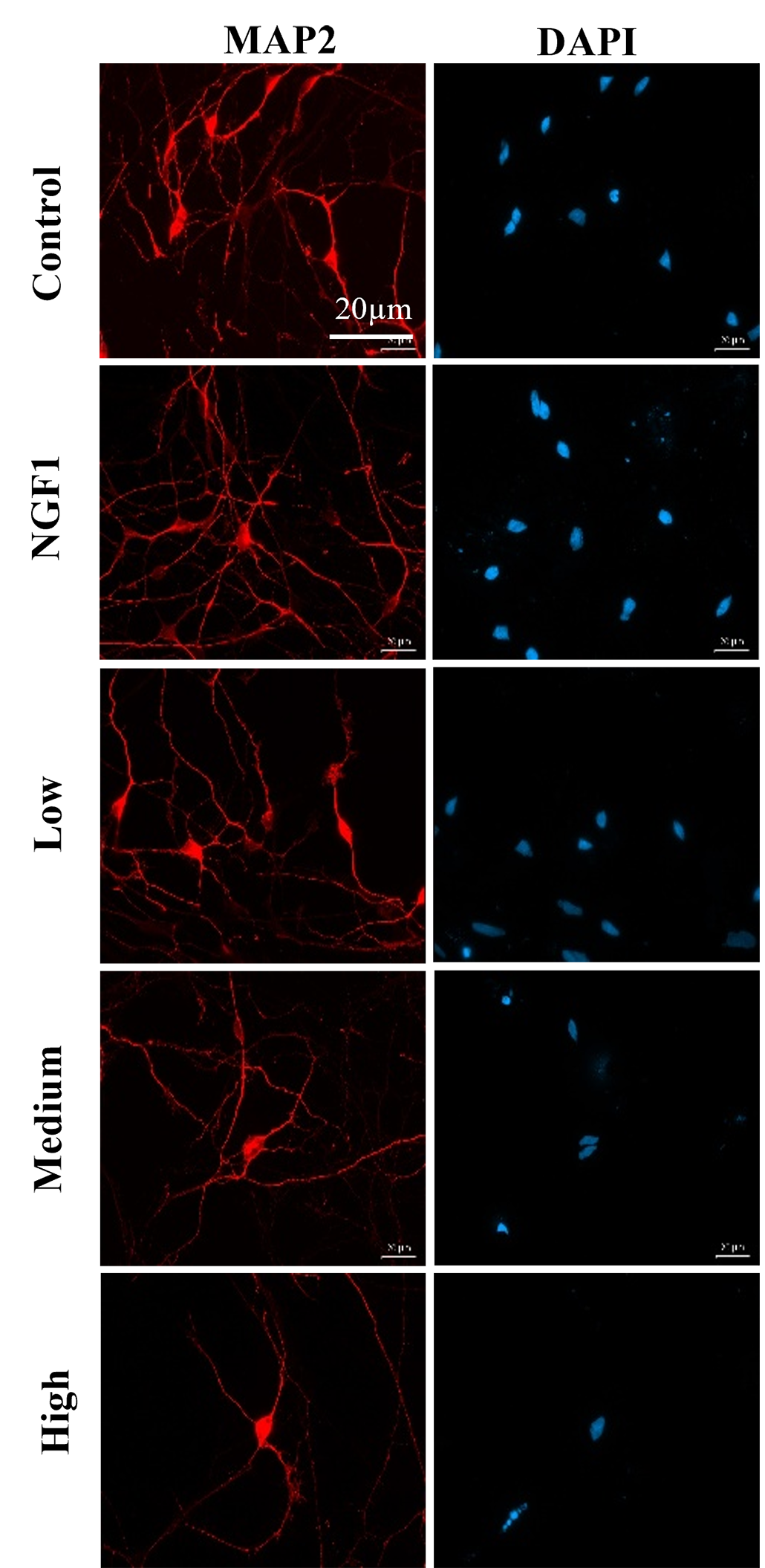
a series of images showing how rat neurons responded to various levels of caffeine exposure
These images show a decrease in rat neuron complexity with increasing caffeine exposure. The “control” group was exposed to no caffeine. “NGF” neurons were exposed to nerve growth factor, which is expected to promote neuron growth. Other neurons were exposed to low, medium and high levels of caffeine. MAP2 is a protein on the surfaces of cells. Red fluorescent dye binds to those proteins to reveal neurons’ shapes and growth. DAPI is a fluorescent dye that highlights the nuclei of cells. Together, these colorful tags reveal that with increasing levels of caffeine, fewer neurons grow and fewer connections form between neurons. S. Zeng

The first thing Sophia did was look at past data on caffeine. She analyzed published studies with the goal of identifying new patterns. This approach is called bioinformatics.

Sophia focused on studies that had looked at how caffeine affected the brains of adult mice. Her analysis suggested that caffeine could alter which genes in a mouse’s DNA were active, or expressed.

Next, Sophia took her work to a lab. She grew rat brain cells in petri dishes for six days, then exposed the cells to different levels of caffeine. Two days later, she compared the neurons under a microscope.

To make the neurons show up clearly, Sophia used fluorescent dyes that bind to the cells. Dyes can highlight important features, such as how the cells are forming connections with one another. Those neuron-to-neuron connections are called synapses.

Dishes exposed to more caffeine contained fewer neurons, Sophia found. What’s more, those neurons showed fewer connections. Caffeine seemed to make neuronal networks simpler and scarcer.

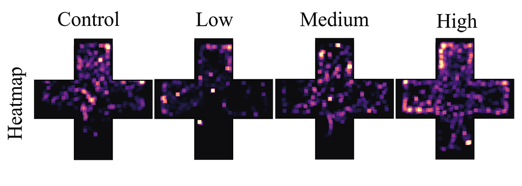
In another set of experiments, Sophia studied young zebrafish that had been exposed to different levels of caffeine. She looked at how far the fish swam and where. More caffeinated fish swam more, she says. They also made erratic choices, spending time in regions of their tank that calm fish typically avoided. Both of these findings hinted that the fish that got more caffeine were more anxious.

But Sophia didn’t stop there. She looked at what changes in the fishes’ brains might have led to this behavior. Sophia measured the activity of four genes involved in brain development. All four were less active in the caffeine-exposed fish, she found.

Takeaways for caffeine drinkers

Interestingly, some genes in the young zebrafish were turned down, or less active, in a way not seen in the neurons from adult rats. So, caffeine may affect the brain differently at different ages, Sophia says. But together, the mouse brain data, the rat neuron experiment and the fish studies all point to the same pattern. Caffeine seems to affect gene activity in neurons. And that in turn may impair brain development and behavior. 

a heat map showing how movement patterns of zebrafish shifted with caffeine exposure
Past caffeine exposure changed the movement patterns of zebrafish. As caffeine exposure increased, fish moved more erratically throughout the four zones of their aquarium. (The bright dots on the heatmaps show where the fish swam.) This evidence points to behavior changes and increased anxiety linked to caffeine.S. Zeng

“I feel that we really don’t talk about caffeine, even though we drink it,” Sophia says. It’s in so many everyday drinks, she adds. Between soda, tea and other beverages, young people may be consuming a lot more of it than they realize.

Sophia suggests greater skepticism about using this drug. People tend to think about caffeine as a helpful tool for focus, she notes, and dismiss potential downsides. But her findings highlight that caffeine comes with trade-offs — and those trade-offs may harm young brains more than we know.

A Surprising Discovery Suggests Autism Is Not One Condition

Autism Confused Girl Stickers FaceBrain scans have uncovered two biologically distinct forms of autism hidden within the spectrum. An international team of researchers has identified at least two biologically distinct forms of autism by examining how different regions of the brain communicate with one another. The findings could help advance more precise and personalized approaches to autism care and [...]

Scientists Uncover Surprising Secrets of a Mysterious Man Buried in Finland 400 Years Ago

10 June 2026 at 02:56
Historical Sámi BurialA multidisciplinary study has uncovered new details about a centuries-old burial in northern Finland, revealing genetic links to Sámi populations and evidence of a remarkably mobile life. A new study by the University of Turku and its partners has revealed new details about a man buried near Lake Kitka in Kuusamo, Finland, around the turn [...]

This Deep-Sea Predator Can Live for Centuries—Now Geneticists Are Close to Hacking the Secrets of Its Longevity

8 June 2026 at 13:39


The mystery of the Greenland shark’s incredible longevity may finally be unraveling, as a team of international researchers has completed the first comprehensive genome map of the creature.

Native to the cold waters between Greenland, Iceland, and Canada, Greenland sharks boast one of the longest vertebrate lifespans on Earth, approaching 400 years. In a recent paper published in the Proceedings of the National Academy of Sciences, researchers led by Shigeharu Kinoshita of the University of Tokyo have identified some of the genetic basis of that extraordinary lifespan, which they say future researchers may exploit to treat cancer and age-related illnesses.

Greenland Sharks Longevity

As genetic sequencing technology has improved and costs have fallen, there has been a major increase in the number of sequences from long-lived species for comparative analysis. Discoveries have included identifying increases in genes related to the immune system and growth hormone receptors in rockfish and bat species noted for their longevity.

With an entire lifecycle in slow motion, Greenland sharks’ longevity can be traced to their prolonged development: they take 150 years to reach sexual maturity, growing only 1 centimeter per year. Despite being one of the slowest swimmers in the ocean, the species undertakes large-scale migrations spanning over 1,000 kilometers.

Genome Mapping

Large genome sizes, like those of the Greenland shark, pose enormous mapping challenges, full of many repetitive sequences. Despite their importance for management and conservation, few whale and shark species have been sequenced due to the difficulty involved. To fill data gaps in the genetic causes of longevity, aquatic species evolution, and the risks of biodiversity loss, the team chose the Greenland shark as their subject.

The University of Tokyo-led team mapped roughly 5.9 billion DNA base pairs, accounting for 96.7% of the shark’s genome. Among the discoveries were the identification of specific genetic features likely related to the Greenland shark’s longevity. 

In their paper, they wrote of discovering “genetic traits associated with cancer, immune response function, genome stability, and cardiac function, deep-sea adaptations, and population size dynamics,” in the new research.

Longevity Results

One of the most crucial of these traits is how the shark appears to mitigate the genetic strain that contributes to aging by stabilizing the building blocks of chromosomes. Chromatin is a structure consisting of DNA, RNA, and proteins, which makes up chromosomes.

In the Greenland shark, the histone H1.0 protein, which binds DNA in chromatin, undergoes an amino acid substitution that may enhance stability and reduce the effects of aging.

Another genetic tool aiding the shark’s longevity is the presence of 59 copies of the FTH1b gene, which researchers discovered on pseudochromosome 33, far above the number seen in other shark or fish species. This helps regulate ferroptosis, a form of programmed cell death, thereby mitigating cell damage. Since the gene controls iron storage and ferroptosis is iron-dependent, the researchers say this multiplication of FTH1b could prevent oxidative cell damage.

Beyond those specific expressions, researchers also found expansion of entire gene families associated with DNA repair, immune functions, and cancer resistance.

Although limited to a single species, the research does advance general understanding of the genetic factors involved in extended lifespans. However, it will be up to future researchers to identify how this new information can be applied to human concerns.

The paper, “The Greenland Shark Genome: Insights into Lifespan Extremes and Population Dynamics,” appeared in Earth and Planetary Science Letters on May 19, 2026.

Ryan Whalen covers science and technology for The Debrief. He holds an MA in History and a Master of Library and Information Science with a certificate in Data Science. He can be contacted at ryan@thedebrief.org, and follow him on Twitter @mdntwvlf.

Ancient Europeans Ate Some Pretty Gross Things, but The Ickiest One Was Probably by Accident

8 June 2026 at 13:16


An analysis of dental tartar containing traces of human diets dating back over 33,000 years has revealed that ancient Europeans and many early Asian populations likely only ate insects by accident, perhaps as larvae in the carcasses of dead animals.

Conversely, the new study found that intentional insect consumption, called entomophagy, was more common in tropical regions and among Neanderthal populations.

Although the difference has some cultural and religious roots, the team found evidence that the abundance of insects in the tropics and the decreasing ability of humans migrating away from the tropics to digest insects likely led to the modern-day separation over thousands of years.

The Institute of Evolutionary Biology (IBE), a joint center of the Spanish National Research Council (CSIC) and Pompeu Fabra University (UPF), research team behind the new findings suggests their research could help explain why some modern populations still consume insects as food while other parts of the world consider it gross or taboo, especially given the increasing food demands of a rising world population.

Insect Consumption in Ancient Northern Europeans Likely Accidental

According to a statement announcing the new research, hundreds of millions of people currently include insects in their diet. However, they also note that most of the Western world continues to “show an aversion” to including any of the more than 1,611 insect species designated as edible by the Food and Agriculture Organization of the United Nations (FAO) in theirs. Although some theories suggest the difference is almost purely cultural, the team concedes that “its origin remains unknown.”

To explore the origins of human entomophagy, the IBE analyzed 745 samples of ancient human dental tartar dating back to 33,000 before present. According to the team’s results published in the journal Science Advances, dental tartar (sometimes called dental ‘calculus’) is a critical archaeological tool because it preserves the DNA of the species these human ancestors consumed during their lifetimes.

Although the analysis found evidence for entomophagy amongst virtually all ancient populations sampled in the study, the levels varied dramatically. Notably, the evidence suggested that ancient humans in northern Eurasia did not routinely eat insects in measurable quantities. Instead, any consumption was likely incidental, including consumption of insect larvae in rotting food.

Curious whether the lack of entomophagy in ancient European and Central and Northern Asian populations was purely cultural or whether genetic and geographic factors were involved, the team studied human genes involved in chitin digestion. Chitin, which can be found in some marine animal shells, is also part of an insect’s hard exoskeleton.

According to the study authors, the analysis revealed that the ‘chitinase’ genes in North Eurasian human populations “carry mutations that confer a reduced capacity to digest insect exoskeletons.” Their analysis revealed that this reduced capacity to digest insect exoskeletons has persisted in the genomes of modern Eurasian populations “for the last 9,000 years,” which would coincide with the widely accepted date for the origin of agriculture.

Pablo Librado, study leader and a principal investigator at the IBE, said the “scarce presence of insects in the diet of northern Eurasians” suggests that the cause may not lie solely in more recent cultural factors but also in ecological and evolutionary components dating back several millennia. 

Neanderthals and Denisovans Probably Ate Insect Larvae in Rotting Meat

When the team compared data from ancient human tartar with that of ancient Neanderthals, they found that Neanderthals engaged in much higher levels of entomophagy than humans living in the same regions. According to the study authors, Neanderthal insect consumption levels were similar to those found in western chimpanzee populations, which the researchers note “rely on entomophagy to supplement their diet on the savanna, especially during periods of drought.”

Further analysis of the extracted insect DNA showed that the group most commonly consumed by the Neanderthal specimens included flies and mosquitoes. Although the team only analyzed the DNA, they noted that a recent hypothesis suggests Neanderthals ate carcasses laden with mosquito larvae.

“The abundance of mosquito remains reinforces the possibility that the carcasses of their prey were kept in ponds and marshy areas, where mosquitoes lay their eggs,” the team’s statement explained.

An analysis of the Neanderthal DNA and the DNA from the lone Denisovan study subject revealed that both ancient hominid populations could digest chitin. This result reinforced the genetic basis for increased or reduced entomophagy beyond cultural factors. 

Humans Ate Fewer Bugs When They Moved Away from Tropical Regions

One geographic component identified by the chitinase DNA analysis showed higher levels of digestive enzyme expression among humans living in or near the tropics. However, the expression of these enzymes gradually decreased as populations moved away from tropical regions. The team said this variation, which has been maintained for at least the last 9,000 years, “reflects the abandonment of entomophagy in European populations.”

Manuel Piñero, a predoctoral researcher at the IBE and first author of the study, noted that large numbers of insects must be consumed to offset the “high caloric cost” of collecting them, which might explain the higher levels of entomophagy in this region.

“In the tropics, there is a greater availability of social insects, such as termites and locusts,”  Piñero  explained, adding that the biomass and diversity of tropical edible insects enable “sustainable exploitation throughout the year.” The researcher also noted that insect consumption “adds to pest control.”

When discussing the implications of their findings, Librado highlighted the DNA and geographic factors the IBE team uncovered.

“Beyond cultural or religious factors, our results suggest that the reduced availability of insects in non-tropical areas may have been a key factor in the abandonment of entomophagy, leading to a reduced capacity to digest insect exoskeletons,” the researcher explained.

Christopher Plain is a Science Fiction and Fantasy novelist and Head Science Writer at The Debrief. Follow and connect with him on X, learn about his books at plainfiction.com, or email him directly at christopher@thedebrief.org.

Tiny Genetic Change Turns Female Mice Into Males, Scientists Discover

7 June 2026 at 16:58
X Chromosome DNA StrandA new study reveals that an extremely small change in a non-coding region of DNA can dramatically alter how sex develops in mice. How much can a single letter of DNA matter? According to new research from Bar-Ilan University, one misplaced genetic “letter” can completely change the course of sexual development. In a study published [...]

This Deep Sea ‘Giant’ Can Go Five Years Without Eating—Now Scientists Are Unlocking the Genetic Secrets of Its Extreme Survival

6 June 2026 at 14:03


Scientists have uncovered the genetic secrets behind a novel energy-use strategy that allows a deep-sea “giant” to survive without food for over five years, in one of nature’s most stunning survival stories.

Supergiant bathynomids, also known as giant isopods, present a bizarre biological paradox: they exhibit pronounced body gigantism as megafaunal species yet inhabit a deep-sea habitat with extremely sparse nutrient availability.

In a recent paper in Cell, researchers from the Institute of Oceanology of the Chinese Academy of Sciences (IOCAS) revealed how these large deep-sea creatures subsist on such limited food, using a novel genetic strategy unique to bathynomids.

A Complex Genetic Approach

The IOCAS team employed a multi-omic strategy to address the problem, combining genomics with other disciplines, such as morphology, physiology, behaviorology, and metagenomics. Doing so led to a novel discovery: that supergiant bathynomids don’t have just one, but two techniques to manage their limited access to nutrients.

Bathynomus giganteus
One of the large species of Bathynomus seen in the Gulf of Mexico (Image Credit: NOAA)

First, isopods have a massive stomach that can store vast amounts of nutrients for later use. Second, they have an exceptionally low basal metabolic rate (BMR), which is genetically driven.

To diversify their research, the team focused on two species that occupy different depths. At around 300 meters, the Bathynomus doederleini is the shallower of the two, while the Bathynomus jamesi is found at 898 meters. Despite the difference in depth, both are technically considered “deep-sea” inhabitants, as the boundary is defined at 200 meters.

A Powerful Stomach

Roughly two-thirds of a bathynomid’s body is stomach—a figure far beyond that of related intertidal and shallow water species. The contents of those enormous stomachs are also unusual, filled with a mud-like mixture of highly digested food. This finely ground nutrient sludge has a low proportion of digestive bacteria, but a high amount of lipid-storing Chlamydiae. 

With a bacterial ratio leaning toward preservation rather than just digestion, this creates a food reserve that can be slowly digested and utilized as the creatures lower their BMR to endure long periods between feedings.

Genetic Findings

Another major finding was that bathynomids benefit not only from vertically transferred genes between parents and offspring, but from the horizontally transferred ND1 gene they acquired from an exogenous symbiotic bacterium. Intriguingly, the transfer defied typical horizontal transfer limitations, achieving an ultra-high level of expression in the bathynomid genome. 

These isopods use epigenetic histone modifications to regulate energy-related gene expression, thereby improving efficiency, conservation, and control. An example of this is histone acetylation regulating ND1 expression at an ultra-high level, which the team investigated in a laboratory test. Researchers suspected this gene was essential to energy metabolism, as it is a component of Complex I in the electron transport chain.

ND1 Genetic Tests

Using zebrafish, nematodes, and human 293T cells, the IOCAS team tested the gene’s impact on energy usage. At normal temperatures, the team found that ND1 actually decreased starvation tolerance by increasing energy metabolism. Yet when the team lowered temperatures to reflect those found in a typical deep sea environment, they found the opposite effect, with ND1 decreasing mitochondrial activity and energy metabolism rates, to improve zebrafish starvation tolerance an astounding 37%.

The IOCAS team concluded that ND1 provides a solution to the long-standing paradox of bathynomid gigantism in the context of their nutrient-poor environment. By fine-tuning the degree of metabolic depression, the gene can adjust the mitochondrial metabolic network to preserve energy during extended periods without sustenance.

Never before has such a strategy for energy efficiency, combining horizontal gene transfer and epigenetic optimization, been identified in a deep-sea megafaunal species.

“Our work not only deciphers the mystery of ultra-long starvation tolerance in deep-sea isopods,” said lead author Yuan Jianbo, “but also provides an important paradigm for understanding how life balances growth and survival in extreme environments.”

The paper, “Deep-Sea Megafauna Co-Opts Microbial Energy Metabolism Genes to Withstand Ultra-Long Starvation,” appeared in Cell on June 5, 2026.

Ryan Whalen covers science and technology for The Debrief. He holds an MA in History and a Master of Library and Information Science with a certificate in Data Science. He can be contacted at ryan@thedebrief.org, and follow him on Twitter @mdntwvlf.

Scientists Discover Rogue Gene That Could Unlock New Cancer Treatments

5 June 2026 at 16:02
DNA Mutation Cancer Genetics Disease BioengineeringA gene normally responsible for repairing DNA may have a darker side when produced in excess. Cancer treatment is increasingly moving beyond where a tumor forms and focusing instead on the genetic flaws that drive it. Now, researchers at Penn State College of Medicine have identified an unexpected DNA repair gene that could help expand [...]

Scientists Uncover the Earliest Brain Changes That May Predict Alzheimer’s Decades Before Symptoms

Genetic Brain Disorders DNA Helix Mental HealthScientists have uncovered an early brain mechanism that may help explain why people carrying the APOE4 gene face a higher risk of Alzheimer’s disease. For millions of people who carry the APOE4 gene, the strongest known genetic risk factor for Alzheimer’s disease, changes in brain activity may start years before memory problems become noticeable. Researchers [...]

Why Some Cancers Turn Deadly: Researchers Uncover a Hidden Trigger

3 June 2026 at 16:26
Cancer Cell Spread Abnormal GrowthA closer look at cancer cells with extra chromosomes uncovered surprising traits linked to faster-growing, more dangerous tumors, pointing to potential new indicators of disease severity. Cancer cells are notorious for breaking the rules of biology. One of the most dramatic violations occurs when a cell suddenly doubles its entire genetic library, creating a chromosome-packed [...]
❌