Normal view

What happens inside your body during a hot flash

26 May 2026 at 14:01

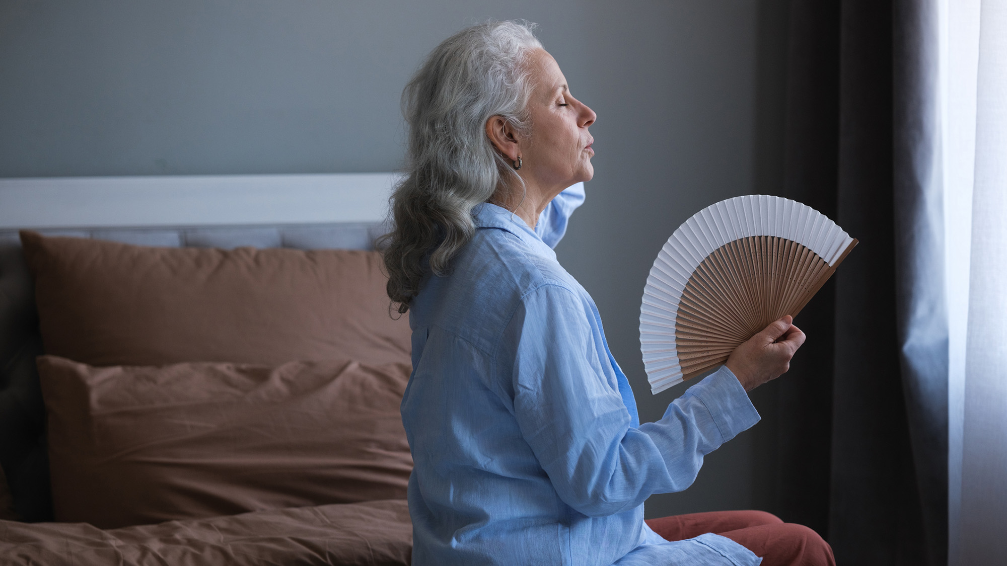
For a woman in her mid-40s to mid-50s, it arrives without warning. She wakes up, overheated, wondering why it’s so hot in the house—until she sees the thermostat is set for 70 degrees, same as always. Or, she’s midway through a work presentation when heat rises from her chest to her face, and she wonders if the flush on her cheeks is visible to everyone in the room. 

It’s a hot flash—a rite of passage for the majority of women in either perimenopause, the years leading up to menopause, or the years beyond it. Menopause itself is diagnosed after 12 consecutive months without a period, but the hot flashes don’t always get the memo.

Here’s everything doctors currently know about hot flashes.

What is a hot flash, and who gets them?

Hot flashes are a sudden heat flare up often paired with flushed skin and sweating. They don’t usually last long, between a minute and five minutes in duration.

Most women experience a hot flash about four and a half to five years after their last period, Dr. Monica Christmas, an OB/GYN at University of Chicago Medicine and director of its menopause program tells Popular Science. She also is the associate medical director of the nonprofit Menopause Society, which provides healthcare professionals with tools and resources to support women through the transition.

Women have grappled with hot flashes—whether simply annoying or genuinely debilitating—for centuries. In 1582, Dr. Jean Liebault of France was among the first to document the phenomenon. But while we know much more about hot flashes and night sweats than Liebault ever did, one question still stumps experts. 

“What we can’t answer is why doesn’t everybody get them,” Christmas says. “Because everybody doesn’t get them. I have patients that will say, ‘I don’t know,’ if I say, ‘Are you having any hot flashes or night sweats?’ And as soon as they say that, I’m like, ‘You’re not having them.’” 

What’s actually happening inside women’s bodies during a hot flash? 

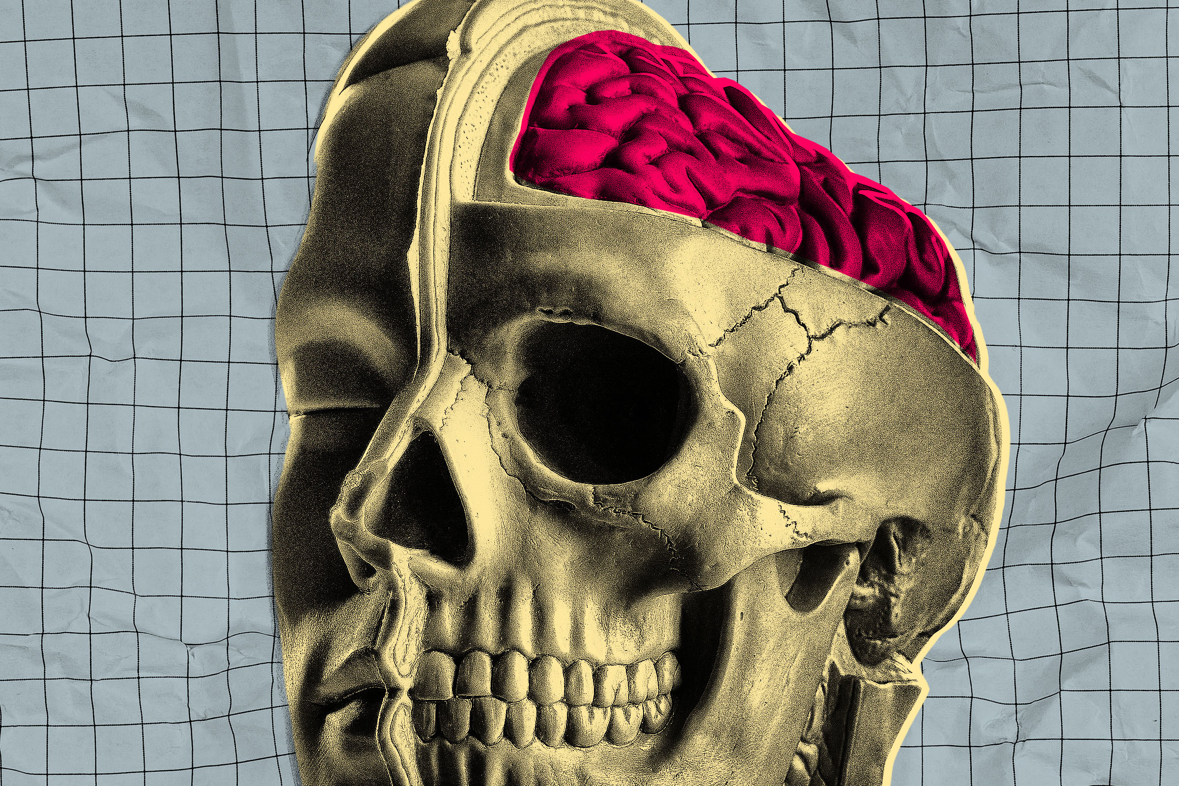
During a hot flash, a woman might feel like she’s spiking a high fever, but physiologically, that’s not what is happening. As women approach menopause and the ovaries begin to make less estrogen, the brain’s internal thermostat—the hypothalamus—becomes hypersensitive to even small shifts in temperature, Christmas says.

The body “thinks” it’s overheating, even when the actual temperature hasn’t changed much. In response, our bodies try to cool us down. Blood vessels dilate, which is supposed to help dissipate some of that heat, but then that triggers a sweating reflex.

“Many people will say, ‘I feel this out of nowhere, this surge of warmth that typically is from the nipple line up,’” she says. “And then as soon as the heat came on, and I felt like I was internally heated up or on fire, I start to sweat.” 

Related 'Ask Us Anything' Stories

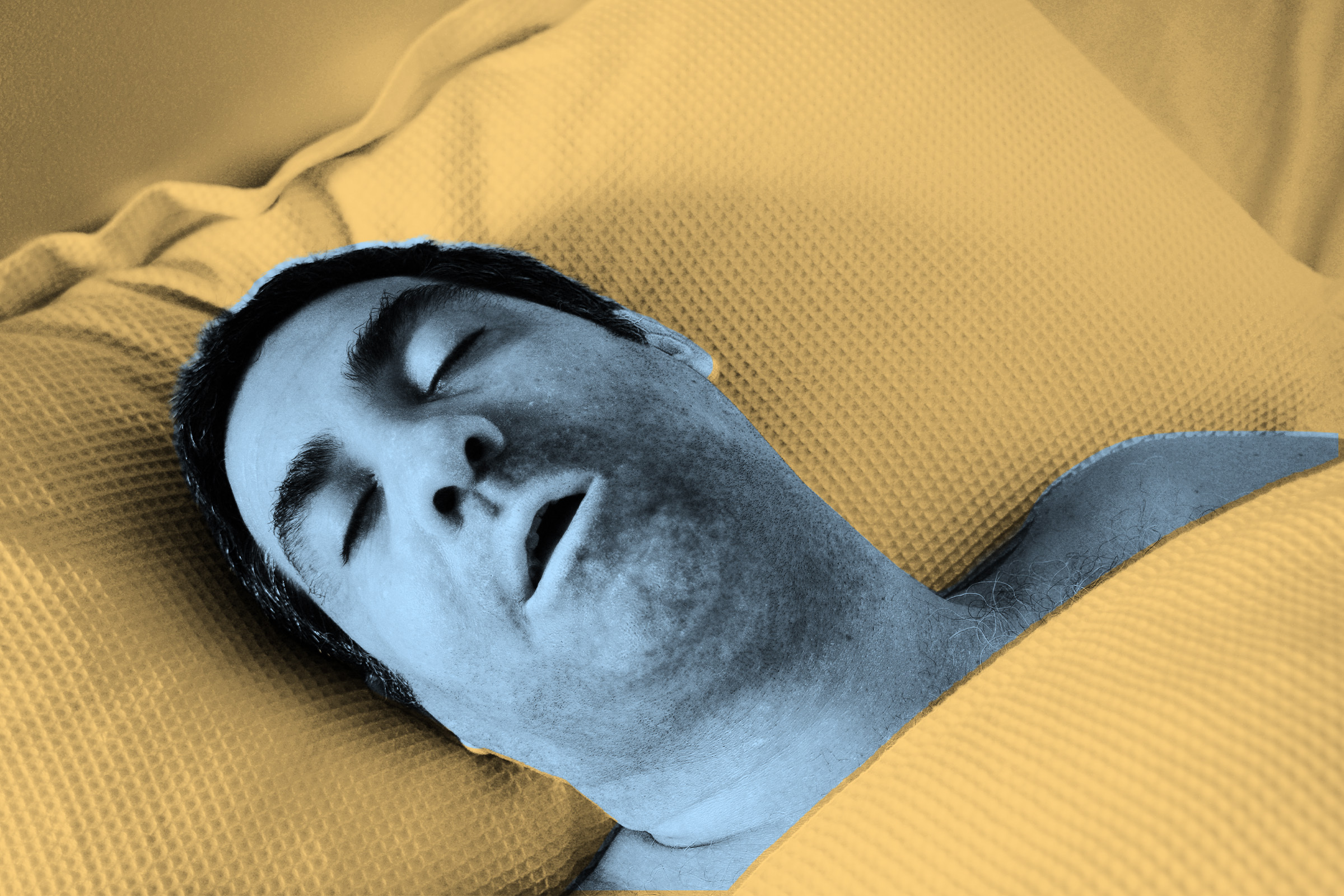
How do women experience hot flashes differently? 

Exactly how an individual woman experiences hot flashes varies wildly. Some describe very mild symptoms. Others grapple with profuse sweating. Some experience only hot flashes during the day, while some have regular night sweats. About four in five women experience them at some point during the menopause transition, according to the American College of Obstetricians & Gynecologists.

“There’s a lot of variability,” Christmas says. Common triggers include alcohol, caffeine, high-sugar and highly processed foods, along with stress.

Black women also are more likely to experience more severe and longer-lasting symptoms, sometimes up to 11 years, she says. And research also shows that women with more severe, longer-lasting hot flashes and night sweats appear to be at higher risk of cardiovascular disease.

That doesn’t mean treating hot flashes automatically lowers heart risk, Christmas says. But it does reinforce that these women deserve particularly careful attention to blood pressure, cholesterol, and lifestyle. “I want to make sure I’m doing everything possible to minimize that risk,” she says when she treats her patients. 

There’s more to hot flashes than hormonal changes

For decades, the entire process was blamed purely on estrogen loss, Christmas says. But that explanation left some unanswered questions. 

“That doesn’t explain why every menopausal woman doesn’t have night sweats,” she says. “And it also doesn’t quite explain why we can sometimes start to experience them during the perimenopause transition because during perimenopause, people still have some estrogen.” 

Newer research now is telling a more complex story. When the brain recognizes that a woman’s estrogen levels are low, nerve cells in the hypothalamus called KNDy neurons (pronounced “candy”) become overactive, releasing neurotransmitters, which are chemical signals the brain uses to send messages throughout the body. These neurotransmitters include kisspeptin, dynorphin, and neurokinin B. 

“It’s actually those neurotransmitters that seem to have more of an impact on our ability to regulate our internal temperature,” Christmas says. “They’re not hormones.” 

What to do if you get a hot flash

For women in the middle of their hot flash years—along with the 10 percent of menopausal women who continue to experience them—there are treatments. 

Estrogen-based hormone therapy can help, but not every woman, including those with a history of blood clots or breast cancer, can take hormone therapy. 

Hormone therapy can help alleviate hot flashes. Video: Hormone therapy – Four things a Mayo Clinic women’s health specialist wants you to know., Mayo Clinic

Fortunately, researchers’ new understanding about the role of KNDy neurons has allowed for new treatments that block the brain signals that trigger hot flashes in the first place. The FDA approved a new drug called Veozah (it’s chemical name is fezolinetant) in 2023. It targets the neurokinin 3 receptor, which plays a key role in regulating body temperature. 

Lynkuet, another drug (with the chemical name elinzanetant), came along in 2025. It blocks both the neurokinin 1 and neurokinin 3 receptors, interrupting the process that triggers hot flashes at two points instead of one. 

Other medications can also provide relief, though weren’t originally developed for hot flashes, Christmas says. Some SSRIs and SNRIs; gabapentin, a neurologic medication; and oxybutynin, used for overactive bladder, are all used off-label for hot flashes and night sweats. 

Cognitive behavioral therapy and hypnosis also have been shown to reduce hot flashes. “I’m menopausal, too, so I know if I’m under a lot of stress or in a stressful situation, I’m going to probably have more hot flashes than not,” Christmas says. 

“So there’s certainly something about being able to calm our central nervous system down that seems to have an impact, too.”

If you’re struggling with hot flashes, Christmas recommends seeing your healthcare provider for help. Treatments are available. What’s more, in some cases, hot flashes or night sweats could signal other issues, including thyroid disorders, cancer, and infections, among others. 

But bottom line, when it comes to hot flashes, you don’t have to sweat them out.

In Ask Us Anything, Popular Science answers your most outlandish, mind-burning questions, from the everyday things you’ve always wondered to the bizarre things you never thought to ask. Have something you’ve always wanted to know? Ask us.

The post What happens inside your body during a hot flash appeared first on Popular Science.

Startup Testing Drugs on Freshly Extracted Human Brains That Are Kept On Life Support

31 May 2026 at 11:45

A biotech startup called Bexorg is doing something that sounds like it was ripped straight from the pages of a cyberpunk novel — or from the script of “RoboCop,” for that matter.

The company is extracting human brains just hours after their owners died and then hooking them up to specialized life support machines, Science reports. While the masses of pink mush no longer host electrical activity, most of their key functions remain intact, allowing scientists to test experimental drugs, such as potential treatments for Alzheimer’s disease, like never before.

You’d hope that the disembodied cerebrums are most assuredly dead. But according to the reporting, an extracted brain hooked up to one of Bexorg’s proprietary life support machines, BrainEX, “hovers between life and death.” There’s no spark of consciousness, and yet the brains are kept running on an artificial lung, kidney oxygenate, blood, and other fluids.

Perhaps you can put this ambiguity down to the startup being deliberately enigmatic to provoke attention. Or maybe it’s a reflection of how the distinction between life and death is uncomfortably blurry.

But you can put those doubts out of your very-much embodied mind, assures Brendan Parent, one of Bexorg’s six ethicists. The extracted brains are almost devoid of the coordinated neural firing necessary for minimal consciousness, he told Science. To prevent the eerie implausibility that some the brains produce electrical activity, the brains are also dosed with anesthetic propofol. Of course, that such a measure has to be taken in the first place may actually be less assuring and more unsettling.

Ethics aside — not a statement that should be made lightly — the scientific possibilities that these extracted brains afford may well hold promise. Bexorg CEO Zvonimir Vrselja said that the brains come with decades of environmental exposures, histories of drug treatments, and other factors that make them a more realistic testing medium for drugs. “You get cells that have been there for 60 to 80 years,” Vrselja told Science.

Bruna Bellaver, who studies neurodegeneration at the University of Pittsburgh, was also effusive. 

“It’s a huge step up from mouse models,” she told Science.

Bexorg is the same startup that demonstrated, over six years ago, that it could keep decapitated pig brains alive for 36 hours using a prototype of its BrainEX machine.

Today, its human brains aren’t kept running in perpetuity. After 24 hours, they’re sliced into hundreds of pieces so they can be analyzed by scientists. The company plans to use a robotic arm to slice up to 1,600 brains per year.

Though Bexorg hasn’t itself published any papers on its work with human brains, other companies have already been eagerly experimenting with them. The pharmaceutical firm Biohaven has used 130 of its brains to test drugs, according to Science, including a potential treatment for Parkinson’s disease, and plans to launch a clinical trial for another drug using data it gathered from those experiments.

More on neuroscience: Scientists Say Test Subjects Were Able to Quit Smoking After They Blasted Their Brains With a Huge Magnet

The post Startup Testing Drugs on Freshly Extracted Human Brains That Are Kept On Life Support appeared first on Futurism.

Woman Alarmed When Her Trusted Therapist Starts Recording Her With AI

30 May 2026 at 18:30

Therapy is predicated on trust. You can’t be honest and vulnerable, and share how you’re really feeling, if you don’t believe in the embodied-concerned-frown sitting in the armchair across from you.

So you can understand why one woman, 31-year-old Molly Quinn, was taken aback when her trusted therapist suddenly whipped out an AI model to start recording their private conversations, NPR reports

“She wasn’t taking notes like she usually did,” Quinn recalled realizing halfway through one session. “The iPad was just propped up.”

Where were her words being processed and stored? Will they one day become training data? It’s not something you have to ask yourself when your therapist jots stuff down on a clipboard. But those questions were now racing through Quinn’s head, leaving her uneasy.

“The more I thought about it, the more I just started getting more and more sick to my stomach,” she told NPR. “This person who I’m supposed to be able to trust with some very private and very intense emotions had just completely disregarded something I said I was not comfortable with. I felt completely violated.”

Though her therapist offered to stop using the AI tool, Quinn cut her off and found another one.

“The trust was gone,” she told NPR.

Like doctors, therapists across the country are adopting AI tools for notetaking and generating transcripts. AI companies offering these services frame it as a way of cutting down on the drudgery of paperwork and other administrative tasks, freeing up more time to focus on patients — a permutation of a common AI industry refrain: let us do the tedious stuff for you. 

The reliability of AI tools remains fairly dodgy, though, and even setting aside questions of hallucinations creeping into clinical notes — which is something we’re already seeing happen — it’s not clear whether patients are even comfortable with the tech yet. In a YouGov survey cited by NPR, only 11 percent of Americans said they would be open to using AI in mental health care. An even slimmer eight percent said they would trust AI being used this way, while 40 percent said they don’t trust the technology at all.

“Even the presence of AI changes the therapeutic experience,” Marisa Cohen, a couples and sex therapist in New York, told NPR. “Clients know or feel like something else is listening to them. That awareness can subtly alter their disclosure.”

“When you introduce something that’s being stored electronically, it raises additional questions about trust and safety,” Cohen added. “It’s essentially a third party.”

Tal Salman, the CEO a popular AI scribe tool for therapists called Berries, insists that conversation recordings are deleted immediately and that transcripts are stored on HIPAA compliant servers in the US. Even if this is true, if AI companies’ tools are to ever have a place in private mental health settings, they need the trust of patients — and that’s something the AI industry clearly hasn’t earned yet. Quinn fears that AI-recorded conversations could one day be exposed by hackers.

“We’re going to see breaches,” she told NPR. “Maybe not tomorrow, maybe not next week. But in a few years? I think we’re going to see them. And I don’t want my therapy session to be part of that.”

More on AI: The Pope Just Low Key Declared Holy War on Artificial Intelligence

The post Woman Alarmed When Her Trusted Therapist Starts Recording Her With AI appeared first on Futurism.

Apocalyptic “Fungal Storms” Are Now Surging Across the US

30 May 2026 at 11:00

Dust storms are becoming increasingly common sights throughout much of the US, fueled by hot and arid conditions brought about by global warming. While there are the ever-present dusters in states like Arizona, those living in regions like the Midwest and Pacific Northwest are now learning what it means to be swallowed by these great walls of sand and dirt.

These dust storms are dangerous enough on their own, leading to spikes in emergency room visits and disrupting agricultural economies. But according to writing climate scientist Bill McGuire in the BBC‘s Science Focus, dust storms are now becoming vectors for massive clouds of fungal spores, an event he calls “fungal storms.”

Far-out as that may sound, McGuire writes that fungus and bacteria traveling along with dust particles has been strongly correlated with outbreaks of bacterial meningitis across the Sahel region of Africa. In the continental US, they’re becoming increasingly tied to outbreaks of Valley Fever, a lung infection caused by the spores from the fungus Coccidioides.

One 2017 research paper published in Geophysical Research Letters noted that incidents of Valley Fever ballooned by over 800 percent from 2000 to 2011. In two geographical areas with high concentrations of the infection, dust storms where “found to better correlated with the disease than any other known controlling factor.”

Though some have argued the term “fungal storm” is more media hyperbole than a well-defined scientific phenomenon, further research has noted “abundant evidence” pointing to dust storms as a vector for pathogens like Valley Fever.

Whatever you call it, it’s clear the dangers of dust storms are growing right before our eyes.

As secretary general of the World Meteorological Organization Celeste Saulo told McGuire, “sand and dust storms do not just mean dirty windows and hazy skies. They harm the health and quality of life of millions of people and cost many millions of dollars through disruption to air and ground transport, on agriculture and on solar energy production.”

More on climate change: The Upcoming El Niño Is About to Unleash Devastation, Experts Warn

The post Apocalyptic “Fungal Storms” Are Now Surging Across the US appeared first on Futurism.

Kids Are Flying Into Lunatic Rages When Their iPads Are Taken Away

29 May 2026 at 15:57

Parents say their kids are going ballistic when they take their iPads away from them, leaving them unsure of what normal behavior might be — and whether there’s something sinister going on with their child’s connection to the devices.

Rachel, a mother of two, tried limiting her son Jonah’s screen time by warning him that he had put down his iPad to leave for a birthday party at 11 AM. Despite the repeated warnings the day before and several reminders before the hour mark, when it came time to leave, Jonah had a meltdown.

“He just left his body,” the mother told The Cut of her son, who hurled the electronics and started screaming: “You said I had until 11! It’s not 11 yet! You’re always doing this!”

Jonah followed her around the house, distraught, until finally collapsing on the kitchen floor and refusing to move.

“I remember standing there thinking, I don’t know this person,” she recalled. “I genuinely did not recognize him.”

Thirty minutes later, he tied his shoes, got in the car, and acted like nothing had happened.

“That’s the part that really messes with you,” Rachel says. “How fast they come back.”

Think that’s bad? Hear what Nora had told The Cut about her 13-year-old son when she asked to check his phone settings: he accused Nora of ruining his life, before dropping a grenade in conversation.

“You make me want to kill myself,” he lashed out.

And while getting dinner last month at an Italian restaurant, Rachel told the outlet that she allowed her daughter Maya to watch YouTube Kids on the phone. When it was time too leave, she took the phone back. Maya went rigid, screamed, and hid under the table.

What’s going on here? Are these the kind of extreme tantrums that kids are prone to throw no matter the toy they’re being deprived of? Is everyone a bad parent, or at least not handling this the right way? Or are apps and the devices they run on uniquely addictive, somehow impacting a child’s development in novel and frightening ways?

The research into this area is still emerging, as are the generation of children raised on YouTube Shorts, Roblox, and other mobile games. That’s to say that we’re still a long way from grasping the long-term cognitive effects of being a so-called “iPad kid.” And the latest Silicon Valley horror, AI chatbots, are an even bigger question mark.

What evidence we do have, however, is alarming. The Cut cites a recent University of Washington study that found that 22 percent of parents’ attempts to cut down screen time sparked a negative reaction from kids under five. And in another study from Brigham Young University, 93 percent of parents reported that their toddlers would sometimes whine or throw tantrums when “transitioning away from media.”

Experts are mixed on whether the devices are provoking some newly negative response. 

“We frequently hear from parents who say, ‘When I ask my child to get off technology, they get very mad at me.’ That is true of almost anything that children find reinforcing,” Dave Anderson, a senior psychologist at the Child Mind Institute in New York City, told The Cut.

Anderson was skeptical of using the word addiction to describe what’s fueling iPad rages, noting that withdrawal symptoms of actual addiction don’t disappear within minutes. Kid’s minds just aren’t developed enough to handle having their favorite toy taken away from them, she said.

Stanford psychiatrist Anna Lembke, however, hasn’t hesitated to invoke the specter of addiction, calling screen devices a “digital drug” in an interview with Oprah. And Sarah Coyne, a professor of human development at Brigham Young, seemed to consider equating post-iPad rages to tantrums related to other pleasures outrageous. 

“I’m not sure how many children are struggling to function because their parents tell them to be done with their ice cream,” she told The Cut, adding that she’s seen addiction-like behavior in kids using devices as young as two years old.

If describing these patterns as signs of outright addictive behavior goes too far, there’s certainly there is a lot of evidence painting screen time’s cognitive effects. One study found that the more  babies and toddlers looked at screens each day, the more likely they were to miss key development goals, including fine motor skills and social skills.

The effects are no less worrying in older children. A study that followed tweens over four years found that increased screen time was a reliable predictor of ADHD diagnoses. Beyond iPad rages and worrying cognitive trends, there are other behaviors that illustrate the impact of device usage on children. In a survey of UK preschool school teachers, the teachers on average estimated that a third of their pupils didn’t know how to correctly use books — as in they literally couldn’t figure out that they had to turn the page. Instead, some reportedly tried to swipe or tap them.

More on mental health: Influential Tech Founder Says His Peers Are Suffering From Mass AI Psychosis

The post Kids Are Flying Into Lunatic Rages When Their iPads Are Taken Away appeared first on Futurism.

Influential Tech Founder Says His Peers Are Suffering From Mass AI Psychosis

28 May 2026 at 16:46

It’s no secret that many of the world’s top CEOs are obsessed with AI. By pursuing lofty goals of complete AI automation, these executives have created one of the largest financial bubbles in recent memory while transforming the job market into a barren wasteland, with little to show for their efforts so far.

As the top tech companies have yet to find a way to turn AI into a profitable venture, those decisions to go all-in on AI are looking increasingly delusional. According to Aaron Levie, CEO and founder of the massive cloud computing company Box, there’s a simple explanation for it: many of his colleagues are suffering from AI psychosis.

“CEOs are uniquely prone to AI psychosis because they’re sufficiently distant from the last mile of work that still has to happen to generate most value with AI,” Levie wrote on X-formerly-Twitter. Translation: AI-happy CEOs are out of touch with the rank-and-file workers tasked with making their AI ambitions come to life.

As an example, Levie offers cases in which corporate executives say “look I made this awesome product prototype” with an AI chatbot. “Yes but you didn’t have to review the code before it went into production and fix a bunch of issues,” he retorts.

Whether “AI psychosis” is the best metaphor for this concept is up for debate. Arguably the most common definition of AI psychosis is that it’s a phenomenon where extreme interactions with AI triggers or amplifies delusions or paranoia, sometimes already existing and sometimes seemingly newly cooked up with the AI. The symptoms can be extreme, with AI chatbots convincing victims that they’re communing with God-like entities, or have singlehandedly uncovered a grave threat to humankind.

There are indeed some executives who seem to fit the bill. Last year, Futurism reported that colleagues of Geoff Lewis, managing partner of the multi-billion dollar investment firm Bedrock, were concerned that he was suffering from a break with reality after spending too much time with ChatGPT (ironically, Bedrock was an early investor in OpenAI.) In that case, Lewis had claimed to be mapping an incomprehensible “non-governmental system” that was designed to disrupt his life.

That said, there’s a major gap between an exec believing they’re targeted by a vast conspiratorial network and an exec buying into AI hype. The phenomenon Levie is identifying might better fall under “organizational blindness,” a known phenomenon where leaders of a company find themselves disconnected from the reality of work on the ground. Coupled with a ravenous hunger for profit, this kind of tunnel vision seems to be exactly what we’re seeing in companies around the globe.

In today’s world, many executives and managers operate at an abstract level, working via spreadsheets, emails and Zoom meetings. This is different from concrete labor, meaning the specific, friction-heavy tasks that workers perform, like writing code or wiring server racks. When a board-room full of executives loses sight of this tangible labor — by failing to consider the kinds of tasks AI chatbots are actually good at, for example — it can certainly create a break from material reality, though one driven by social factors rather than psychological.

In other words, there are two possibilities: either the world’s CEOs are losing their minds, or they’re just succumbing to the latest manifestation of capitalism run amok. Occam’s razor probably suggests the latter.

More on AI and CEOs: 99 Percent of CEOs Are Preparing to Lay Off Workers and Replace Them With AI Within Two Years, Survey Finds

The post Influential Tech Founder Says His Peers Are Suffering From Mass AI Psychosis appeared first on Futurism.

More Than This Many Hours of Sleep Is Linked to Early Death, Scientists Find

27 May 2026 at 16:59

How much should we sleep each night? It’s the age-old question that makes almost nobody happy, since most of us know at the back of our chronically shut-eye deprived minds that we don’t get enough of it

But on the flip-side, new research suggests there’s such a thing as excessive sleep, too. In a study published in the journal Nature, scientists narrowed down a “sweet spot” of between 6.4 and 7.8 hours of sleep per night. Sleep durations that fall too much on either side of that, the study found, were associated with accelerated aging.

This cuts against the traditional wisdom that everyone should get around eight hours of sleep per night, and it also notably contrasts with some studies that found that less than seven hours of sleep per night is associated with a higher risk of negative health outcomes like high blood pressure and heart disease.

“Too little sleep is bad and too much sleep is bad,” Mark Lachs, co-chief of the Division of Geriatrics and Palliative Medicine at Weill Cornell Medicine and New York-Presbyterian Hospital, who wasn’t involved in the study, told the Washington Post. “It is a Goldilocks kind of phenomenon.”

The optimal amount of sleep is highly dependent on the individual, with some needing as few as six hours, and others as much as nine. A rare few — less than one percent of the population — thrive off of just four hours of sleep per night with no health consequences; scientists are still trying to understand why, with current research focusing on a mutation in a gene that modulates the production of orexin, a hormone that regulates sleep. (Whatever the cause, we’re envious.)

In this latest work, the researchers analyzed biomedical data on 500,000 volunteers collected from another long-term study, the UK Biobank, with the goal of developing a biological aging clock for the body’s organs. They examined data including self-reported sleep durations, MRI images of organs, and blood plasma and metabolomics data.

“The hypothesis is that different organs, even within the same person, age at different rates,” lead author Junhao Wen, an assistant professor of radiology at Columbia University, told WaPo.

Their analysis found a U-shaped pattern between sleep and biological age gaps, with smaller deviations from the sweet spot associated with less aging, and larger deviations with more.

Both sides of the U were intriguing. Less than six hours of sleep was associated with increased risk of disease and all-cause mortality, which is lower than the typical seven hours of sleep that other studies have set as the minimum. And on the other side, these negative effects were also associated with more than eight hours of sleep.

There are limitations to the study. The UK Biobank data set skews mostly toward people of White European ancestry. And the researchers say there’s more of a direct link with the effect of short sleep, because they can’t rule out that a worse biological aging clock, or essentially poorer health, could be partially causing longer sleep instead of vice versa, as when someone who’s sick or depressed may need to sleep longer than someone who’s healthy.

Chances are that you need to get a little more sleep, but, according to these findings, probably not as much as you think.

“I would treat this as guidance,” Wen told WaPo. “The key point is consistent sleep time, around 6 to 8 hours per day. We know that’s going to do good for your overall health.”

More on health: They Held a New Olympics Where Athletes Can Take as Many Drugs and Steroids as They Want, and the Funniest Possible Thing Happened

The post More Than This Many Hours of Sleep Is Linked to Early Death, Scientists Find appeared first on Futurism.

They Held a New Olympics Where Athletes Can Take as Many Drugs and Steroids as They Want, and the Funniest Possible Thing Happened

26 May 2026 at 15:40

For years now, organizers of a controversial sporting event called the Enhanced Games have been promising to push the limits of human athleticism by allowing participants to use whatever performance enhancing drugs they want.

The event, backed by Silicon Valley billionaire Peter Thiel and fellow billionaire biohacker Christian Angermayer, was meant to prove a highly contentious point: that regimens of stimulants, growth hormones, and peptides — many of which can be bought directly through the event’s website, naturally — can unlock previously unattainable levels of human performance and beat world records in the process.

Unfortunately for them, the spectacle didn’t go according to plan. The event, which took place over the weekend, saw dozens of athletes go head to head in a number of Olympic disciplines with the hope of proving that synthetically enhancing their bodies would allow them to swim and sprint faster, not to mention lift heavier weights.

But instead, as The Guardian reports, three of the event’s winners weren’t actually taking any banned substances at all — a hilarious development that put a major dent into the organizers’ boisterous marketing.

However, there was one widely-disputed claim of a world record, which won’t be recognized by international sporting bodies. Greek athlete Kristian Gkolomeev beat Australian swimmer Cameron McEvoy’s 50 meter freestyle record by a mere 0.07 seconds, covering the distance in just 20.81 seconds. And even that claim is a bit muddy: while Gkolomeev was using several banned substances, he was also relying on a special swimming suit that was banned in professional sports over a decade ago.

Organizers were seemingly desperate to run a victory lap in their efforts to paint the event as the “Olympics of the future.”

“We have arrived in mainstream culture,” said Enhanced Games CEO ­Maximilian Martin in a statement. “We are here to stay. We have changed the world tonight.”

“With the power of enhancements we can prove we are the best we can ever think of and you are ­living proof of that,” he added while addressing an audience of influencers and biotech investors.

Other athletes were far less impressed. McEvoy, who broke the 50 meter freestyle swimming world record in March, shot back following Gkolomeev’s performance.

“Seriously?! That’s all you got!” a meme he posted to Instagram following the event reads.

Meanwhile, Icelandic strongman Thor Bjornsson, of “Game of Thrones” fame, failed to beat his own deadlifting record of 1124 pounds, further putting a damper on the event.

In short, the Enhanced Games had embarrassingly little to show in terms of pushing the envelope with the use of potentially dangerous and highly controversial performance enhancing drugs. If anything, the event appears to have had the opposite of the intended effect.

“The whole pitch was that drugs would shatter the limits of clean sport,” one user tweeted. “Instead they proved the gap between juiced and clean is now seven hundredths of a second — in a suit banned 17 years ago.”

“The only thing they actually proved was how good the clean athletes already are,” the user added.

More on the games: Peter Thiel Funding New Olympics Where Athletes Can Take Performance Enhancing Drugs

The post They Held a New Olympics Where Athletes Can Take as Many Drugs and Steroids as They Want, and the Funniest Possible Thing Happened appeared first on Futurism.

Man Drives Cybertruck Into Lake to Test Elon Musk’s “Boat” Claims, and It Went About as Well as You’d Guess

23 May 2026 at 14:45

Longtime Cybertruck watchers might remember a peculiar day back before the brutalist pickup was even released, when Tesla CEO Elon Musk randomly tweeted that the vehicle would function as a rudimentary flotation device.

“It will even float for a while,” he wrote at the time.

It wasn’t a one-off claim. Musk later boasted that the vehicle would be able to “traverse at least 100m [330 feet] of water as a boat.”

“Mostly just need to upgrade cabin door seals,” he claimed, writing at another point that the “Cybertruck will be waterproof enough to serve briefly as a boat, so it can cross rivers, lakes and even seas that aren’t too choppy.”

The Cybertruck finally did make it to market, where it’s suffered a seemingly endless parade of recalls, embarrassing incidents, and dismal sales figures.

Unsurprisingly, all Musk’s bluster about the truck serving as a makeshift schooner turned out to be flimflam. In fact, it quickly emerged that just getting wet in a car wash could brick the thing.

To muddy the waters further, the company ended up adding what it calls “Wade Mode” to the vehicles, which sets the truck’s ride height to the highest level, ostensibly so it can ford creeks and streams.

All that mixed messaging clearly got jumbled for a Texas man, though, who activated Wade Mode and drove his Cybertruck into a lake. Unsurprisingly, things didn’t go well for him.

“Yesterday, [Grapevine Police Department] and [Grapevine Fire Department] were dispatched to Grapevine Lake, where a Tesla Cybertruck was stranded in the water,” police in Grapevine, Texas, wrote on X-formerly-Twitter. “The driver drove into the lake to use the ‘Wade Mode’ feature when the vehicle became disabled.”

Not only is the man’s vehicle swamped — as the cops showed in an amazing attached photo — but he’s in legal trouble as well.

“The passengers abandoned the vehicle and the driver was arrested,” they wrote.

More on the Cybertruck: Cybertruck Recalled to Keep Its Wheels From Flying Off While Driving

The post Man Drives Cybertruck Into Lake to Test Elon Musk’s “Boat” Claims, and It Went About as Well as You’d Guess appeared first on Futurism.

The 700-million-year journey of human blood cells

31 May 2026 at 12:24

Every second, millions of blood cells travel through your body, carrying oxygen, fighting infections, and helping keep you alive. But according to a new study, these cells have a much older story than anyone might imagine—one that stretches back about 700 million years to some of Earth’s earliest life forms. Researchers from Kyoto University wanted […]

The post The 700-million-year journey of human blood cells appeared first on Knowridge Science Report.

New Parkinson’s Discovery May Stop the Disease Before It Gets Worse

31 May 2026 at 12:12

Parkinson’s disease is one of the most common brain disorders in the world. It affects movement, balance, and coordination, and it gradually becomes worse over time. More than one million people in the United States live with Parkinson’s disease, and thousands of new cases are diagnosed every year. Although doctors have treatments that can help […]

The post New Parkinson’s Discovery May Stop the Disease Before It Gets Worse appeared first on Knowridge Science Report.

A Promising New Drug May Treat Root Cause of Fatty Liver Disease

31 May 2026 at 12:11

Fatty liver disease has quietly become one of the world’s most common chronic health conditions. As obesity, type 2 diabetes, and metabolic disorders continue to rise, doctors are seeing increasing numbers of patients develop liver damage caused by excess fat accumulation. Unfortunately, many people do not discover the problem until the disease has already progressed. […]

The post A Promising New Drug May Treat Root Cause of Fatty Liver Disease appeared first on Knowridge Science Report.

Impact of Immigration Restrictions on the US Healthcare Workforce

31 May 2026 at 00:34

In recent years, the United States healthcare system has increasingly relied on immigrant healthcare professionals to address workforce demands. However, a groundbreaking study published in JAMA Network Open reveals a concerning trend that threatens this dynamic: the number of physicians and nurses immigrating from countries subjected to complete immigration bans has risen significantly over the last decade. In 2023 alone, these nations accounted for nearly 24,000 physicians and 56,000 nurses who contributed to the intricate fabric of the U.S. healthcare workforce. This surge, paradoxically, coincides with increasing immigration restrictions, presenting a complex challenge with far-reaching implications.

The detailed study delves into the demographic flux within the healthcare workforce, focusing specifically on foreign-trained medical professionals originating from countries facing rigorous immigration prohibitions. The researchers’ analysis demonstrates a critical link between these immigrant healthcare providers and the persistent healthcare shortages experienced in underserved regions of the United States. Communities hosting significant numbers of physicians and nurses from banned countries appear disproportionately affected by workforce deficits, exacerbating existing disparities in healthcare access.

These findings emerge against a backdrop of evolving immigration policies that impose blanket bans on entire nations. Such policies have the unintended consequence of undermining the replenishment of healthcare personnel in the U.S. by constricting the inflow of skilled medical workers. The study’s authors emphasize that workforce shortages are not merely academic concerns but translate directly into diminished healthcare delivery, especially in rural and socioeconomically disadvantaged areas where reliance on immigrant practitioners is often highest.

From a methodological perspective, the research utilized a comprehensive dataset spanning several years, cross-referencing immigration records with healthcare workforce registries to quantify the impact of bans on the physician and nursing populations. This approach allowed for granular insights into regional workforce composition and identified correlations between the presence of banned-origin healthcare workers and persistent shortages. Importantly, the study controlled for other variables such as population growth, healthcare infrastructure, and policy changes, strengthening its conclusions regarding causality.

The implications of this research extend beyond workforce statistics and policy debates, hinting at profound consequences for public health outcomes. With fewer professionals available to meet demand, delays in care, reduced patient-provider interaction times, and increased burnout among the remaining workforce are anticipated. Such conditions heighten the risk of medical errors and contribute to poorer health outcomes, particularly among vulnerable demographics who already face systemic barriers to care.

Moreover, the study’s insights should galvanize policymakers and healthcare administrators to critically reevaluate current immigration frameworks. Rather than blanket bans that strip the system of essential human resources, more nuanced approaches could preserve the integrity of the healthcare pipeline while addressing legitimate national security concerns. The authors underscore the potential benefits of targeted visa programs, expedited credential recognition, and bilateral agreements to facilitate the ethical recruitment of healthcare workers from affected regions.

The broader sociopolitical context is also salient. The healthcare profession has long been a path to economic and social integration for immigrants, fostering community stability and intercultural exchange. Removing these opportunities through stringent immigration controls risks eroding these benefits, compounding social inequities, and destabilizing local healthcare ecosystems. The study indirectly warns that exclusionary policies may inadvertently weaken the very communities they aim to protect.

Further technical analysis within the study examines the intricate relationships between immigration restrictions, workforce dynamics, and healthcare delivery metrics. Researchers employed advanced econometric models to simulate future scenarios under varying policy regimes. These models predict that ongoing bans could lead to a contraction of the healthcare workforce by several percentage points within the next decade, with disproportionately severe impacts in regions already struggling with provider shortages.

In terms of nursing, the findings highlight a particularly troubling trend. Nurses from banned countries constitute a sizable share of the U.S. nursing workforce, many serving in critical care, home health, and long-term care settings. Given the aging American population and increasing chronic disease burdens, the loss of these professionals would significantly impair capacity and quality of care, underscoring an urgent need for policy reassessment.

Corresponding author Hao Yu, PhD, advocates for evidence-based policy adjustments grounded in rigorous empirical research. He suggests that harmonizing immigration and healthcare strategies could create pathways to bolstered workforce resilience and improved access to care. Collaborative efforts involving government agencies, academic institutions, and healthcare organizations are essential to crafting sustainable solutions that protect public health while honoring international migration realities.

Presented at the 2026 AcademyHealth Annual Research Meeting, this study invites a critical discourse on the intersection of immigration policy and healthcare system sustainability. As the U.S. grapples with evolving demographic trends and healthcare needs, integrating findings like these into legislative and operational frameworks will be paramount. The healthcare community and policymakers must work in tandem to ensure that restrictive immigration policies do not inadvertently fuel shortages that compromise patient care and community health.

In conclusion, the rise in immigrant physicians and nurses from banned countries over the past decade underscores a paradox within U.S. immigration and healthcare policy. While these professionals provide vital services essential for the functioning of healthcare delivery, restrictive bans threaten to reverse progress and widen disparities. It is imperative that informed, balanced policy responses are crafted to safeguard both national security and the health of underserved populations across the country.


Subject of Research: The impact of complete immigration bans on the U.S. healthcare workforce, specifically the contribution of immigrant physicians and nurses from banned countries.

Article Title: Not provided.

News Publication Date: 2026.

Web References: Not provided.

References: doi:10.1001/jamanetworkopen.2026.18999

Image Credits: Not provided.

Keywords: Health care, United States population, Community stability, Nursing, Physician scientists, Legislation

Innovative AI Technique Predicts Radiation Dosage Prior to Treatment in Advanced Prostate Cancer

31 May 2026 at 00:28

A groundbreaking advancement in the realm of metastatic castration-resistant prostate cancer (mCRPC) therapy has emerged from a recent study involving machine learning and molecular imaging. Researchers have developed an innovative predictive model capable of estimating the radiation dose that tumors and critical organs might absorb during ^177Lu-PSMA radiopharmaceutical therapy, a leading treatment modality for mCRPC. This pioneering approach leverages data derived from pre-therapy ^18F-PSMA PET/CT scans, fundamentally transforming treatment planning by enabling more accurate, patient-specific predictions prior to the commencement of therapeutic intervention.

Dosimetry—the precise measurement of absorbed radiation dose—remains an indispensable component in refining and optimizing radionuclide therapies such as ^177Lu-PSMA. Traditionally, dosimetric evaluation relies heavily on imaging conducted post-treatment, which poses significant challenges due to its labor-intensive nature and the extensive resources required. The advent of a pre-therapy predictive tool utilizing widely available ^18F-PSMA PET/CT imaging represents a major leap forward by potentially circumventing these constraints. This shift not only promises to streamline clinical workflows but also extends the possibility of tailoring treatment intensity to individual patient profiles, thus maximizing therapeutic benefit while minimizing adverse effects.

The research, spearheaded by Dr. Amit Nautiyal and colleagues at the University Hospital Southampton and the University of Southampton, UK, employs a sophisticated machine learning framework combining mixed-effects modeling with multi-parametric data inputs. The model assimilates PET uptake metrics, radiomic features—which capture spatial and textural heterogeneity of lesions—and relevant clinical biomarkers. By integrating these multidimensional variables, the algorithm can accommodate inter-patient variability and predict absorbed dose distributions in tumors alongside vital organs such as salivary glands and kidneys with promising accuracy.

This proof-of-concept study analyzed data from nine mCRPC patients undergoing ^177Lu-PSMA therapy. Across these individuals, 57 tumors, 36 salivary glands, and 18 kidneys were evaluated, offering a robust dataset for model training and validation. The comparison of predicted absorbed doses with those calculated via conventional post-therapy imaging demonstrated the model’s potential in accurately forecasting dosimetric outcomes prior to treatment initiation. Such validation underscores how comprehensive image-derived quantitative features, when harnessed through machine learning techniques, can revolutionize personalized treatment planning in nuclear medicine.

One of the critical advantages of this approach lies in its capacity to inform patient selection. By predicting which patients are likely to receive optimal radiation doses in tumors while sparing normal tissue, clinicians can better stratify candidates for ^177Lu-PSMA therapy. This strategic selection inherently reduces the risk of treatment-associated toxicity and enhances the likelihood of favorable clinical responses. Furthermore, this predictive capacity may serve as an invaluable decision support tool during multidisciplinary team discussions, where tailored therapeutic regimens are formulated based on individual risk-benefit assessments.

The integration of radiomics—a burgeoning field that quantitatively analyzes medical images beyond conventional visual interpretation—marks a significant step forward in nuclear oncology. The nuanced information extracted from texture, shape, and intensity patterns within the ^18F-PSMA PET/CT images provides a rich dataset that machine learning algorithms can exploit to uncover complex relationships correlating with dosimetric parameters. When combined with patient-specific clinical biomarkers, this multifaceted modeling embodies the essence of precision medicine, ensuring treatment is dynamically adapted to each patient’s unique biological landscape.

Dr. Nautiyal emphasizes the transformative potential of this methodology, suggesting that, pending corroboration through larger cohort studies, it could redefine pre-treatment assessment strategies globally. Such validation would not only affirm the reproducibility and scalability of the model but also encourage its adoption into routine clinical practice. The ability to anticipate radiation dose distributions before therapy confers tangible benefits, including reduced need for extensive post-therapy imaging, diminished patient burden, and expedited initiation of treatment cycles.

The current research represents a foundational step in a comprehensive five-year initiative aimed at expanding the training dataset, refining the predictive accuracy of the model, and conducting rigorous external validation using multi-center patient cohorts. This longitudinal program aspires to establish a robust, clinically deployable tool capable of stratifying patients effectively and personalizing ^177Lu-PSMA radiopharmaceutical therapy. Importantly, the ongoing collaboration across institutions highlights the multidisciplinary nature of this endeavor, spanning nuclear medicine, radiology, oncology, and data science.

From a technical perspective, the employment of mixed-effects models within the machine learning framework allows for the accommodation of both fixed effects related to PET and clinical features and random effects capturing patient-specific variabilities. This statistical architecture enhances the model’s flexibility and adaptability across heterogeneous patient populations, which is paramount given the variability inherent in tumor biology and organ susceptibility. It also mitigates potential biases that might arise from limited sample sizes, fostering generalizability.

The implications of this work extend beyond prostate cancer and ^177Lu-PSMA therapy. The demonstrated feasibility of using pre-treatment imaging combined with advanced computational analytics to predict treatment dosimetry could inspire similar approaches across various theranostic applications. This positions imaging not merely as a diagnostic modality but as a dynamic, integral component of personalized therapy planning, bridging the gap between molecular visualization and actionable clinical insights.

In conclusion, this compelling study from the University of Southampton consortium delivers a visionary framework for enhancing the precision and efficacy of radionuclide therapy in advanced prostate cancer. By harnessing routinely acquired ^18F-PSMA PET/CT data through machine learning innovation, the research charts a path toward individualized treatment strategies that promise to improve patient outcomes significantly. As this technology progresses toward clinical translation, it heralds a paradigm shift in nuclear medicine, where therapy is foreseen and optimized well before a radioactive agent is administered.

Subject of Research: Machine learning for pre-therapy prediction of tumor and organ absorbed dose in ^177Lu-PSMA radiopharmaceutical therapy using ^18F-PSMA PET/CT radiomics and clinical biomarkers.

Article Title: Machine Learning-Based Pretherapy Prediction of Tumor and Organ Absorbed Dose in ^177Lu-PSMA Therapy Using ^18F-PSMA PET/CT Radiomics and Biomarkers

News Publication Date: 2026 (presented at SNMMI 2026 Annual Meeting)

Web References:

References:

  • Nautiyal A., Crabb S., Martinez Camacho R., Sundram F., Saad Z., Michopoulou S., Dewaraja Y., Dickson J. Machine Learning-Based Pretherapy Prediction of Tumour and Organ Absorbed Dose in ^177Lu-PSMA Therapy Using ^18F-PSMA PET/CT Radiomics and Biomarkers. SNMMI 2026 Annual Meeting, Abstract 262138.

Image Credits: Courtesy of SNMMI

Keywords: molecular imaging, positron emission tomography, radiopharmaceutical therapy, prostate cancer, ^177Lu-PSMA therapy, ^18F-PSMA PET/CT, dosimetry, machine learning, radiomics, personalized medicine, metastatic castration-resistant prostate cancer, nuclear medicine

COXFA4L2 Boosts Cytochrome C Oxidase in Leigh Syndrome

31 May 2026 at 00:09

In a groundbreaking study published in Nature Communications, researchers have unveiled a striking compensatory mechanism that could revolutionize the understanding and treatment of mitochondrial disorders, particularly Leigh-like encephalopathy linked to mutations in the COXFA4 gene. This research elucidates the role of a previously underappreciated mitochondrial protein, COXFA4L2, whose upregulation appears to preserve cytochrome c oxidase activity despite genetic impairments, offering new hope for patients grappling with this debilitating neurodegenerative condition.

Leigh-like encephalopathy is a devastating disorder characterized by progressive neurodegeneration arising from defects in mitochondrial respiratory chain complexes. The cytochrome c oxidase complex, also known as complex IV, plays a crucial role in cellular respiration by facilitating electron transfer to oxygen, thereby driving ATP production. Mutations in the COXFA4 gene, integral to complex IV assembly or stability, severely disrupt this process, leading to energy deficits in neurons. Until now, treatment options have been limited, largely supportive, and ineffective in halting disease progression.

The newly published research by Falabella, Lopez Calcerrada, Aref, and colleagues dives deep into mitochondrial homeostasis, focusing on how the cell compensates for COXFA4 dysfunction. They discovered that COXFA4L2, a paralogous protein sharing structural similarity with COXFA4, experiences notable upregulation in cells harboring COXFA4 mutations. This expression enhancement was not only observed in cellular models but also validated in patient-derived samples, underscoring its biological relevance.

Functionally, COXFA4L2 appears to integrate into the cytochrome c oxidase complex, partially substituting for the defective COXFA4 subunit. Biochemical analyses revealed that mitochondria expressing higher levels of COXFA4L2 maintain a residual level of complex IV activity, preserving oxidative phosphorylation capacity to a greater extent than previously believed possible under such genetic constraints. This residual activity correlates with improved cellular viability and suggests a natural resilience mechanism the cell employs in face of mitochondrial distress.

From a molecular standpoint, the study utilized cryo-electron microscopy (cryo-EM) to resolve the structural incorporation of COXFA4L2 within the complex IV superstructure. The data illuminated subtle conformational adaptations in the complex permitting COXFA4L2 substitution without significantly compromising enzymatic function. This structural insight highlights an elegant evolutionary adaptation allowing mitochondrial function to persist when canonical components are impaired.

The implications of this investigation extend beyond Leigh-like encephalopathy. By unraveling how COXFA4L2 mediates functional rescue, these findings open avenues for targeted therapies that could enhance or mimic this compensatory effect. Gene therapy approaches aiming to upregulate COXFA4L2 or small molecules designed to stabilize its incorporation within complex IV could represent transformational strategies in managing mitochondrial respiratory deficiencies.

Moreover, the research team explored regulatory pathways controlling COXFA4L2 expression, identifying transcription factors responsive to mitochondrial stress signals that drive its induction. This mechanistic understanding presents additional pharmacological targets to amplify the body’s intrinsic protective response to mitochondrial dysfunction. Future studies are poised to examine these regulatory cascades across diverse mitochondrial pathologies to assess generalizability.

Clinically, the discovery of COXFA4L2’s role raises the potential for biomarkers reflective of this compensatory response, aiding in early diagnosis and prognostic evaluation of Leigh-like encephalopathy. Quantifying COXFA4L2 levels or activity in patient biofluids could provide a minimally invasive means to monitor disease status or therapeutic efficacy in real time, enhancing personalized medicine efforts.

The epidemiological context also warrants attention. Mitochondrial disorders collectively affect millions worldwide yet remain underdiagnosed due to their complex phenotypic presentations. Insights from this study encourage renewed screening initiatives in genetically at-risk populations, particularly focusing on COXFA4 mutations where COXFA4L2 upregulation might serve as both a diagnostic and therapeutic marker.

Beyond translational and clinical perspectives, this compelling work enriches foundational mitochondrial biology. It exemplifies how gene paralogs can evolve to furnish adaptive flexibility in critical bioenergetic processes, ensuring cellular survival amidst genetic perturbations. Such plasticity is likely a widespread but underexplored phenomenon in mitochondrial function that warrants further exploration.

The interdisciplinary team combined molecular genetics, biochemistry, high-resolution imaging, and clinical neurology expertise to deliver comprehensive insights into this complex biological problem. Their integrative approach exemplifies the power of cross-field collaboration to decode sophisticated cellular phenomena with direct human health implications.

In summation, the revelation that COXFA4L2 upregulation preserves residual cytochrome c oxidase activity in COXFA4-related Leigh-like encephalopathy constitutes a paradigm shift. It not only expands the molecular understanding of mitochondrial disease pathogenesis but also heralds tangible pathways toward innovative treatments capable of mitigating neurodegeneration and improving patient quality of life.

As the scientific community digests these striking findings, the path forward is clear: accelerate translational research focusing on COXFA4L2, optimize therapeutic modalities harnessing its protective properties, and amplify efforts to identify patients who stand to benefit. The promise of enhancing mitochondrial resilience through leveraging endogenous compensatory pathways offers a beacon of optimism in an arena historically marked by therapeutic paucity.

The future holds exciting prospects for mitochondrial medicine, inspired and propelled by discoveries such as these. By unveiling nature’s own molecular adaptations, we edge closer to conquering diseases once deemed inexorable, reaffirming the profound potential residing within cellular biology to inform and transform clinical care on a global scale.


Subject of Research: Mitochondrial dysfunction and compensatory mechanisms in COXFA4-related Leigh-like encephalopathy

Article Title: COXFA4L2 upregulation preserves residual cytochrome c oxidase activity in COXFA4-related Leigh-like encephalopathy

Article References:
Falabella, M., Lopez Calcerrada, S., Aref, J. et al. COXFA4L2 upregulation preserves residual cytochrome c oxidase activity in COXFA4-related Leigh-like encephalopathy. Nat Commun (2026). https://doi.org/10.1038/s41467-026-73455-9

Image Credits: AI Generated

Precise Gene Control Using FDA-Approved RNA Splicing Drug

30 May 2026 at 23:08

In a groundbreaking advance poised to reshape gene therapy and molecular medicine, researchers have unveiled a novel strategy for precise gene regulation via RNA splicing modulation, utilizing a clinically approved small molecule. This pioneering approach, reported in a recent Nature Communications publication, marks a significant paradigm shift in how we can control gene expression post-transcriptionally, with vast implications for treating genetic disorders and beyond. The ability to finely tune gene activity by manipulating splicing patterns, using an already established drug, offers unprecedented versatility and safety for future therapeutic applications.

At the core of this innovation lies the intricate process of RNA splicing—a fundamental biological mechanism where precursor messenger RNA (pre-mRNA) transcripts undergo selective removal of non-coding introns and the joining of coding exons. Alternative splicing expands the proteomic repertoire of cells, enabling a single gene to produce multiple protein isoforms. However, dysregulation of this mechanism is implicated in various human diseases, including cancers, neurodegenerative conditions, and inherited genetic disorders. Thus, the capacity to externally modulate RNA splicing opens up transformative potential for correcting aberrant gene expression profiles.

The team, led by Mendel, Schwarz, and Sun, has shown that a small molecule, already in clinical use for unrelated indications, can be repurposed to manipulate splicing outcomes by binding to specific components of the spliceosome complex, the cellular machinery responsible for RNA splicing. This binding event shifts the splicing equilibrium, promoting the inclusion or exclusion of targeted exons, effectively turning gene expression ‘up’ or ‘down’ with remarkable precision. Unlike gene editing techniques which rely on altering the DNA code itself, this RNA-centric approach allows reversible, adjustable, and more nuanced gene control without permanent genomic changes.

One of the remarkable facets of this discovery is the tunability of gene expression control. The researchers demonstrated that varying the concentration and exposure duration of the small molecule enabled graded responses in splicing patterns, translating to dose-dependent changes in protein production. This tunability was confirmed across multiple gene targets and cell types, suggesting broad applicability. Moreover, because the compound in question is already clinically approved, it carries an established safety profile, potentially accelerating the transition from bench to bedside.

Mechanistically, the small molecule’s binding alters the conformational dynamics of spliceosomal proteins involved in recognizing and processing splicing sites. By stabilizing or destabilizing certain spliceosome intermediates, the molecule effectively ‘redirects’ the splicing machinery towards alternative splice site usage. Detailed biochemical assays and structural studies corroborated these findings, elucidating the molecular interactions at play and paving the way for rational design of next-generation splicing modulators with enhanced specificity.

Beyond the fundamental science, the therapeutic implications of this technology are vast. Genetic diseases caused by splicing defects, such as spinal muscular atrophy or certain forms of cystic fibrosis, stand to benefit immensely from a modality that can restore normal splicing patterns. Additionally, cancers driven by aberrant splicing isoforms could be sensitized to treatment by selectively switching splice variants. The reversible nature of this control also mitigates risks associated with permanent genetic modifications, offering a safer therapeutic window.

Further experiments using patient-derived cells demonstrated functional rescue of disease phenotypes following treatment with the small molecule. Correction of faulty splicing resulted in restoration of normal protein function and amelioration of cellular deficits associated with disease. These results not only validate the clinical promise but also highlight the adaptability of the approach for personalized medicine where gene expression patterns need tailored modulation.

Importantly, the study also delved into potential off-target effects and long-term safety. Comprehensive transcriptomic analyses revealed a high degree of specificity, with minimal unintended splicing changes beyond the intended gene targets. Chronic exposure studies indicated that cells maintain viability and normal function, alleviating concerns of toxicity. Nonetheless, the researchers emphasize that ongoing vigilance and refinement will be essential as this technology advances towards clinical trials.

From a broader perspective, this work represents a conceptual leap in the field of synthetic biology and gene regulation. It integrates deep molecular understanding with practical therapeutic insights, demonstrating how modulating RNA processing pathways can serve as a powerful lever to control gene function dynamically. This opens exciting possibilities for developing small molecule libraries capable of targeting diverse splicing events to manipulate cellular phenotypes at will.

The collaboration across disciplines—combining structural biology, chemical pharmacology, genomics, and clinical expertise—was critical to achieving this milestone. Cutting-edge experimental platforms such as cryo-electron microscopy and high-throughput RNA sequencing played pivotal roles in deciphering the mechanism and breadth of splicing control. This multidisciplinary blueprint sets a new standard for how complex molecular therapies can be developed efficiently and rationally.

Looking ahead, the research team envisions expanding this platform to include combinatorial control of multiple splicing events simultaneously, enabling sophisticated gene expression programming akin to biological circuits. Such capabilities could revolutionize regenerative medicine, oncology, and even neurotherapeutics by allowing environment-responsive or temporally gated interventions.

In addition to therapeutic applications, the insights gained from this study deepen our fundamental understanding of spliceosome plasticity and its regulation by small molecules. This knowledge could inspire targeted chemical biology tools aimed at mapping intricate RNA networks and decoding disease-associated splicing alterations at unprecedented resolution.

As this innovative approach matures, the convergence of safe, tunable splicing modulators with precision medicine infrastructure holds promise for transforming how we diagnose, treat, and potentially cure myriad genetic conditions. By harnessing the power of RNA, a more flexible and accessible layer of gene regulation emerges, heralding a new era in molecular therapeutics.

In summary, the discovery that a clinically approved small molecule can be repurposed to exert tunable control over gene expression by modulating RNA splicing represents a landmark breakthrough. It provides a versatile, precise, and safe platform to manipulate cellular function with direct clinical relevance. The implications extend from fundamental biology to personalized therapies, offering hope for addressing previously intractable genetic diseases with elegance and efficiency.


Subject of Research: Gene regulation through RNA splicing modulation using a clinically approved small molecule.

Article Title: Tunable gene control via RNA splicing with a clinically approved small molecule.

Article References:
Mendel, M., Schwarz, D., Sun, T. et al. Tunable gene control via RNA splicing with a clinically approved small molecule. Nat Commun (2026). https://doi.org/10.1038/s41467-026-73673-1

Image Credits: AI Generated

Society of Nuclear Medicine and Molecular Imaging Unveils 2026 Fellows

30 May 2026 at 22:39

Los Angeles—In a distinguished ceremony at the Society of Nuclear Medicine and Molecular Imaging (SNMMI) 2026 Annual Meeting, six eminent professionals were inducted as new SNMMI Fellows, an accolade that honors exceptional contributions to nuclear medicine and molecular imaging. Since its inception in 2016, the SNMMI Fellowship has become one of the most prestigious recognitions awarded to members who have demonstrated extraordinary dedication to advancing the field through service, innovation, education, and clinical excellence.

The SNMMI Fellowship reflects a rigorous selection process that emphasizes not only distinguished volunteer service to the society but also outstanding achievement in scientific discovery, educational impact, or clinical practice. These criteria ensure that the honorees represent the pinnacle of expertise and leadership, fostering the ongoing evolution of nuclear medicine and molecular imaging techniques that are central to modern precision medicine.

One of the newly inducted Fellows, Dr. Gholam Reza Berenji, currently directs nuclear cardiology at the VA Greater Los Angeles Healthcare System. His academic role as an adjunct professor at the University of Victoria in Canada underscores his commitment to fostering interdisciplinary knowledge transfer. Dr. Berenji’s involvement in multiple SNMMI councils, including the Academic and Cardiovascular Councils and specialized centers of excellence, positions him at the forefront of facilitating cutting-edge research and practice integration in cardiovascular molecular imaging modalities.

Dr. Mehdi Djekidel, another inductee, serves as associate professor of radiology at the Zucker School of Medicine at Hofstra University and practices diagnostic radiology and nuclear medicine at Northwell Health. His leadership roles within the Theranostics Leadership Group and other critical committees highlight his active participation in the development and oversight of radiopharmaceutical therapies and brain imaging initiatives, contributing significantly to the refinement of neuroimaging and personalized treatment paradigms.

In Washington, D.C., Dr. Giuseppe Esposito presides as chief of nuclear medicine at Medstar Georgetown University Hospital and co-directs nuclear medicine services at Medstar Medical Group Radiology. His stewardship on the SNMMI Board of Directors and as chair of the Scientific Program and Education Committee reflects his dedication to advancing scientific education and orchestrating high-impact sessions at annual meetings that disseminate the latest research breakthroughs and clinical protocols widely across the nuclear medicine community.

Distinguished for his contributions to oncologic imaging, Dr. Homer Macapinlac holds the James E. Anderson Distinguished Professorship of Nuclear Medicine at the University of Texas MD Anderson Cancer Center. His longstanding leadership within the SNMMI PET Center of Excellence, including serving as its president, underscores his pivotal role in promoting positron emission tomography applications in cancer diagnostics and therapy management, fostering innovations that enhance tumor detection sensitivity and treatment monitoring.

Professor John Prior, based at Lausanne University Hospital in Switzerland, is renowned for his expertise in nuclear medicine and molecular imaging, where he heads the related department. His multifaceted contributions as a society leader, educator, and prolific speaker at SNMMI conferences have significantly influenced the international scientific discourse, particularly emphasizing molecular imaging’s capacity to revolutionize disease detection and therapeutic strategies on a global scale.

Recognizing the importance of patient advocacy in advancing nuclear medicine, Josh Mailman was honored as an Honorary Fellow. An internationally respected advocate for neuroendocrine tumor patients, Mailman’s pivotal role as the inaugural chair of SNMMI’s Patient Advocacy Advisory Board exemplifies his efforts to bridge the gap between patient communities and medical practitioners, ensuring that patient narratives inform therapeutic innovation and regulatory policies alike.

The 2026 Fellowship also acknowledged the career of Dr. Libero (Lou) Marzella, a former director at the FDA Division of Imaging and Radiation Medicine. Dr. Marzella’s contributions have been instrumental in shaping regulatory frameworks that govern PET radiopharmaceutical drug development. His expertise has not only guided policy in the United States but has also fostered international collaborations that streamline PET imaging agent approval, proving vital for translational research and clinical trial success worldwide.

The upcoming SNMMI president for 2025-26, Dr. Jean-Luc Urbain, will receive Fellowship status after his term, recognizing his extensive leadership across multiple domains within the society. Dr. Urbain’s commitment to international collaboration and educational outreach continues to drive innovation by integrating research, clinical application, and global partnerships, enabling nuclear medicine to address challenges in personalized diagnostics and tailored therapies comprehensively.

Throughout these recognitions, SNMMI reiterates its mission to promote nuclear medicine and molecular imaging as indispensable tools in precision medicine. These imaging techniques exploit radiopharmaceuticals to visualize and measure biological processes at the molecular and cellular levels, providing unparalleled insights into disease mechanisms while facilitating the tailored treatment of conditions ranging from cardiac disorders to complex malignancies.

The integration of theranostics—where diagnostic imaging and therapeutic delivery are fused—represents a paradigm shift in patient care, enabling clinicians to predict, monitor, and optimize treatments based on individualized biological data. The honored Fellows’ varied expertise across PET, radiopharmaceutical therapy, and clinical oncology underscores the dynamic and interdisciplinary evolution of this field.

The SNMMI’s emphasis on Fellow recognition not only celebrates individual excellence but also highlights the collaborative and translational efforts necessary to push the boundaries of nuclear medicine. By fostering a vibrant community of innovators, educators, and advocates, SNMMI ensures that molecular imaging continues to impact patient outcomes profoundly, influencing future healthcare practices globally.

The 2026 Annual Meeting itself, a cornerstone event for the nuclear medicine community, provides an invaluable platform for sharing advancements, debating challenges, and forging partnerships that accelerate scientific discovery. The induction of these Fellows symbolizes the ongoing quest for excellence and the relentless pursuit to harness molecular insights for groundbreaking clinical applications.

As the SNMMI Fellowship cohort grows, the society reinforces its commitment to recognizing those who enhance the knowledge base, clinical capabilities, and patient-centered focus of the nuclear medicine and molecular imaging fields. This prestigious designation serves as an inspiration to both emerging and established professionals dedicated to improving diagnostics and therapies through cutting-edge science.

Subject of Research: Nuclear Medicine, Molecular Imaging, Theranostics, Positron Emission Tomography, Radiopharmaceutical Therapy

Article Title: SNMMI Honors New Fellows Advancing Nuclear Medicine and Molecular Imaging Innovation at 2026 Annual Meeting

News Publication Date: June 2026

Web References: http://www.snmmi.org

Keywords: Nuclear Medicine, Molecular Imaging, Theranostics, Positron Emission Tomography, Radiopharmaceutical Therapy, Personalized Medicine, Oncology Imaging, Regulatory Science, Patient Advocacy

Two Plasmodium vivax Proteins Block Liver Stage

30 May 2026 at 22:07

In a groundbreaking study set to redefine our understanding of malaria pathology, researchers have identified two RNA-binding proteins expressed specifically during the hypnozoite stage of Plasmodium vivax that play a crucial role in inhibiting liver stage replication. This discovery, published in Nature Communications in 2026, offers unprecedented insight into the elusive biology of the hypnozoite, the dormant form of the parasite responsible for malaria relapses, and opens new avenues for therapeutic intervention in one of the most persistent forms of malaria affecting millions worldwide.

Plasmodium vivax has long been a challenging parasite to study because of its unique ability to form hypnozoites—dormant forms that can reactivate weeks, months, or even years after the initial infection. Unlike the more lethal Plasmodium falciparum, P. vivax can evade complete eradication by sequestering itself in the liver, escaping the immune system and antimalarial drugs. Understanding the molecular mechanisms that maintain this hypnozoite state is essential for developing strategies to prevent relapses, which are a significant obstacle in malaria control and elimination efforts.

The authors, Vo, van Biljon, Zanghi, and colleagues, employed advanced transcriptomic and proteomic techniques to isolate and characterize the RNA-binding proteins (RBPs) that are selectively expressed during the hypnozoite phase of the parasite’s life cycle. These proteins, previously undetected in blood-stage parasites, exhibit high affinity for specific RNA motifs that are thought to regulate the translational repression necessary for maintaining dormancy in liver cells. The identification of these RBPs is a pivotal breakthrough in malaria biology, as it reveals how the hypnozoite arrests its growth and evades host defenses.

Using innovative single-cell RNA sequencing combined with crosslinking immunoprecipitation (CLIP) assays, the research team delineated the RNA interactome of each RBP. These data indicate that the proteins bind to transcripts encoding crucial cell cycle and replication factors, effectively silencing their translation and thereby halting progression into the replicative schizont stage. This insight into post-transcriptional regulation adds a new layer of complexity to the malaria parasite’s developmental control, highlighting the sophistication of its dormant state management.

Furthermore, the study demonstrated through gene knockdown experiments conducted in a humanized liver mouse model that suppression of these RNA-binding proteins leads to a premature reactivation of the hypnozoite and uncontrolled replication of liver-stage parasites. This phenomenon, while potentially catastrophic for the parasite’s survival strategy, offers a tantalizing therapeutic target. If drugs can be developed to destabilize these RBPs or alter their RNA-binding capacity, it may be possible to flush out dormant hypnozoites, making radical cure of P. vivax malaria a feasible objective.

The implications of these findings extend beyond basic parasitology into the realms of drug discovery and public health policy. Currently, the only approved drug for hypnozoite eradication, primaquine, carries significant toxicity risks and requires prolonged treatment regimens, limiting its use in vulnerable populations. Targeting the RNA-binding proteins introduced in this study could yield safer, more effective therapeutics that minimize side effects and improve patient compliance, potentially revolutionizing malaria treatment protocols worldwide.

The researchers also postulate that these RBPs might interact with host cell factors to modulate the liver microenvironment, promoting parasite survival during dormancy. This hypothesis stems from observed alterations in hepatocyte gene expression profiles subsequent to parasite invasion. Deciphering these parasite-host interactions is a promising future direction that could uncover additional biomarkers or drug targets essential for controlling P. vivax infections.

Moreover, evolutionary analysis conducted as part of the investigation shows that these RNA-binding proteins are highly conserved among P. vivax strains but are absent or significantly divergent in P. falciparum and other Plasmodium species that do not produce hypnozoites. This specificity underscores their unique adaptation to dormancy and relapse biology and may explain why P. vivax malaria remains problematic even in regions with substantial malaria control efforts.

In the broader context of infectious disease research, these findings contribute to a growing recognition of RNA-binding proteins as critical regulators of pathogen life cycles. Similar mechanisms controlling dormancy or latency have been observed in viruses and bacteria, suggesting that post-transcriptional control strategies may be a widespread evolutionary solution to balancing persistence and replication in hostile host environments.

The study’s methodological rigor is noteworthy, integrating cutting-edge molecular techniques with in vivo validation in models that closely mimic human liver biology. The team’s use of clinically relevant parasite isolates and minimally manipulated liver cultures enhances the translational potential of their results, offering a reliable platform for future drug screening and vaccine development.

Vo and colleagues emphasize that while these discoveries lay the foundation for novel therapeutic approaches, significant challenges remain. The complexity of hypnozoite biology and the fine balance it strikes between dormancy and activation require a deep mechanistic understanding before safe and effective interference is possible. Additionally, the technical difficulties in maintaining and studying hypnozoites in vitro reiterate the importance of developing robust model systems to accelerate research.

Experts in the field hail this work as a milestone in tackling P. vivax malaria. Dr. Helena Martinez, a leading malariologist not involved with the study, comments, “The identification of functionally critical RNA-binding proteins specific to hypnozoites is a paradigm shift. This research unveils a molecular Achilles’ heel in the parasite’s lifecycle that could finally enable us to eliminate the dormant reservoirs that have long thwarted eradication efforts.”

As the global health community continues to push for malaria eradication by 2030, research such as this will be instrumental in addressing the distinct challenges posed by P. vivax. The discovery of these RBPs enriches the toolkit available to scientists and healthcare providers seeking to deliver radical cures that preclude relapse, reduce transmission, and save millions of lives in endemic regions.

Overall, this study represents a vital leap forward in malaria biology, merging molecular parasitology with translational research to bring us closer to a future where P. vivax infections can be definitively controlled and ultimately eliminated. It is a testament to the power of interdisciplinary collaboration and technological innovation in solving one of the world’s oldest and deadliest diseases.

Subject of Research: Two hypnozoite-specific RNA-binding proteins in Plasmodium vivax that inhibit liver stage replication and maintain dormancy.

Article Title: Two Plasmodium vivax hypnozoite-expressed RNA-binding proteins inhibit liver stage replication.

Article References:
Vo, K.C., van Biljon, R., Zanghi, G. et al. Two Plasmodium vivax hypnozoite-expressed RNA-binding proteins inhibit liver stage replication. Nat Commun (2026). https://doi.org/10.1038/s41467-026-73666-0

Image Credits: AI Generated

Oliceridine and Propofol-Etomidate in Geriatric Gastroscopy

30 May 2026 at 22:05

In the ever-evolving realm of anesthesiology and geriatric medicine, a new study protocol is set to challenge conventional sedation methods during painless gastroscopy procedures in elderly patients. The innovative research, recently published in BMC Geriatrics, details a randomized controlled trial exploring the combination of oliceridine with traditional propofol-etomidate sedation. This approach aims to optimize sedation efficacy while minimizing adverse reactions often encountered in geriatric outpatients, thereby enhancing patient safety and procedural comfort in a demographic notoriously vulnerable to sedation complications.

Painless gastroscopy, a diagnostic and potentially therapeutic procedure, is frequently performed in older adults to investigate gastrointestinal issues. Sedation is critical for patient comfort and procedural success; however, anesthesia in the geriatric population is fraught with challenges, including heightened sensitivity to sedatives and an increased risk of respiratory and cardiovascular complications. Propofol and etomidate are well-established agents commonly used in sedation due to their rapid onset and recovery profiles. Yet, both agents carry risks, including hypotension, respiratory depression, and adrenal suppression, which are especially pronounced in elderly patients.

Enter oliceridine, a novel opioid receptor agonist designed to provide the analgesic benefits of opioids with a reduced risk profile for respiratory depression and gastrointestinal side effects. Unlike traditional opioids that activate both G-protein and beta-arrestin pathways leading to side effects, oliceridine selectively engages the G-protein pathway, theoretically offering potent analgesia with fewer adverse reactions. This property makes it an attractive candidate for enhancing sedation regimens, particularly in vulnerable populations such as the elderly undergoing gastrointestinal endoscopy.

The forthcoming randomized controlled trial outlined in the study protocol aims to evaluate the safety, efficacy, and recovery profiles of sedation combining oliceridine with propofol-etomidate versus the traditional sedation methods alone. The research will enroll geriatric outpatients slated for painless gastroscopy to generate robust data regarding hemodynamic stability, respiratory parameters, sedation depth, recovery time, and patient satisfaction. Researchers anticipate that the adjunctive use of oliceridine will reduce the requirement for propofol and etomidate, thereby mitigating their dose-dependent side effects.

Sedation strategies in geriatric medicine continue to demand a delicate balance, given the patients’ often-limited physiological reserves. Cardiopulmonary instability, reduced hepatic and renal clearance, and polypharmacy are common aspects complicating anesthetic management. By refining sedation techniques to attenuate these risks, this trial could significantly influence clinical guidelines and best practices for endoscopic sedation, enhancing outcomes for a demographic that is rapidly expanding due to global aging trends.

The underlying pharmacodynamics of oliceridine’s selective receptor engagement could revolutionize perioperative analgesia and sedation. Differentiating from traditional opioids, oliceridine’s ability to circumvent beta-arrestin recruitment — implicated in opioid-related adverse effects like respiratory depression and constipation — may mark a paradigm shift. This molecular targeting could be leveraged not only in gastroscopic procedures but across a spectrum of interventions requiring sedation in complex populations.

Moreover, the inclusion of etomidate in the sedation cocktail brings along its unique anesthetic profile. Known for its cardiovascular stability, etomidate is favored for induction in patients at risk of hypotension. However, its dose-dependent suppression of adrenal steroidogenesis could raise concerns in elderly patients. By incorporating oliceridine, clinicians potentially can lower the required dosage of etomidate, thereby lessening its impact on the hypothalamic-pituitary-adrenal axis and reducing biochemical stress in older adults during procedures.

Clinical sedation practices demand constant reassessment in light of emerging evidence and pharmacotechnological advancements. This protocol embodies a forward-thinking approach integrating pharmacological innovation with clinical pragmatism aimed at tailoring sedation to patient-specific vulnerabilities. The complex interplay between sedative agents, patient comorbidities, and procedural variables underscores the necessity of such trials to generate evidence-based sedation pathways in geriatrics.

Technological enhancements in monitoring sedation depth and respiratory function are also intertwined with this research trajectory. Real-time feedback systems assessing sedation levels and respiratory status can synergize with optimized pharmacology to mitigate risks. The anticipated results from this trial may help inform the development of predictive models for individualized sedation dosing regimens, leveraging artificial intelligence and machine learning to refine anesthetic care further.

Patient-centered outcomes remain the cornerstone of this investigative effort. Beyond physiological metrics, the study’s emphasis on patient-reported comfort, incidence of nausea or vomiting, and post-procedural cognitive function highlights the holistic vision behind the trial. Older adults often experience prolonged cognitive recovery or delirium post-sedation; thus, strategies mitigating these sequelae are paramount for preserving quality of life and reducing healthcare burden.

The implications of incorporating oliceridine with propofol-etomidate sedation extend to healthcare economics as well. By potentially reducing adverse events, shortening recovery room durations, and enhancing overall procedural efficiency, this combined sedation approach could lead to meaningful cost savings. In an era where healthcare systems grapple with cost containment alongside quality improvement, such innovations are welcomed.

Additionally, the trial’s design reflects rigorous methodological standards, including robust randomization, blinding procedures, and defined endpoints, essential for generating high-quality data. The multicenter nature of the study adds generalizability, allowing findings to resonate across diverse clinical settings, thus bolstering the applicability of the results to routine clinical practice.

The research team, led by Xu, Gao, Meng, and colleagues, pioneers a comprehensive evaluation that may prompt reconsideration of opioid and sedative combinations in geriatric sedation. Their collaborative expertise straddles anesthesiology, gerontology, and pharmacology, providing a multidisciplinary perspective necessary for addressing the intricate challenges of sedation in elderly outpatients.

In conclusion, this upcoming trial investigating the combination of oliceridine with propofol-etomidate sedation represents a significant stride towards enhancing the safety and efficacy of painless gastroscopy in geriatric patients. By leveraging novel pharmacological agents and meticulous clinical study design, this research could set new standards in sedation care, improving patient outcomes, procedural success, and healthcare resource utilization. The trial’s outcomes may pave the way for broader applications of oliceridine-enhanced sedation protocols, redefining perioperative management in vulnerable populations worldwide.


Subject of Research: Sedation efficacy and safety in geriatric outpatients undergoing painless gastroscopy using a combination of oliceridine with propofol-etomidate.

Article Title: Oliceridine combined with propofol-etomidate sedation in geriatric outpatients undergoing painless gastroscopy: study protocol for a randomized controlled trial.

Article References:
Xu, N., Gao, H., Meng, X. et al. Oliceridine combined with propofol-etomidate sedation in geriatric outpatients undergoing painless gastroscopy: study protocol for a randomized controlled trial. BMC Geriatr (2026). https://doi.org/10.1186/s12877-026-07744-9

Image Credits: AI Generated

❌