
❌

Normal view

NASA’s Lucy Reveals Wobbling, Peanut-Shaped Asteroid

18 June 2026 at 19:04

Even small asteroids lead complex lives. During its flyby of the asteroid Donaldjohanson last year, NASA’s Lucy spacecraft revealed the asteroid to be a wobbly, peanut-shaped body that has undergone a lot of activity in its relatively short history. Formed as fragments coalesced after a violent collision 155 million years ago, the asteroid was transformed by the small but inexorable force of the Sun’s radiation, all while retaining signs of the brief presence of liquid water in its distant past.

Zooming through the main asteroid belt toward one of the Jupiter Trojan asteroid groups, the Lucy spacecraft collected the first close-up images and other data at Donaldjohanson on April 20, 2025, as it passed 650 miles away from the asteroid. The data revealed that, instead of spinning simply around one axis like most other asteroids and planets, Donaldjohanson has a more complicated two-axis rotation. Scientists also saw Donaldjohanson’s peanut shape and the craters and ridges on its surface.

A timelapse video made from images taken by NASA’s Lucy spacecraft as it approached the asteroid Donaldjohanson on April 20, 2025. The L’LORRI (Lucy Long Range Reconnaissance Imager) instrument, the spacecraft’s high-resolution black-and-white imager, collected these images over two hours as the spacecraft rapidly closed in on the asteroid from an initial separation of more than 58,000 miles (93,000 km), until the spacecraft passed a mere 650 miles (1000 km) from the 5-mile- (8 km-) wide asteroid.
NASA/Goddard/SwRI/JHU-APL

Lucy’s encounter with the asteroid was planned as a dress rehearsal for the spacecraft and mission team before its primary asteroid encounters, which begin with Lucy’s flyby of the Trojan asteroid Eurybates on Aug. 12, 2027. The instruments performed as expected, and, as a bonus, scientists got a rare opportunity to study a previously unexplored asteroid up close and to compare it to two asteroids with similar compositions but different histories: Bennu, the target of NASA’s OSIRIS-REx sample-return mission, and Ryugu, the site of JAXA’s (Japan Aerospace Exploration Agency) Hayabusa2 sample-return mission.

Here’s what Lucy’s science team has learned so far from Lucy’s encounter with Donaldjohanson, as reported on June 18 in the journal Science.

Wobbling rotation

With Earth-based telescopes, observers saw fluctuations in the light Donaldjohanson reflects, regular patterns of peaks and valleys, typical of an elongated object rotating once every 10.5 Earth days. But Lucy’s data revealed another pattern: Donaldjohanson appears to be rotating like a wobbly top. Paper authors reported that the asteroid rotates end-over-end once every 10.5 Earth days, and wobbles back and forth around its long axis once every 26.5 days.

The asteroid Donaldjohanson is shown slowly rotating in a tumbling, non-principal axis motion, with its angular momentum vector and rotation axes indicated. The surface is colored by gravity slope, which measures the angle between the local surface and the direction of gravity. Higher values (warmer colors) indicate steeper terrain relative to the local gravitational pull. Regions with limited stereo image coverage have been masked out where the shape model is less well constrained.
Kel Elkins/NASA’s Science Visualization Studio/DLR

Peanut shape

While the Earth-based observations hinted at Donaldjohanson’s elongated shape, the Lucy flyby revealed a “bilobate” structure: two lobes connected by a neck, like a peanut. These lobes are likely two fragments from an asteroid collision that gently came together afterward by their mutual gravity.

Donaldjohanson likely rotated at least 10 times faster when it formed, having slowed to its current rate in the last 20 to 60 million years, the team estimates. As it slowed, the balance between the centrifugal force pushing things apart and gravity pulling things together changed and loose rocky material slid down slopes creating the worn-down appearance of many craters, as the flyby images showed.

The paper’s authors say that the asteroid’s slowing rotation is likely caused by a subtle consequence of solar heating known as the YORP effect. Each part of the asteroid’s Sun-warmed surface radiates heat away as infrared light, and that radiation imparts a tiny recoil force to the surface. Because the asteroid’s shape isn’t symmetric, this results in a net torque, or twist, that can change the asteroid’s rotation. Thus, YORP can slow asteroid spins down or speed them up, as in the case of Bennu (once every four hours) and Ryugu (once about every seven hours), which both likely used to rotate much slower than they do today.

Fleeting water

As it passed by Donaldjohanson at 30,000 mph, Lucy recorded the signatures of iron-rich clay minerals on the surface. These clays must have formed in the distant past with the help of liquid water. However, the exposure must have been brief, Lucy scientists concluded, because iron in clays tends to be replaced with other elements, such as magnesium, as water lingers.

Indeed, scientists saw magnesium-rich clays at Bennu and Ryugu, which suggested prolonged water exposure, perhaps lasting millions of years, when they were still part of larger asteroids.

This difference in water exposure history, and other characteristics, may mean that the parent bodies of these asteroids formed at different times or in different regions of the solar system before relocating to the main belt.

Compare, contrast

Donaldjohanson is thought to be made from rocky remnants of a larger, carbon- and water-rich asteroid that collided with another object in the main asteroid belt. Bennu and Ryugu are thought to have formed in the same way and in the same region.

But Donaldjohanson is different. At 155 million years old, it is much younger than Bennu and Ryugu, which formed 1 to 2 billion years ago. Donaldjohanson also has remained in the asteroid belt since birth, while its wandering cousins migrated into orbits around the Sun that bring them close to Earth’s orbit about once a year (which made them perfect close targets for sample return missions).

Black background graphic with a large white title at top. Upper left shows a gray, pea-nut-shaped, rock-like object. Upper right shows a rectangular inset image of a dark textured rock fragment. Near the center are a mission patch logo and a small globe with Antarctica highlighted. Lower half contains two side-by-side charts with axes, tick marks, and blue and green plotted lines with scattered blue data points. Additional white, blue, and green labels appear throughout the graphic.
During its April 20, 2025, encounter with the main-belt asteroid Donaldjohanson, NASA’s Lucy spacecraft discovered evidence for iron-rich clays on the surface using its infrared spectrometer. These clays, which are similar to those found in carbon-rich meteorites such as QUE 97990, indicate that water was briefly present in the asteroid during the distant past.
NASA/Goddard/SwRI/Dan Gallagher

“It’s helpful for scientists to compare Donaldjohanson with asteroids like Bennu and Ryugu, which are seemingly similar asteroids, because every subtle difference is another clue to our origin story,” said Simone Marchi, Lucy deputy principal investigator and lead author of the study at the Boulder, Colorado, office of the Southwest Research Institute.

“Once we start learning more about the Trojans, a completely different population of space rocks with very different histories, our understanding of solar system formation is destined to be challenged,” said Marchi.

Named after a fossilized skeleton of a human ancestor discovered in Ethiopia in 1974, NASA’s Lucy will be the first mission to explore Jupiter’s Trojan asteroids, a population of well-preserved space rocks that formed early in our solar system’s history and could help scientists understand how the planets formed and moved around before settling in their current configuration.

Download story graphics from NASA’s Scientific Visualization Studio.

About Lucy:

Lucy’s principal investigator is based out of the Boulder, Colorado, branch of Southwest Research Institute, headquartered in San Antonio. NASA’s Goddard Space Flight Center in Greenbelt, Maryland, provides overall mission management, systems engineering, and safety and mission assurance. Lockheed Martin Space in Littleton, Colorado, built the spacecraft. Lucy is the 13th mission in NASA’s Discovery Program. NASA’s Marshall Space Flight Center in Huntsville, Alabama, manages the Discovery Program for the agency’s Science Mission Directorate in Washington.

For more information on NASA’s Lucy mission, visit:

By Lonnie Shekhtman

NASA’s Goddard Space Flight Center, Greenbelt, Md.

and

Katherine Kretke

Southwest Research Institute, Boulder, Colo.

Media Contacts:

Karen Fox / Molly Wasser

Headquarters, Washington
240-285-5155 / 240-419-1732
karen.c.fox@nasa.gov / molly.l.wasser@nasa.gov

Sarah Frazier

NASA’s Goddard Space Flight Center, Greenbelt, Md.
202-853-7191
sarah.frazier@nasa.gov

Simplified Summary

Zooming through the main asteroid belt toward one of the Jupiter Trojan asteroid groups, the Lucy spacecraft collected the first close-up images and other data at Donaldjohanson on April 20, 2025, as it passed 650 miles away from the asteroid. The data revealed that, instead of spinning simply around one axis like most other asteroids and planets, Donaldjohanson has a more complicated two-axis rotation. Scientists also saw Donaldjohanson’s peanut shape and the craters and ridges on its surface.

The post NASA’s Lucy Reveals Wobbling, Peanut-Shaped Asteroid appeared first on NASA Science.
NASA’s Lucy Reveals Wobbling, Peanut-Shaped Asteroid

18 June 2026 at 19:04

Even small asteroids lead complex lives. During its flyby of the asteroid Donaldjohanson last year, NASA’s Lucy spacecraft revealed the asteroid to be a wobbly, peanut-shaped body that has undergone a lot of activity in its relatively short history. Formed as fragments coalesced after a violent collision 155 million years ago, the asteroid was transformed by the small but inexorable force of the Sun’s radiation, all while retaining signs of the brief presence of liquid water in its distant past.

Zooming through the main asteroid belt toward one of the Jupiter Trojan asteroid groups, the Lucy spacecraft collected the first close-up images and other data at Donaldjohanson on April 20, 2025, as it passed 650 miles away from the asteroid. The data revealed that, instead of spinning simply around one axis like most other asteroids and planets, Donaldjohanson has a more complicated two-axis rotation. Scientists also saw Donaldjohanson’s peanut shape and the craters and ridges on its surface.

A timelapse video made from images taken by NASA’s Lucy spacecraft as it approached the asteroid Donaldjohanson on April 20, 2025. The L’LORRI (Lucy Long Range Reconnaissance Imager) instrument, the spacecraft’s high-resolution black-and-white imager, collected these images over two hours as the spacecraft rapidly closed in on the asteroid from an initial separation of more than 58,000 miles (93,000 km), until the spacecraft passed a mere 650 miles (1000 km) from the 5-mile- (8 km-) wide asteroid.
NASA/Goddard/SwRI/JHU-APL

Lucy’s encounter with the asteroid was planned as a dress rehearsal for the spacecraft and mission team before its primary asteroid encounters, which begin with Lucy’s flyby of the Trojan asteroid Eurybates on Aug. 12, 2027. The instruments performed as expected, and, as a bonus, scientists got a rare opportunity to study a previously unexplored asteroid up close and to compare it to two asteroids with similar compositions but different histories: Bennu, the target of NASA’s OSIRIS-REx sample-return mission, and Ryugu, the site of JAXA’s (Japan Aerospace Exploration Agency) Hayabusa2 sample-return mission.

Here’s what Lucy’s science team has learned so far from Lucy’s encounter with Donaldjohanson, as reported on June 18 in the journal Science.

Wobbling rotation

With Earth-based telescopes, observers saw fluctuations in the light Donaldjohanson reflects, regular patterns of peaks and valleys, typical of an elongated object rotating once every 10.5 Earth days. But Lucy’s data revealed another pattern: Donaldjohanson appears to be rotating like a wobbly top. Paper authors reported that the asteroid rotates end-over-end once every 10.5 Earth days, and wobbles back and forth around its long axis once every 26.5 days.

The asteroid Donaldjohanson is shown slowly rotating in a tumbling, non-principal axis motion, with its angular momentum vector and rotation axes indicated. The surface is colored by gravity slope, which measures the angle between the local surface and the direction of gravity. Higher values (warmer colors) indicate steeper terrain relative to the local gravitational pull. Regions with limited stereo image coverage have been masked out where the shape model is less well constrained.
Kel Elkins/NASA’s Science Visualization Studio/DLR

Peanut shape

While the Earth-based observations hinted at Donaldjohanson’s elongated shape, the Lucy flyby revealed a “bilobate” structure: two lobes connected by a neck, like a peanut. These lobes are likely two fragments from an asteroid collision that gently came together afterward by their mutual gravity.

Donaldjohanson likely rotated at least 10 times faster when it formed, having slowed to its current rate in the last 20 to 60 million years, the team estimates. As it slowed, the balance between the centrifugal force pushing things apart and gravity pulling things together changed and loose rocky material slid down slopes creating the worn-down appearance of many craters, as the flyby images showed.

The paper’s authors say that the asteroid’s slowing rotation is likely caused by a subtle consequence of solar heating known as the YORP effect. Each part of the asteroid’s Sun-warmed surface radiates heat away as infrared light, and that radiation imparts a tiny recoil force to the surface. Because the asteroid’s shape isn’t symmetric, this results in a net torque, or twist, that can change the asteroid’s rotation. Thus, YORP can slow asteroid spins down or speed them up, as in the case of Bennu (once every four hours) and Ryugu (once about every seven hours), which both likely used to rotate much slower than they do today.

Fleeting water

As it passed by Donaldjohanson at 30,000 mph, Lucy recorded the signatures of iron-rich clay minerals on the surface. These clays must have formed in the distant past with the help of liquid water. However, the exposure must have been brief, Lucy scientists concluded, because iron in clays tends to be replaced with other elements, such as magnesium, as water lingers.

Indeed, scientists saw magnesium-rich clays at Bennu and Ryugu, which suggested prolonged water exposure, perhaps lasting millions of years, when they were still part of larger asteroids.

This difference in water exposure history, and other characteristics, may mean that the parent bodies of these asteroids formed at different times or in different regions of the solar system before relocating to the main belt.

Compare, contrast

Donaldjohanson is thought to be made from rocky remnants of a larger, carbon- and water-rich asteroid that collided with another object in the main asteroid belt. Bennu and Ryugu are thought to have formed in the same way and in the same region.

But Donaldjohanson is different. At 155 million years old, it is much younger than Bennu and Ryugu, which formed 1 to 2 billion years ago. Donaldjohanson also has remained in the asteroid belt since birth, while its wandering cousins migrated into orbits around the Sun that bring them close to Earth’s orbit about once a year (which made them perfect close targets for sample return missions).

Black background graphic with a large white title at top. Upper left shows a gray, pea-nut-shaped, rock-like object. Upper right shows a rectangular inset image of a dark textured rock fragment. Near the center are a mission patch logo and a small globe with Antarctica highlighted. Lower half contains two side-by-side charts with axes, tick marks, and blue and green plotted lines with scattered blue data points. Additional white, blue, and green labels appear throughout the graphic.
During its April 20, 2025, encounter with the main-belt asteroid Donaldjohanson, NASA’s Lucy spacecraft discovered evidence for iron-rich clays on the surface using its infrared spectrometer. These clays, which are similar to those found in carbon-rich meteorites such as QUE 97990, indicate that water was briefly present in the asteroid during the distant past.
NASA/Goddard/SwRI/Dan Gallagher

“It’s helpful for scientists to compare Donaldjohanson with asteroids like Bennu and Ryugu, which are seemingly similar asteroids, because every subtle difference is another clue to our origin story,” said Simone Marchi, Lucy deputy principal investigator and lead author of the study at the Boulder, Colorado, office of the Southwest Research Institute.

“Once we start learning more about the Trojans, a completely different population of space rocks with very different histories, our understanding of solar system formation is destined to be challenged,” said Marchi.

Named after a fossilized skeleton of a human ancestor discovered in Ethiopia in 1974, NASA’s Lucy will be the first mission to explore Jupiter’s Trojan asteroids, a population of well-preserved space rocks that formed early in our solar system’s history and could help scientists understand how the planets formed and moved around before settling in their current configuration.

Download story graphics from NASA’s Scientific Visualization Studio.

About Lucy:

Lucy’s principal investigator is based out of the Boulder, Colorado, branch of Southwest Research Institute, headquartered in San Antonio. NASA’s Goddard Space Flight Center in Greenbelt, Maryland, provides overall mission management, systems engineering, and safety and mission assurance. Lockheed Martin Space in Littleton, Colorado, built the spacecraft. Lucy is the 13th mission in NASA’s Discovery Program. NASA’s Marshall Space Flight Center in Huntsville, Alabama, manages the Discovery Program for the agency’s Science Mission Directorate in Washington.

For more information on NASA’s Lucy mission, visit:

By Lonnie Shekhtman

NASA’s Goddard Space Flight Center, Greenbelt, Md.

and

Katherine Kretke

Southwest Research Institute, Boulder, Colo.

Media Contacts:

Karen Fox / Molly Wasser

Headquarters, Washington
240-285-5155 / 240-419-1732
karen.c.fox@nasa.gov / molly.l.wasser@nasa.gov

Sarah Frazier

NASA’s Goddard Space Flight Center, Greenbelt, Md.
202-853-7191
sarah.frazier@nasa.gov

Simplified Summary

Zooming through the main asteroid belt toward one of the Jupiter Trojan asteroid groups, the Lucy spacecraft collected the first close-up images and other data at Donaldjohanson on April 20, 2025, as it passed 650 miles away from the asteroid. The data revealed that, instead of spinning simply around one axis like most other asteroids and planets, Donaldjohanson has a more complicated two-axis rotation. Scientists also saw Donaldjohanson’s peanut shape and the craters and ridges on its surface.

Advanced Tech on Station Informing Space-Designed Health Treatments

18 June 2026 at 18:46
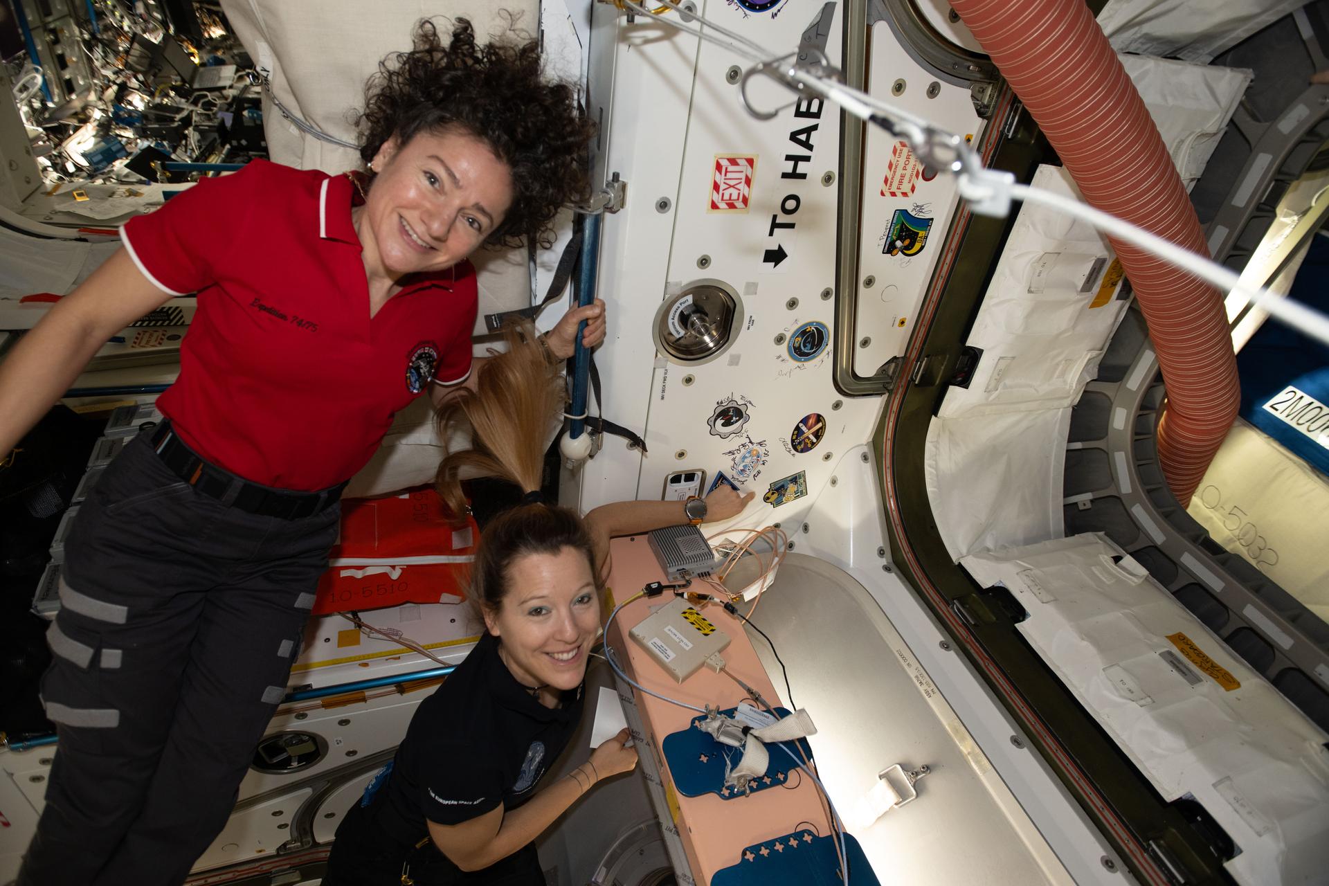
Expedition 74 flight engineers, from left, Jessica Meir of NASA and Sophie Adenot of ESA (European Space Agency), show off a Cygnus mission sticker for Northrop Grumman's 24th cargo mission to the International Space Station. The sticker is affixed to the vestibule between the orbital outpost's Earth-facing port on the Unity module and the company’s Cygnus XL cargo spacecraft.
Astronauts, from left, Jessica Meir and Sophie Adenot show off a Cygnus mission sticker for Northrop Grumman’s 24th cargo mission to the International Space Station.
NASA/Chris Williams

The Expedition 74 crew explored how weightlessness affects cartilage growth and the digestive system on Thursday to protect crew health and improve patient care on Earth. The orbital residents are also gearing up for a robotics maintenance spacewalk at the end of the month on the International Space Station.

Conducting research in the microgravity environment of the orbiting laboratory provides unique insights unobtainable in Earth’s gravity. This helps scientists, doctors, and engineers develop space-influenced therapies, medicines, and products leading to advances in human health, industrial processes, spacecraft designs, and more.

Cartilage cells are growing aboard the orbital outpost that may inform new ways to treat disabilities and repair injuries. NASA flight engineers Jessica Meir and Chris Williams worked together on the biotechnology investigation using the Kibo laboratory module’s Life Science Glovebox. Williams retrieved the cartilage cell samples preserved in a science freezer then thawed them to begin scientific operations. Next, Meir nourished the cell samples inside the glovebox and stowed them inside a research incubator so they could begin growing. Manufacturing cartilage tissues in space could lead to self-repairing implants on Earth and advanced fitness techniques for astronauts on a long-term spaceflight.

Roscosmos cosmonauts Sergey Kud-Sverchkov and Sergei Mikaev joined each other after breakfast and scanned their abdominal region using an ultrasound device for a digestion study. The real-time biomedical data will give doctors insight into how microgravity affects the blood flow and shape of a crew member’s digestive organs after a meal. Results may help advance methods to monitor and improve digestive health both on Earth and in space.

Meir and flight engineer Sophie Adenot of ESA (European Space Agency) also conducted ultrasound scans using ESA’s EchoFinder-2 device to image the major blood vessels and key organs in their abdomen. The human research experiment uses augmented reality to assist with the scans and artificial intelligence to recognize organs. The lightweight, easy-to-use gear may enable independent crew health monitoring on spacecraft travelling to the Moon, Mars, and beyond.

Adenot then joined NASA flight engineer Jack Hathaway and studied procedures necessary to prepare astronauts for a spacewalk. The duo reviewed spacesuit handling techniques, Quest airlock pressurization and depressurization steps, and emergency responses. Adenot also partnered with Meir and Williams and serviced the lithium-ion batteries that power the spacesuits. NASA will soon announce the two astronauts who will exit the space station on June 30 to repair a wrist joint on the Canadarm2 robotic arm.

Roscosmos flight engineer Andrey Fedyaev had a busy day of science and maintenance beginning his shift downloading and reading data collected from a station radiation detector. Afterward, Fedyaev transferred water from the Progress 94 resupply ship into tanks inside the orbital outpost’s Roscosmos segment. Finally, the two-time cosmonaut wrapped up his shift testing artificial intelligence tools to boost crew efficiency and communications in space.

Learn more about station activities by following the space station blog, @space_stationon X, as well as the ISS Facebook and ISS Instagram accounts.

Get the latest from NASA delivered every week. Subscribe here.

The post Advanced Tech on Station Informing Space-Designed Health Treatments appeared first on NASA Science.

Scientists Find a New Way to Fight Alzheimer’s After 20 Years of Research

18 June 2026 at 12:22

Alzheimer’s disease gradually steals memories, independence, and quality of life from millions of people worldwide. Despite decades of research, scientists still struggle to find treatments that can stop the disease from progressing. Most available medications provide only modest benefits and cannot fully prevent the loss of brain cells that characterizes the condition. A new study […]

The post Scientists Find a New Way to Fight Alzheimer’s After 20 Years of Research appeared first on Knowridge Science Report.

AI Model Emerges as a Game-Changer in Tumor Assessment: Advancing Care for Mesothelioma Patients and Physicians

18 June 2026 at 00:40

Physicians and researchers at the Netherlands Cancer Institute have unveiled a groundbreaking artificial intelligence (AI) model that fundamentally reshapes how treatment responses in pleural mesothelioma—a notoriously challenging cancer—are evaluated. This model, called ARTIMES, excels beyond traditional clinical methods, surpassing expert human judgment in accuracy and efficiency. By precisely measuring the entire tumor volume instead of relying on conventional diameter-based assessments, ARTIMES promises to revolutionize patient care and accelerate clinical research in this difficult-to-treat disease.

Pleural mesothelioma poses unique diagnostic challenges because it develops as a thin, irregular layer along the lining of the lungs rather than forming discrete masses. This morphology renders existing international standards like the RECIST (Response Evaluation Criteria in Solid Tumors) inadequate. RECIST depends primarily on simple diameter measures, which poorly represent the tumor’s true progression or regression in mesothelioma’s diffuse growth pattern. Clinicians have expressed frustration and uncertainty in evaluating treatment efficacy using these parameters, highlighting a pressing need for a more refined and reliable approach.

To address these limitations, an interdisciplinary team of AI scientists, radiologists, and pulmonologists collaborated at the Netherlands Cancer Institute. Leveraging an extensive dataset comprising over 11,000 computed tomography (CT) scans from more than 2,000 patients across 121 hospitals worldwide, they developed ARTIMES, an AI-driven volumetric response evaluation tool. Unlike humans, who face near-impossible challenges in manually delineating tumor boundaries at the pixel level on complex images, ARTIMES can effortlessly segment entire tumors and calculate their true volume with exceptional precision.

Pulmonologist Sjaak Burgers emphasizes that ARTIMES advances clinical practice by eliminating tedious and error-prone manual tumor assessments. While verifying the AI’s output remains essential, the review process is far less labor-intensive. This reduces interobserver variability and enables clinicians to obtain more consistent and objective insights into tumor dynamics. The ability to evaluate the full tumor burden rather than a single diameter mark dramatically increases sensitivity for detecting true positive or negative treatment responses.

The scientific community is witnessing a milestone with ARTIMES being the first AI model worldwide to demonstrably outperform clinicians in assessing treatment outcomes for pleural mesothelioma. Kevin Groot Lipman, lead author and technical physician, highlights that their study, published in The Lancet Oncology, cements AI’s potential to become an integral clinical decision support tool. Importantly, ARTIMES enhances rather than replaces physician judgment, interfacing smoothly into existing workflows while enabling rapid, data-driven decision-making.

Beyond measuring tumor volume, the researchers have undertaken the critical task of integrating ARTIMES measurements into actionable clinical guidelines. Since knowing the tumor size alone does not dictate specific treatment changes, these criteria empower pulmonologists to determine when to modify or cease therapies. This synergy ensures that patients receive individualized care tailored to their tumor behavior patterns, reducing exposure to ineffective treatments and unnecessary side effects while optimizing healthcare resources.

One of ARTIMES’s most transformative capabilities is its ability to detect non-response to therapy earlier than ever before. This timely recognition allows physicians to pivot treatment plans sooner, offering patients alternative therapeutic avenues or sparing them from futile and potentially harmful continuation of ineffective regimens. The combination of predictive accuracy and clinical oversight represents a major leap forward in precision oncology for pleural mesothelioma patients.

Currently, EU regulations restrict ARTIMES’s use exclusively to the Netherlands Cancer Institute under an in-house exemption, given that the model was developed internally. Nonetheless, the research team is actively pursuing regulatory approval to deploy ARTIMES globally in other hospitals. There is hopeful anticipation surrounding proposed EU frameworks aimed at streamlining the certification process for AI-enabled medical devices, which could accelerate widespread adoption and patient benefit.

The advent of ARTIMES is poised to deliver a shockwave across oncology fields by demonstrating the tangible superiority of AI over human evaluators in complex tumor assessments. To foster transparency and collaborative innovation, the Netherlands Cancer Institute has made the mesothelioma AI model publicly accessible online, enabling researchers worldwide to explore and extend its applications. This open science approach is expected to catalyze new studies and adaptations for other tumor types with challenging morphologies.

Already, the NKI team is extending their AI methodologies to address lung cancer and brain metastasis tumor evaluations. The success of ARTIMES signals the dawn of a new epoch in oncological imaging, where volumetric and morphological complexities that previously hindered precise quantification become tractable. Such breakthroughs unlock significant potential to enhance clinical trial design by furnishing robust, reproducible endpoints that more faithfully capture therapeutic impact.

Clinical trials for novel treatments stand to gain markedly from ARTIMES’s introduction. Using data from eight distinct trials, the research team validated that AI-guided volumetric criteria yield significantly improved accuracy relative to traditional RECIST assessments. This refined precision enables better evaluation of an investigational drug’s efficacy, ultimately accelerating regulatory approval timelines and facilitating faster patient access to promising therapies.

In summary, ARTIMES exemplifies how synergistic integration of AI and clinical expertise can surmount longstanding challenges in tumor measurement. By transitioning from simplistic unidimensional diameter metrics to comprehensive volumetric analysis, this technology brings unprecedented clarity and confidence to oncological decision-making. As this AI model becomes more widely disseminated and refined, it heralds a paradigm shift in cancer treatment evaluation, with rippling benefits for patients, clinicians, and research worldwide.


Subject of Research: People

Article Title: Development and validation of artificial intelligence-assisted volumetric response criteria in pleural mesothelioma (ARTIMES): a retrospective, multicohort, multicentre study

News Publication Date: 17-Jun-2026

Web References:

References:
Groot Lipman K, Burgers S, et al. Development and validation of artificial intelligence-assisted volumetric response criteria in pleural mesothelioma (ARTIMES): a retrospective, multicohort, multicentre study. The Lancet Oncology, 2026.

Image Credits: ©Netherlands Cancer Institute

Keywords: Cancer treatments, Artificial intelligence, Imaging analysis, Pleural mesothelioma, Tumor volumetrics, Clinical trials, Precision oncology

iSCORE-PD: Stem Cells Advance Parkinson’s Research

17 June 2026 at 23:57

In an unprecedented advancement for neurodegenerative disease research, a team led by Busquets, Li, Syed, and colleagues has unveiled iSCORE-PD, an innovative isogenic stem cell repository designed explicitly to accelerate Parkinson’s disease studies. Published in Nature Communications in 2026, this comprehensive collection represents a pivotal leap toward resolving the complex pathophysiology of Parkinson’s, a condition affecting millions worldwide yet still eluding curative therapies. The iSCORE-PD platform promises to redefine experimental paradigms by providing scientists with genetically matched cellular models that mirror the intricate genetic underpinnings of Parkinson’s disease.

Parkinson’s disease is characterized primarily by the progressive loss of dopaminergic neurons in the substantia nigra pars compacta, leading to debilitating motor symptoms such as tremors, rigidity, and bradykinesia, alongside a spectrum of non-motor complications. Despite extensive research, there remains a conspicuous gap between clinical observations and mechanistic understanding at the cellular and molecular levels. Existing animal models and conventional stem cell lines often fall short in recapitulating the exact human pathology. The iSCORE-PD initiative addresses this deficiency head-on by harnessing isogenic stem cell lines, which are genetically identical except for defined Parkinson’s-relevant mutations, thus enabling unprecedented control over genetic variables.

The technical foundation of iSCORE-PD relies on advanced genome editing methodologies, most notably CRISPR/Cas9 technology. By precisely introducing or correcting mutations in key genes implicated in Parkinson’s pathogenesis such as SNCA, LRRK2, PINK1, and PARKIN, researchers have generated isogenic pairs of induced pluripotent stem cells (iPSCs). These iPSC lines can subsequently be differentiated into dopaminergic neurons or other relevant cell types, facilitating side-by-side comparisons that isolate the effects of specific genetic alterations without the confounding noise of inter-individual genetic variability.

One of the remarkable features of the iSCORE-PD collection is its extensive genomic validation pipeline. Whole genome sequencing and transcriptomic analyses are systematically employed to ensure the integrity and authenticity of the edited lines, confirming that off-target effects are minimized and that the genetic landscape conforms precisely to intended edits. This meticulous validation is crucial for downstream experiments intending to dissect subtle cellular phenotypes attributable to Parkinson’s mutations with a high degree of confidence.

The availability of these isogenic cell lines dramatically enhances the fidelity of disease modeling. Scientists can now observe, in real time and within a human cellular context, the cascade of pathological events triggered by individual mutations. For instance, iSCORE-PD lines harboring the G2019S mutation in LRRK2 have already demonstrated distinct lysosomal dysfunction and mitochondrial impairment, hallmarks of Parkinson’s-related neurodegeneration. These insights provide vital clues about the molecular mechanisms that underpin neuronal vulnerability and death.

Beyond mechanistic studies, iSCORE-PD serves as an invaluable platform for drug discovery and therapeutic screening. High-throughput assays conducted on these standardized cellular models allow for robust evaluation of candidate compounds’ efficacy and toxicity. The isogenic background ensures that drug responses can be directly attributed to genetic contributors and not extraneous genomic differences, thereby accelerating the identification of promising therapeutic leads with increased translational potential.

Moreover, the iSCORE-PD repository supports integrative omics approaches. By coupling proteomics, metabolomics, and epigenomic profiling with the genetically defined cell lines, researchers can generate comprehensive molecular atlases elucidating the multifaceted alterations engendered by Parkinson’s-associated mutations. Such data-rich resources augment our understanding of disease heterogeneity and may reveal novel biomarkers for early diagnosis and progression monitoring.

The initiative also pioneers robust methodologies for modeling non-cell autonomous effects in Parkinson’s pathology. By co-culturing isogenic dopaminergic neurons with glial cells or other brain cell types derived from similarly engineered iPSC lines, investigators can explore the cell–cell interactions and inflammatory pathways that contribute to disease propagation. These complex in vitro systems increasingly resemble the in vivo brain microenvironment, bridging translational gaps.

In addition to its scientific hinge, iSCORE-PD represents a significant step forward in reproducibility and collaborative science. By providing open-access to a rigorously characterized and genetically standardized stem cell collection, the project promotes harmonization of experimental models across laboratories worldwide. This standardization mitigates discrepancies often observed in Parkinson’s research due to genetic heterogeneity of patient-derived cells and disparate culture conditions.

Ethical considerations have been front and center in the development and dissemination of the iSCORE-PD lines. The team has implemented stringent protocols to ensure donor anonymity, informed consent, and compliance with international guidelines for stem cell research. This ethical diligence is fundamental to fostering public trust and enabling broad scientific utilization with responsibility.

The iSCORE-PD research consortium envisions future expansion of the collection to encompass additional mutations, gene variants, and epigenetic modifications implicated in Parkinson’s disease, thereby creating an ever-more comprehensive toolkit for the field. This evolving repository will facilitate the study of gene–environment interactions and provide models for the sporadic forms of Parkinson’s that constitute the majority of cases.

Importantly, the development of iSCORE-PD aligns synergistically with advances in single-cell sequencing and live-cell imaging technologies. These combined approaches empower real-time monitoring of neuronal dynamics, synaptic connectivity, and intracellular trafficking in disease-relevant contexts. Such capabilities promise to unravel the temporal progression of neuronal dysfunction in unprecedented detail.

The translational impact of iSCORE-PD extends beyond fundamental research, holding profound implications for personalized medicine. Patient-specific isogenic lines can be derived, enabling evaluation of individualized drug responses and prediction of disease progression trajectories. This precision medicine paradigm could revolutionize clinical management and therapeutic development for Parkinson’s patients.

Finally, iSCORE-PD serves as a testament to the power of interdisciplinary collaboration, integrating expertise in stem cell biology, genome editing, neuroscience, bioinformatics, and clinical neurology. This concerted effort exemplifies how cutting-edge technologies, when harnessed collectively, can illuminate complex diseases and hasten the arrival of effective treatments.

In sum, the iSCORE-PD isogenic stem cell collection stands as a groundbreaking resource that transforms the landscape of Parkinson’s disease research. By providing genetically precise, highly validated cellular models, it enables unparalleled mechanistic insights, drug discovery opportunities, and personalized medicine applications. As the Parkinson’s field grapples with its most urgent challenges, iSCORE-PD promises to be a beacon guiding innovative interventions and ultimately improving patient outcomes worldwide.


Subject of Research: Parkinson’s disease; isogenic stem cell models; neurodegeneration; genome editing; induced pluripotent stem cells.

Article Title: iSCORE-PD: an isogenic stem cell collection to research Parkinson’s disease.

Article References:

Busquets, O., Li, H., Syed, K.M. et al. iSCORE-PD: an isogenic stem cell collection to research Parkinson’s disease.
Nat Commun (2026). https://doi.org/10.1038/s41467-026-74355-8

Image Credits: AI Generated

New Study Reveals Mechanisms Behind High Iron Levels in Colorectal Cancer Cells

17 June 2026 at 23:16

University of Michigan Rogel Cancer Center researchers have unveiled a groundbreaking metabolic mechanism that colorectal cancer cells exploit to maintain exceptionally high iron levels, a discovery that opens promising avenues for targeted cancer therapies. Published recently in Cell Metabolism, this study provides an unprecedented insight into how tumor cells sidestep iron toxicity and evade a form of cell death known as ferroptosis, potentially revolutionizing the understanding of metal metabolism in cancer biology.

Colorectal cancer cells are known to harbor elevated iron concentrations, far surpassing those found in healthy cells. Iron is a double-edged sword in cellular physiology—essential for processes like DNA synthesis and cell proliferation but lethal in excess due to its propensity to generate harmful reactive oxygen species. Ordinarily, cells with excessive iron succumb to ferroptosis, a specialized form of oxidative cell death driven by iron-mediated lipid peroxidation. Tumor cells subvert this natural safeguard, sustaining iron overload without triggering their own demise, but how they achieve this has remained elusive—until now.

The investigative team led by Dr. Yatrik Shah, Horace W. Davenport Collegiate Professor of Physiology at Michigan Medicine, employed a metabolism-directed CRISPR screening approach to systematically dissect the pathways protecting colorectal cancer cells from iron-induced oxidative damage. Surprisingly, canonical ferroptotic enzymes, previously presumed central to this resistance, were found non-essential for tumor survival. This redirected focus led the scientists to explore mitochondrial metabolism more profoundly.

Their research unveiled that the mitochondrial enzyme complex II plays a pivotal role in safeguarding cancer cells from iron-induced toxicity. Complex II regulates coenzyme Q (CoQ) within mitochondria, a key antioxidant molecule that quells oxidative stress. By fine-tuning CoQ’s redox state, complex II effectively buffers the destructive potential of accumulated iron, preventing ferroptosis and enabling cancer cell proliferation. When researchers knocked out complex II in colorectal cancer models, iron toxicity became unmanageable for the tumor cells, leading to widespread cell death and marked tumor growth inhibition.

Crucially, complex II’s protective mechanism appears specific to the high-iron environment of cancer cells. In mouse models, disruption of complex II elicited negligible adverse effects on normal tissues, underscoring the therapeutic potential of selectively targeting this mitochondrial axis in colorectal cancer. This specificity addresses a significant hurdle in oncology: minimizing treatment toxicity while maximizing antitumor efficacy.

Further intricacies emerged as the study revealed a feedback loop wherein iron itself modulates complex II activity, suggesting a sophisticated regulatory axis that maintains iron homeostasis within tumors. This bidirectional interaction offers additional molecular targets for disrupting iron tolerance in cancer cells and deepening our understanding of tumor metabolism.

These findings represent a paradigm shift from earlier hypotheses centered on canonical ferroptosis regulators, highlighting the necessity of focusing on mitochondrial metabolism in cancer research. By leveraging sophisticated genome-editing tools and bioenergetic profiling, the Rogel Cancer Center team charted a novel course for drug discovery efforts aimed at crippling iron addiction—a hallmark of not only colorectal but potentially many other malignancies.

The next phase of this research endeavors to identify and develop potent inhibitors of complex II or its associated metabolic pathways. Given that dysregulated iron metabolism is a common vulnerability across diverse cancer types, these interventions could herald a new era of broad-spectrum anticancer strategies. Moreover, detailed characterization of iron complex II interplay might uncover additional metabolic dependencies exploitable for therapeutic gains.

This transformative research underscores the intricate metabolic adaptations that empower colorectal cancers to circumvent intrinsic iron toxicity constraints. By illuminating the heme-complex II axis as a linchpin in maintaining oxidative balance amid iron overload, it offers a highly selective target for the design of next-generation anticancer agents.

Researchers anticipate that combining complex II inhibition with other therapies may amplify treatment responses and overcome resistance mechanisms. As the quest to outsmart cancer evolves, this study reinforces the vital role of mitochondrial metabolism understanding in crafting innovative clinical interventions.

In sum, the University of Michigan team’s discovery of complex II’s role in buffering iron toxicity not only deciphers a long-standing biological enigma but also charts a compelling translational pathway. It epitomizes how fundamental metabolic insights can accelerate the development of precision treatments conferring hope to millions affected by colorectal cancer worldwide.

Subject of Research: Cells
Article Title: Iron addicted colorectal cancers exploit heme-complex II axis to resist oxidative cell death
News Publication Date: June 17, 2026
Web References: http://dx.doi.org/10.1016/j.cmet.2026.04.020
References: “Iron addicted colorectal cancers exploit heme-complex II axis to resist oxidative cell death,” Cell Metabolism, DOI: 10.1016/j.cmet.2026.04.020
Image Credits: Shah Lab, Rogel Cancer Center
Keywords: Colorectal cancer, iron metabolism, ferroptosis, complex II, coenzyme Q, mitochondrial metabolism, oxidative cell death, tumor metabolism, cancer therapy

New Study Suggests Microplastics Could Aggravate Fatty Liver Disease

17 June 2026 at 23:15

Microplastics: Unseen Threats to Liver Health Revealed Through Cutting-Edge Spatial Transcriptomics

In recent years, microplastics have emerged as pervasive environmental contaminants, infiltrating virtually every corner of the globe. These minuscule plastic particles, derived from the breakdown of larger plastic debris, have invaded air, water, and soil, exposing humans to continuous contact via inhalation, ingestion, and dermal absorption. Despite the ubiquity of microplastics, understanding their direct impact on biological systems has remained a formidable challenge within the scientific community. A groundbreaking study by researchers at the University of Oklahoma, recently published in the journal Science Advances, delves into this critical area by examining how microplastics particularly affect liver health under dietary stress conditions.

Tae Gyu Oh, Ph.D., an assistant professor of oncology science at the University of Oklahoma College of Medicine and lead author of the study, highlights the pressing concern: “Exposure to microplastics is inevitable. Their presence in human tissues has been confirmed across multiple studies. However, we wanted to investigate how microplastic exposure interacts with a high-fat, high-cholesterol diet, known to independently induce liver damage.” The study offers compelling evidence that the combination of a typical Western diet and microplastic exposure exacerbates liver injury, potentially accelerating the progression of metabolic liver diseases.

Central to the study is the focus on polyethylene, the most prevalent plastic polymer globally, commonly found in everyday items such as plastic bags and milk containers. The research team administered polyethylene microplastics to mice over eight weeks, with one cohort receiving a standard diet and another subjected to a diet mimicking metabolic dysfunction-associated steatohepatitis (MASH). This severe form of fatty liver disease is characterized by inflammation and liver cell damage, often culminating in cirrhosis and liver failure if untreated.

The findings were striking: mice consuming the high-fat diet alongside microplastic exposure exhibited blood markers indicating liver injury more than twice as elevated as those on a standard diet experiencing similar exposure. This synergistic effect underscores the intricate interplay between environmental pollutants and diet-induced metabolic stressors, intensifying hepatic damage beyond what each factor causes independently.

To unravel the molecular and cellular underpinnings of this phenomenon, the research employed an array of sophisticated analytical techniques, culminating in the use of spatial transcriptomics. Unlike conventional bulk transcriptomic approaches, which average gene expression across millions of cells and can obscure localized responses, spatial transcriptomics enables researchers to map transcriptional activity within intact tissue sections at near single-cell resolution. This technique revealed precise “hot spots” of inflammation and tissue injury within the liver, a breakthrough insight unattainable by earlier methodologies.

Analysis of gene regulatory networks through spatial transcriptomics indicated a pivotal role for PPAR-alpha (peroxisome proliferator-activated receptor-alpha), a nuclear receptor that orchestrates fat metabolism and energy homeostasis in liver cells. PPAR-alpha appears to engage in cross-talk with Anxa2, a gene implicated in tissue repair and membrane dynamics. The altered activity of this axis in microplastic-exposed livers suggests that microplastics may disrupt the liver’s natural defense and regenerative processes, impairing its capacity to recover from metabolic insults.

This discovery has profound implications for understanding the mechanistic pathways by which environmental contaminants like microplastics contribute to liver pathology. The perturbation of PPAR-alpha and Anxa2 signaling potentially links microplastic exposure with the dysregulation of lipid metabolism and compromised repair, exacerbating the severity of conditions such as nonalcoholic fatty liver disease (NAFLD) and MASH.

While these findings were generated in a murine model, they establish an essential framework that informs potential human health risks. Given the parallels between murine and human liver physiology, it is plausible that microplastic exposure combined with high-fat diets could similarly predispose humans to aggravated liver damage. However, the researchers caution that further studies are necessary to confirm this translation and to elucidate the long-term implications for populations worldwide.

Dr. Oh emphasizes the broader relevance of this research: “Microplastics are now inextricably linked to daily life, yet their biological impact is only beginning to be understood. Through advanced spatial transcriptomic mapping, we have visualized the precise loci of hepatic damage induced by microplastics, revealing a novel environmental dimension to liver disease pathogenesis.” This nuanced comprehension paves the way for future investigations targeting environmental and dietary risk factors in liver health.

Moreover, these insights open avenues for therapeutic targeting. Modulating the PPAR-alpha-Anxa2 pathway could become a strategy to mitigate microplastic-induced liver injury or fortify the liver’s resilience against environmental toxins. Understanding such molecular crosstalk also facilitates improved diagnostic markers sensitive to environmental damage, allowing for earlier intervention in vulnerable populations.

This pioneering study exemplifies the intersection of environmental health, genomics, and hepatology, demonstrating how innovative technologies can elucidate complex biological interactions. It underscores the urgent need to address microplastic pollution not only as an ecological crisis but as a public health priority, particularly in societies where high-fat diets are prevalent.

As humanity grapples with escalating plastic waste and its fragmentary descent into invisible pollutants, research such as this serves as a clarion call for comprehensive strategies. Reducing plastic production, enhancing waste management, and fostering healthier dietary practices collectively form the cornerstone of mitigating hidden dangers to liver health and overall well-being.

The University of Oklahoma study, titled “Spatial Transcriptome Mapping Identifies Ppara-Anxa2 Crosstalk in Microplastic-Induced Hepatotoxicity,” stands as a seminal contribution offering unprecedented mechanistic clarity. Through employing spatial transcriptomics, the researchers have achieved a level of resolution that redefines how environmental toxicology and metabolic disease research can coalesce to confront emergent health threats posed by our plastic-saturated environment.

Subject of Research: Animals
Article Title: Spatial transcriptome mapping identifies Ppara-Anxa2 cross-talk in microplastic-induced hepatotoxicity
News Publication Date: 17-Jun-2026
Web References: https://doi.org/10.1126/sciadv.aec8681
References: Oh, T.G., Jung, W., Joshi, A.D., et al. Spatial Transcriptome Mapping Identifies Ppara-Anxa2 Crosstalk in Microplastic-Induced Hepatotoxicity. Science Advances, 2026.
Image Credits: University of Oklahoma
Keywords: Microplastics, Liver Disease, Fatty Liver Disease, Polyethylene, High-Fat Diets, Spatial Transcriptomics, PPAR-alpha, Anxa2, Hepatotoxicity, Environmental Health, Metabolic Dysfunction, Inflammation

Scientists Awarded $4 Million to Enhance Endangered Species Management on Military Lands

17 June 2026 at 23:14

Across the expansive military installations of the United States, where substantial tracts of restricted-access natural lands persist, an evolving challenge has taken shape: how to effectively conserve endangered species amid overlapping and often conflicting recovery plans. These lands, which serve dual purposes of defense readiness and environmental stewardship, present a unique ecological puzzle. Amid this complexity, researchers at Virginia Tech’s College of Natural Resources and Environment have embarked on pioneering projects funded by two $2 million grants, aiming to redefine conservation strategies on military properties by emphasizing ecosystem-wide management rather than isolated species-specific interventions.

Historically, wildlife management on military bases has heavily relied on individual species’ recovery plans, a methodology that becomes problematic when multiple endangered species inhabit the same territory. Each species’ plan often prescribes distinct conservation actions, potentially leading to redundant efforts, increased management complexity, and sometimes conflicting directives. Recognizing these inefficiencies, the Virginia Tech research initiative seeks to develop integrated strategies that optimize land management actions to benefit diverse species assemblages simultaneously. This integrative approach has the potential not only to streamline operational practices on military lands but also to serve as a scalable model for ecosystems worldwide grappling with multifaceted conservation demands.

One cornerstone of this research is led by Assistant Professor Elizabeth Hunter, who operates within the U.S. Geological Survey’s Virginia Cooperative Fish and Wildlife Research Unit. Hunter’s project area in southeastern Georgia—including large military bases characterized by fire-managed ecosystems—examines the application of prescribed burning as a habitat management tool. Prescribed fire has traditionally been tailored around a handful of focal species, such as the red-cockaded woodpecker, leaving many other species with differing ecological needs inadequately addressed. Hunter’s work challenges this paradigm by employing cutting-edge monitoring technologies to survey a wide spectrum of taxa, from avian and mammalian species to amphibians, reptiles, insects, and crucial pollinators, aiming to capture a comprehensive biological response to various fire regimes.

By integrating extensive biodiversity data into a sophisticated multi-objective optimization framework, Hunter’s team can evaluate how different prescribed burning strategies affect numerous species simultaneously. This analytical tool allows for the assessment of trade-offs and synergies in conservation outcomes, addressing complex ecological interactions. The methodology asks crucial questions such as whether it is possible to adjust burn frequency, intensity, and spatial patterns to enhance habitats for multiple species concurrently. Such questions are vital for military land managers, who must balance ecosystem health with operational imperatives. Collaborations with organizations like the Orianne Society and the National Council for Air and Stream Improvement anchor this work in both academic and applied conservation contexts.

Parallel to Hunter’s fire ecology focus, Associate Professor Haldre Rogers directs research tackling the inefficiencies inherent in the current species-by-species recovery plan framework under the U.S. Endangered Species Act. Rogers’ project, focused on the Marianas Islands, explores whether ecosystem-wide habitat management can serve as an effective surrogate for the management of multiple individually listed species. Given that military lands often harbor dozens of endangered species with overlapping but separate recovery plans, the question arises: can habitat proxies be identified that allow for effective ecosystem-level conservation without the cumbersome requirement of separate interventions and monitoring for each species?

Rogers utilizes innovative ecosystem hypergraph modeling to encapsulate the intricate web of species interactions, habitat connectivity, and life-history traits, thereby identifying clusters of species that might respond similarly to shared management actions. This approach strives to uncover ecological congruencies and potential conflicts, aiming to inform management choices that maximize conservation benefits while minimizing inadvertent harm to co-occurring species. The project acknowledges inevitable ecological trade-offs but aspires to equip land managers with a landscape-scale understanding that promotes more efficient and informed decision-making.

The ecological importance of military installations in the United States cannot be overstated. Many contain well-preserved habitats that have been safeguarded from urban development, offering refugia for imperiled species. Activities such as prescribed burning and restricted access have inadvertently preserved biodiversity hotspots. At the same time, these installations must fulfill critical defense and training missions, underscoring the need for a delicate balance between operational readiness and environmental stewardship. The innovations proposed by Hunter, Rogers, and their teams promise a new frontier in reconciling these dual mandates through science-driven, scalable conservation models.

Their research partnership traverses multiple disciplines and institutions, spanning Virginia Tech’s Department of Fish and Wildlife Conservation and Statistics, collaborations with Rice University, Brown University, MIT, and contributions from postdoctoral scholars. This transdisciplinary approach enriches the projects with a fusion of ecological expertise, advanced computational modeling, and applied management perspectives. By leveraging emerging technologies and sophisticated analytical tools, these projects are poised to generate transformative insights relevant to conservation practitioners, policymakers, and military land managers alike.

In essence, these Virginia Tech-led studies embody a paradigm shift: transitioning from fragmented, species-specific management plans to holistic ecosystem-based strategies is both a necessity and an opportunity. This shift acknowledges the complex interdependencies among species and their habitats, calling for innovative methodologies that embrace ecological complexity rather than oversimplify it. For military lands, where the intersection of conservation and operational use is most pronounced, such research can catalyze both more effective biodiversity protection and enhanced land-use efficiency.

By integrating large-scale biological monitoring with computational optimization and ecosystem modeling, this research presents a robust framework for addressing the mounting challenges of endangered species management amid competing land uses. The implications extend far beyond military properties, offering a strategic blueprint adaptable to diverse ecosystems facing overlapping conservation mandates. The collective efforts spearheaded by Hunter and Rogers represent a significant leap forward in safeguarding the nation’s biological heritage through science-guided stewardship, setting the stage for broader applications in the global conservation community.

As endangered species faces continue to escalate globally, the insights arising from military installation ecosystems can inform best practices worldwide. The integration of multi-species data into management decisions, the recognition of ecosystem processes over isolated species needs, and the embracing of technological and analytical innovation underscore the future of conservation science. In a world of finite conservation resources and competing priorities, finding operationally feasible yet ecologically sound approaches is paramount.

These projects, supported by the Strategic Environmental Research and Development Program, exemplify such an approach, bridging theoretical science and practical land management. Their success will not only enhance the resilience of endangered species populations on military lands but also serve as a clarion call to redefine conservation paradigms universally. Ultimately, their work invites a transformative dialogue around how humanity navigates the complex stewardship of nature amid modern societal demands.


Subject of Research:
Endangered species management on military lands through ecosystem-based strategies and multi-species conservation optimization.

Article Title:
Rethinking Conservation: Integrated Ecosystem Management for Endangered Species on Military Installations

News Publication Date:
Not provided

Web References:

Image Credits:
Photo courtesy of Elizabeth Hunter.

Keywords:
Endangered species, Extinction, Wildlife management, Conservation biology, Ecosystem management, Prescribed fire, Habitat connectivity, Multi-species optimization, Military land conservation, Ecosystem hypergraphs.

Radiation Therapy Boosts Immune Response in Brain Metastases, Enhancing Treatment Effectiveness

17 June 2026 at 23:10

In a groundbreaking study led by The University of Texas MD Anderson Cancer Center, researchers have unveiled compelling insights into how radiation therapy can profoundly influence the immune landscape of brain metastases, opening new avenues for combinational immunotherapeutic strategies. Brain metastases, secondary tumors that originate from cancers elsewhere in the body and colonize the brain, represent one of the most formidable clinical challenges in oncology. Their treatment has been notoriously difficult due to the highly immunosuppressive nature of the brain tumor microenvironment and the protective blood-brain barrier, which together blunt the effectiveness of conventional immunotherapies.

Despite the advances in immune checkpoint inhibitors and cellular therapies that have revolutionized treatment for many cancers, these modalities have shown limited success in brain metastases. The prevailing concept attributes this resistance largely to the “cold” immune microenvironment within the brain, characterized by suppressed immune activation and poor T cell infiltration. Radiation therapy, traditionally employed for its cytotoxic effects on cancerous cells, has garnered interest as a potential immunomodulatory agent. However, the precise mechanisms by which radiation alters immune dynamics within brain metastases remained elusive until now.

Leveraging an integrative approach encompassing RNA sequencing and T cell receptor (TCR) profiling on samples from over 300 patients harboring brain metastases sourced predominantly from breast and lung primaries, the research team decoded the complex interplay between radiation and immune activation. Their findings, recently published in Clinical Cancer Research, reveal that pre-operative radiation not only directly damages tumor DNA but also reshapes the tumor microenvironment through enhanced recruitment and activation of cytotoxic T lymphocytes. This dual impact fundamentally transforms the previously immunologically inert metastatic lesions into immunologically responsive entities.

Mechanistically, radiation induces the release of tumor-associated antigens and danger signals, which act as potent stimulators for antigen-presenting cells and infiltrating lymphocytes. This cascade prompts an upregulation of inflammatory cytokines and immune checkpoints, effectively increasing tumor visibility to the immune system. Interestingly, radiation also modulates tumor vasculature, facilitating enhanced permeability and immune cell trafficking into the tumor bed. Concurrently, suppressive myeloid populations within the microenvironment are either depleted or reprogrammed, further alleviating immunosuppression.

One of the study’s pivotal revelations centers on T cell receptor diversity as a prognostic biomarker. Enhanced TCR diversity corresponds with better immunotherapeutic outcomes, suggesting that radiation-induced augmentation of this diversity might predict patient responsiveness. Such biomarkers hold immense clinical promise, enabling personalized treatment algorithms that optimize radiation-immunotherapy combinations tailored to individual tumor immune profiles.

Dr. Jason Huse, M.D., Ph.D., a leading pathologist at MD Anderson and co-senior author, emphasizes the transformative nature of these findings. “Our results demonstrate that instead of merely bypassing the blood-brain barrier, we should recalibrate our therapeutic focus toward the tumor microenvironment itself. Radiation effectively ‘primes’ these metastases, enabling robust immune engagement that was previously unattainable. This paradigm shift paves the way for clinical trials harnessing synergistic radiation and immunotherapy regimens.”

This shift is particularly significant given the limitations of systemic therapies in penetrating brain lesions. By directly modifying the local immune milieu, radiation circumvents longstanding pharmacologic barriers and leverages the body’s own immune arsenal for tumor eradication. The implications extend beyond localized control; emerging evidence hints at the potential for abscopal effects where immunity induced in the irradiated site mediates systemic antitumor responses.

The research incorporated tissue specimens from a contemporaneous clinical trial spearheaded by Dr. Debra Nana Yeboa, which contrasted pre-operative and postoperative radiation regimens. This comparative analysis reinforced the immunomodulatory superiority of pre-operative radiation, highlighting its capacity to bolster immune infiltration and activation before surgical tumor resection.

While the experimental findings are preliminary and retrospective, the investigators are actively validating these observations in larger, prospective clinical cohorts. The overarching goal is to delineate optimized therapeutic windows and dosing sequences that exploit radiation’s immunogenic potential in concert with checkpoint inhibitors, vaccines, or adoptive cell therapies.

In summary, this study delineates a critical and previously underappreciated function of radiation therapy in rendering brain metastases immunologically “hot.” By integrating radiation with immunotherapeutic modalities, there is a tangible prospect of overcoming the entrenched resistances posed by the brain’s unique protective and suppressive environment. This work marks a pivotal step toward improved survival rates and quality of life for patients afflicted with metastatic brain tumors.

As the oncology community eagerly anticipates forthcoming clinical trials, this research provides an urgent biological rationale for a comprehensive therapeutic strategy that targets both tumor cells and their microenvironment. The convergence of radiation biology and immunotherapy represents a new frontier with the capacity to redefine standard-of-care paradigms for brain metastases.


Subject of Research: The immunomodulatory effects of radiation therapy on brain metastases and the tumor immune microenvironment.

Article Title: Ionizing Radiation Enhances Prognostically Relevant Immune Signatures in Brain Metastases, Paving the Way for Combined Radiation-Immunotherapy Approaches.

News Publication Date: June 17, 2026

Web References:

Image Credits: The University of Texas MD Anderson Cancer Center

Keywords: Brain metastases, Tumor microenvironment, Radiation therapy, Immunotherapy, T cell receptor diversity, Immune modulation, Blood-brain barrier, Cytotoxic T cells, Immune checkpoints, Biomarkers, Clinical oncology, Cancer treatment

New Study Reveals Ebola Virus Persistence in the Central Nervous System, Led by Mount Sinai Microbiologists

17 June 2026 at 23:08

Ebola Virus Finds Sanctuary in the Brain: Breaking Ground with Human Cerebral Organoids

In a landmark study published in Nature Microbiology, researchers from the Icahn School of Medicine at Mount Sinai and the Bernhard Nocht Institute for Tropical Medicine (BNITM) have uncovered how the deadly Ebola virus can persist and replicate stealthily within the human central nervous system. By leveraging cutting-edge cerebral organoid models—three-dimensional brain-like structures derived from human pluripotent stem cells—the team has shed light on the viral mechanisms that allow long-term survival, raising critical implications for treatment and prevention strategies of Ebola virus disease (EVD).

It has long been a harrowing clinical observation that survivors of acute Ebola infections sometimes suffer relapses or develop inflammatory syndromes months or even years after recovery. This enduring threat is attributed to the virus’s ability to hide out in “immune-privileged” regions of the body—such as the central nervous system (CNS)—where the immune surveillance is deliberately restrained to protect vulnerable tissue from damaging inflammation. This latest research elucidates the ways Ebola virus exploits this sanctuary and endures prison-like conditions inside cerebral tissue without fully succumbing to immune clearance.

The team engineered cerebral organoids by coaxing human induced pluripotent stem cells (iPSCs) into forming spherical, differentiated structures composed of multiple neural cell types: neurons, astrocytes, and microglia alike. These organoids mimic the cellular environment of the human brain closely enough to serve as a viable model to explore viral persistence with unprecedented resolution and relevance. Unlike traditional animal models, these human-derived organoids enable direct examination of host-virus interactions in a context that recapitulates human neuroanatomy and immunology.

Longitudinal infection experiments revealed that Ebola virus and related filoviruses—including Sudan, Reston, and Marburg viruses—could sustain active replication within these cerebral organoids for up to 120 days. More intriguingly, Ebola virus infected not just neurons but also astrocytes and microglia, the brain’s resident immune cells. The virus exhibited two spreading modalities: direct cell-to-cell contact transmission as well as classical budding from host cells. This productive persistence clearly places the virus in an infectious, replicative state rather than a latent or inactive one.

Despite the robust production of pro-inflammatory cytokines by infected organoids, the innate immune responses orchestrated within this model failed to clear the infection entirely. The study observed an upregulation of immune and inflammatory markers at later stages, mirroring clinical presentations seen in Ebola virus disease survivors who develop delayed meningoencephalitis and other inflammatory sequelae. The data affirm that persistent filovirus infection in immune-privileged tissues contributes to localized inflammation, fostering neurological disease conditions even after primary viremia subsides.

At the molecular level, whole-genome sequencing of viral populations recovered from persistently infected organoids uncovered the presence of defective viral genomes (DVGs) and a spectrum of mutations emerging during chronic replication. DVGs are truncated or mutated forms of viral genomes known to modulate replication dynamics and immune evasion in other viral families. These defective genomes may allow the Ebola virus to dampen replication while maintaining a foothold inside host tissue, facilitating long-term survival without triggering overwhelming immune destruction.

Several mutations identified in the viral sequences corresponded to variants previously reported to attenuate replication in natural infections, while others were novel and uncharacterized. This emerging mutational landscape hints at an ongoing evolutionary arms race as the virus adapts to the CNS environment and immune pressures. Such adaptations could be critical determinants in the capacity of Ebola virus to establish persistent reservoirs and cause relapses.

This cerebral organoid model’s faithful recapitulation of human Ebola virus infection patterns underscores its utility as a powerful platform for mechanistic studies of viral persistence. Unlike animal models, which often fail to mimic intricate human neural immunology adequately, organoids offer a nearer-to-human experimental system. This breakthrough has potential ramifications for testing and optimizing targeted antiviral therapies aimed specifically at reservoirs residing in immune-privileged compartments.

Moreover, cerebral organoids provide an ethically favorable and scalable alternative to animal experiments, fostering more humane and potentially faster progress in infectious disease research. This innovation aligns with global efforts to reduce animal use in biomedical studies and to increase translational fidelity to human conditions.

Looking ahead, the research team emphasizes expanding their investigations to include other lesser-studied filoviruses such as Taï Forest, Bombali, and Bundibugyo viruses. Comprehensive deciphering of host–virus dynamics in such a model may unravel universal persistence mechanisms across filovirus species, informing cross-cutting therapeutic strategies. Crucially, this work also lays groundwork for exploring how viral persistence in the CNS intersects with immune modulation, neuroinflammation, and long-term neurological sequelae in EVD survivors.

Persistent Ebola infection in the brain highlights a formidable challenge for public health, given the potential for virus reactivation or even transmission from immune-privileged sanctuaries. This study delivers a compelling window into that hidden battleground, leveraging state-of-the-art human cellular models to confront a deadly pathogen’s stealth strategies. Unlocking the biology of Ebola’s cerebral persistence may ultimately pave the way for interventions that not only save lives during acute infection but also circumvent post-recovery complications and community outbreaks.

As our understanding deepens, harnessing cerebral organoids as investigative tools promises to revolutionize the study of neurotropic viral infections. The Mount Sinai and BNITM collaboration exemplifies how multidisciplinary, translational research can illuminate the dark corners of viral pathogenesis and guide clinical innovation for diseases once deemed intractable.


Subject of Research: Animals (via human-derived cerebral organoids)

Article Title: Host–virus determinants of Ebola virus persistence in a human cerebral organoid model

News Publication Date: 12-Jun-2026

Web References:
https://www.nature.com/articles/s41564-026-02388-2
http://dx.doi.org/10.1038/s41564-026-02388-2

Keywords: Ebola virus, cerebral organoids, central nervous system, viral persistence, filoviruses, neuroinflammation, antiviral therapy, immune evasion, defective viral genomes, virus-host interaction

❌