Scientists in Arkansas have developed a machine-learning approach that can identify potentially disease-causing strains of a poultry bacterium by analyzing not only which genes are present, but also how those genes are arranged across the genome. The method focuses on the “genetic neighborhoods” surrounding genes, offering a new way to distinguish harmful strains of Enterococcus cecorum from closely related strains that do not cause disease.
E. cecorum is commonly found in poultry environments, and many of its strains are harmless. However, pathogenic strains can cause arthritis, bone infections and lameness, creating serious animal-welfare problems and economic losses for poultry producers. Because pathogenic and nonpathogenic strains can share many of the same genes, identifying the genetic features associated with disease has been challenging.
The research team, led by Aranyak Goswami of the Arkansas Agricultural Experiment Station’s Center for Agricultural Data Analytics, examined the organization of genes within genomic islands. These are segments of DNA that bacteria often acquire from other microorganisms through horizontal gene transfer. Genomic islands can carry genes involved in antibiotic resistance, bacterial survival, movement of DNA and virulence, making them important regions for understanding how pathogens emerge.
Rather than treating the bacterial genome as a simple list of genetic components, the researchers analyzed it as a structured map. They investigated which genes occurred near one another, the order in which they appeared and the way groups of neighboring genes formed recurring units known as genomic-island cassettes. This architecture provided information that could be missed when genes are examined individually.
“The machine-learning model recognizes patterns in gene order much like it recognizes patterns in language,” Goswami said. In the same way that a language model can learn that certain words frequently appear together, the computational system learned that particular combinations and arrangements of bacterial genes were associated with pathogenic strains. The approach therefore captures relationships among genes rather than relying only on the presence or absence of individual DNA sequences.
For the proof-of-concept study, the researchers analyzed the genomes of 145 E. cecorum strains isolated from poultry. The dataset included 95 nonpathogenic strains and 50 pathogenic strains capable of causing illness. The analysis found that disease-associated strains were more likely to contain genomic islands enriched in genes linked to antimicrobial resistance and the transfer of genetic material between bacteria.
Those findings do not mean that every bacterium carrying such genes will necessarily cause disease. Instead, the researchers describe the system as an exploratory classification and research tool. It identifies patterns that may help scientists investigate why some bacterial lineages become harmful, while also providing clues about how resistance and virulence traits move through bacterial populations.
Current monitoring methods for E. cecorum often depend on culturing the bacterium or screening for specific genes. Such techniques remain valuable, but they may overlook broader genomic relationships. The new pipeline could complement these approaches by evaluating the wider context in which genes occur. Additional testing with larger and more geographically diverse datasets will be necessary before the method can be used for routine flock surveillance or diagnostic decision-making.
The researchers believe the strategy could eventually be adapted to other bacterial species affecting animals, humans, wildlife and plants. Goswami’s team is beginning to examine Enterococcus faecalis, a close relative of E. cecorum and a frequent cause of hospital-acquired infections in humans. They also plan to apply the pipeline to Escherichia coli and other bacteria to study how nonpathogenic lineages acquire the genomic configurations associated with disease. The study, published in Frontiers in Microbiology, demonstrates how machine learning and comparative genomics can reveal hidden signals in bacterial DNA and may help researchers track the evolution of emerging pathogens.
Subject of Research: Animals
Article Title: Genomic-island cassette architecture provides interpretable signal for exploratory classification of poultry-associated Enterococcus cecorum lineages
Cardiovascular health in midlife may influence mental function later in life. A person’s activity level and blood sugar in middle age may help shape how quickly thinking skills decline decades later. Research from UT Health San Antonio, the academic health center of The University of Texas at San Antonio, found that physical activity and better [...]
Clearing amyloid plaques may break the chain reaction that fuels brain cell destruction in Alzheimer’s disease. One Alzheimer’s brain offered researchers something they almost never get to see: a natural side-by-side experiment. In some regions, years of anti-amyloid treatment had removed most of the sticky plaques associated with Alzheimer’s disease (AD). Those areas contained little [...]
A battery-free sweat sensor could make levodopa monitoring faster and more personalized for people with Parkinson’s disease. For people with Parkinson’s disease, the effect of a levodopa dose can fade abruptly, allowing movement problems to return before the next scheduled dose. Engineers and neuroscientists at the University of California San Diego have developed a soft [...]
In a perspective paper published in The American Journal of Clinical Nutrition, a team of researchers and health experts calls for a ‘paradigm shift’ in how public health agencies address hypertension.
Editor's note: This is a developing story and may be updated.
Michigan has reported two deaths in its ongoing Cyclospora outbreak, a wave of parasitic infections that has affected over 11,000 people in the state.
Since early May, 193 people in Michigan have been hospitalized with cases of Cyclospora cayetanensis infection, called cyclosporiasis. The parasite can enter the body when someone consumes food or water contaminated with the organism, and the cases in Michigan have been linked to iceberg lettuce sourced from central Mexico.
These two deaths are the first reported in the Michigan outbreak, as well as the first deaths documented in any of the ongoing Cyclospora outbreaks in the United States.
Multiple outbreaks have been detected in the country in recent months, but not all have been linked to a specific source of exposure, such as lettuce. Since May, the Centers for Disease Control and Prevention has confirmed over 6,700 cases of cyclosporiasis acquired within the U.S. (In some states, case counts are typical of what's usually seen this time of year, while in others, case counts are higher than normal.)
Regarding the deaths in Michigan, "according to medical records, both individuals had significant underlying health conditions that may have been impacted by cyclosporiasis and dehydration," the state's Department of Health and Human Services (HHS) said in a statement. "No additional information will be provided on these two cases."
Cases of cyclosporiasis range from mild to severe, with severe cases being more likely in young children, older adults, and people with weakened immune systems. Symptoms include loss of appetite, fatigue, low-grade fever, vomiting and diarrhea. The infection is treated with antibiotics, which block the parasite's ability to multiply.
In mild cases, the infection can sometimes resolve on its own, but without treatment, the symptoms can linger for over a month. Severe and prolonged infections can raise the risk of dehydration due to vomiting and diarrhea. Severe dehydration can then cause various complications, such as electrolyte imbalances and hypovolemia (severe fluid loss).
While these complications can be dangerous — causing kidney failure or shock, for instance — cyclosporiasis is not usually life-threatening. For instance, over 2,200 cases were reported in the U.S. between 2011 and 2015, but there were no associated deaths.
"Death resulting from cyclosporiasis is uncommon in the United States," Michigan HHS representatives noted in the statement.
This article is for informational purposes only and is not meant to offer medical advice.
Scientists have discovered that the sleep problems seen in Alzheimer’s disease may not be caused directly by harmful protein plaques as previously believed. Instead, the brain’s own immune system may be playing a much larger role, opening the door to new treatment possibilities. Sleep problems are one of the earliest and most common symptoms of […]
Alzheimer’s disease may be leaving a more precise anatomical signature in the brain than scientists once realized. A new study published in Translational Psychiatry investigates regional changes in the fine structure of brain tissue immediately beneath the cerebral cortex, known as the juxtacortical region, and links those changes to patterns of gene expression associated with neurodegeneration. The work, led by J. Zhang, L. Sun, X. Yang and colleagues, offers a combined view of Alzheimer’s biology that connects what can be detected through brain imaging with the molecular programs active inside vulnerable neural tissue.
The juxtacortical zone is a narrow but biologically important interface. It lies directly beneath the cortex, the outer layer responsible for functions including memory, language, perception and decision-making. Although much Alzheimer’s research has focused on cortical thinning, large-scale tissue loss and abnormalities in deeper brain structures, the tissue bordering the cortex may reveal earlier or more regionally specific changes. Its organization includes densely packed axons, dendrites, neurons, glial cells and blood vessels, all of which can be affected as the disease progresses. Subtle disruption in this area could therefore provide clues before dramatic anatomical damage becomes visible.
The study’s central idea is that Alzheimer’s disease does not alter every brain region in the same way. Instead, neurodegeneration follows a complex geography shaped by cellular composition, connectivity, metabolism and genetic vulnerability. By examining regional juxtacortical microstructural changes, the researchers seek to move beyond a simple question of whether tissue has shrunk. Microstructure refers to the organization of brain components at scales smaller than conventional anatomical imaging can directly resolve. Variations in water diffusion, tissue organization and local architecture can serve as indirect markers of changes to axons, membranes, extracellular space and other microscopic features.
This regional perspective matters because the same imaging signal can arise from different biological processes. A change in tissue organization may reflect loss of neuronal connections, damage to myelinated fibers, altered glial activity, inflammation or shifts in the balance of cellular compartments. Imaging alone can reveal where abnormalities are concentrated, but it cannot always explain why they occur. The researchers therefore paired their investigation of juxtacortical structure with gene-expression information, creating a bridge between regional anatomy and molecular biology. That combination can help identify which biological pathways may underlie the imaging patterns seen in Alzheimer’s disease.
Gene expression describes the activity of genes within cells and tissues: which genetic instructions are being read, and at what levels. In the Alzheimer’s brain, these molecular signatures can point toward processes such as synaptic dysfunction, immune activation, impaired energy production, protein handling and cellular stress. Mapping gene-expression patterns onto regions showing microstructural alterations may reveal whether vulnerable areas share particular molecular characteristics. It may also help distinguish tissue changes related directly to neurodegeneration from those associated with aging or general brain atrophy. The result is a more layered portrait of disease, linking microscopic organization to the biological machinery that maintains it.
The findings described by the paper’s title suggest that these two forms of evidence—regional microstructure and gene activity—provide complementary insights into Alzheimer’s pathology. Rather than treating the brain as a uniform organ undergoing gradual damage, the research emphasizes a mosaic of vulnerability. Some areas may show distinctive structural disruption alongside gene-expression profiles associated with neuronal injury or altered support-cell function, while other regions may be relatively resilient. Such differences could help explain why symptoms vary between individuals and why memory, language, attention and other abilities can deteriorate along different trajectories.
The work could also influence how scientists think about biomarkers. A biomarker is a measurable biological feature that helps indicate disease presence, progression or response to treatment. Traditional markers of Alzheimer’s include abnormal amyloid and tau proteins, brain volume loss and changes in metabolism. Juxtacortical microstructural measures, especially when interpreted alongside molecular data, could eventually add regional detail to this toolkit. They might help researchers identify tissue at risk, track the spread of degeneration or evaluate whether an experimental therapy is protecting neural architecture even before clinical improvement becomes obvious.
However, the study should not be interpreted as proof that a single imaging signature can diagnose Alzheimer’s disease or predict an individual’s future with certainty. Microstructural measurements are indirect, and gene-expression data often come from specialized datasets that may not capture every cell type or every stage of disease. Differences in age, disease severity, medication, vascular health and individual brain organization can also influence results. The strength of the approach lies not in one isolated measurement, but in the convergence of regional imaging and molecular evidence. Together, these methods can generate testable hypotheses about how and where neurodegeneration unfolds.
The broader message is that Alzheimer’s research is moving toward increasingly integrated maps of the human brain. The study by Zhang, Sun, Yang and colleagues presents the juxtacortical region as a potentially informative frontier, where microscopic tissue organization and gene regulation intersect. If future research confirms these relationships in larger and more diverse groups, the approach could sharpen disease staging and improve the design of targeted therapies. For now, the findings add weight to a crucial principle in neuroscience: understanding Alzheimer’s requires knowing not only which brain regions are damaged, but also how their cellular architecture and molecular identity make them vulnerable.
Subject of Research: Regional juxtacortical microstructural changes and gene expression in Alzheimer’s disease
Article Title: Regional juxtacortical microstructural changes and gene expression provide insights into neurodegeneration in Alzheimer’s disease
Article References: Zhang, J., Sun, L., Yang, X. et al. “Regional juxtacortical microstructural changes and gene expression provide insights into neurodegeneration in Alzheimer’s disease.” Translational Psychiatry (2026). https://doi.org/10.1038/s41398-026-04292-z
LONDON — In the packed halls of the recent Alzheimer's Association International Conference in mid-July, over 10,000 experts in brain research discussed strategies to beat dementia. But in one room, attendees gathered to watch a video from a nonscientist named Isabel Beltre, whose life was transformed by a recent trial.
Beltre was one of 1,200 participants ages 60 to 77 who took part in the Latin American Initiative for Lifestyle Intervention to Prevent Cognitive Decline (LatAm-FINGERS), the first trial to test a culturally adapted lifestyle intervention to prevent cognitive decline in Latin America. Rather than attempting to treat dementia after it has developed, the trial tackled risk factors that raise the likelihood of the condition, like smoking and a sedentary lifestyle.
In her native Spanish, Beltre thanked both God and the project for its impact on her life. There was a celebratory air in the conference room as the trial's results were released. The lifestyle interventions, which included exercise programs and brain training, had statistically improved participants' thinking skills, compared with a control group who received only bare-bones support, like general health advice.
Trials like LatAm-FINGERS — including a related U.S.-based trial called the POINTER study — ultimately aim to cut rates of dementia around the world. The LatAm trial is the most successful of these trials to date, raising hopes that this approach could help to prevent dementia en masse.
Whether that promise pans out remains to be seen, though, experts told Live Science.
The lifestyle factors that increase dementia risk
When it comes to dementia prevention, few guidelines are as influential as those written by the Lancet Commission, an expert-led review of the available evidence on dementia. Three editions of this review have provided comprehensive overviews of risk factors; the most recent, published in 2024, identified 14 factors, including smoking, infrequent social contact, and a lack of physical activity.
The commission estimated that, together, the identified risk factors contribute to 45% of dementia cases at a population level.
Long before the 2024 report, Dr. Miia Kivipelto, a professor of clinical geriatrics at the Karolinska Institute in Sweden, wanted to tackle these risk factors at the individual and population level in her work as a medical doctor and epidemiologist.
"I felt that I wanted to move on to interventions to really show that if you do something, you can change the risk," Kivipelto told Live Science.
The two-year project recruited 1,260 participants ages 60 to 77 and divided them into two groups. The first group received general health advice, such as tips on healthy eating and exercise, at the start of the study and then again after six and 12 months.
The other group additionally received a multipart intervention consisting of meetings with nutritionists to discuss personalized meal plans, brain training, exercise programs, social activities, and metabolic and vascular tests. These programs lasted throughout the study period, with the brain training alone including 144 sessions over 12 months of the study.
At the end, the two groups' cognitive skills, such as thinking speed and memory, were tested; the trial did not track dementia rates, only overall cognition. In both groups, these skills improved by the end of the study. But the headline finding was that in the group given the comprehensive intervention, overall brain function was judged to be 25% stronger than in the control group.
What did that difference mean in concrete terms? It turns out, the improvement was relatively small.
I felt that I wanted to move on to interventions to really show that if you do something, you can change the risk.
Miia Kivipelto, professor of clinical geriatrics at the Karolinska Institute
It was calculated in terms of a statistical measure called a z-score, which represented the extent of the improvement in brain-function test scores in each group. In the end, the difference in z-scores between the two groups was just 0.04 points, which a 2021 review defined as "small."
However, Kivipelto maintained that the improvement is significant. "It's as big as anti-amyloid treatments," she said.
Despite criticism of the trial's moderate effects, Kivipelto's FINGER intervention has since been repeated all over the world, including by the U.S.-based POINTER study, and, most recently, LatAm-FINGERS.
Dr. Gill Livingston, a psychiatrist at University College London who led the 2024 Lancet Commission report, told Live Science that the data from these trials shows that dementia interventions need to be tailored to the risks the participants actually face.
In the POINTER study, 70% of the participants had a college degree, compared with roughly 43% of the wider U.S. population. "People who are usually in randomized controlled trials are usually highly educated people who are very interested in health," Livingston noted. "And that means they're at less risk of developing dementia, and therefore, less can be done to help them" through lifestyle interventions.
The difference between the test group and the control group in the POINTER study was even smaller than in the original FINGER trial — just 0.029 points.
LatAm-FINGERS tried to maximize flexibility in its approach by better tailoring its interventions to its study participants, said study first author Dr. Lucía Crivelli, a neuropsychologist at the neurology center FLENI in Argentina. Dietary advice reflected the produce available in each region. Exercise programs considered local environmental factors; for example, sessions were held outside, avoiding hot gyms during the sweltering summers in Ecuador and Mexico.
Regular exercise and social interaction are associated with a lower risk of dementia, compared to infrequent exercise and social contact. (Image credit: FG Trade Latin via Getty Images)
The trial recruited volunteers across Argentina, Brazil, Bolivia, Chile, Colombia, Costa Rica, Ecuador, Mexico, Peru, Puerto Rico, the Dominican Republic and Uruguay. The participants were less highly educated than the POINTER cohort had been; 37% had no education beyond a high school diploma, compared to just 5% in the POINTER study. That likely made the study participants more representative of people who might benefit from the trial's interventions.
When Crivelli announced the results of the trial at the conference, a large cheer filled the room. The intervention had achieved a difference of 0.11 points between the test and control groups' z-scores — more than twice the difference seen in the FINGER trial.
That difference corresponded to a relative 55% improvement in overall brain function in the test group compared to the control group at the end of the study.
What made the difference?
Crivelli, Kivipelto and Livingston all pointed to one key factor in explaining the project's relative success. "In Latin America, there are more modifiable risk factors," Kivipelto summarized.
The study participants had lower academic attainment and higher cardiometabolic risk factors, such as high BMI, than the participants in previous studies. The trial had reaped the rewards of delivering the intervention to people who needed it most, experts concluded.
But the approach wasn't perfect. said Dr. Claudia Suemoto, a neuroscientist at the University of São Paulo who presented a detailed breakdown of the trial's results. They found that the intervention didn't improve men's overall cognitive ability, compared with the control group. Only women benefited.
The difference may be that men simply didn't engage in the trial as much as women did, Suemoto's data suggested. Livingston pointed to the trials' exercise classes as an example of an activity that men may not be as likely to participate in, although that's an assumption rather than being based on the trial data.
Experts think that women may be more likely than men to participate in these types of lifestyle interventions. (Image credit: FG Trade Latin via Getty Images)
Can we really prevent dementia?
The LatAm-FINGERS results show that addressing the dementia risk factors that people face is the most effective way to prevent dementia, Livingston said.
And Livingston is leaning into the idea. She's planning a dementia prevention trial in the U.K. that will ask participants about their risk factors and then give them tailored interventions based on the risks they can realistically avoid.
"We ask them to choose which ones they want," Livingston said. "If somebody doesn't want to stop drinking or smoking, then they're not going to."
While LatAm-FINGERS has been the most successful of the FINGER trials to date, it's still unclear whether such projects actually reduce dementia rates.
Only two previous preventiontrials have attempted to measure dementia incidence, and neither saw a notable change in their test group. That said, neither trial included cognitive training interventions like FINGER did; they instead focused on cardiovascular risk factors, such as low exercise rates and high body weight.
Measuring changes in dementia rates requires trials much longer than two years, and they also need massive recruitment. Implementing an intensive study like LatAM-FINGERS in the wider community "is not practical," Crivelli said. Instead, she said the study's interventions would need to be distilled into their essential parts so that they could be implemented at scale.
Kivipelto is optimistic that advances in Alzheimer's screening, including new blood-based tests, could provide an early biological measure to show that FINGERS-style interventions can help stave off dementia. This would be an indirect measure but wouldn't require as long a trial as tracking dementia rates.
Some of the risk factors the Lancet Commission identified, like air pollution, can't be modified at an individual level, Livingston noted. A combination of personal and governmental change could address this, she said, as it has in the realm of infectious-disease prevention.
While the scientific field still has uncertainty about the scale of change that trials like FINGERS can bring about, Beltre has little doubt about the trial's impact.
While she previously relied on help from family members like her grandson to complete everyday activities, she said, "I do everything myself now."
This article is for informational purposes only and is not meant to offer medical advice.
See how much you know about the most complex organ in the human body with our brain quiz!
LONDON — New blood tests for Alzheimer's disease aim to catch the condition in its earliest stages. Scientists hope that, someday, by spotting very early signs of dementia and enabling earlier treatments, these tests could significantly improve patients' prognoses.
But how well do current blood tests work for that purpose?
At the Alzheimer's Association International Conference in London on July 12-15, Live Sciencespoke with researchers and clinicians who study and use blood tests for Alzheimer's disease. Here's their take on the current state of these tests and their practicality for real-world use
Scientists' big hopes for the tests
People diagnosed with Alzheimer's disease face an incurable condition, but their outlook is marginally more hopeful than it was 10 years ago. That's because the first "disease-modifying" drugs for Alzheimer's, which target the disease's causes rather than its symptoms, have been approved in recent years.
Alzheimer's disease researchers hotly debate the exact causes of the condition, but they are united on the idea that early intervention is likely to produce the best possible outcome for patients. The aim is to remove disease-linked proteins before they can damage the brain.
That's why new blood tests for Alzheimer's disease, some of which have been approved in the past year, are exciting so many scientists. These tests, which measure chemical signals of the disease circulating through the bloodstream, are currently paired with other clinical exams to provide an accurate Alzheimer's diagnosis for patients with early symptoms, such as subtle changes to memory or personality.
The hope is that if these patients could be identified at this stage, they could access clinical trials and treatments when they might be the most helpful.
New blood tests for Alzheimer's can help patients get diagnosed earlier, which experts think could help boost the degree to which treatments help them. (Image credit: Iparraguirre Recio via Getty Images)
What do Alzheimer's blood tests measure?
The U.S. Food and Drug Administration (FDA) has approved two blood tests for Alzheimer's disease: Roche's Elecsys pTau181 assay and Fujirebio's Lumipulse G pTau217/ß-Amyloid 1-42 Plasma Ratio test.
An additional test from Roche (the Elecsys pTau217 test) has received a basic safety certification in the European Union and U.K., which allows it to be sold but is not equivalent to FDA approval. This test is not approved in the U.S. yet. A second test from Fujirebio (the Lumipulse G pTau 217 Plasma assay) is in the same boat.
Plus, there are a host of other tests made in specific clinical laboratories; these are not FDA-approved but they are available in certain specialist clinics.
All of these tests in varying stages of approval measure "a core part of the pathology of Alzheimer's disease in the brain," said Dr. Sebastian Palmqvist, a clinical neurologist at Lund University in Sweden. These include two proteins that build up in the brains of Alzheimer's patients: tau and amyloid.
The Elecsys tests and the FDA-approved Lumipulse test measure levels of tau in the blood, while the other Lumipulse test measures the ratio of tau to amyloid. Both measurements can be used to indirectly assess the presence of amyloid plaques, sticky deposits in the brain that are associated with the disease's progression.
It's important to note that the blood tests alone aren't diagnostics for Alzheimer's disease, said Dr. Jemma Hazan, an old-age psychiatrist at University College London. Instead, "the test is telling you whether you have certain abnormal proteins in your brain." This, she said, makes it important to use the test in a setting where patients can be properly directed to the next steps of diagnosis and treatment.
How are Alzheimer's disease blood tests used?
Currently in the U.S., Alzheimer's blood tests are approved for use only for adults ages 55 and older who have signs of cognitive impairment, like memory problems. Palmqvist said these tests are best used by specialists with knowledge of dementia, such as doctors in memory clinics. They aren't approved for use by primary care doctors in the U.S.
"The clinicians working there [at memory clinics] have many, many years of experience of interpreting these types of biomarkers," he explained.
Before the advent of these blood tests, memory clinics had to rely solely on expensive brain scanning technology or invasive lumbar punctures to identify physical signs of Alzheimer's disease. The new tests greatly simplify diagnosis, Palmqvist said, but they still need to be used in combination with other tests.
So currently, if a patient receives a positive blood test, they must undergo further cognitive testing, brain scans or invasive biomarker analyses to confirm the diagnosis.
While for now the blood tests are best used by specialists, data that Palmqvist presented at the conference suggested that access to a blood test result may improve a primary care physician’s diagnostic accuracy to levels closer to a memory clinic specialist's. That hints that the test may eventually be usable in primary care, without more invasive diagnostics.
The blood tests look for Alzheimer's-related proteins, including tau and amyloid, which form tangles in the brains of patients with the condition. (Image credit: wildpixel via Getty Images)
How well do the approved Alzheimer's tests work?
In clinical trials, the approved Lumipulse test had a sensitivity of 97.6% and a specificity of 90.8%. Sensitivity refers to the percentage of people who do have amyloid in their brains that test positive — so it's the rate of true positives. Specificity refers to the percentage of healthy people who test negative, meaning true negatives.
However, the test was recalled in February because it returned an unusually high percentage of false-positive results when it was used in the real world, compared to clinical trials. Palmqvist said there's now a newer version of the same test that avoids this problem.
Analyses comparing pTau217 and pTau181, the two tau biomarkers that Roche uses in its tests, have concluded that pTau217 is a more accurate indicator of Alzheimer's disease. That means it more closely matches results from gold-standard amyloid brain scans.
Dr. Ashton Harper, global medical affairs lead for neurosciences at Roche Diagnostics, said the Roche pTau217 test correctly spotted Alzheimer's-linked proteins 96.1% of the time and accurately ruled out that pathology 94.7% of the time in trials. By comparison, pTau181 — the Roche test approved in the U.S. — correctly identified Alzheimer's pathology 83.6% of the time and ruled out pathology in healthy brains 72.2% of the time.
Patients who take this more-accurate test are given one of three possible results based on two cutoff points, Harper said. If their tau level comes back above the upper cutoff — giving a positive result — clinicians can be "highly certain" that the patient has disease-linked amyloid in their brain, he said.
Below the lower of the two cutoffs, clinicians can be confident that their patient doesn't have any disease-linked amyloid in their brain. If the reading comes back in between the two cutoffs, though, the result is uncertain.
Someday, the blood tests could help flag patients in the earliest stages of Alzheimer's before they show any symptoms, scientists hope. (Image credit: stefanamer via Getty Images)
Current guidelines set by the Alzheimer’s Association require that patients end up in that gray area no more than 20% of the time, and the pTau217 test meets that requirement, Harper said. As with patients who get a positive test result, patients who receive these uncertain results are also advised to undergo further testing, such as scans or cognitive tests.
There are other reasons a person without Alzheimer's disease might get a positive test result; rare cases of severe kidney disease, for example, can artificially inflate the test's readings.
Harper said early data suggests that Roche's pTau217 test is roughly as accurate at detecting Alzheimer's disease proteins in cognitively unimpaired people as it is in those with symptoms. This may someday open the door to prevent Alzheimer's in patients who have not yet developed symptoms, he suggested, clarifying that Roche's test is not intended for that use yet.
How do Alzheimer's tests actually affect patients' care?
In the U.S., these tests can help patients get a diagnosis more quickly, but treatment options for the condition remain limited.
The approved antibody drugs are available through both private and government insurers, but they still cost the patient thousands of dollars each month. And again, these drugs only slow, rather than stop, the progression of the disease.
Researchers hope that early treatment with the drugs will enhance their effectiveness, but as of yet, there's no published data to back this idea up.
Diagnostic accuracy is important in its own right so that people feel more certain about the diagnosis and also there are management implications.
Dr. Jemma Hazan, an old-age psychiatrist at University College London
In the U.K., disease-modifying drugs like the antibodies have not been approved by the U.K.'s National Health Service (NHS) yet. Because the drugs only modestly slow Alzheimer's progression, ultimately, the NHS determined that they were too costly to cover without clearer signs of efficacy. That means British patients cannot access the drug for free.
Still, Hazan said a positive test could be beneficial for patients.
"Diagnostic accuracy is important in its own right so that people feel more certain about the diagnosis and also there are management implications," Hazan said. While they can't access antibody drugs, patients do have access to drugs that help ease the condition's cognitive and behavioral symptoms, as well as non-pharmacological treatments like behavior management.
Diagnosed patients can also access one of the many clinical trials for the condition. It's estimated that 50,000 patients are needed to populate these ongoing trials worldwide.
Hazan and Palmqvist agreed that, despite the blood tests' accuracy, they should not be used to preventively screen for Alzheimer's in people with no symptoms. That's because testing in a wider population, where fewer people will have Alzheimer's disease, naturally increases the number of false-positive results, even while the assay itself retains the same level of accuracy. This could lead many healthy people to be incorrectly diagnosed with Alzheimer's disease, which would cause stress for patients and have significant financial implications for healthcare systems.
Newer tests that are still in development may help reduce the likelihood of false positives by measuring only a subset of tau protein that comes directly from the brain rather than from the rest of the body, Palmqvist said. But for now, mass screening remains a goal, rather than a reality, for these blood-based Alzheimer's diagnostics.
"I think that's the next frontier," Palmqvist said.
This article is for informational purposes only and is not meant to offer medical advice.
See how much you know about the most complex organ in the human body with our brain quiz!
Bats have long posed an immunological mystery: They can harbor viruses that cause severe diseases in other animals, including humans, yet they rarely show signs of illness. Now, scientists have discovered a big clue as to why that is.
Researchers uncovered an unusual feature of bat immunity that could help to explain how these mammals evade infections. Vesper bats, the world's largest family of bats, may possess two separate genetic systems for producing the building blocks of antibodies, the immune proteins that latch onto germs. This configuration has never been found before in mammals.
"It was really surprising," study co-author Hannah Frank, an associate professor of ecology and evolutionary biology at Tulane University, told Live Science. "The only other vertebrates we see something similar in are fish."
The findings, published Wednesday (July 29) in the journal Science Advances, could open new avenues for studying viral infections and the evolution of immunity.
"The study provides a novel view into the evolution and diversity of adaptive immunity in bats," said Daniel Becker, an associate professor of biology at the University of Oklahoma who was not involved in the study. "The duplication seen in vesper bats is really intriguing and suggests that we need more immunological study of this globally distributed family of bats," Becker told Live Science in an email.
'More diversity in their antibodies'
Antibodies are Y-shaped proteins that help the immune system recognize and neutralize foreign invaders, including viruses, as well as other threats, like toxins. Each antibody contains two "heavy chains" and two "light chains," structures that work together to recognize and bind to specific features of viruses, bacteria or other targets.
The tips of the Y shape are responsible for recognizing the targets, while other parts of the antibody help determine how the immune system responds — by blocking pathogens from infecting cells or flagging them for destruction by other immune cells, for instance.
In humans and most other mammals, the genes that build antibody heavy chains are organized into a single region of the genome, called a locus. Cells create a wide range of antibodies by mixing and matching the proteins encoded in different segments of that locus.
But when Frank and her team examined 26 bat species, they found two complete heavy-chain loci located on different chromosomes. The team then showed that both sets of genes were functional and actively used by immune cells to build antibodies. The finding was initially surprising enough that the researchers considered whether it could be an error in how they'd assembled the bats' genomes after sequencing them.
However, Frank's collaborator and former graduate student Dr. Taylor Pursell thought the finding might be genuine, rather than an experimental fluke.
"She said, 'No, Hannah; this is really weird — like, this doesn't happen,'" Frank recalled. "And I said, 'Oh my God; you're right. This is not just an experimental annoyance. This is actually super cool.'"
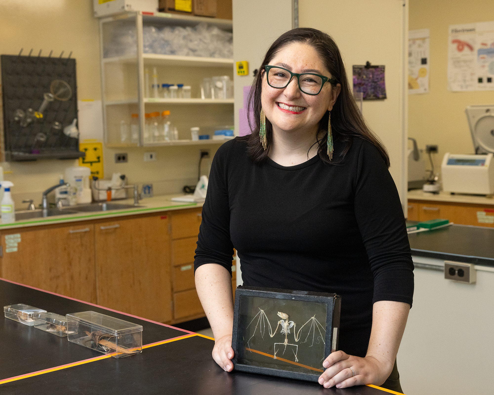
Hannah Frank holds a bat skeleton in her laboratory. (Image credit: Kenny Lass/Tulane University)
Using genetic analysis, the team found that one of the two loci contained a larger and more varied collection of gene segments that could be used to create a broad, ready-made repertoire of antibodies. The other had fewer of these building blocks but appeared to rely more heavily on a process called somatic hypermutation, in which antibody-producing cells make small genetic changes after encountering a pathogen.
Those changes can help antibodies become more "customizable" to whatever threatens the immune system, Frank said.
She compared this system to the different layers of defense seen in the 1998 Disney movie "Mulan."Upon seeing invaders at the Great Wall of China, guards raised an alarm to alert others without yet knowing the specific threat; this is like the innate immune system, the body's first, broad line of defense. The adaptive immune system is more like the heroine Mulan: highly targeted and capable of responding to a specific enemy, but slower to develop.
This study suggests bats have more diversity to start with, which may allow for faster and more effective responses from the start of an infection.
Michael Letko, molecular virologist at Washington State University
These two layers of immune defense are not unique to bats, but bats' two antibody systems may provide an additional layer of protection.
From each loci, bats can create a "cache of trainable, customizable cells," Frank said. This degree of customization could help the bats immune system ward off a wider range of threats than other mammals can.
"This extra set of genes gives the animals more diversity in their antibodies," said Michael Letko, a molecular virologist at Washington State University who was not involved in the study. "This study suggests bats have more diversity to start with, which may allow for faster and more effective responses from the start of an infection," Letko told Live Science in an email.
Letko added that the genetic analysis in the study was impressive and lends credibility to the results. "It is hard to dispute the validity of information solidly founded on the genetic organization of these bats," he said.
That interpretation remains a hypothesis, though; Frank and her team will need to study how these two systems behave when bats are exposed to viruses before they can determine exactly what advantages they provide.
"We think this discovery is an important piece of the puzzle," Frank said in a statement. "It doesn't fully explain why bats are such effective viral reservoirs, but it reveals a level of immune variety we didn't know existed and gives us an entirely new direction to explore."
Understanding how bats tolerate viruses could help scientists predict which kinds of bats are most likely to affect human health and how we can mitigate those risks, Becker noted. Separately, bats have a notably low incidence of tumors and cancer, Letko said, and he wonders if their unique immune systems might hold clues as to why.
While bats are often viewed through the lens of the infectious diseases they carry, their story is far more complex, Frank said.
"Let's learn from the guys who've been evolving with them [viruses] for millions and millions of years," Frank said. "But let's try not to villainize them for surviving for millions and millions of years."
A large British study spanning more than 70 years has found that people who experience chronic money struggles over their working lives show measurable declines in memory and processing speed by their 50s, along with signs of accelerated brain shrinkage decades later.
Researchers have developed a magnetic method for influencing deep-brain circuits without permanently implanted electrodes. A magnetic field may offer a new way to reach circuits buried deep within the brain without permanently implanting electrodes. An international research team injected magnetic nanoplatelets into a targeted brain region and used them to improve movement problems in mice [...]
Brain immune cells appear to drive reversible sleep loss in mice with Alzheimer’s-like disease. A fire starts in one room, but instead of containing the danger, the sprinkler system floods the entire house. The response causes far more damage than the original threat. Something similar may happen in Alzheimer’s disease. Amyloid plaques, the sticky protein [...]
Alzheimer’s disease is the most common form of dementia and affects millions of people worldwide. It slowly destroys memory, thinking, and the ability to carry out everyday activities. Although scientists have developed medicines that can ease some symptoms, there is still no cure, and one of the biggest challenges is that treatments do not work […]
An outbreak of parasitic illness linked to iceberg lettuce is affecting nine U.S. states, federal health officials have announced. That's up from the five affected states that were identified in earlier stages of the officials' investigation.
This lettuce-related outbreak is one of several in the U.S. that are being caused by the parasite Cyclospora cayetanensis, which can enter the body when someone consumes food or water contaminated with the organism. The federal government has not officially identified a source of contamination in the other Cyclospora outbreaks yet, although some state officials have mentioned potential culprits. (North Carolina officials have pointed to cilantro and parsley, for example.)
The nine-state outbreak linked to iceberg lettuce includes cases in Illinois, Indiana, Kansas, Kentucky, Michigan, Ohio, Oklahoma, Pennsylvania and West Virginia, according to the Food and Drug Administration (FDA). To date, 1,947 people have been infected across these states and reported eating Taco Bell before they got sick. There have been 98 hospitalizations and no deaths.
Specifically, the infections have been linked to lettuce sourced from central Mexico, prompting Taylor Farms to remove all of its iceberg lettuce from that region from the U.S. market. The company has also issued a recall. A complete list of recalled products can be found on the FDA's website.
The recall affects products that were distributed in the nine states listed above, as well as additional states, all of which are listed on the FDA's website. Consumers, restaurants and retailers that purchased or received recalled lettuce are advised to discard it immediately and not consume it.
"We also expect to continue to see the number of confirmed cases rise, despite the recall, due to the fact that it can take as long as six weeks for CDC [Centers for Disease Control and Prevention] and state officials to determine if a sick person is part of this outbreak," the FDA's latest update notes. "FDA will continue to work with federal and state partners to investigate this multistate outbreak and ensure products implicated in this outbreak have been removed from the market."
This article is for informational purposes only and is not meant to offer medical advice.
Ten years ago, a pioneering AI scientist predicted that computers would replace human experts as readers of medical images. It hasn’t exactly worked out that way.

 Login