Normal view
A Massive Study Reveals the Brain Is Far More Flexible Than We Thought
Why Dreaming Leaves the Brain Running Low on Energy
The Immortal Soul | Neuroscience vs. Materialism


Inflammation may trigger persistent 'out-of-body' experiences, emerging research hints
Inflammation may be behind a mysterious phenomenon known as dissociation, a mental state that includes "out-of-body" experiences and the feeling that the world is "unreal," emerging research suggests.
Scientists have long known that this altered state of consciousness is often triggered by trauma. But new research hints at how: Trauma triggers a stress response, which dials up chronic inflammation. The findings are part of a broader shift in psychiatric research as experts increasingly examine how the immune system may play a role in mental health conditions.
The research, which has been accepted for publication and is currently under peer review, is still preliminary, and it is based on correlations between blood markers and symptoms, which cannot prove inflammation causes dissociation. But if follow-up work shows inflammation fuels dissociation, that raises the possibility that anti-inflammatory drugs could treat disorders in which people spend much of their time feeling detached from themselves or reality, experts told Live Science. Interventions that broadly target reducing inflammation could also be beneficial, the study suggests.
Research in this area could provide a "whole new avenue for potential treatment options," study lead author Evelyn Dilkes, a psychology researcher at the University of Essex in England, told Live Science.
A common experience, a rare disorder
Dissociation encompasses both out-of-body experiences, known as depersonalization, and the feeling that everything around you is unreal, called derealization, said Dr. Ruth Lanius, a psychiatry professor and Harris-Woodman chair of mind-body medicine at Western University in Ontario. Both of these experiences are related to a person's sense of self and embodiment.
As a short-term phenomenon, it is common; between a quarter and three-quarters of the population may experience dissociation at some point in their lives. It can also be a symptom of many conditions, such as schizophrenia, depression, anxiety, panic disorders and epilepsy.
In around 1% of the population, people may experience prolonged dissociation, in what's known as depersonalization-derealization disorder (DPDR). People with this condition may frequently feel detached from their body or from reality for hours, weeks, months or years at a stretch.
Research suggests DPDR is often associated with past trauma, such as a life-threatening event or childhood abuse. During a traumatic event, dissociation may be helpful because it may offer "a psychological escape when a physical escape is not possible," Lanius told Live Science. It may also help a person stay calm and focused on the threat at hand.
The problem arises when dissociation continues after the threat has passed.
"[Dissociation] helps you to survive, but in the aftermath of trauma, it leaves you detached from all of your emotions, positive and negative, and so often individuals lose the capacity to feel fully alive," Lanius said.
So why does dissociation linger long past the immediate trauma for some people?
From stress to inflammation and then dissociation
Past research showed that inflammation, which can be the immune system's response to infection or injury, seems to be dysregulated in individuals with dissociative conditions. So Dilkes wondered whether the link between trauma and prolonged dissociation runs through the body's response to stress as a kind of "biological embedding," in which life experiences alter a person's physiology. That hypothesis proposes that trauma initially triggers a stress response. When that stress response doesn't subside, the body's stress system stays activated, continually releasing the hormone cortisol from the adrenal glands.
Cortisol is typically anti-inflammatory in short exposures, but when it remains elevated over long periods, it can contribute to chronic inflammation. This can show up in blood levels of interleukin-6 and C-reactive protein.
So Dilkes' lab looked for those inflammatory markers in the blood of children in the U.K. who were followed, starting in the 1990s, as part of the Avon Longitudinal Study of Parents and Children. The study collected samples at age 9 and 12 and again at age 17 and 24. The study asked participants when they were adults whether they “ever felt that they were not a real person, not part of the living world,” or if they “ever felt that the world was unreal, that things around them were like a stage set.”
Early results suggest that high interleukin-6 levels at age 9 seem to be correlated with people experiencing depersonalization in adulthood, while elevated C-reactive protein levels in earlier childhood, but lower ones in adulthood, are linked with experiencing derealization as an adult.
In the latter case, the study authors suggest that chronic stressors could dampen the immune response, leading C-reactive protein levels to drop over time even as symptoms linger.
The researchers also speculated that interleukin-6 and C-reactive protein may differentially act on the different brain regions affected in depersonalization and derealization, respectively. These findings may reflect differences in how stress and immune activity shift across the lifespan following trauma. "It's suggestive of a very intricate system," Dilkes said.
Affected brain regions and networks
Dilkes' research can't answer which brain regions and networks are likely affected, but past research suggests dissociation involves emotional blunting in the brain. In this response, the prefrontal cortex, which is involved in planning and decision-making, acts as a brake on the limbic system, which helps people experience emotions, Lanius said.
According to this theory, dissociation occurs because circuits in the amygdala, the fear-processing part of the brain, hyperactivate the prefrontal cortex. The overactive prefrontal cortex then disrupts normal signaling to the limbic system. The overall result is a dampening of emotions, which may enable people to stay focused and rational during a scary event.
This brain activation pattern, which is characteristic of DPDR, is the opposite of that classically found in post-traumatic stress disorder (PTSD), in which the amygdala is hyperactive, while parts of the prefrontal cortex are underactive. (There is, however, a subtype of PTSD associated with dissociation.)
Some early research hints that the default mode network and the temporoparietal junction, which are thought to be involved in maintaining a sense of self, may be affected in dissociation.
In the new study, the researchers speculate that interleukin-6 exposure in youth may influence the development of the default mode network, which could result in depersonalization symptoms later in life.
Many unanswered questions
While Dilkes' findings are intriguing, outside experts cautioned against overinterpreting early results.
Extrapolating results from studies that investigate dissociative disorders is difficult because patient groups with different diagnoses differ a lot from each other, said Judith K. Daniels, a professor of clinical psychology at the University of Groningen in the Netherlands. What's more, many symptoms can overlap between different dissociative disorders. Dissociation fueled by childhood abuse may differ biologically from dissociation found in a panic disorder, but current research isn't able to distinguish these groups.
What's more, biomarkers of inflammation like C-reactive protein and interleukin-6 are often elevated in other inflammatory and autoimmune conditions — such as rheumatoid arthritis, lupus and multiple sclerosis, while, most people who experience trauma do not develop a dissociative disorder, which suggests that additional genetic or environmental factors are likely involved, Lanius said.
The nonspecific nature of these blood markers would also make it challenging to use them to diagnose DPDR, though in the future they could potentially be used alongside traditional DPDR screening tools, experts told Live Science.
One big limitation is that scientists do not yet fully understand how the brain constructs conscious reality in the first place.
"In my opinion, if we don't understand altered states of consciousness and dissociation, we can't understand psychopathology fully," Lanius said.
For now, more research is needed to determine whether therapies targeting inflammation would benefit people with dissociative disorders. Conditions like DPDR are thought to be rare, but symptoms are often overlooked, which can make it hard to both determine how common it is and to find study participants. But Dilkes thinks dissociation is underdiagnosed.
"So many people experience this, but no one has the words or knowledge of what it is," Dilkes said.
Editor's Note: This article was produced as part of the Dalla Lana Fellowship in Journalism and Health Impact program at the University of Toronto.
See how much you know about the most complex organ in the human body with our brain quiz!
- The Debrief
- The Brain Has a Flexible Sense of Time That Can Split Across Regions, New Research Reveals
The Brain Has a Flexible Sense of Time That Can Split Across Regions, New Research Reveals
When the brain tracks time, it must decide whether its regions should rely on a shared timing signal or tack time separately. A recent study in Nature Communications provides evidence that the brain uses both approaches, switching between them depending on the situation.
A group at the Institute of Science Tokyo simultaneously measured the activity of thousands of neurons in two mouse brain regions and found that the areas sometimes shared timing information but maintained more independent representations at other times. The researchers, led by Associate Professor Riichiro Hira, maintained that this flexibility enables the brain to remain stable as it adapts to changing requirements.
A Task Built to Catch the Brain Switching Modes
Hira’s team created a task to highlight this timing mystery. They trained mice to expect rewards at intervals that switched between 6 and 12 seconds, without any break to mark the end of one trial and the start of the next. Since there was no pause, the mice had to keep updating their internal sense of time throughout the experiment, instead of resetting their mental stopwatch for each new trial.
The mice eventually figured out the pattern. At first they expected a reward every six seconds even if the time they had to wait was longer than this, but by the end of the training session, their behavior indicated that they had learned to anticipate both reward intervals. This showed that the mice were capable of keeping track of both durations and of predicting which one would come next.
While the mice performed the task, the researchers recorded activity in their secondary motor cortex and the posterior parietal cortex using wide-field two-photon calcium imaging. Both regions play key roles in planning, prediction, and working memory.
Two Regions, Two Kinds of Mistakes
The neurons fired in sequence in each brain area, with different cells becoming active at different times. This sequence showed that each region was monitoring the passage of time.
The researchers also identified two kinds of errors in the neural activity. In some cases, both areas made the same mistake when assessing the passage of time. These coherent errors suggested that both regions sometimes shared the same timing drift. In other instances, only one region was inaccurate while the other remained correct. These independent errors suggested that one region could temporarily maintain a different timing representation from the other. Independent errors were more frequent than coherent ones, and this ratio remained the same in all the mice examined.
If the brain regions were just randomly out of sync, you’d expect the ratio to change. However, it didn’t, which suggests that the brain has a built-in way to balance working together and working independently.
Modeling the Switch Between Coherence and Independence
To identify this mechanism, the team built a computational model with two recurrent neural networks linked by sparse, long-range connections and exposed to the same background noise. The model produced the same 2-to-1 ratio of independent to coherent errors seen in the mice.
The model also discovered that the limited connections between the networks tended to cause them to become synchronized, while the common noise caused them to move in separate directions. Hira provided an explanation of the interaction saying, “We found sparse inter-regional connectivity promotes synchronization between the two regions, whereas widespread fluctuations prevent complete synchronization, allowing each region to preserve its own temporal representation.” Neither force takes precedence; instead, a compromise is reached which allows communication between the regions.
The findings challenge the idea that brain regions rely on a single synchronized timing system. It indicates that the brain can adjust how different parts are coordinated, rather than just turning that coordination on or off.
What a Flexible Clock Could Mean for the Brain
Hira’s team believes that there are implications beyond simple timing circuits. “In the future, our results may contribute to a better understanding of cognition, neurological disorders involving disrupted inter-regional coordination, and the development of brain-inspired artificial intelligence and robotic control systems that combine stability with flexibility,” said Hira.
This last idea may prove most important in the years ahead. Engineers building AI and robotic systems face a similar challenge: how to let separate parts work on their own when needed, yet still come back into sync. The mouse brain shows that sparse connections and shared background fluctuations can achieve this balance. This could point to a design principle that even applies to systems with no biological components.
Austin Burgess is a writer and researcher with a background in sales, marketing, and data analytics. He holds an MBA, a Bachelor of Science in Business Administration, and a data analytics certification. His work focuses on breaking scientific developments, with an emphasis on emerging biology, cognitive neuroscience, and archaeological discoveries.
Decades of Financial Hardship Linked to Faster Brain Aging
A large British study spanning more than 70 years has found that people who experience chronic money struggles over their working lives show measurable declines in memory and processing speed by their 50s, along with signs of accelerated brain shrinkage decades later.
The post Decades of Financial Hardship Linked to Faster Brain Aging appeared first on Sci.News: Breaking Science News.
- Live Science
- Diagnostic dilemma: Rare 'spinal stroke' struck an 8-year-old and temporarily paralyzed her
Diagnostic dilemma: Rare 'spinal stroke' struck an 8-year-old and temporarily paralyzed her
The patient: An 8-year-old girl in the U.S.
The symptoms: The girl arrived at an emergency department after waking up with sudden, severe pain in her upper back. Within hours of the pain emerging, she developed rapidly worsening weakness in all four limbs, making it impossible for her to stand or walk.
She also reported painful tingling and loss of sensation in her hands, and doctors discovered that she had diminished reflexes and sensory changes beginning at her upper chest and moving down her body. In multiple instances at the hospital, she lost control of her bladder and bowels.
The combination of acute paralysis, severe back pain and incontinence suggested damage to the spinal cord, but the cause wasn't immediately clear, her doctors wrote in a report of the case.
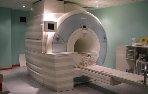
What happened next: The medical team launched an extensive search for the underlying cause. An MRI of her neck and torso showed evidence of extensive inflammation or injury that stretched from her lower neck to the middle of her back. The scan also showed a small tear in one of the disks in her lower neck, with a small amount of disk material pushing outward.
However, the girl and her parents did not report any recent falls or injuries, and the MRI alone couldn't determine what had caused this apparent damage.
The girl's medical team then performed blood tests, a spinal fluid analysis and more imaging to look for signs of infections, autoimmune diseases or other inflammatory conditions that can trigger acute paralysis in children. These tests all came back negative.
While clinicians continued investigating, the girl was treated with intravenous steroids and underwent a procedure called plasmapheresis, during which blood is drawn from the patient into a machine which separates blood plasma (the liquid portion of blood) from blood cells and then returns the blood cells to the patient mixed with fresh plasma from donors. This process helps to filter out harmful antibodies and abnormal proteins that can be present in people with autoimmune or blood disorders.
Over the following days, the girl gradually regained her ability to feel, walk and control her bladder. However, repeat MRI scans showed worsening inflammation, which didn't seem to match the girl's improving condition.
The diagnosis: Throughout her hospital stay, the medical team suspected a diagnosis of transverse myelitis, which is a rare neurological disorder that involves swelling in the spinal cord. However, as infectious and autoimmune causes became increasingly unlikely, the physicians changed their minds.
They instead concluded that the girl had experienced a spinal cord infarction, or essentially a stroke affecting the spinal cord. This blood-flow blockage was most likely caused by an exceptionally rare condition called fibrocartilaginous embolism (FCE).
In FCE, a small piece of jelly-like material from the soft center of an intervertebral disk breaks off, enters the bloodstream and blocks one of the arteries supplying the spinal cord. The resulting loss of blood flow can cause sudden paralysis, sensory loss and bowel and bladder dysfunction.
It's unknown exactly how many people experience FCE, as it's difficult to diagnose and frequently underreported. One literature review found only 30 suspected cases and 41 confirmed cases of FCE that had been documented in the U.S.
The case study notes that FCE is most common in children and young adults ages 10 to 20, and that rate also jumps up later in adulthood, around ages 40 to 60. It is slightly more common among female patients than male. While slightly younger than the most commonly affected demographic, the 8-year-old patient in this case otherwise fit this description.
FCE is difficult to diagnose because there is no definitive laboratory or imaging test for it. Instead, physicians must rule out more common causes of spinal cord injury while looking for imaging findings and clinical signs and symptoms that might fit the diagnosis of FCE.
The treatment: There isn't a specific treatment for FCE; instead, care focuses on managing the symptoms caused by the spinal stroke.
The patient was treated with corticosteroids to reduce inflammation, medications to manage pain and plasmapheresis. After a few weeks, and with extensive rehabilitation therapy, the girl regained nearly all of her strength and functional abilities. Her only lingering symptom was weakness in her hands.
What makes the case unique: The case study notes that FCE is often linked to minor trauma to the spine, such as that caused by heavy lifting or other activities that briefly increase pressure inside the spinal disks. In this case, however, the medical team was unable to identify a clear triggering event, and the girl was otherwise healthy and had no reported history of physical trauma. The girl simply woke up with symptoms, which made the diagnosis even more unexpected.
The case also highlights that, although FCE is rare and often carries a poor prognosis — as many patients experience long-term disability and some may even die from complications — meaningful recovery is also possible. The case report authors concluded that, with early recognition and supportive treatment, patients with FCE may achieve improved outcomes and a return to their normal activities.
For more intriguing medical cases, check out our Diagnostic Dilemma archives.
This article is for informational purposes only and is not meant to offer medical advice.
Kakadiya, J., Lakhani, D. A., Gaddamanugu, S., Goncalves, F. G., & Mullen, A. J. (2026). Presumed fibrocartilaginous embolism causing spinal cord infarct in 8-year-old female: A case report. Radiology Case Reports. https://doi.org/10.1016/j.radcr.2026.03.047
Can you guess the diagnosis in these strange medical cases? Find out with our diagnostic dilemma quiz!
Magnetic Nanoparticles Restore Movement in Mice With Parkinson’s-Like Symptoms
Scientists Found What May Be Keeping the Alzheimer’s Brain Awake
Scientists Reveal How Exercise May Protect the Aging Brain
Long COVID Brain Scans Reveal Damage to Dopamine Neurons
- The Debrief
- The 5,700-Year-Old Psychedelic Drug Mescaline Finally Got a Modern Brain Scan, And The Results Were Unexpected
The 5,700-Year-Old Psychedelic Drug Mescaline Finally Got a Modern Brain Scan, And The Results Were Unexpected
The first proper brain scan involving the effects of mescaline found the drug ignoring the regions where psychedelic research has spent a decade studying, settling instead on the one structure no one really expected.
Sometime around 3,700 BC, someone in what is now southwest Texas cut two tops off a peyote cactus and dried them. They spent most of the twentieth century in a drawer at the Witte Museum in San Antonio, and when chemists finally analyzed them in the early 2000s, the mescaline was still detectable — roughly 2% alkaloids by weight, 5,700 years on.
Those dried-out cactus tops, where the drug compound is concentrated, remain the oldest plant drug ever to give up a major bioactive compound to chemical analysis. And with thousands of years of use, mescaline remains a bit of a scientific mystery.
Mescaline became a Schedule 1 substance under the Controlled Substances Act in 1970, and the classification effectively ended research on it. LSD and psilocybin eventually clawed their way back into laboratories for scientific research, but mescaline, the older compound by several thousand years, doesn’t have the same level of lab testing.

Now, a new study in Neuroscience Bulletin has become the first to properly look, and the answer is not what most expected.
Researchers at Northeastern University’s Center for Translational Neuroimaging scanned 24 rats, evenly split by sex. After a single dose of mescaline, while the animals were awake (anesthesia flattens the very signals a drug study is trying to read), the drug largely ignored the forebrain. It went almost entirely for the cerebellum, which is a strange location for a psychedelic.
The cerebellum is the lump at the back and bottom of the brain where balance and motor coordination live. It also holds more than half of all the neurons in the brain, and everything it computes leaves through just three narrow output channels.
An interpretation, proposed by neuroscientist Anna Devor in 2002, is that the cerebellum is the “Great Gate.” As the brain receives sensory information, it acts as a filter that suppresses the sensory information that the brain already predicted and passes along only what it did not expect. A working brain spends most of its time discarding what it saw coming.
Under mescaline, the gate appeared to come off its hinges.
Activity inside the cerebellum dropped sharply, while its connections outward multiplied to the hippocampus, the thalamus, the sensory cortex, and the midbrain. Quieter on the inside, wired to far more on the outside. The researchers read this as a filter that has stopped filtering and started broadcasting, pushing raw sensory information into regions that normally receive it neatly packaged.
“Wouldn’t it be devastating if you didn’t ‘gate’ all of that sensory information out?” senior author Craig Ferris explained in a university press statement.
The clearest evidence for that reading came from a smell test. Rats were given almond odor, a reliably rewarding stimulus, and in the control group, it lit up 48 of the 169 brain regions the team tracked. Under mescaline, the response was essentially abolished, flattened close to nothing across the board.
Broken down by modality, smell, touch, and hearing were all measurably blunted, while the visual system showed no significant change at all — a peculiar result for a compound whose entire cultural reputation rests on what it does to sight.
The authors put the discrepancy down to species differences, reasonably enough given that rats navigate by nose and whisker, but it remains one of the odder findings in the paper. What makes this significant is the comparison.
Mescaline, LSD, and psilocybin all act on the same target, the 5-HT2A serotonin receptor, and all three produce broadly similar experiences in people, which has encouraged researchers to treat findings from one as transferable to the others. However, brains don’t seem to cooperate.
Earlier work from this same lab found LSD acting on the prefrontal cortex, thalamus, and hippocampus, and psilocybin engaging cortico-striato-thalamic circuits. Mescaline’s acute signature sat in neither place.
“The power of mescaline to me is that it is doing something different than other psychedelics,” said lead author Noah Cavallaro. “Essentially, the cerebellum is being shut down internally, but it’s talking more to other parts of the brain.”
The distinctiveness is narrower than it first appears, though. The lab’s own psilocybin work also produced pronounced cerebellar hyperconnectivity, so the sprawling network changes are not unique to mescaline; what looks specific to it is the acute suppression, the cerebellum falling quiet while the rest of the brain carried on largely unaffected.
“If two compounds produce similar hallucinogenic symptoms through different neural pathways, comparing them could help us pinpoint what’s actually causing those specific experiences,” Cavallaro said.
That being said, there are some cautions offered in the paper. First, these were rats, and rat and human brains differ most in the cerebellar-cortical wiring. Second, only one dose was tested. Third, the team did not monitor heart rate or breathing during scanning, so ordinary cardiovascular effects cannot be cleanly separated from neural ones.
Lastly, the researchers also flagged recent work showing that the scanner signal itself can run opposite to a brain region’s actual oxygen use, which complicates reading any of these images as direct neural activity. The one previous attempt to image mescaline in humans, a small study decades ago, found a different pattern entirely.
With all that being said, the paper presents a key social issue. Mescaline’s status as a drug has left it largely untested, and only now are we learning about what it can do to the brain, outside of the anecdotes of its users.
After fifty years of regulatory silence, scientists are just starting to answer a question people have arguably been asking since the Neolithic.
MJ Banias is a co-founder of The Debrief, covering science and technology news. You can email him at mj@thedebrief.org or follow him on LinkedIn.
Does Watching TV Really Rot Your Brain?
Generations of parents have a new piece of scientific evidence to combat couchpotatoism
The post Does Watching TV Really Rot Your Brain? appeared first on Nautilus.

- The Debrief
- New Research Reveals How Traveling Brain Waves Allow Us to Predict, Reconstruct, and Perceive the World
New Research Reveals How Traveling Brain Waves Allow Us to Predict, Reconstruct, and Perceive the World
Scientists from the Salk Institute have determined that brain waves in the visual cortex serve as a computational engine, allowing the brain to construct internal representations of the external world that enhance prediction, reconstruction, and perception capabilities.
While the scientists behind the new study focused their work on the visual cortex, they suspect that similar brain waves washing over other areas of the brain’s surface likely serve a similar role for other sensory systems.
Unraveling the Role of Traveling Brain Waves in the Visual Cortex
The human brain uses electricity to communicate between synapses. Research has shown that this electrical activity can travel in waves across the brain’s surface. Called neural traveling waves, the phenomenon’s role in brain function and consciousness is not completely understood.
Previous studies have established that traveling brain waves play a significant role in behavior and attention. In 2020, Salk neuroscientist John Reynolds, PhD, successfully identified traveling brain waves in the visual systems of animals during a wakeful state.
According to a statement from Reynolds, the senior and co-corresponding author on the new paper, and colleagues, the 2020 research effort also found a direct correlation between traveling brain waves in the visual cortex and an animal’s ability to perceive objects directly in front of them. The authors suggest this discovery explained the “classic conundrum” of searching for something like a pair of keys or a phone that was right in front of you the whole time.
Although the connection between traveling brain waves and an awake animal’s ability to perceive objects seemed solid, Reynolds said he was left wondering why.
Process is Analogous to LLM’s Like ChatGPT
In their newly proposed theory, Reynolds and colleagues suggest that traveling brain waves are not simply “electrical noise” resulting from the brain’s normal relaying of signals. Instead, the Salk Institute team proposes that the neural connections responsible for the generation of traveling brain waves change their physiological structure, or “synaptic weights,” to more accurately reflect the outside world.
“Each sight, smell, sound, and action of an animal alters the connections that generate these waves, building the neural circuitry that the brain uses to construct an internal representation of the external world,” the team’s statement explains.
In the visual cortex, the part of the brain responsible for receiving and processing visual information, the team proposed that traveling brain waves perform four distinct functions. These include modulating visual perception from moment to moment and turning recent visual sensory inputs into “internal representations” of objects, people, and surroundings.
The team proposes that traveling brain waves also help humans generate short-term predictions about their surroundings, and “store and replay patterns that represent memories of events unfolding in time.” Reynolds said the process laid out by his team is analogous to how ChatGPT and other LLMs translate and interpret data.
“They learn statistical structure from language and use that knowledge to generate meaningful and appropriately structured text that reflects the patterns of language,” the researcher explained. “The brain may be doing something functionally similar—a biological generative model built from the ground up by experience.”
“One Step Closer” to Understanding How Our Brains Compute the World
When detailing the potential impact of their proposed role for traveling brain waves in the visual cortex, the team highlights the contrast between a rich and complex world and its general predictability.
“Objects around you exist in 3D space, you see the world in retinal images that change with your eye and body movements, and the laws of physics and physiology overlay this all,” they explain.
Reynold’s team says that traveling brain waves are merely the brain’s way of internalizing these predictable “regularities” and encoding them in its synapse network. If correct, they said that this encoding would allow the brain to “infer the likely causes of sensory input and construct an internal model of the world.”
“This framing brings us one step closer to understanding how our brains compute the busy, messy world around us, turning a complicated sensory onslaught into behaviors and experiences,” they conclude.
The study “Neural traveling waves in cortex: network mechanisms and potential roles in neural computation” was published in Neuron.
Christopher Plain is a Science Fiction and Fantasy novelist and has spent the last six years as Associate News Editor and Head Science Writer at The Debrief. Follow and connect with him on X, learn about his books at plainfiction.com, or email him at christopher@thedebrief.org
- Live Science
- Neuroscience findings often can't be replicated — and it's a big problem for what we know about the brain
Neuroscience findings often can't be replicated — and it's a big problem for what we know about the brain
One of the central assumptions of modern neuroscience research is that the brain's shape and structure affect behavior. Scientists have linked a thicker cortex (the brain's outer layer) to higher intelligence, certain brain wave patterns to better volleyball ability, and higher connectivity between parts of the brain to chess-playing skills. There's a vast number of these so-called brain-wide association studies (BWAS).
But when experts repeat these studies, they can't replicate the results.
This "replication crisis" suggests that a substantial amount of brain-behavior imaging research rests on a shaky foundation. There are myriad problems affecting this area of research, said Randy Ellis, a senior scientist at Oracle who wrote about these issues while working as a biomedical informatician at the Icahn School of Medicine at Mount Sinai in New York.
Some of these issues aren't unique to neuroscience. They begin with what Ellis called the "original sin" of science: Academics are put under huge pressure to publish positive research findings.
But some of these factors do apply specifically to this field.
The problems are big enough that they are "halting the growth and development of science and the curing of diseases," Ellis told Live Science.
Tiny differences, small samples
Several brain studies that could not be reproduced in follow-up work show how problems can creep in. Each of the original papers has over 500 citations, with the number of citations reflecting how much these studies may influence thinking in the field.
For example, a landmark study in 2007 found that the brains of kids with attention-deficit/hyperactivity disorder took longer to mature. But a study published earlier this year found that this link vanished once the different rates of aging between boys and girls were taken into account. "That's what made the whole house of cards topple," Matthew Albaugh, co-author of the replication paper and a clinical neuroscientist at the University of Vermont, previously told Live Science.
In 2011, researchers at University College London published a study that found that the density of gray matter, the part of the brain where brain cell bodies are found, in several brain areas was linked to participants' number of Facebook friends. (In 2011, this was a vital social metric.)
A 2015 paper failed to replicate this finding, but the authors of the original paper argued that was because the follow-up study didn't follow the same protocol.
"It's hard to do exactly what was done in the original studies," said Sarah Genon, a neuroscientist at the Research Center Jülich in Germany who was not involved in either the Facebook study or its replication.
So, in 2019, Genon and her colleagues tried a different type of replication analysis.
They used a vast database and conducted more than 10,000 analyses of MRI scans from hundreds of volunteers. The median neuroimaging study sample size was around 25, so they divided this larger dataset into smaller ones and then looked for significant associations within each.
If one of these micro-studies reported a significant association between a behavior and a brain structure, the team tried to reach the same findings using a different subset of the data. Very few of the repeat studies replicated the results of the first. "It was clear evidence that the replicability of those brain-behavior associations is relatively weak," Genon said.
One of the key problems is that in healthy volunteers, variation among individual brains is relatively small, which means you need a large number of participants to detect average differences that tie to behavior, Genon said. That's similar to genome-wide association studies, which pick up the very tiny effects of individual genes, and they must sample tens or hundreds of thousands of people to find robust effects. To detect meaningful differences in brain-wide imaging studies, only sample sizes in the thousands would do the trick, a 2022 paper suggested. Because brain scans are expensive and time-consuming to capture, most studies have relied on much smaller samples.
Everybody always says that replication is great. But then when I suggest that their specific studies could be replicated, often they are a bit more skeptical
Luca Kämmer, doctoral student at the Max Planck Institute for Human Cognitive and Brain Sciences
Additionally, the behavioral tests used to establish psychological variables can be inconsistent, Genon said. What's more, some of these studies may be operating on the outdated assumption that small brain regions control complex characteristics like intelligence, she added. In reality, however, "those types of abilities are usually relatively distributed across the brain," she said. When researchers analyze multiple small brain areas with that assumption in mind using separate statistical tests, it can raise the risk of producing mirage associations.
Some of these studies may be flawed at the outset. A 2017 brain imaging study by researchers at Weill Cornell Medical College explored whether MRI data could be used to stratify patients with depression. The researchers found that brain activity clustered into four "biotypes" of depression, each of which featured distinct, unusual activity patterns in key brain networks.
But a follow-up study found that the differences among the clusters weren't statistically significant, meaning they could have occurred by chance. Richard Dinga, a neuroscientist at the Friedrich Schiller University Jena in Germany who worked on the follow-up study, said the main problems lay in its design.
Its statistical approach, he said, essentially guaranteed that significant links would be detected. That's because of a problem called overfitting, in which a model is so well tuned to one set of data that it struggles to find real patterns in other datasets. This design correlated 17 clinical features of depression with 30,000 brain imaging features, but Dinga said the authors could have used "30,000 coin flips" and still produced the same strong correlation.
Conor Liston, a psychiatrist at Weill Cornell Medicine and co-author of the original paper, acknowledged the model's susceptibility to overfitting. "That was not at all our intention, but that was the result," Liston told Live Science
Still, he argued that the biotype finding they identified was real, pointing to later studies his team had published to back up their original finding. When asked why his team hadn't updated the original paper to acknowledge that their analysis method had serious flaws, Liston said their subsequent publications were sufficient to correct the record. "I think the message is out there, and I think the solutions to those overfitting issues are very clear," he said.
Solving the problem
The results of many studies that share the same issues as these papers will never be replicated and yet will be left to stand. For one thing, there's no money in replication efforts. "Grant agencies wouldn't be really excited" to fund a study whose goal was to confirm past work, Genon said. And even when these studies get done, they aren't as likely to get published when they undermine prior findings.
For Dinga, the way forward is to collect more data. "That's what solved the same irreproducibility problem in genetics," he said, referring to genome-wide association studies that have produced important findings over the past two decades by using sample sizes that have reached the millions. But it's also important to improve study design, he added, since bigger datasets won't fix studies with incorrect analysis approaches. "The methods just don't have a chance to produce something useful," he said.
Some researchers are leveraging large brain imaging datasets. Martin Hebart, a group leader at the Max Planck Institute for Human Cognitive and Brain Sciences in Germany, and Luca Kämmer, a doctoral student in Hebart's lab, are developing re:vision, a project that is collecting imaging data — the largest initiative yet to capture how the brain responds to visual stimuli. They plan to make this information available to other researchers, who will then explore whether hypotheses tested in other papers can be replicated in re:vision's dataset.
Hebart and Kämmer said they have already received over a dozen applications for their project. Reception has been particularly positive among younger researchers in the field. But convincing senior scientists to retest their data might take more work because they have more to lose if their long-standing finding is ultimately not replicated, Kämmer said.
"Everybody always says that replication is great," he told Live Science. "But then when I suggest that their specific studies could be replicated, often they are a bit more skeptical."
See how much you know about the most complex organ in the human body with our brain quiz!
- The Debrief
- China Clears Brain-Computer Interfaces for Commercial Use: Is It Time for a Global Coalition?
China Clears Brain-Computer Interfaces for Commercial Use: Is It Time for a Global Coalition?
Brain-computer interface (BCI) technology is advancing rapidly worldwide. Far more than just the innovative technology promoted by prominent advocates involved in its expansion, like Elon Musk, BCIs are being developed in countries including the United States, Canada, Australia, Germany, Spain, and China.
Beyond their life-changing potential for treating neurological conditions, BCIs could fundamentally reshape what it means to be human, and soon. If that’s the case, is a global BCI coalition needed?
Before Elon Musk: Jacques Vidal and Thomas Oxley
A common misconception is that Elon Musk and his company, Neuralink, launched the BCI revolution we see today. In reality, the origins of the technology date back to the 1960s. Jacques Vidal published the first paper introducing the term “brain-computer interface” in 1973, demonstrating how electroencephalographic (EEG) signals could be used for direct communication. Vidal’s pioneering work laid the foundation for all modern BCI research.
Fast forward to 2012, when Australian-born physician, neuroscientist, and Synchron founder and CEO Thomas (Tom) Oxley entered the BCI conversation. Oxley was among the first physicians to bring BCI technology into clinical practice at Mount Sinai Hospital. With early funding from DARPA, he and his team began clinical trials in Australia in 2019. Oxley’s minimally invasive Stentrode device attracted significant business and media attention well before Neuralink’s highly publicized demonstrations in 2023.
What the BCI community can thank Elon Musk for is the unprecedented level of global investor attention he has brought to the field. Without that influx of funding, BCI technology might still be in its infancy, and fewer people would have access to devices capable of addressing life-changing healthcare needs.
China’s Rapid Advancement into BCI
Since 2010, China has moved aggressively to advance BCI technology while making clear that the field is a national priority—one viewed as nearly as important as breakthroughs in quantum computing.
Recently, the Chinese BCI company NEO announced approval for the country’s first commercial use of a coin-sized brain-computer interface for patients living with paralysis caused by spinal cord injuries.
Tsinghua University and Shanghai-based Neuracle Technology designed the brain chip to be less invasive than many competing technologies. Rather than penetrating deep into brain tissue, the NEO device is implanted beneath the skull, where its electrodes are attached to the dura mater—the protective membrane surrounding the brain.
The first Chinese-designed and developed brain chip was implanted last November in volunteer Dong Hui. Hui, 39, sustained a spinal cord injury in a car accident six years earlier that left him paralyzed. During a video interview with MIT Technology Review, Hui said excitedly, “I couldn’t believe I was able to write again. I was so excited, I even missed a stroke in my name.”
This past March, Hui became the first person in the world to receive an invasive brain-computer interface approved for use beyond a clinical trial. But what does “commercial” use actually mean?
The NEO device, developed by Neuracle Technology, was reportedly approved by Chinese regulators for use outside clinical trials in certain patients with spinal cord injuries. In practical terms, that means the technology has moved beyond the experimental stage and can now be offered in clinical settings to eligible patients. Hospitals may provide it as a treatment, Neuracle can manufacture and sell the system commercially, and qualifying patients may receive the implant as part of standard clinical care rather than solely through an experimental research program, representing a major milestone for BCI technology.
Another competitor in China’s BCI market is Gestalt, founded by Phoenix Peng. Surprisingly, the two companies are pursuing very different approaches. NEO’s system places sensors directly on the brain’s protective covering, allowing the electrodes to measure electrical activity generated by neurons. This approach can provide stronger and faster signals but requires neurosurgery.
Gestalt, by contrast, is attempting to eliminate the need for surgery. According to Peng, the company aims to use phased-array ultrasound to both read brain activity and stimulate specific neural pathways without requiring invasive procedures. If successful, this approach could make brain-computer interfaces safer, more accessible, and available to a much broader population.
In a recent podcast interview, Peng told The Debrief that this is why he and others in the industry have begun pursuing ultrasound-based BCIs. He noted that such systems are now being developed not only in China but also by several companies in the United States. “The whole technology is just rising up,” Peng said.
Competitors Chime In
With the Chinese government investing heavily in and prioritizing BCI technology, it is becoming increasingly clear that the country is positioning itself to set the “gold standard” for BCI development worldwide. Last July, the Chinese government announced its intention to establish a global standard, outlining plans to do so through new guidelines issued by the Ministry of Industry and Information Technology, along with the creation of a committee to oversee the development and implementation of those standards.
“China progresses fast and is likely to win the race,” said Dr. Martin Schüttler, the CTO, co-founder, and managing director of CorTec Neuro, in an email to The Debrief. “However, young Chinese implantable BCI companies might run into challenges when demonstrating long-term robustness and efficacy, which validations are difficult to accelerate.”
“From my perspective, the Chinese governmental orchestration of the BCI race provides a major advantage over Europe and the USA, where each BCI company struggles individually with similar challenges,” he adds. As an example, he notes China’s move to define reimbursement of implantable BCI, which occurred months before Neuracle received its market approval. “Therefore, the Chinese government also published a definition of a BCI and issued ethical guidelines for BCI technology,” Schüttler said.
“It also funds BCI initiatives and companies, leading to a boom of start-ups in the field,” he says, though noting that this is not strictly restricted to implants.
Florian Solzbacher, PhD, co-founder of Blackrock Neurotech, adds more context on BCI commercialization by telling The Debrief that “this is more than just a technology race. This is about which nations and values will lead the world and likely set standards in a completely new, strategically important field of technology.”
“There are several aspects of the current processes for translation and regulatory approval that could be adjusted to new technological capabilities without throwing safety overboard,” Solzbacher adds. “And with all the concern about China’s pace, speed to commercialization alone is not the right metric. The concern, in any market, is whether long-term safety and security, technological maturity, clinical evidence, regulatory oversight, and patient support can keep pace with deployment.”
“One can only hope that China’s pace wakes up our own nation and creates incentives and pressure for the US and EU to coordinate far more effectively and to support translational neurotechnology with far more conviction and focus, without jeopardizing patient safety,” he says.
High Volume in 2026?
This recent commercial BCI announcement from China comes on the heels of Elon Musk declaring that Neuralink will ramp up its efforts in a “high volume” attempt for 2026.
“This is a big deal,” Musk’s posting on X read.
But what does high volume really consist of? This past January, Barcelona, Spain-based BCI company and co-founder Carolina Aguilar, INBRAIN Neuroelectronics, one of Neuralink’s competitors, told The Debrief, “At this stage, we interpret ‘high-volume’ realistically as hundreds moving toward low thousands of implants per year.” She also notes that it could eventually reach “tens of thousands”.
Solzbacher said the challenges presented with scaling “usually come in all areas.”
“It starts with capability, the processes, and the capacity to actually make that many devices,” Solzbacher said in an email to The Debrief, adding that that number—should it reach “tens of thousands, or hundreds of thousands of devices” would require “well-qualified, validated processes.”
With China accelerating the commercial deployment of BCI technology, how do European and American BCI companies view the pace of its advancements, and what impact could this have on the global race for brain technology leadership?
Defense Applications
The commercialization of brain-computer interfaces is now at the forefront of the brain-chip revolution. Citizens around the world should be engaged in discussions about how this technology will be implemented within their countries. At the same time, the conversation should also acknowledge the role the military-industrial complex has played in funding BCI research through DARPA, as well as how the technology has been explored for potential defense, intelligence, and counterintelligence applications.
DARPA has been one of the leading funders of BCI research for decades. Programs (that we know of) such as Revolutionizing Prosthetics (LUKE Arm), NESD (Neural Engineering System Design), and N3 (Next-Generation Nonsurgical Neurotechnology were created with the goals of restoring movement to wounded service members, enabling direct communication between the brain and computers, and developing high-bandwidth, minimally invasive, or noninvasive neural interfaces. Collectively, these efforts aim to improve decision-making, situational awareness, pilot workload management, and human-machine teaming.
It remains unclear how other nations are integrating BCI technology into their own military-industrial complexes or how these systems may ultimately be employed. That uncertainty underscores the growing need for international collaboration and the establishment of a global BCI framework sooner rather than later. Because of this, continued oversight and transparency are necessary to ensure future applications respect civil liberties and ethical standards, and many believe safeguards should be established to prevent any future civilian-targeted programs from emerging.
Ethics of BCI and Global Counsel
Humanity may be approaching a tipping point in its biological and technological evolution. Some would describe this as a “singularity”—a historic moment when technology and human development converge in a transformative way, fundamentally reshaping what it means to be human.
On October 8, a private Morgan Stanley report titled Neuralink: AI in Your BrAIn focused on Elon Musk’s Neuralink. The report argues that Musk and his BCI team are positioned at the forefront of a broader technological shift—one for which society may be unprepared. The implications could be profound, potentially changing the fabric of society itself.
The report warns that “the ability for human beings to ‘keep up’ and communicate with the AI apparatus may deteriorate at an exponential rate,” noting that “one of Elon Musk’s many goals in founding Neuralink is to ‘give humanity a chance’ to keep up with AGI.” It also suggests that Musk is using a “back door” into healthcare to enter the consumer market and, in turn, create a “Neuro Elite.”
Such a development could further widen existing wealth and educational gaps, particularly in emerging economies already struggling to keep pace with wealthier Western nations.
“Many countries, particularly those in the Global South, have limited involvement in BCI research and are understandably focused on more immediate public health, economic, and social priorities. As a result, they may have little incentive to invest in developing or enforcing BCI-specific regulations,” says Kerry Bowman, PhD, a bioethicist, environmentalist, and professor at the University of Toronto, in an email to The Debrief.
“At the same time, researchers seeking to avoid restrictive oversight may simply relocate their work to jurisdictions with few or no regulations, creating opportunities for regulatory arbitrage,” Bowman adds. “Furthermore, meaningful global consensus is complicated by profound cultural differences regarding individual autonomy, collective welfare, human enhancement, and the role of the state. Recognizing these challenges, it remains a good and positive step in the right direction.”
With these concerns on the horizon, Senate Majority Leader Chuck Schumer, along with Senators John Cornyn and Ron Wyden, introduced the Management of Individuals’ Neural Data Act of 2025, or MIND Act, on September 24. The proposed legislation seeks to establish safeguards for brain-computer interface technology by directing federal agencies to develop guidelines addressing ethical concerns, safety, and privacy risks associated with neural data.
As BCIs advance, concerns about their potential ability to influence thoughts, memories, and emotions have become increasingly central to policy discussions.
Without global safeguards and international agreements governing BCI development, scientists could potentially collaborate with foreign adversaries, black-market organizations, or extremist groups seeking to advance radical visions of human enhancement. Technologies developed through such collaborations could then be sold illegally or distributed commercially around the world.
For instance, former Harvard professor Charles Lieber, 67, who was convicted in the United States of concealing ties to a Chinese talent program, is now leading a state-funded brain-computer interface laboratory in Shenzhen, China. Lieber currently directs the Institute for Brain Research, Advanced Interfaces and Neurotechnologies, or i-BRAIN, raising renewed concerns about the geopolitical competition surrounding emerging technologies.
This may appear to be the work of a single controversial scientist, but it also illustrates how quickly advanced technologies, expertise, and intellectual property can move between nations and research communities. The question, therefore, remains: Where do we go from here if a “singularity” arrives, and what might it look like?
“Carbon-based lifestyles and silica-based lifestyles may merge,” Peng offered for this potential event in a podcast interview. “Artificial intelligence and neuroscience are two sides of the same coin. BCI will be the collector between these two shapes of life, probably because AI will have consciousness.”
A Global Coalition?
One proposed approach would be to create a global coalition of academics and experts representing a broad range of disciplines, including physics, philosophy, neurology, psychology, psychiatry, and other hard and social sciences. What might such a global BCI coalition look like, and how could it be structured to guide research, governance, ethics, and the responsible development of brain-computer interface technologies?
Schüttler suggests that if a global BCI coalition enabled technologies developed in China, the United States, and Europe to be shared and approved across regions, its impact would depend largely on who was involved.
For patients, such a framework could mean faster access to breakthrough treatments, particularly because, according to Schüttler, “Chinese approval processes can be substantially faster.”
For U.S. and European companies, however, it could disrupt existing business models. Companies that have invested heavily in Western clinical trials may face significant challenges if Chinese data becomes widely accepted, since he says “a coalition could risk their business model once they are already committed to costly approval studies.”
The shift could prove even more disruptive for clinical research organizations, since trials in China can often be “cheaper and faster to conduct,” Schüttler says. Ultimately, a global BCI partnership could provide substantial benefits or create major disruptions, depending on “who you are in the game, and at what stage you are.”
By contrast, Solzbacher believes a “coalition needs to be informed and driven by a deep understanding of patient and caregiver needs as well as the underlying technologies and their associated risks and opportunities. Practical and multi-stakeholder, with international representation.”
“Regulators, clinicians, engineers, neuroscientists, companies, ethicists, advocates, and, above all, patients and caregivers,” he says, would allow such a coalition to “work alongside national regulators and support their authority, setting shared expectations on trial design, safety reporting, post-market surveillance, cybersecurity, interoperability, patient access, and long-term device stewardship.”
“The center of gravity must stay on the patients. This field will carry strategic and economic weight regardless,” Solzbacher says. “We need to get this technology to the people who could benefit most, responsibly and with as much urgency as possible.”
The next phase of BCI advancement cannot be driven by speed alone. The ability to connect the human brain with machines raises profound questions: Who owns neural data? Who controls access to these technologies? And how can society ensure that tools designed to enhance human potential do not create new forms of inequality, coercion, or exploitation?
Countries around the world will need to work together to confront these questions before the technology advances beyond the safeguards intended to govern it.
Chrissy Newton is a PR professional and the founder of VOCAB Communications. She currently appears on The Discovery Channel and Max and hosts the Rebelliously Curious podcast, which can be found on YouTube and on all audio podcast streaming platforms. Follow her on X: @ChrissyNewton, Instagram: @BeingChrissyNewton, and chrissynewton.com. To contact Chrissy with a story, please email chrissy @ thedebrief.org.

Science word of the day: Neuroplasticity
Pronunciation: Nur-roh-pla-STISS'-ih-tee
What it means: Neuroplasticity is the ability of the nervous system to adapt to the environment or to injuries by forming new connections and modifying and strengthening existing connections. Neuroplasticity can happen after an injury, such as a stroke or trauma to the brain, but it also occurs during ordinary learning. Just by reading this definition, you may be making new, sustainable brain connections.
How to use it in a sentence: Scientists used to think neuroplasticity was an ability only the the young had, but researchers are increasingly learning that aging brains can adapt throughout the lifespan, much to everyone's relief.
Can you crack our science word of the day puzzle, Chain Word?