Normal view
Queen Odor Mediates Reproductive Suppression in Eusocial Mammals
Naked mole-rats have long puzzled biologists with their striking eusocial structure, where most adults remain reproductively suppressed while a queen controls the colony’s future. Now, a new study identifies a chemical “queen odour” that links social status to endocrine control, revealing a mammalian analogue to insect queen pheromones.
The molecule, termed IPM, is enriched by the queen and acts as the key mediator of reproductive suppression in non-breeding females. When IPM is present, circulating prolactin rises while progesterone is suppressed, locking the colony into a stable reproductive hierarchy characterized by reduced conflict and maintained group cohesion.
Crucially, the team connects this behavioural outcome to the brain. Using multiple approaches—including non-invasive functional ultrasound imaging (fUSI)—they show that IPM provokes strong activation of olfactory neurons and central neural circuits. This provides a neural substrate for how a chemical cue can trigger colony-wide physiological change, rather than relying on social learning or passive association.
The experiments also clarify sensory routing. Ablating olfaction abolishes aggression initiation and lowers prolactin levels, directly tying IPM detection to reproductive suppression. While the findings are consistent with sensing through the main olfactory epithelium, the authors note that the vomeronasal system may still contribute.
Queen removal normally destabilizes colonies, leading to aggression, reproductive competition, prolactin decline, and, in extreme cases, mortality. Yet when IPM is added to queenless bedding, the colony remains stable for months. During this period, fecal progesterone metabolite levels stay low, indicating sustained ovarian quiescence alongside elevated prolactin.
The study also addresses a longstanding paradox: transferring soiled bedding and urine from parent colonies fails to maintain suppression in isolated individuals. The authors propose that IPM’s chemistry helps explain this discrepancy. As a long-chain fatty acid ester with low volatility, IPM may persist within the colony environment but fail to travel effectively over distance or survive transfer pathways that preferentially carry other chemicals.
Comparative work extends beyond naked mole-rats. IPM is detected in breeding females of four additional Fukomys species. Although reproductive suppression mechanisms may differ across species, the presence of a similar queen-linked odorant suggests convergent chemical strategies for managing reproductive skew in eusocial mammals.
Overall, the research reframes reproduction control as a chemosensory-to-endocrine pathway: IPM activates olfactory–hypothalamic signalling, elevating prolactin and inhibiting GnRH release. This “social hormone” model offers a new principle for social evolution—chemical cues can synchronize behaviour and physiology at the scale of an entire mammal colony.
Subject of Research: Reproductive suppression and social hierarchy in eusocial mammals (naked mole-rats and related Fukomys species)
Article Title: A queen odour mediates reproductive suppression in a eusocial mammal
Article References:
Khallaf, M.A., Hart, D.W., Luo, W. et al. A queen odour mediates reproductive suppression in a eusocial mammal.
Nature (2026). https://doi.org/10.1038/s41586-026-10772-5
Image Credits: AI Generated
DOI: https://doi.org/10.1038/s41586-026-10772-5
Keywords: IPM; queen odour; reproductive suppression; prolactin; GnRH; olfaction; eusociality; functional ultrasound imaging; Fukomys
- The Debrief
- The Brain’s Language System Is Far More Expansive Than Scientists Thought, New Study Finds
The Brain’s Language System Is Far More Expansive Than Scientists Thought, New Study Finds
Neuroscientists have long believed that language processing was only found in regions of the brain’s left hemisphere, including the well-known Broca’s area. However, new evidence suggests the brain’s language network is far more widespread than previously thought.
Researchers at the Massachusetts Institute of Technology (MIT) analyzed functional magnetic resonance imaging (fMRI) scans from 772 participants and identified 17 additional brain regions involved in language comprehension. These newly discovered areas are distributed across the brain, including the cerebellum, hippocampus, amygdala, and several regions of the cerebral cortex. In total, this accounts for five percent of the adult brain volume (roughly the size of a large strawberry).
“Even though there are all these distant components, it’s pretty restricted in terms of volume. You don’t need that much of the brain to do language,” says senior author Evelina Fedorenko, an MIT associate professor of brain and cognitive sciences and a member of MIT’s McGovern Institute for Brain Research, in a statement.
Locating the Brain’s Hidden Language Centers
To find these hidden language-related regions in the brain, the researchers revisited years of brain imaging data collected since 2013. Additionally, participants completed a standard “language localizer” task, during which they read or listened to meaningful sentences and sequences of meaningless nonwords. By comparing brain activity between these two conditions, the research team identified regions that responded more strongly to language.
“When we initially started looking at language, in the first couple of papers, I tried to be comprehensive and include anything that seemed consistent across participants, and there was a huge amount of resistance,” said Fedorenko. “People would say things like, ‘Well, we know those are not language areas, so please focus on the language areas.’”
The team decided to adopt a more flexible statistical approach to detect weaker signals and identify previously overlooked language-sensitive regions beyond the classical language areas. They then compared these regions’ responses during a spatial working memory task to distinguish language-specific areas from those involved in what would be considered general cognitive functions.
Cerebellum Discoveries
Interestingly, five of the newly identified language regions are located in the cerebellum, a structure traditionally associated with movement and coordination. Some of these cerebellar regions were also active during non-language tasks, suggesting they may help integrate information across different brain systems rather than process language alone.
“It’s a very simple paradigm that lets you identify this core language system in individual brains,” says MIT postdoc Agata Wolna, the lead author of the paper.
Additional language-sensitive regions were identified in the hippocampus and amygdala, areas better known for their roles in memory and emotion. Their involvement suggests that understanding language may rely more on integrating memory, emotional context, and higher-order cognitive functions than previously understood in science.
“We always see this frontal temporal network, but there’s quite a lot of evidence that there are other regions that are also critical for language processing,” said Wolna. “By using a laxer threshold and zooming in on areas with weak MRI signal, we tried to maximize the chances of finding small and weakly responsive regions outside of this left frontal temporal system.”
The findings challenge long-held assumptions about the brain’s organization and can open new avenues for research into how language processing is understood in neuroscience.
The recent study was published in the Journal of Neuroscience.
Chrissy Newton is a PR professional and the founder of VOCAB Communications. She currently appears on The Discovery Channel and Max and hosts the Rebelliously Curious podcast, which can be found on YouTube and on all audio podcast streaming platforms. Follow her on X: @ChrissyNewton, Instagram: @BeingChrissyNewton, and chrissynewton.com. To contact Chrissy with a story, please email chrissy @ thedebrief.org.
OU Researchers Enhance Imaging Technology for Better Donor Liver Assessment
A groundbreaking imaging technology developed by researchers at the University of Oklahoma promises to revolutionize liver transplant evaluations by enabling more comprehensive and rapid assessments of donor organ health. With thousands of patients awaiting liver transplants nationwide, transplant teams urgently need tools that can deliver swift, accurate insights without invasive procedures. The team, led by Dr. Qinggong Tang, has adapted polarization-sensitive optical coherence tomography (PS-OCT)—an advanced imaging modality originally utilized in retinal scans—to noninvasively evaluate donor livers at high resolution.
Traditional liver assessments rely heavily on biopsies, where small tissue samples are extracted and examined for signs of disease or damage. While considered the gold standard, biopsies are inherently limited, providing only a microscopic snapshot of a vast organ that may vary in condition. This narrow sampling can cause uncertainty, sometimes leading to the discard of viable organs. The OU team sought to overcome these limitations by employing PS-OCT, which not only captures detailed cross-sectional images of liver tissue but also measures its polarization properties, revealing subtle structural and biochemical features linked to liver health.
The engineers designed a portable PS-OCT system optimized for clinical and operating room environments, allowing transplant teams to scan liver surfaces quickly and noninvasively. The imaging data are then analyzed using advanced artificial intelligence algorithms, co-developed with Dr. Chongle Pan’s computer science lab. These AI tools quantify critical parameters indicative of hepatic steatosis (fat accumulation) and inflammation, two primary factors influencing liver viability. By training their models on datasets from over thirty clinical cases—comparing PS-OCT results with biopsy outcomes and post-transplant organ function—the researchers demonstrated strong correlations between imaging biomarkers and actual clinical metrics.
Beyond providing a more holistic view of donor liver condition, PS-OCT could enhance decision-making speed, potentially expanding the pool of organs deemed suitable for transplantation by identifying healthy tissue regions missed by biopsy. Such improvements may reduce organ discard rates and improve transplant success. Encouraged by initial findings, the OU team plans a large multisite clinical trial involving institutions like UT Southwestern and the Cleveland Clinic to validate and refine this approach across diverse patient populations.
This work exemplifies OU’s interdisciplinary innovation platform that integrates optical bioimaging, machine learning, pathology, and organ perfusion technology to translate engineering advances directly into clinical practice. The researchers also intend to extend PS-OCT evaluation methods to other organs, including kidneys and lungs, widening the impact of this technology.
Supported by National Science Foundation grants and institutional partnerships, the team’s efforts mark a significant step toward redefining how transplanted organs are assessed. Dr. Tang emphasizes that this is just the beginning, envisioning a future where noninvasive, AI-enhanced imaging facilitates safer, faster, and more effective organ transplantation worldwide.
Subject of Research: Donor liver evaluation using advanced imaging techniques
Article Title: Human donor liver viability evaluation with polarization-sensitive optical coherence tomography
News Publication Date: 24-Jun-2026
Web References: http://dx.doi.org/10.1126/scitranslmed.adv7124
Image Credits: University of Oklahoma / Ellie Pinter
Keywords
Organ transplantation, Optical coherence tomography, Polarization-sensitive imaging, Liver viability, Artificial intelligence, Biomedical imaging, Noninvasive diagnostics, Medical innovation
Cutting Through Optical Noise: A Clearer Method to Image the Eye
In the realm of modern ophthalmology, Optical Coherence Tomography (OCT) has revolutionized the way eye diseases are diagnosed and monitored. This non-invasive imaging technology allows clinicians to peer through the layers of the retina, producing detailed cross-sectional images that reveal the structural integrity of this delicate tissue without the need for surgical intervention. However, despite OCT’s widespread adoption and profound clinical impact, the technology is inherently limited by physical constraints that occasionally obscure or degrade the crucial information contained within these images. The newly developed Spatio-Temporal Optical Coherence Tomography (STOC-T) promises to address these limitations by fundamentally altering data acquisition methods, thereby enhancing image quality and diagnostic precision at the earliest possible stage.
OCT functions by directing light into the eye and interpreting the backscattered photons to construct images of internal structures like the retina and choroid. But the journey of photons through tissue is fraught with challenges. When light encounters complex biological matrices, it scatters unpredictably, producing signals that blend with those carrying useful information. This phenomenon, known scientifically as “optical crosstalk,” causes image blur and loss of contrast, making it difficult to discern fine detail that could be critical for early disease detection. Optical crosstalk is essentially a breakdown of coherence within the returning light, as photons from a single spatial point arrive dispersed across multiple detector pixels. For ophthalmologists and patients alike, this translates to potentially missed or delayed diagnosis of conditions where subtle changes herald disease onset.
The breakthrough reported by Professor Maciej Wojtkowski and his team from the International Centre for Translational Eye Research (ICTER) introduces STOC-T, an innovative approach that goes beyond conventional image enhancement techniques. Unlike software filters applied post-hoc to clean up images, STOC-T reforms the optics of data collection itself—imposing controlled, repetitive phase modulations onto the illuminating light during scanning. By employing distinct spatial phase masks that alter the wavefront of the illuminating beam, the system captures a series of images, each with a differently patterned illumination. Scattered photons, which respond chaotically to these phase shifts, become decorrelated and diminish upon averaging multiple frames. Conversely, photons faithfully reflecting true tissue architecture maintain coherent behavior, thus strengthening their visibility in the final reconstructed image.
This methodology can be likened to differentiating a single voice in a bustling crowd. Just as a consistent voice can be isolated amid random background chatter using adaptive recording strategies, STOC-T distinguishes meaningful optical signals from noise in real-time. This preemptive discrimination obviates the need for computational post-processing to “clean” images after data acquisition, which historically cannot fully recover information lost to scattering. Defensive design of the imaging process ensures that interfering signals are suppressed from the outset, preserving delicate cellular-level details of the retina and choroid that are indispensable for early diagnosis.
STOC-T’s performance has been rigorously tested both in laboratory settings and on living tissue. In one compelling demonstration, a standard imaging target obscured by a highly scattering artificial medium and even by rat skin rendered virtually invisible under conventional OCT. Upon integrating STOC-T’s phase modulation technique, the obscured structural features resurfaced with remarkable clarity. This experiment underscores the scale of the optical crosstalk problem and the potency of STOC-T, since the objects remained physically present but were previously hidden by noise.
The true clinical significance comes from STOC-T’s application to human retinal imaging, where it achieves a lateral resolution near five micrometers—a scale sufficiently fine to resolve individual photoreceptors and ganglion cells as well as intricate vasculature within the choroid. This extraordinary level of detail enables prospective monitoring of microscopic disease processes with an acuity far surpassing existing OCT methods. Additionally, STOC-T provides new insights through optoretinography (ORG), capturing functional responses of photoreceptors to flickering light stimuli at frequencies up to 45 Hz. These functional measurements resemble electrophysiological studies conducted via invasive patch-clamp techniques, suggesting that STOC-T could non-invasively monitor cellular function—a significant leap for early detection of retinal dysfunction before structural damage occurs.
The potential clinical impact of this technology cannot be overstated. Visual impairment affects more than 2.2 billion individuals worldwide, with over a billion cases attributable to conditions amenable to early diagnosis and intervention. Diseases such as glaucoma, diabetic retinopathy, and age-related macular degeneration often become irreversibly severe due to delayed detection. STOC-T’s capacity to enhance diagnostic accuracy at the cellular and functional levels augments the ophthalmologist’s ability to initiate timely therapies, potentially halting or reversing vision loss before clinical symptoms manifest.
Despite these promises, STOC-T remains an experimental technique, currently limited by technical demands. The system requires cutting-edge hardware, including a high-speed CMOS camera capable of capturing quarter-million frames per second and a tunable near-infrared laser source spanning 800 to 870 nanometers in wavelength. The immense data volume generated—exceeding 8.5 gigabytes per acquisition—also presents significant computational challenges for real-time processing and analysis. These hurdles explain why STOC-T is not yet in widespread clinical use, although ongoing advances in photonic hardware and data science are likely to mitigate these barriers.
Looking forward, the research team envisions enhancing the system’s flexibility using multimode optical fibers for phase modulation. Such fibers, with diameters around 50 micrometers and lengths extending to hundreds of meters, support hundreds of propagation modes. They offer the theoretical potential to reduce optical crosstalk noise by a factor approaching 30 without complex electronic controls, simplifying implementation while preserving image quality improvements.
Professor Wojtkowski emphasizes that STOC-T represents a transformative conceptual advance rather than a final product. The roadmap to widespread application involves optimizing speed, minimizing data volume, refining phase encoding strategies, and automating image reconstruction workflows. The foundational principle—shaping the acquisition process to preempt noise contamination—opens avenues not only for ophthalmology but also for diverse biomedical imaging fields where light scattering impairs image fidelity. This innovation exemplifies how deep physical understanding combined with technical ingenuity can reshape diagnostic imaging, making previously invisible biological details accessible and advancing patient care.
In summary, STOC-T addresses a critical bottleneck in OCT imaging by employing spatio-temporal phase modulation to separate meaningful tissue signals from scattered noise at the data collection stage. This technique enhances resolution, contrast, and functional imaging capability, holding significant promise for earlier and more accurate diagnosis of a broad spectrum of vision-threatening diseases. Although currently laboratory-bound due to technical demands, STOC-T’s theoretical and experimental foundations herald a new era in optical imaging, where noise is never blindly accepted but actively prevented from degrading our view of living tissue.
Subject of Research: Human tissue samples
Article Title: Spatio-temporal optical coherence imaging and tomography for in vivo applications
News Publication Date: 25-May-2026
Web References: DOI: 10.1117/1.JBO.31.11.113504
Image Credits: Optica
Keywords
Optical coherence tomography, STOC-T, ophthalmology, retinal imaging, optical crosstalk, phase modulation, optoretinography, photoreceptors, biomedical optics, imaging noise reduction, high-resolution microscopy, optical imaging innovations
Vantor selects BAE Systems to build next-generation imaging satellites

WASHINGTON — Vantor has chosen BAE Systems to build its next generation of high-resolution imaging satellites. The selection reunites Vantor with the former Ball Aerospace business that helped develop DigitalGlobe’s […]
The post Vantor selects BAE Systems to build next-generation imaging satellites appeared first on SpaceNews.
Uncovering Competitive Reactions in Li–S Batteries
In the ever-evolving landscape of energy storage technologies, the lithium–sulfur (Li–S) battery stands out for its promise to deliver unmatched energy density and reduced costs compared to traditional lithium-ion batteries. However, progress toward commercial viability has been hampered by complex electrochemical challenges, particularly those involving interfacial charge transfer dynamics. A new study, recently published in Nature, unveils groundbreaking insights into the intricate reaction pathways occurring at solid–liquid interfaces within Li–S batteries, illuminating mechanisms that govern lithium sulfide deposition under realistic, lean electrolyte conditions.
The difficulty in understanding and controlling the conversion of lithium polysulfides (LiPSs) during battery cycling stems largely from the dynamic and highly concentrated nature of reactant distributions at the electrode–electrolyte interface. In these environments, the balance between surface-bound and dissolved species dictates the overall electrochemical performance. The new research deploys advanced in situ liquid-cell electron microscopy techniques to directly visualize these sophisticated concentration gradients, a feat that has eluded scientists due to the liquid phase’s complexity and the scales involved.
What emerges from this study is a revelation of concentration-driven phase segregation phenomena right at the interfacial layers, termed high-concentration interfacial layers (HCILs). These HCILs form as LiPSs aggregate in the immediate vicinity of the electrode surface during cycling, dramatically altering the local reaction environment. This segregation is not merely a passive occurrence; it actively directs charge-transfer dynamics by establishing competitive conditions between surface-mediated and solution-based pathways. The delicate interplay in these zones influences where and how lithium sulfide (Li₂S) nucleates and grows, directly impacting the battery’s capacity retention and cycling stability.
Complementing the microscopy observations, computational explorations using density functional theory (DFT) provide a molecular-level understanding of how LiPS clusters modify their electronic structure upon aggregation. The study identifies that the molecular geometries and orbital hybridizations evolve as LiPSs coalesce, resulting in enhanced electronic interactions that facilitate charge transfer through the concentrated clusters. This phenomenon fundamentally challenges previously held models which treated LiPS species largely as isolated entities, offering a new paradigm that accounts for collective behavior in highly concentrated solutions.
The practical implications of these mechanistic insights are far-reaching. By comprehending how HCILs govern interfacial reaction pathways, the researchers have engineered electrodes that strategically balance surface and solution reactions amidst high LiPS concentrations. These meticulously optimized electrodes enable impressive fast-charging capabilities—reaching rates up to 4 C with current densities of 26.8 mA cm⁻²—without compromising energy density, which itself surges beyond 400 Wh kg⁻¹. Such milestones represent a substantial leap forward in the achievable performance metrics for Li–S battery technology.
This study’s significance extends beyond the immediate Li–S chemistry, addressing a broader challenge in electrochemical energy conversion: capturing and controlling complex interfacial phenomena under dynamically varying concentrations. The in situ liquid-cell electron microscopy method establishes a powerful approach for observing reactive species and phase evolution directly in their native electrochemical environment. As such, it holds promise to transform research across a host of energy storage and catalysis systems where interfacial reactions dictate overall function.
Notably, this work also clarifies why lean electrolyte conditions—essential for practical high-energy devices—have historically been so difficult to optimize. The rich detail uncovered concerning phase segregation and charge transfer competition suggests that managing HCIL formation and stability may be the key to unlocking robust Li₂S deposition and dissolution cycles. Hence, this strategy could mitigate common pitfalls like cathode passivation and capacity fading, which have limited long-term operational viability.
The fusion of experimental visualization with robust theoretical modeling provides a comprehensive framework that future researchers and engineers can exploit to rationally design electrode interfaces. Moving past empirical trial-and-error, the ability to predict and control molecular aggregation behavior and its effects on electronic properties opens new avenues for tuning battery chemistry on an atomic scale. Such sophistication could accelerate the sustainable deployment of lithium–sulfur batteries within electric vehicles, grid storage, and portable electronics.
Moreover, the study emphasizes the profound impact of molecular-level phenomena on macroscopic performance metrics such as charging speed and energy density. By showing that the electrochemical environment at the nanoscale directly modulates reaction kinetics and phase evolution, this research bridges a critical knowledge gap between fundamental science and technological application. It highlights the necessity for integrated approaches combining real-time imaging, theoretical insight, and materials engineering.
Looking ahead, the insights gained are poised to influence material selection, electrolyte formulation, and cell architecture design. Tailoring these elements to favor desirable HCIL characteristics could lead to batteries that not only charge rapidly and store vast amounts of energy but also maintain stability over many cycles. Furthermore, the foundational principles elucidated may inspire innovations beyond lithium–sulfur systems, potentially transforming a range of emerging battery chemistries where interface complexity rules.
In conclusion, this remarkable study marks a pivotal advance in understanding and harnessing the subtleties of interfacial charge transfer within lithium–sulfur batteries. By unraveling the competitive reactions shaped by concentration gradients and molecular aggregation, it charts a pathway toward high-performance batteries ready for real-world demands. As energy storage solutions continue to underpin the clean energy transition, such elegant fusion of fundamental science and engineering innovation will be indispensable for propelling next-generation technologies.
Subject of Research: Charge transfer and reaction mechanisms at solid–liquid interfaces in lithium–sulfur batteries.
Article Title: Revealing competitive interfacial reactions in high-energy Li–S batteries.
Article References:
Zhou, S., Pei, F., Zheng, Q. et al. Revealing competitive interfacial reactions in high-energy Li–S batteries. Nature (2026). https://doi.org/10.1038/s41586-025-09473-2
Image Credits: AI Generated
Retinal Imaging Reveals Key Predictors of Alzheimer’s Disease Risk
Often hailed as “the window to the soul,” the human eye has long fascinated scientists and philosophers alike. However, beyond its poetic allure, emerging research suggests that the eyes provide a vital window into the health of the brain. A groundbreaking new study spearheaded by the University of Florida’s biomedical engineering department has unveiled a transformative approach to predicting Alzheimer’s disease risk factors by analyzing retinal photographs with cutting-edge deep learning algorithms. This development promises to revolutionize early detection and intervention for one of the most devastating neurodegenerative diseases.
Traditionally, diagnosing Alzheimer’s disease has been a challenge due to its insidious onset and prolonged development over decades. Most diagnostic protocols focus on detecting the disease in its later stages, by which time significant, irreversible brain damage has already occurred. Recognizing this limitation, Dr. Ruogu Fang and her interdisciplinary team leveraged advances in artificial intelligence and biomedicine to explore the retina—a part of the central nervous system accessible via non-invasive imaging—as a potential biosensor for Alzheimer’s risk.
The study capitalized on a rich database of over 40,000 retinal images collected from the UK Biobank, a comprehensive health resource with longitudinal patient data. Retinal photography is commonly conducted during routine eye exams, diabetes management, glaucoma monitoring, and cataract assessments. By harnessing these widely available, inexpensive images, the research team circumvented the need for costly, invasive, or logistically complex diagnostic tools such as MRI or PET scans, thus holding promise for scalable, population-wide screening.
Employing deep learning, a subset of machine learning characterized by neural networks modeled after the human brain, the researchers developed an AI framework that scrutinized retinal morphology. This model was able to discern subtle variations in retinal arteries, veins, and the optic nerve head, features imperceptible to the human eye but correlated strongly with known risk factors for Alzheimer’s disease. Such morphological markers serve as proxies for neurovascular health, which is crucial given Alzheimer’s recognized association with vascular impairment.
One of the most striking revelations of this research was the model’s capacity not only to predict intrinsic biological traits such as sex and blood pressure but also modifiable lifestyle factors that influence Alzheimer’s risk. The AI effectively inferred behaviors like smoking, alcohol consumption, and even insomnia from retinal images alone. This objective measurement circumvents the notorious unreliability of self-reported lifestyle data, often plagued by bias and inaccuracies, thereby enriching risk assessment precision.
As Dr. Fang points out, retinal morphology functions less like a mere clinical questionnaire and more like an integrative biological sensor reflecting a cumulative lifetime burden of neurovascular and lifestyle insults. This conceptual shift underscores the retina’s role in encapsulating decades of physiological data, transforming retinal imaging into a powerful tool for capturing both present health and future vulnerability.
Furthermore, the implications extend beyond risk prediction. Identifying individuals with early signs of retinal changes could trigger timely clinical interventions long before cognitive symptoms manifest. Such preclinical detection enables the implementation of protective lifestyle adjustments, pharmacological therapies, or cognitive training programs designed to delay or mitigate Alzheimer’s progression.
The pioneering work built on previous findings from Fang’s group that demonstrated retinal images’ utility in detecting active Alzheimer’s disease cases. However, this new model pioneers the field by focusing on prodromal markers and risk factors instead of established dementia, bridging a critical gap in neurodegenerative diagnostics.
Importantly, the use of retinal photographs democratizes neurovascular health monitoring. Since retinal imaging is non-invasive, accessible, and affordable, it offers the prospect of integrating brain health assessment into routine eye care, thus expanding preventive neurology beyond specialized clinical settings.
This research also exemplifies the synergy between computational science and biomedical engineering. By combining sophisticated image analysis algorithms with large-scale patient data, it transcends traditional diagnostic constraints, marrying technology with medicine in a profoundly impactful manner.
While challenges remain, including the need for further validation across diverse populations and integration with other biomarkers, this study sets a new paradigm for early Alzheimer’s disease risk stratification. The possibility of preemptive interventions guided by retinal AI analysis heralds a future where the devastating trajectory of Alzheimer’s may be altered through timely and personalized healthcare.
In summary, the novel application of deep learning to retinal photographs opens an unprecedented window into brain health, heralding a new era of precision neurodiagnostics. It moves us closer to the long-sought goal of identifying Alzheimer’s risk decades in advance, fostering opportunities for early intervention and ultimately, better patient outcomes.
Subject of Research: People
Article Title: Prediction of Alzheimer’s disease risk factors from retinal images via deep learning: Development and validation of biologically relevant morphological associations in the UK Biobank
News Publication Date: 16-Jun-2026
Web References:
DOI: 10.1177/13872877261457650
Keywords
Alzheimer’s disease, Neurodegenerative diseases, Neurological disorders, Eye, Retina, Artificial intelligence, Image analysis, Deep learning, Biomedical engineering, Neurovascular integrity, Retinal biomarkers, Predictive modeling

LiON: A Fluorescent Probe for Monitoring Iron and Oxygen Dynamics in Single Cells
A groundbreaking fluorescent reporter enabling real-time visualization of biologically active iron and oxygen at the single-cell level inside living organisms has been developed by researchers at the Institute of Science Tokyo. This revolutionary tool, named LiON (labile iron and oxygen notifier), ushers in a new era for cellular biology by offering unprecedented insight into the dynamic interplay and distribution of these essential elements within living tissues. Published in the esteemed journal Cell Reports Methods, this innovation promises to transform the study of fundamental physiological processes and complex diseases, from oncology and liver pathology to neurodegenerative disorders and aging.
Iron and oxygen are indispensable to cellular function, acting as core contributors to energy production, metabolic regulation, and DNA synthesis. Despite their vital roles, maintaining the delicate balance and spatial distribution of labile ferrous iron and reactive oxygen species remains critical, as disruptions often lead to cellular damage and disease. Historically, the challenge has been the absence of imaging tools capable of monitoring these elements within living cells at sufficient resolution, particularly differentiating biologically active forms from inert reservoirs.
Traditional methods of detecting iron, such as histochemical staining or mass spectrometry, require fixed tissue samples, offering only static snapshots devoid of temporal context. Although advanced clinical imaging modalities like magnetic resonance imaging enable in vivo iron mapping, they lack cellular resolution and fail to discriminate between active and stored iron pools. This limitation impairs the ability to study cellular heterogeneity and microenvironmental influences critical to disease pathogenesis.
To bridge this gap, the team led by Professor Toshiro Moroishi and doctoral researcher Ayato Maeda ingeniously exploited the iron- and oxygen-sensitive regulatory domain of FBXL5, a protein known to undergo degradation in low-iron and low-oxygen conditions. By genetically fusing this domain to a red fluorescent protein while coupling it with a stable green fluorescent protein, they engineered a ratiometric biosensor. Variations in red-to-green fluorescence ratios directly reflect intracellular labile iron and oxygen levels, enabling precise, quantitative assessments at single-cell resolution in living tissues.
Generating transgenic mice expressing LiON, the investigators revealed remarkable gradients and heterogeneity in iron and oxygen distribution across various organs and within homogeneous cell populations. For instance, they identified spatially distinct iron accumulation proximal to portal veins in the liver, highlighting areas susceptible to oxidative stress and metabolic dysfunction. These nuanced observations suggest complex regulatory mechanisms governing iron homeostasis that could not previously be elucidated in vivo.
Beyond fundamental biology, the capacity to monitor labile iron and oxygen in living systems has profound clinical implications. Iron dysregulation and hypoxia are hallmarks of cancer progression, neurodegeneration, and age-related tissue decline. The ability to dynamically track these parameters within specific cell types allows for the investigation of pathophysiological processes at an unprecedented scale, facilitating the development and evaluation of targeted therapies.
Importantly, the modular genetic design of LiON enables conditional activation in defined tissues or cell populations using well-established genetic tools. This versatility empowers researchers to tailor investigations according to disease models or biological questions, drastically expanding the utility of this fluorescent reporter across scientific disciplines. It also paves the way for high-throughput screening approaches and longitudinal studies previously unattainable.
Technically, the LiON sensor leverages competitive degradation pathways mediated by FBXL5’s hemerythrin-like domain, which senses iron and oxygen availability to regulate protein stability. When iron and oxygen are sufficient, FBXL5 remains stable, maintaining the red fluorescence. Under conditions of deficiency, FBXL5 is degraded, reducing red fluorescence and shifting the fluorescence ratio. By normalizing against stable green fluorescence, this ratiometric approach cancels out confounding factors such as expression levels or photobleaching, ensuring robust quantification.
Further development of LiON may enable integration with multiphoton or super-resolution microscopy, amplifying the ability to investigate cellular metabolism and microenvironmental niches in unprecedented detail. Moreover, its application in live imaging models can accelerate translational research by offering real-time insights into therapeutic interventions and disease progression, a vital step toward precision medicine.
The researchers emphasize that understanding heterogeneity in iron and oxygen management at the single-cell level will clarify diverse cellular responses within tissues, facilitating the identification of vulnerable or resistant cell populations. This information is invaluable for unraveling mechanisms underpinning complex diseases, optimizing drug delivery, and minimizing off-target effects.
As the first fluorescent reporter capable of in vivo, single-cell resolution imaging of labile iron and oxygen, LiON represents a transformative leap forward. The integration of this tool into mainstream biomedical research has the potential to catalyze breakthroughs spanning from fundamental cell biology to clinical therapeutics, establishing a new paradigm in the visualization and understanding of elemental biology.
In the words of Professor Moroishi, “LiON opens a window into cellular microenvironments that were previously invisible, enabling us to observe and quantify critical biochemical landscapes that dictate health and disease. We anticipate that this technology will become an indispensable asset across many branches of life science research.”
With the establishment of the Institute of Science Tokyo, a visionary new institution formed through the union of Tokyo Medical and Dental University and Tokyo Institute of Technology, this breakthrough exemplifies a commitment to pioneering innovative science aimed at enhancing human well-being. The convergence of multidisciplinary expertise and cutting-edge technology exemplified by LiON underscores the promise of collaborative research to address some of the most pressing biomedical challenges of our time.
Subject of Research: Animals
Article Title: In vivo visualization of bioactive iron and oxygen using LiON, the labile iron and oxygen notifier
News Publication Date: 8-May-2026
Web References: DOI: 10.1016/j.crmeth.2026.101431
Image Credits: Institute of Science Tokyo
Keywords: Life sciences, Cell biology, Fluorescent proteins, Biomolecules, Biochemistry, Iron, Oxygen, Human health, Diseases and disorders, Neurodegenerative diseases, Cancer research, Clinical medicine
Researchers Spot a Rare Glowing, Growing Planet
![]()
Have you ever eaten a meal so quickly that you started to glow? Although this behavior likely isn’t relatable for most humans, astronomers are beginning to find more and more baby planets doing just that.
Planetary Glow Up
Planets are, famously, quite large. But astronomers know that they don’t start out that way, and that instead, stars and planetary systems start as wispy collections of dust and gas. This raw material eventually compresses itself into stars and planets and life capable of contemplating such things, but how exactly that happens is not perfectly understood.
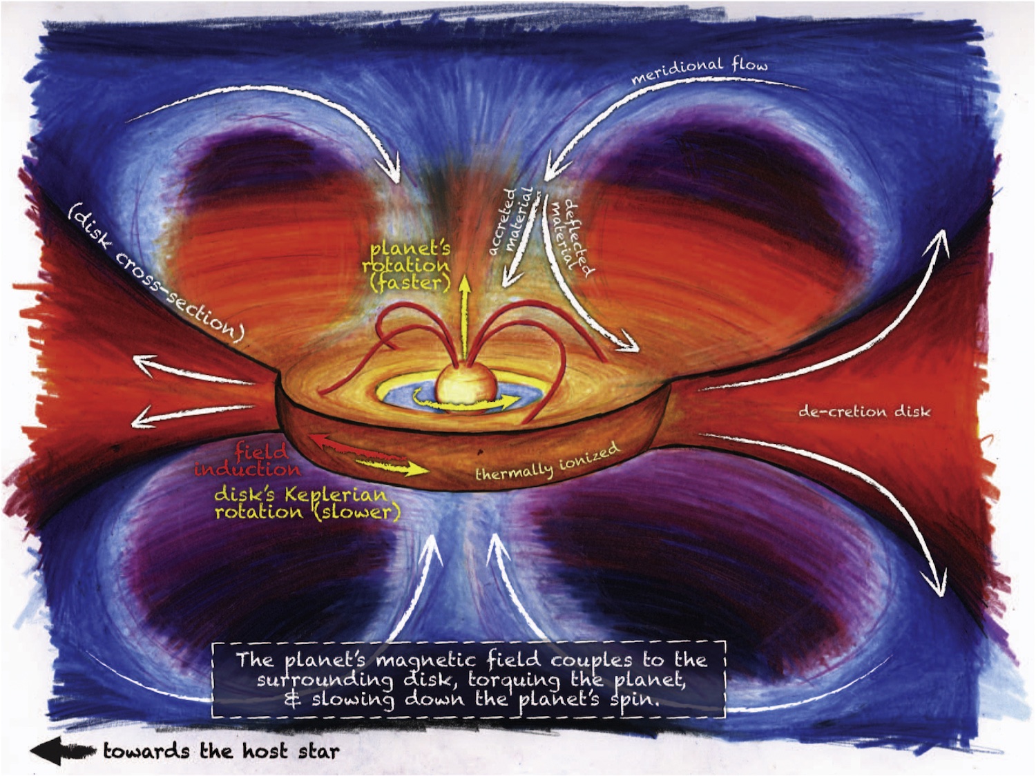
A schematic of some of the processes involved during giant planet formation. Click to enlarge. [Batygin 2018]
A number of different mechanisms predict that during this process, the protoplanet should emit light at a certain frequency associated with electrons jumping between energy levels in hydrogen atoms — what astronomers call Hα emission. In other words, they should glow at that specific wavelength only when they are accreting, though how strongly and consistently will depend on exactly what is happening around the planet.
These ideas existed only on paper until 2019, when a team first observed two glowing, growing planets around the star PDS 70. Recently, however, a powerful new instrument has begun collecting data, and astronomers just announced it has found another similar protoplanet: WISPIT-2b.
A New Discovery
A team of researchers led by Laird Close (University of Arizona) announced the discovery of this new world after analyzing images taken with the powerful new MagAO-X instrument on one of the 6.5-meter Magellan telescopes in Chile. The team outfitted the camera with a narrow filter that only admitted the Hα emission associated with accretion, then collected thousands of images over about two hours in April 2025. After stacking and post-processing their data, a faint dot jumped out beside the central star, right in the gap between two bright rings in the star’s disk.
This protoplanet, which the authors name WISPIT-2b in honor of their Wide Separation Planets in Time (WISPIT) survey, is likely about five times the mass of Jupiter and only 5 million years old, making it a baby on astronomical timescales. Based on how strongly it’s glowing, the team estimates that it’s accreting the equivalent of about 100 times the mass of Phobos, Mars’s largest moon, each year. This process won’t continue forever, as eventually the remains of the star’s disk will disperse and the planet will have to go hungry.This remarkable find is only the third protoplanet unambiguously found to be embedded within a disk, and the first to be found in the gap between rings in a disk. With MagAO-X collecting data and the WISPIT survey underway, we’re likely in store for many other discoveries in the coming years. With enough observations and examples of these growing worlds, astronomers will fill in their models of how exactly giant planets as we know them are formed.
Citation
“Wide Separation Planets in Time (WISPIT): Discovery of a Gap Hα Protoplanet WISPIT 2b with MagAO-X,” Laird M. Close et al 2025 ApJL 990 L9. doi:10.3847/2041-8213/adf7a5
The post Researchers Spot a Rare Glowing, Growing Planet appeared first on AAS Nova.
- AAS Nova
- Weather Forecast for the Habitable Worlds Observatory: Cloudy with a Chance of Biosignatures
Weather Forecast for the Habitable Worlds Observatory: Cloudy with a Chance of Biosignatures
![]()
Editor’s Note: Astrobites is a graduate-student-run organization that digests astrophysical literature for undergraduate students. As part of the partnership between the AAS and astrobites, we occasionally repost astrobites content here at AAS Nova. We hope you enjoy this post from astrobites; the original can be viewed at astrobites.org.
Title: Clouds Can Enhance Direct-Imaging Detection of O2 and O3 on Terrestrial Exoplanets
Authors: Huanzhou Yang, Michelle Hu, and Dorian S. Abbot
First Author’s Institution: University of Chicago
Status: Published in ApJ
In the hunt for life beyond Earth, studying exoplanet atmospheres is one of our most powerful tools. The atmosphere of an exoplanet acts as a window into its environment and allows astronomers to take a guess at what processes are shaping the planet’s climate, chemistry, and thus, its potential for habitability. The gases present in the planet’s atmosphere impart a unique spectral fingerprint on the light that passes through the atmosphere and allows us to identify what gases might be present and look for possible signs of life.
JWST is already hard at work looking at distant worlds and gathering spectroscopic data for us to analyse. It has already observed several exoplanets and recorded the light that passed through their atmospheres. This has not only sparked a debate over what could be interpreted as the first possible signs of biological activity, but has also given us a glimpse of worlds that are remarkably different from our own.
However, JWST is mostly limited to observing the atmospheres of transiting exoplanets. That means planets that orbit close to their host star and are more or less directly edge-on, as viewed from us, so that they transit across the face of the star. The only planets that the telescope is able to observe directly are big, young, and hot planets farther from their host star, at distances more resembling those of the outer planets in our solar system, like Saturn or Uranus. If only we had a telescope powerful enough to image a potentially habitable planet directly and analyse the light reflected from it.
The Habitable Worlds Observatory
This is where the Habitable Worlds Observatory (HWO) will come in. Now, I know what you are thinking. Why must astronomers always look towards greener pastures bigger telescopes when we already have perfectly functional space telescopes in orbit or practically on the launch pad? Well, it is true that the next flagship space telescope from NASA, the Nancy Grace Roman Space Telescope, is designed to directly image exoplanets and analyse their atmospheres. But space telescopes can take decades to plan and are often built on lessons learned from previous missions. Roman, with its 2.4-meter primary mirror and coronagraph, will be able to observe planets similar in size and orbital distance to Jupiter and will get us some of the way, for sure, but not all the way. At three times the mirror size, HWO, NASA’s proposed successor to Roman that is planned for the 2040s, will carry the torch even further and be able to directly image Earth-like planets in the habitable zone, where liquid water can exist on the planet’s surface.
But It Might Be Cloudy Tomorrow
This brings us to today’s article. If the HWO is going to potentially observe Earth analogues, we might expect it to run into the same issue that plagues everyone living in the UK for most of the year — clouds. Clouds in the atmosphere of an exoplanet can sometimes cause trouble when trying to analyse the light. Cloud particles scatter different wavelengths of light differently, and cloud variability creates dynamic changes in the observed brightness that can make it hard to know what exactly you are looking at. Clouds can also mean varying levels of opacity to look through depending on cloud composition, and they can hide underlying atmospheric properties such as water content beneath a thick cloud layer. For transmission spectroscopy, even a thin layer of clouds can disrupt the measurements as the light has to travel a long distance through the atmosphere. However, for direct imaging, the effect of clouds is perhaps more nuanced. While a thick cloud deck will block the signal from whatever gas lies beneath it, it will also increase the overall albedo of the planet and make it easier to detect the gas above. This means that cloud height and type are both critical variables in understanding atmospheric properties of exoplanets, which is what the authors of today’s article set out to investigate.
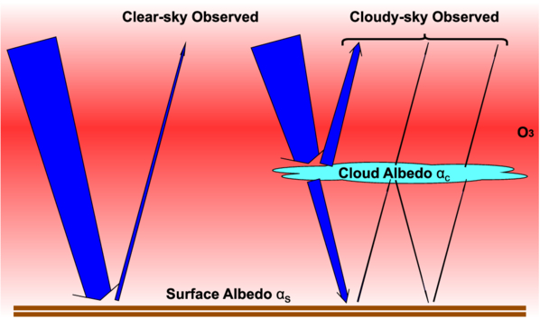
To understand how clouds affect the ability to detect signs of life on distant Earth-like planets, the authors used computer simulations. They focused on two gases, molecular oxygen (O2) and ozone (O3). Both gases are often regarded as biosignatures, although their astrobiological significance has been debated. The authors first used a tool that simulates how a telescope like the HWO might observe an Earth-like planet around a star 15 parsecs away. This tool predicts how light bounces off a planet’s surface and atmosphere, and whether the two gases would show up clearly in the reflected light. Next, they created realistic cloud conditions using a cloud microphysics model. This model calculates how clouds might form and behave depending on factors like surface pressure or how much sunlight the planet gets. With this they were able to change things like cloud height and particle size. They then combined the two tools by adding the simulated clouds into the observations model and building a mathematical model to help explain what they saw in the simulations. The sketch for their model can be seen in Figure 1.
Figure 1: Clouds can affect the amount of reflected light (albedo) from the imaged planet. Depending on the cloud height and particle size, this can have a big impact on the observability of different species of gas in the atmosphere. Here the blue arrows indicate the amount of light entering the atmosphere and being reflected. The arrows get thinner from tail to head, representing absorption in the atmosphere. Depending on the presence of clouds, a varying amount is reflected back. The red colour indicates the assumed distribution of ozone, O3, which is concentrated at a high altitude. [Yang et al. 2025]
A Cloudy Sky Can Actually Be a Good Thing
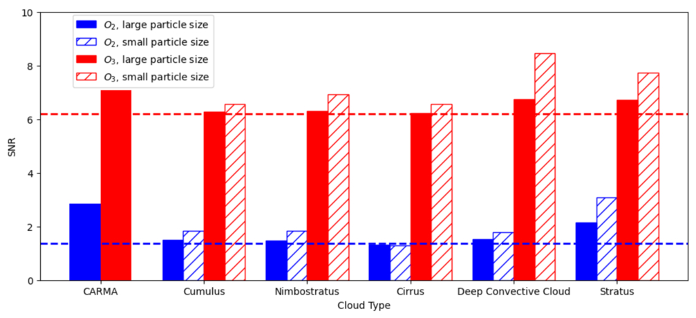
The researchers discovered that low clouds can actually make gases like oxygen and ozone easier to detect, because these clouds reflect more light without blocking the gases above them. On the other hand, high clouds can make it harder since they can hide the gases from view. They tested different cloud types using real data from Earth as seen in Figure 2. They looked at five common cloud types, like stratus (flat and low), cirrus (high and wispy), and deep convective clouds (tall, thunderstorm-like), and compared them to their model clouds (labelled as CARMA in the figure).
Figure 2: The figure shows how different types of clouds affect our ability to detect oxygen (blue) and ozone (red) on an Earth-like planet. The dashed lines show the results without clouds, while the bars show what happens when clouds are present. Solid bars represent larger cloud droplets, and striped bars represent smaller ones, which reflect more light. The figure shows that most cloud types — especially low clouds like stratus and thick storm clouds — actually make it easier to detect these gases, particularly when droplets are small. Only high, thin clouds (cirrus) slightly reduce oxygen detection, and even then, the effect is small. This is the article’s main finding: while clouds can block light in some observation methods, for direct imaging of reflected light, they often help us see signs of life better. The cloud types are based on real Earth data while the authors’ simulated cloud model is labelled as CARMA. [Yang et al. 2025]
Original astrobite edited by Maria Vincent.
About the author, Kasper Zoellner:
I have a Master of Science in astronomy and I am currently working towards a PhD in physics and educational science. My greatest passion is the search for exoplanets and how stellar variability may influence the possibility of life. I am also interested in science outreach, education, and discussing what sci-fi novel to read next!
The post Weather Forecast for the Habitable Worlds Observatory: Cloudy with a Chance of Biosignatures appeared first on AAS Nova.
A Dusty Disk Points to a Potential Planet
![]()
JWST observations of a nearby star’s debris disk recently revealed what may be one of the lowest-mass planets ever imaged.
How to Find a Small Planet
Though it remains a formidable technical challenge, astronomers have gotten fairly good at taking images of planets around other stars by carefully blocking the light of the host and searching for the small points of light that remain. However, though this technique is well-suited for discovering large, bright, high-mass planets, their lower-mass cousins below the size of Jupiter remain challenging to detect. To get pictures of these smaller worlds, astronomers must resort to detective work and search for signs of their presence through indirect means.
One promising approach is to look not for the planet itself, but rather its effect on the dusty disk of material around the star. These structures, called debris disks, are constantly replenished by planetesimals colliding and grinding one another to dust. If a planet happens to orbit within this disk, it will “stir” the dust into distinctive patterns including rings and spiral arms. If astronomers observe that a star has a debris disk with gaps or spirals, they can analyze those substructures and deduce where a planet might be hiding.
The Star of this Show: TWA 7
A mid-infrared image of TWA 7’s debris disk and candidate planet. [ESA/Webb, NASA, CSA, A.M. Lagrange, M. Zamani (ESA/Webb); CC BY 4.0]
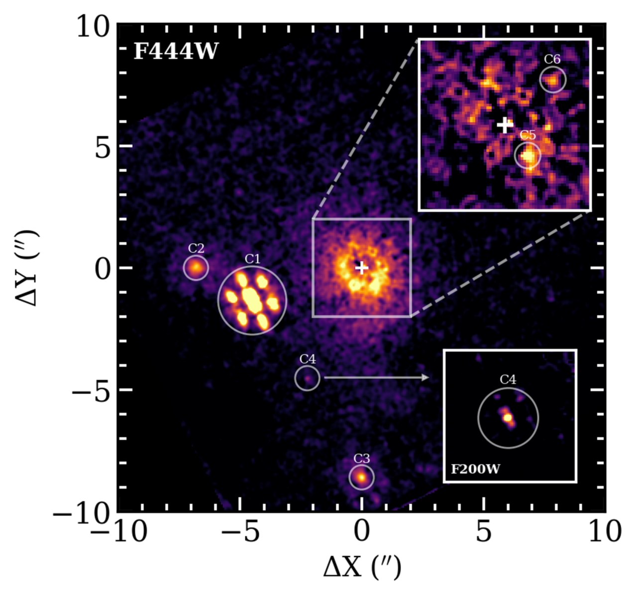
This prediction led to a search with JWST last summer. Initial observations taken at mid-infrared wavelengths revealed a bright dot sitting near the predicted location of the planet. However, with just these observations, it was hard to confidently say that this source wasn’t just a distant background galaxy that happened to appear there by chance. To help settle the matter, JWST took another look in two different near-infrared wavelength bands a few weeks later.
New JWST observations of TWA 7. The sources labeled C5 and C6 are planet candidates. C6 is located at the same place as the planet candidate identified in mid-infrared observations, making it a strong candidate. C5 requires further observations to understand if it is real or an artifact. Click to enlarge. [Crotts et al. 2025]
Revisiting TWA 7
A team led by Katie Crotts (Space Telescope Science Institute) recently published these later observations. These new data show a dot in the exact same place as before, and with a color that’s much more planet-like than galaxy-like.
While this adds plenty of evidence to the planetary interpretation, the team cautions that one more set of follow-up observations is needed to be confident that this is, in fact, a planet. Assuming future observations back up these first hints, however, this would be the lowest-mass planet ever imaged, and a happy conclusion to a detective story that started with dust.
Citation
“Follow-Up Exploration of the TWA 7 Planet–Disk System with JWST NIRCam,” Katie Crotts et al 2025 ApJL 987 L41. doi:10.3847/2041-8213/ade798
The post A Dusty Disk Points to a Potential Planet appeared first on AAS Nova.
Potential Discovery of a World Next Door
![]()
By pushing JWST to its limits, astronomers may have just spotted a planet around the nearest Sun-like star, Alpha Centauri A. Though another look at the star is needed next year to confirm the finding, if the detection holds up, this discovery easily earns several superlatives: the planet would be the nearest, oldest, coldest, and lowest-mass world ever directly imaged beyond our solar system.
Hi, Neighbor!
The scale of our Milky Way galaxy is hard to comprehend with our Earth-bound minds that are more attuned to measuring distances in miles than in light-years. However, even though our suburban stellar neighborhood is tens of thousands of light-years from the galactic downtown, it is not empty: there are a handful of nearby systems that offer unique opportunities to study stars and their potential planet children “up close.”
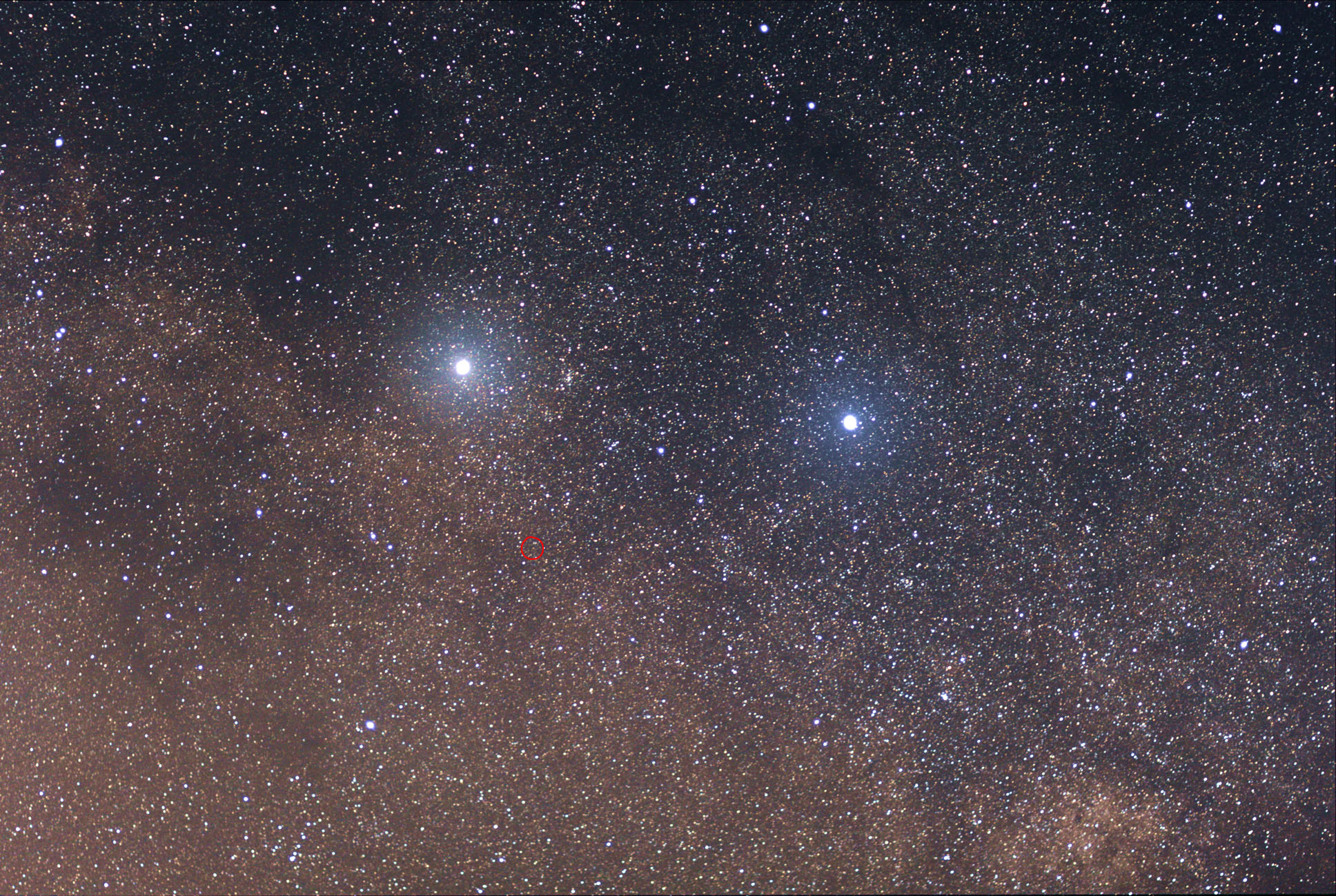
Alpha Centauri A and B (bright stars), along with Proxima Centauri, circled in red. [Wikipedia; CC BY-SA 3.0]
Though Proxima Centauri is the most famous of the trio, since it’s technically the closest star to the Sun, its larger siblings are arguably better targets for searching for planets. Since these stars are more massive, their habitable zones are large enough that astronomers can block out the star itself and search the surrounding area for planets using a tool called a coronagraph.
Challenging Observations
Back in 2019, a team of astronomers using a coronagraph on the Very Large Telescope uncovered tantalizing evidence of a giant planet orbiting Alpha Centauri A. However, confirming or rejecting this initial peek using telescopes on Earth’s surface was challenging. When JWST finally launched in 2021, following up on this hint was a top priority. Astronomers first attempted to point JWST toward the Alpha Centauri system in 2023, but unfortunately, the observations from that attempt were unusable.
Though one might think aiming a telescope at a nearby star is easier than taking a look at something more distant, this is unfortunately not the case. For one thing, the stars of the Alpha Centauri system are bright enough to overwhelm JWST’s sensitive detectors without careful calibration. For another, keeping the telescope locked on target and the star behind the coronagraph requires continuous tweaks to account for the Sun’s motion relative to Alpha Centauri, JWST’s motion relative to the Sun, and Alpha Centauri A’s motion relative to Alpha Centauri B. The team regrouped, tested a new observing strategy in mid-2024, then took three long observations in August 2024, February 2025, and April 2025.
A Planet Candidate
JWST coronagraph image of the Alpha Centauri system. Alpha Centauri B is the bright labeled star at the top, while Alpha Centauri A is labeled but hidden behind the coronagraph. The inset shows the region around Alpha Centauri A during the three different observations. The planet candidate, labeled S1, is only visible in the August 2024 observation. Click to enlarge. [Beichman & Sanghi et al. 2025]
The authors estimated that the source is a 1–1.1 Jupiter radius planet with a mass of 90–150 Earth masses (0.28–0.47 Jupiter mass) on a 2–3-year orbit. The team stressed that with just one detection, this should still be regarded as a planet candidate, not a confirmed discovery. If the planet is eventually verified, however, it stands out among its peers as the first mature, cold planet ever imaged within its star’s habitable zone. Future detectors may be able to collect spectra of the planet itself, unlocking new insights into giant planet evolution and composition in multiple-star systems with nested orbits, like the Alpha Centauri system.
Now that JWST has a proven strategy for observing this challenging system, it should be possible to take follow-up images. If the planet is real and traveling on its expected orbit, it should reappear beside Alpha Centauri A in August 2026. Hopefully, JWST will be looking when it does, and we can finally confirm that we have a planetary neighbor next door.
Citation
“Worlds Next Door: A Candidate Giant Planet Imaged in the Habitable Zone of α Cen A. I. Observations, Orbital and Physical Properties, and Exozodi Upper Limits,” Charles Beichman et al 2025 ApJL 989 L22. doi:10.3847/2041-8213/adf53f
“Worlds Next Door: A Candidate Giant Planet Imaged in the Habitable Zone of α Cen A. II. Binary Star Modeling, Planet and Exozodi Search, and Sensitivity Analysis,” Aniket Sanghi et al 2025 ApJL 989 L23. doi:10.3847/2041-8213/adf53e
The post Potential Discovery of a World Next Door appeared first on AAS Nova.
Exo-Saturns and Exo-Jupiters Are Within JWST’s Reach
![]()
Of the nearly 6,000 currently known exoplanets, few closely resemble any of the planets in our solar system. New research suggests that JWST is capable of directly imaging exoplanets with temperatures and orbital distances similar to Jupiter and Saturn, placing truly familiar exoplanets within our observational grasp.
Increasingly Cold Discoveries
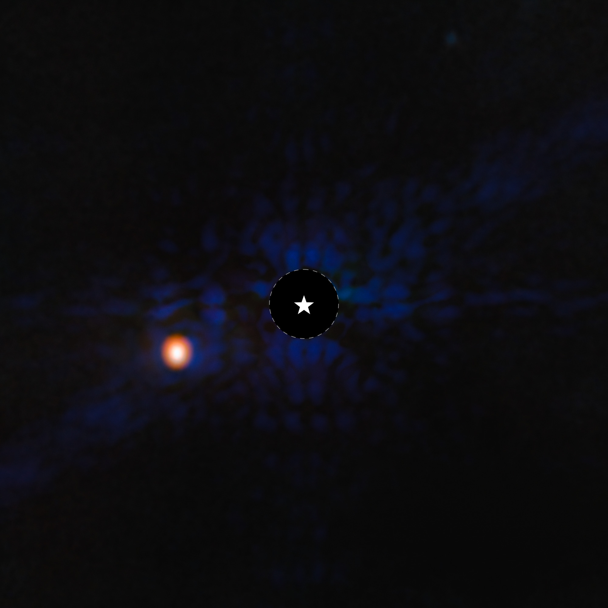
A JWST image of the exoplanet Epsilon Indi Ab, one of the coldest exoplanets to be directly imaged. The planet’s temperature is estimated to be just 275K (35℉/2℃). [NASA, ESA, CSA, STScI, Elisabeth Matthews (MPIA)]
Answering this question requires a deep dive into the abilities of JWST’s instruments. The current go-to method for directly imaging planets with JWST is coronagraphy with its Near-Infrared Camera (NIRCam). In this observing mode, the instrument blocks the light from the star, allowing the fainter thermal glow of the planet to shine through.
But as Rachel Bowens-Rubin (University of Michigan and Eureka Scientific) and collaborators note in a recent research article, this may not be the best way to detect cold giant planets. Models suggest that these planets have cloudy atmospheres, which means that they wouldn’t be bright at NIRCam’s preferred near-infrared wavelengths, and would instead be detected more easily in the mid-infrared, where JWST’s Mid-Infrared Instrument (MIRI) reigns.
Combining Data and Models
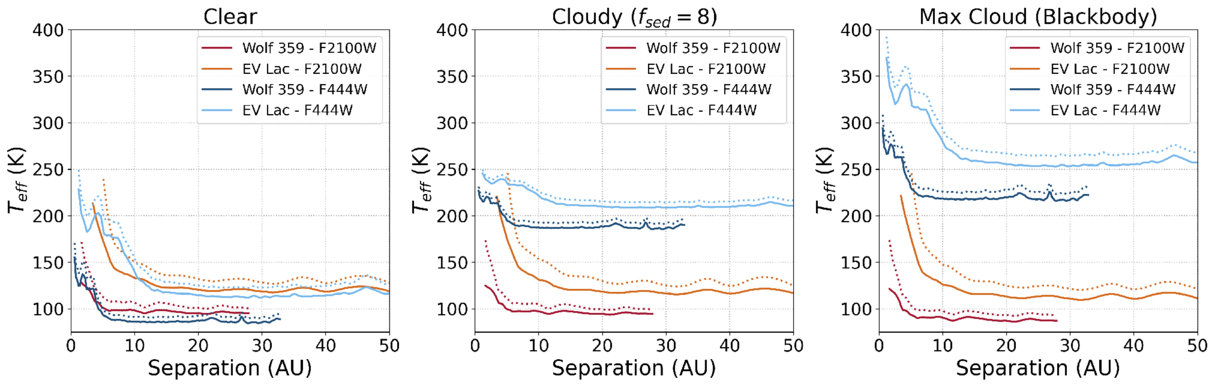
To examine the capabilities of both of these instruments, Bowens-Rubin’s team analyzed JWST observations from the Cool Kids on the Block program, which targets cold, low-mass giant planets around nearby low-mass stars with NIRCam coronagraphy and MIRI imaging. The team used observations of nearby M-dwarf stars Wolf 359 and EV Lac to construct constrast curves: the level of planet–star flux contrast that is detectable by each instrument as a function of distance from each star. These curves depend on the flux of the star and the planet as well as the limitations of the instrument — the detector noise and background noise.
Temperatures of coldest detectable planets as a function of separation from the host star for Wolf 350 and EV Lac. Results are shown for MIRI F2100W imaging and NIRCam F444W coronagraphy. Click to enlarge. [Bowens-Rubin et al. 2025]
NIRCam vs. MIRI
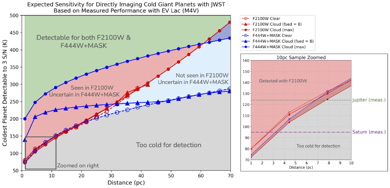
This analysis showed that MIRI is the best choice for directly imaging cold planets around nearby stars (within 65 light-years). MIRI should be able to detect giant planets with temperatures down to 94K around Wolf 359 and 114K around EV Lac — about the temperature of Saturn and slightly colder than Jupiter, respectively. For Wolf 359, sub-100K planets are detectable at orbital distances of at least 4.8 au, meaning these planets could also have similar orbital separations to Jupiter and Saturn.
Temperatures of planets detectable to a signal-to-noise ratio of 3 as a function of distance from Earth. Detection limits for MIRI and NIRCam are shown as red and blue lines, respectively. Click to enlarge. [Bowens-Rubin et al. 2025]
As impressive as these results are already, Bowens-Rubin and coauthors noted that future work, such as developing strategies to mitigate MIRI’s “brighter-fatter effect” that limits sensitivity at small angular separations from the host star, could enhance the search for exo-Saturns and exo-Jupiters even further.
Citation
“NIRCam Yells at Cloud: JWST MIRI Imaging Can Directly Detect Exoplanets of the Same Temperature, Mass, Age, and Orbital Separation as Saturn and Jupiter,” Rachel Bowens-Rubin et al 2025 ApJL 986 L26. doi:10.3847/2041-8213/addbde
The post Exo-Saturns and Exo-Jupiters Are Within JWST’s Reach appeared first on AAS Nova.
Stanford’s Revolutionary New Microscope Reveals Living Cells in Stunning Detail
Chickens without eggs? De-extinction company creates artificial egg.
On Tuesday, biotech startup Colossal announced its newest development on the road to its announced goal: reversing the extinction of species, in this case, avian species. The development itself is essentially an artificial eggshell, one that allows almost the entire developmental process to occur without the shell. The company transferred the contents of eggs to their specially designed container within a day or two of laying and were able to have normal chicks walk away from it.
Beyond its potential utility for Colossal's intended efforts, the work is personally interesting to me because it may solve a problem I faced in my research days. I'm going to start by describing the research problem that Colossal may have solved, before coming back to what it hopes to use its technology to do—and why the company still has a few key hurdles left to overcome.
Watching development
For part of my career, I studied the development of vertebrates using chickens. While they're less closely related to us than something like mice, the basics of their development are largely the same. And, unlike mice, they develop outside of their mother's body. If you're careful, you can chip away a hole in the egg, perform manipulations on the developing embryo, and then seal it back up with some tape. The chicken embryo will keep developing, allowing you to see the impact of what you've done on normal development.


© Colossal