❌

Normal view

Cells from your mother likely infiltrated your brain in the womb, and they could survive for decades, study reveals

19 July 2026 at 12:00

Researchers have discovered that children's brains can contain cells with their mother's DNA and that these cells can persist for decades.

The findings, which were posted to the preprint database bioRxiv June 10 but have not been peer-reviewed yet, are part of a growing body of work showing that a mother and fetus exchange cells during pregnancy ‪—‬ a phenomenon known as "microchimerism." Previously, scientists had found that a mother's brain harbors cells with her children's DNA.

The findings are important for several reasons, said Amy Boddy, co-director of the Microchimerism, Human Health and Evolution Project at the University of California, Santa Barbara, who was not involved in the study. Past work mostly found evidence of maternal microchimerism in infancy, and in blood samples, she told Live Science in an email. "What's exciting here is that it's tissue, not blood; it's real human data, not an animal model; and the methods are cutting-edge."

More broadly, the work reinforces the idea that microchimerism is "a normal process of mammalian biology," Boddy said.

Hunting down maternal cells in the brain

Before this study, there was sparse evidence for maternal microchimeric cells in brains, mostly because it is hard for researchers to get samples of human brain tissue and DNA from both parents and their children.

To overcome this challenge, a team led by Sami Kanaan, a staff scientist at the Fred Hutchinson Cancer Center in Seattle, analyzed brain tissue that had been surgically removed from dozens of children with severe epilepsy as part of their treatment. The patients ranged in age from 28 days to 19 years at the time of their surgery, and their mothers provided DNA samples through cheek swabs.

Kanaan's team used a tool called quantitative PCR to identify and count maternal cells hiding among millions of cells in the children's brains.

Out of 37 mother-child pairs, 26 children ‪—‬ 70% ‪—‬ had their mother's cells in their brains. These maternal cells were distributed across multiple regions of the brain, including the frontal, temporal and parietal lobes, which sit on the brain's outer surface, and the hippocampus, which is buried deep inside. Each sample had, on average, about 2.2 maternal cells per 100,000, though one sample from the hippocampus had 459 maternal cells per 100,000 and 11 children had no evidence of maternal DNA in their brains.

That prevalence is probably underestimated, Boddy said, "due to the limits of detecting rare cells at low frequency with this method."

Being a firstborn child seemed to increase the odds of having maternal cells in the brain. Of the children who carried their mother's cells, 14 were firstborns and 12 were later-born. In contrast, among the children who didn't have these cells, only one was a firstborn and 10 were later-born.

Using a technique called single nucleus RNA sequencing, which reveals what a cell is doing by showing which genes within the cells are switched on, the researchers found that the maternal cells had transformed into several types of brain cells. Likely, these cells were originally leukocytes and stem cells and had been transferred to the fetus via the placenta or during pregnancy or breastfeeding.

The transformed cells included neurons; oligodendrocytes, which produce the protective sheath around neurons; astrocytes, which support many brain functions and help fuel neurons; microglia, the brain's immune cells; and endothelial cells, which line blood vessels.

"Being able to use single-nucleus RNA sequencing to identify what type of cells the maternal microchimeric cells actually are is amazing," Boddy said. "We've been so limited in understanding the function of these cells, and papers like this, with these methods, are getting us closer."

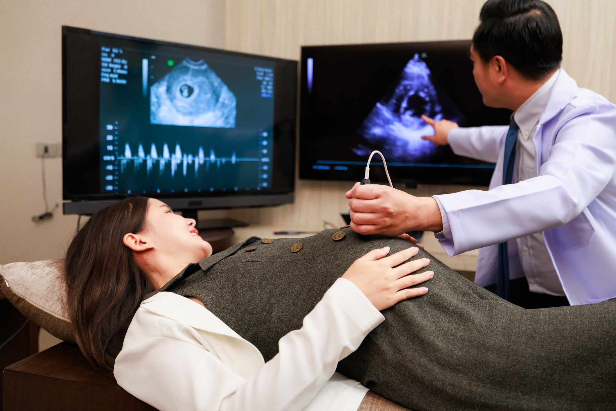
A woman lays on a table with a man giving her an ultrasound

During pregnancy, the mother and fetus exchange cells — a phenomenon called "microchimerism." (Image credit: Rhenizara S via Getty Images)

Checking healthy brains

To see whether these findings also applied to people without epilepsy, the researchers examined brain autopsy data from 29 individuals with no known neurodevelopmental conditions, ranging in age from 22 weeks of gestation to 40 years. They also analyzed brain tissue from three men in their late 80s and early 90s without any known brain conditions originally collected as part of an Alzheimer's disease study.

In total, the researchers found foreign cells in 25 of 32 people — about 78% — including in the brain of a man in his 90s.

The researchers suspect these foreign cells are maternal, but they couldn't confirm it because they didn't have DNA from the mothers. It is possible that the foreign cells came from a twin; an older sibling; a past pregnancy, miscarriage or abortion (in females); or, rarely, from a maternal grandmother, the team wrote in their paper.

As in the epilepsy patients, these cells had matured into several types of working brain cells. In younger brains, they most often became a specific kind of neuron, a layer 2/3 neuron, while in older brains, maternal cells were more likely to be microglia.

Given that these microchimeric cells take on very different roles, it would be interesting to know whether that diversity reflects their origins ‪—‬ for example, whether they came from a mother, an older biological sibling or a maternal grandmother, said Dr. Sing Sing Way, a microchimerism researcher at Cincinnati Children's Hospital Medical Center who was not involved in the study.

The number of maternal cells declined with age, but they never completely disappeared.

Boddy said the findings mostly raise new questions. "Do we maybe need microchimeric cells to 'help out'?" she asked. "Is diversity of cells in the brain important, or is it just a byproduct of being a placental mammal?”

Because microchimeric cells appear to be common, Boddy suspects they "are doing an important job in the brain, so understanding their function could be very important for understanding healthy brain development."

By using new analytical tools to pinpoint microchimeric cells, the study "pushes the boundaries" of previous research, but future studies would benefit from larger and more uniform datasets, Way said. That would mean obtaining more brain biopsies, analyzing more cells from each sample, collecting specimens at different ages, and sampling similar brain regions across individuals to better compare the results.

This article is for informational purposes only and is not meant to offer medical advice.

'RNA can do things which we have never seen before': New study challenges assumptions about what RNA was up to at the dawn of life

17 July 2026 at 18:20

Scientists long thought that when RNA kick-started life on Earth 4 billion years ago, it could form only small, simple structures. But new research shows that naturally occurring RNA molecules can also adopt large, sophisticated geometries, like filaments and cages. Now, scientists wonder whether the structures were present at life's beginning.

According to an idea known as the RNA world hypothesis, RNA-based life-forms preceded modern ones that use DNA and protein. RNA, a molecular cousin of DNA, still plays roles in modern cells but does not serve as the primary genetic material. By comparison, primordial species used RNA to store genetic information and to catalyze reactions as stand-in enzymes.

Proteins eventually dominated as enzymes, perhaps because they can fold into more diverse figures than RNA can. That's because proteins are composed of 20 types of subunits, called amino acids, each with a unique structure, whereas RNA is composed of only four subunits, called nucleotides, that all adopt similar shapes.

Scientists originally thought that only proteins were varied enough to assemble into large structures, but a new paper has demonstrated that RNA — though more limited in its variety — also has the capacity to form these large configurations. The research was posted to the preprint server bioRxiv July 1 and has not been peer-reviewed yet.

"We show RNA can do things which we have never seen before," said study co-author Lin Huang, an RNA biologist at Sun Yat-Sen University in China. "It suggests that at the origin of life RNA could assemble into all kinds of shapes," he told Live Science.

Huang and his colleagues had hypothesized that RNA molecules could link together if they possessed sequences that fold into "kissing stem loops." This occurs when an RNA strand folds over on itself, forming a structure that resembles a loop in a shoelace. If loops from different RNAs bond together, or "kiss," the molecules could link up and form larger complexes, the researchers proposed.

After sifting through a bevy of RNA sequences, the researchers found a family of RNA molecules encoded by bacteriophages ‪—‬ viruses that infect bacteria ‪—‬ that form these loops. They purified several of these RNA molecules in the lab, allowed them to assemble in a dish, and then captured their structures using cryo-electron microscopy.

They discovered that some of the RNA molecules formed long filaments. These resembled protein-based filaments such as the cellular cytoskeleton, a scaffold that participates in many functions, including shaping and moving the cell.

Other RNA molecules assembled into cages as large as common viruses. Some of these took the form of an icosahedron — a 3D shape that is built from 20 equilateral triangles and resembles a soccer ball. Many viruses, including herpesviruses, package their genome into protein-based icosahedra called capsids. This raises a question: Could RNA-based capsids have packaged genomes in the RNA world?

A colorful illustration of an RNA molecule folded

RNA structures assemble into icosahedra as large as protein-based virus capsids. (Image credit: Lin Huang)

This work demonstrates that RNA had the capacity to assemble into these elaborate structures during the RNA world, Huang said, but that doesn't prove it actually happened.

"I definitely think that environmental parameters are a question," Anna Medvegy, an evolutionary biologist at Eötvös Loránd University in Hungary, told Live Science in an email. "Can these structures form in the environment in which the hypothetical RNA World existed?" said Medvegy, who was not involved in the new work.

If scientists could recreate these environmental conditions at the dawn of life, such as high temperatures and low pH, and still observe that these structures take shape, that would strengthen the theory that they could have been present in the RNA world, she said.

Although the RNA cages and filaments were large, Huang's team generated them using only short RNA strands, each no longer than 200 subunits. Medvegy said long RNAs are susceptible to breaking, so if short strands can assemble into these structures, that provides more promise that these multi-tiered molecules could have formed in the RNA world.

Another question is whether these elaborate RNA complexes currently assemble inside the bacteriophage-infected bacteria from which they were derived. So far, Huang's team has only seen these structures form in a lab dish, so they need to determine if factors inside bacteria, such as proteins, would either disrupt or enable their formation inside cells.

Beyond providing insight into life's beginnings, these RNA cages could have potential applications in biotechnology, Huang thinks. Efforts are underway to use DNA folded into "DNA origami" to deliver drugs into cells, and Huang thinks DNA's older cousin, RNA, could one day play a similar role in medicine.

Ancient DNA Reveals the Hidden Origins of China’s Mysterious Shimao Civilization

Carving Stone at Shimao CityAncient DNA from Shimao reveals local origins, broad prehistoric connections, a patrilineal social structure, and gender-specific patterns of human sacrifice. At Shimao, a vast stone-walled settlement in northern China, ancient DNA is filling in details that archaeology alone could not fully resolve. The genetic evidence is helping trace where the people of this late Neolithic [...]

Scientists Discover a Surprising Link Between Sleep, Genes, and Alzheimer’s

14 July 2026 at 15:39
Neuroscience Magnifying Glass Brain Alzheimers ResearchA new study suggests that sleep habits may influence how certain genetic variants affect early brain and cognitive changes associated with Alzheimer’s disease. Sleep may do more than help the brain recover from a busy day. New research suggests it could also influence how certain genes shape the earliest brain changes associated with Alzheimer’s disease. [...]

Japanese Researchers Unlock a Powerful New Way To Edit and Assemble DNA

24 June 2026 at 14:12
Genetic Mutation Damaged Broken DNA StrandSilver nanoparticles provide a more efficient way to cut and assemble DNA, improving recovery rates and boosting DNA joining efficiency for future gene engineering applications. DNA is made of long chains that carry the instructions living things need to grow and function. In genetic engineering, researchers cut DNA at chosen locations and attach the pieces [...]

Epigenetic Locking Drives H. pylori Gastric Cancer

24 June 2026 at 12:02

In a groundbreaking study published in Cell Death Discovery, researchers have unveiled a novel mechanism by which Helicobacter pylori, the notorious gastric pathogen, drives the formation and persistence of gastric cancer through a process termed the “hit-and-run” legacy. This research elucidates how transient infection by H. pylori delivers a lasting epigenetic imprint on gastric epithelial cells, effectively locking these cells into a survival mode that fosters oncogenesis. The implications of these findings are profound, offering fresh insights into the molecular interplay between infection-induced epigenetic modification and cancer development, which could pave the way for revolutionary diagnostic and therapeutic strategies.

Helicobacter pylori has long been established as a principal carcinogen associated with gastric cancer, a malignancy ranking among the world’s deadliest. Traditionally, the carcinogenic potential of H. pylori has been attributed to chronic inflammation and the persistent presence of bacterial virulence factors that continuously disrupt gastric mucosa homeostasis. However, the current study challenges this paradigm by demonstrating that even transient exposure to H. pylori can induce stable, heritable epigenetic alterations that effectively “lock” the cellular genome into an aberrant state, independent of ongoing infection. This “hit-and-run” mechanism underscores the importance of early, possibly even brief, pathogen-host interactions in cancer genesis.

At the heart of this discovery is the concept of epigenetic locking—a process through which transient signals induce durable modifications in DNA methylation and histone patterns, which subsequently enforce gene expression profiles conducive to cell survival and proliferation. The group led by Sun, Hu, and Traub employed state-of-the-art genome-wide methylation profiling alongside chromatin immunoprecipitation sequencing (ChIP-seq) to map the intricate epigenetic landscapes shaped by H. pylori infection. Their findings revealed a marked enrichment of repressive histone marks on tumor suppressor gene loci and a reduction in methylation at key oncogenic enhancers, a dual mechanism that effectively enables gastric epithelial cells to bypass apoptosis and adopt a resilient phenotype.

Intriguingly, the authors identified a “survival switch” embedded within the epigenetic circuitry, which perpetuates cell viability in the hostile gastric environment despite the absence of the pathogen. This survival pathway hinges on the modulation of several critical transcription factors and downstream effectors that drive resistance to oxidative stress and immune checkpoint evasion. The persistence of this switch presents a formidable barrier to therapeutic intervention, explaining the often-observed resistance and relapse seen in conventional anti-H. pylori and chemotherapeutic regimes.

The study further explores the timing and sequence of epigenetic alterations following H. pylori infection. Temporal analysis demonstrated that initial infection triggers a wave of epigenetic remodeling within days, with key changes becoming fixed in the genome over weeks, thus confirming the durability of the modifications. This rapid “imprinting” emphasizes the need for early detection and intervention post infection to prevent the establishment of irreversible oncogenic programs.

Mechanistically, the team delved into the role of bacterial virulence factors such as CagA and VacA in orchestrating these epigenetic changes. They postulate that CagA, upon translocation into host cells, interacts with host chromatin modifiers and signaling pathways like NF-kB and STAT3, initiating cascades that culminate in altered chromatin architecture. VacA further modulates cellular metabolism and stress responses, cumulatively fixing the epigenetic landscape conducive to cancer progression.

The implications of the “hit-and-run” model extend beyond H. pylori-induced gastric cancer, provoking reconsideration of how transient infections may contribute to other epigenetically driven malignancies. This paradigm shift advocates for a broader investigation into the long-term impact of infectious insults at the epigenome level, potentially revolutionizing the standard view of tumorigenesis associated with microbial pathogens.

Therapeutically, the study stimulates the development of epigenetic-targeted approaches aimed at “unlocking” these stable aberrations. By reversing methylation patterns or disrupting the survival networks sustained by epigenetic modifications, it may become possible to sensitize tumor cells to existing chemotherapies or to immune-mediated clearance. The prospect of integrating epigenetic therapy into the clinical management of H. pylori-associated gastric cancer represents a promising frontier.

Moreover, diagnostic innovation could also benefit from these findings. Epigenetic biomarkers identified through this research may serve as early indicators of cancer risk in patients with a history of H. pylori infection, enabling stratification and personalized surveillance. The early detection of epigenetic locking signatures in gastric mucosa could dramatically improve prognosis by facilitating timely therapeutic intervention.

The study’s comprehensive exploration of epigenetic changes was bolstered by the use of gastric organoids and in vivo models, which authentically recapitulated the cellular and tissue contexts of H. pylori infection. This multi-faceted experimental design lent robust physiological relevance to the findings and highlighted the dynamic interplay between pathogen factors and host epigenomic machinery.

Collectively, this research not only deciphers the cryptic molecular legacy left by H. pylori but also refines our understanding of infection-associated carcinogenesis. The “hit-and-run” legacy paradigm sheds light on the complex, often covert, mechanisms through which transient pathogen interactions exert long-lasting oncogenic effects, shifting both scientific insight and clinical approach.

In summary, the work by Sun, Hu, Traub, and colleagues heralds a new era in gastric cancer biology, emphasizing the critical role of infection-driven epigenetic programming. As further investigations build upon this foundation, multidisciplinary efforts encompassing microbiology, epigenetics, and cancer therapeutics will be essential to translate these insights into tangible clinical applications.

The future of combatting H. pylori-associated gastric cancer lies not only in eradicating the bacteria but also in decoding and reversing the epigenetic codes it inscribes. This endeavor carries the promise of transforming patient outcomes, potentially mitigating one of the world’s most lethal cancers with strategies born from the understanding of the pathogen’s epigenetic “hit-and-run” legacy.


Subject of Research: Epigenetic mechanisms underlying the persistence and oncogenic transformation in Helicobacter pylori-associated gastric cancer.

Article Title: The “Hit-and-Run” legacy: epigenetic locking and the survival switch in H. pylori-associated gastric cancer.

Article References:
Sun, J., Hu, P., Traub, B. et al. The “Hit-and-Run” legacy: epigenetic locking and the survival switch in H. pylori-associated gastric cancer. Cell Death Discov. (2026). https://doi.org/10.1038/s41420-026-03210-y

Image Credits: AI Generated

DOI: https://doi.org/10.1038/s41420-026-03210-y

Koalas Nearly Vanished 100,000 Years Ago – Long Before Humans Arrived

22 June 2026 at 00:19
Cute Koala Close UpKoalas survived a climate-driven population crash 100,000 years ago, and new genomic research is helping scientists better protect the species today. A major genomic study has transformed scientists’ understanding of koala (Phascolarctos cinereus) evolution, showing that the species underwent a dramatic population decline about 100,000 years ago, long before humans reached Australia. Researchers found that [...]

Scientists Discover a Gene That Boosts Youth – but It Comes With a Cost

Glowing Green DNA LongevityResearchers have identified a gene that appears to balance a powerful evolutionary trade-off, boosting growth and reproductive success early in life while carrying hidden costs later on. The biological processes that help build a healthy young body may also sow the seeds of aging and cancer. Researchers have now identified a gene that appears to [...]

5,500 Years Ago, Something Was Killing Humans in Prehistoric Siberia—New Research Has Finally Revealed This Ancient Assassin

21 June 2026 at 16:14

A longstanding archaeological mystery has loomed over discoveries at ancient burial sites in eastern Siberia: why were so many children and adolescents among the dead?

Now, according to a recent study in the journal Nature, DNA recovered from human remains found in burial sites at four cemeteries near Lake Baikal in eastern Siberia may finally reveal the answer.

Thousands of years ago, prior to the rise of ancient medieval cities—and their often-rat-infested streets—a disease famously associated with these rodents had already ravaged communities of prehistoric hunter-gatherers.

The research, based on reconstructions of ancient bacterial genomes preserved in the teeth of these ancient Siberians, revealed the presence of previously unknown strains of Yersinia pestis—the bacteria responsible for plague—providing fresh evidence that the lethal disease was already manifesting and killing human populations at least as far back as 5,500 years ago.

“Whether the earliest forms of plague were mild or virulent has been a matter of debate, but our findings demonstrate that these ancient strains were already highly lethal,” said senior author Eske Willerslev of the University of Copenhagen and the University of Cambridge.

New Evidence of an Ancient Siberian Plague

In their study, the team successfully located plague DNA in 18 out of 46 individuals they studied. Based on radiocarbon dating, many of the deaths were confirmed to have all happened within a short span of time, which is consistent with sudden deaths resulting from plague. Additionally, genetic evidence showed that several of these victims had been close relatives.

One key finding in the research involved the number of children and adolescents who were among the dead—a discovery mirroring earlier findings at prehistoric sites in the region, which had remained a longstanding archaeological mystery.

“The unusually high number of children and the short timespan was a real puzzle that we’ve been trying to solve since the 1990s,” said archaeologist Andrzej Weber of the University of Alberta. “Finding out that plague was the cause is extraordinary, but it makes so much sense.”

New Findings Challenge Old Assumptions

Based on the new findings, ancient strains of plague appear to have been more harmful than once thought. In the past, it was presumed that because these early strains lacked genetic adaptations that helped the bubonic plague spread so efficiently through fleas and rodents, they were relatively harmless.

The team’s research now reveals that these newly identified strains carried a specific toxin-producing genetic feature, known as a superantigen, which can trigger severe immune responses that potentially had devastating impacts on mortality rates in the ancient world.

Such deadly outbreaks may have had a far more significant impact on ancient human populations than previously realized, and during periods long before the rise of major urban centers and organized agriculture.

In terms of the origins of the disease, researchers believe that they may have finally traced it to Central or Northeast Asia, where infections may have spread from marmots, large rodents that are still recognized as plague carriers in modern times.

After years of uncertainty, the team’s research offers some of the best evidence to-date that one of the most devastating diseases in human history was already playing a role in shaping human history thousands of years earlier than once thought.

Martin Sikora, associate professor at the University of Copenhagen and the study’s senior author, says his team’s research “changes our understanding of the earliest plague outbreak,” and now confirms that such ancient strains “appear to have carried a potent combination of virulence factors that could make infection highly lethal.”

The team’s study, “Lethal plague outbreaks in Lake Baikal hunter-gatherers 5,500 years ago,” appeared in the journal Nature on June 17, 2026.

Micah Hanks is the Editor-in-Chief and Co-Founder of The Debrief. A longtime reporter on science, defense, and technology with a focus on space and astronomy, he can be reached at micah@thedebrief.org. Follow him on X @MicahHanks, and at micahhanks.com.

Inbreeding Hits Small Males in Threatened Songbird

21 June 2026 at 11:49

Inbreeding depression, a genetic phenomenon where offspring result from the mating of related individuals, has long been understood to compromise individual fitness and elevate the risks of population decline or extinction. However, the depth and nuances of these effects remain largely unexplored in many non-model species, especially those facing dire conservation challenges. A groundbreaking new study investigates this complex relationship in a threatened songbird species, the aquatic warbler (Acrocephalus paludicola), unveiling how inbreeding depression manifests in a phenotype-dependent manner, particularly emphasizing body size as a critical factor in male fitness outcomes.

The aquatic warbler, a habitat specialist bird known for its recent precipitous population decline, poses a unique opportunity to study the genetic intricacies and their ecological consequences. Unlike widely studied model species, this bird’s depleted genetic diversity coupled with habitat specificity creates an ideal system to understand how inbreeding depression impacts survival and reproduction under real-world environmental pressures. Employing cutting-edge genomic tools such as single nucleotide polymorphisms (SNPs), researchers could quantify inbreeding at an unprecedented resolution, offering new insights into the genetic health and viability of this endangered population.

One of the pivotal breakthroughs of this research lies in demonstrating that inbreeding depression is not a uniform or straightforward process but is intricately moderated by the physical and ecological context of the individual. For male aquatic warblers, the genomic markers revealed a pronounced negative effect of inbreeding on breeding success—but only among males characterized by smaller body size, denoted by tarsus length, an established morphological proxy. This finding highlights how phenotypic traits can mediate the severity of inbreeding effects, suggesting that smaller-bodied males experience disproportionately harsh fitness reductions as a consequence of their genetic relatedness.

Equally compelling is the study’s exploration of wing length as another morphological trait potentially influencing inbreeding depression. While the results here were more subtle, a notable weak interaction was found between the degree of inbreeding and shorter wing length in males, correlating with decreased survival probabilities. This interaction suggests that even relatively minor morphological differences may buffer or exacerbate the impacts of genetic relatedness on survival outcomes, adding another layer of complexity to how we understand inbreeding depression in natural populations.

Importantly, the research also reveals a stark contrast between sexes. Female aquatic warblers showed scant evidence of inbreeding depression affecting key fitness components such as clutch size, hatching success, nestling survival, or the number of fledged fledglings. Moreover, no interactions emerged between female fitness measures and phenotypic or environmental variables examined. This asymmetry in inbreeding effects underscores sex-specific pathways in evolutionary and ecological responses to genetic challenges, warranting further inquiry into the mechanistic underpinnings driving such differences.

Prior studies investigating inbreeding depression often relied on relatively simplistic models that did not incorporate potential interactions with individual phenotype or environmental heterogeneity. This omission might explain why many previous estimates of inbreeding load—reflecting the cumulative fitness consequences of deleterious recessive alleles—have been inconsistent or highly uncertain. By integrating detailed phenotypic data with high-resolution genomic analyses, this study advances the field by showing that accounting for individual variation unveils clearer, biologically meaningful patterns of inbreeding depression.

The implications for conservation biology are profound. Small-bodied males, disproportionately burdened by inbreeding loads, might represent a critical demographic subgroup whose reproductive success most strongly influences population trajectories. This insight challenges conventional approaches that treat all individuals as homogenous units within populations, advocating instead for conservation strategies that are finely tuned to the nuanced genetic and phenotypic architectures of endangered species.

The environmental factors considered—timing of breeding and brood size—proved less influential in mediating inbreeding depression compared to phenotypic variables, at least in this particular system. While breeding timing and brood size have been previously hypothesized to influence fitness by affecting resource allocation or offspring survival probabilities, their interaction with genomic inbreeding proved minimal. This result refines our understanding of the complex ecological determinants of fitness and suggests that underlying genetic and phenotypic factors can overshadow the modifying effects of certain environmental variables.

Methodologically, the use of SNP-based genomic inbreeding coefficients represents a significant step forward from traditional pedigree-based measures. Genetic markers enable more precise estimates of genome-wide relatedness and inbreeding, which is especially crucial for populations like the aquatic warbler, where pedigree data are incomplete or unavailable due to logistical challenges in tracking individuals across generations. This technological advance paves the way for more accurate assessments of inbreeding consequences in conservation genetics.

Another noteworthy contribution is the nuanced interpretation of inbreeding load estimates. The study found that models omitting phenotypic interactions yielded highly uncertain inbreeding load figures for both survival and breeding success in males, whereas incorporating body size interactions produced more robust and meaningful estimates. This methodological insight calls for revisiting traditional models that treat inbreeding load as a fixed entity, promoting dynamic frameworks that integrate individual heterogeneity.

The study also contributes to evolutionary theory by demonstrating how inbreeding depression may exert selective pressure that maintains or increases body size in male aquatic warblers. Since smaller males suffer more pronounced fitness detriments due to inbreeding, natural selection may favor larger males, which effectively buffer against the genetic disadvantages. This dynamic adds a rich dimension to sexual selection and life-history evolution, where morphology and genetic fitness interplay to shape population structure.

In broader ecological contexts, understanding conditional inbreeding depression helps elucidate population resilience amid environmental change. As habitats degrade or fragment, and populations become increasingly isolated, the compounding effects of genetic bottlenecks and reduced phenotypic diversity could spell catastrophic declines if certain phenotypic groups disproportionately suffer. This research underscores the urgent need to consider phenotype-genotype-environment interactions in any assessment of vulnerable species.

Moreover, the findings raise compelling questions about the management of breeding programs aiming to mitigate inbreeding effects. Conservation initiatives often emphasize genetic diversity restoration in general terms, yet this study advocates for precision strategies that recognize differential vulnerabilities among phenotypic classes. For the aquatic warbler and similar threatened species, such targeted approaches could optimize reproductive success and survival rates, substantially enhancing recovery prospects.

Overall, this study reshapes scientific paradigms surrounding inbreeding depression by foregrounding the conditionality of its effects. It insists that blanket statements about the genetic risks of inbreeding are oversimplifications. Instead, fitness consequences emerge as a complex tapestry woven from genomic architecture, individual morphology, and ecological context, challenging researchers and conservationists alike to embrace multidimensional models.

Future research inspired by these findings will likely delve deeper into the mechanistic bases for the observed interactions, exploring, for instance, how developmental pathways link body size and genetic load expressions. Additionally, cross-species comparisons incorporating habitat specialists versus generalists may reveal whether this pattern of phenotype-dependent inbreeding depression is widely applicable or highly species-specific.

In conclusion, this landmark study presents a compelling case that it is indeed “hard to be small” for male aquatic warblers facing the genetic drawbacks of inbreeding. Such a phenotype-dependent dimension to inbreeding depression not only enhances scientific understanding but also provides invaluable practical guidance for conserving one of Europe’s most threatened songbirds. As the world grapples with biodiversity loss and climate challenges, integrating genomic, phenotypic, and ecological data remains paramount in crafting effective conservation blueprints.


Subject of Research: Inbreeding depression and its phenotypic and environmental conditionality in a threatened songbird species, the aquatic warbler (Acrocephalus paludicola).

Article Title: It is hard to be small: Inbreeding depression on male breeding success depends on body size in a threatened songbird.

Article References:
Kubacka, J., Arantes, L.S., Herdegen-Radwan, M. et al. It is hard to be small: Inbreeding depression on male breeding success depends on body size in a threatened songbird. Heredity (2026). https://doi.org/10.1038/s41437-026-00859-0

Image Credits: AI Generated

DOI: 21 June 2026

Scientists Just Repeated a Nobel Prize-Winning Experiment in a Creature Older Than Jellyfish

Comb Jellyfish Phylum CtenophoreA century after one of developmental biology’s most influential experiments, researchers have revisited the concept of the embryonic “organizer” in one of the oldest animal lineages alive today. A tiny cluster of cells in an embryo can act like a construction manager for an entire body. It helps determine which end becomes the head, which [...]

This Deadly Disease Was Wiping Out Humans 5,500 Years Ago

20 June 2026 at 15:26
Pathogenic Yersinia pestis Microorganism Plague Disease BacteriaA new study suggests plague was already a deadly threat 5,500 years ago, striking small hunter-gatherer communities long before cities and agriculture emerged. For centuries, plague has been remembered as the disease that devastated medieval Europe, killing millions and reshaping societies. But new research suggests its deadly history stretches much further back than previously thought. [...]

Thinking of Gifting a DNA Test for Father’s Day? This Researcher Warns it Could Lead to Surprising Discoveries

20 June 2026 at 14:45


Have you ever considered buying a DNA kit for a family member as a special gift, such as Father’s Day? On the surface, it may seem like a good idea, as it allows loved ones or friends to peer into their past and family history; however, psychologists have recently warned against it, as it might have profound psychological implications.

In a new book titled The Psychology of Genealogy, Dr. Susan Moore, a retired Australian university researcher in social and developmental psychology, examines the complex emotions associated with DNA testing and uses real-life case studies to illustrate its psychological impact.

“Should you give DNA kits as gifts? It can be fun, [but] it can be risky,” Moore says in a statement. 

Moore cautions that the psychological impact of DNA testing may be far greater than many people anticipate. These tests can reveal unexpected family secrets, uncover cases of misattributed paternity, disclose donor conception, and even link individuals to hundreds of previously unknown half-siblings or relatives they may not want to be connected with.

“Do first think carefully about how your intended recipient might cope with unexpected outcomes,” Moore says.

“Hobbyist family historians are uncovering joyful and inspiring stories of the past as well as sad and unsettling secrets of their ancestry,” Moore explains. “Records and DNA matches provide boosts, challenges, and even threats to one’s sense of self, and this is occurring at unprecedented levels.”

Unexpected DNA results can deeply impact a person’s sense of self and identity, along with family belonging and dynamics—a phenomenon psychologists call “identity disruption.” Moore’s book highlights cases involving revelations of misattributed parentage, late-discovered adoption, and donor conception uncovered through genetic testing, all of which caused more physical harm than good. 

“At the personal level, these may be findings that cause you great distress, that destabilize your sense of who you are, that initially at least, you would have preferred not to know. Once known, however, they cannot be unknown,” says Moore.

DNA testing has also raised privacy concerns in recent years. A 2023 23andMe data breach exposed millions of users’ genetic information, while financial challenges that DNA companies have faced raise questions about what happens to genetic data if these companies fail.

Surprising statistics show that, presently, over 30 million people have tested their DNA through genealogical services like 23andMe and AncestryDNA. 

Still, for many people, even with data breaches and larger companies buying up DNA data sets, it’s still enticing to buy, as their curiosity seems to outweigh the risks.

“How exciting it can be to discover the identity of an ancestor, solve a family mystery, prove or disprove family ‘legends’ or discover previously unknown living relatives,” Moore says. 

“Indeed, genealogists regularly report elation and joy as they unearth lost relatives, discover the identity of a ‘missing’ ancestor, or otherwise break down ‘brick walls’ (genealogical puzzles that seem to resist solutions due to missing, inaccurate, or hard-to-access records).”

“Curiosity is a great motivator,” Moore adds.

Chrissy Newton is a PR professional and the founder of VOCAB Communications. She currently appears on The Discovery Channel and Max and hosts the Rebelliously Curious podcast, which can be found on YouTube and on all audio podcast streaming platforms. Follow her on X: @ChrissyNewton, Instagram: @BeingChrissyNewton, and chrissynewton.com. To contact Chrissy with a story, please email chrissy @ thedebrief.org.

Trigger-Day hCG Impacts DNA Methylation, Neurodevelopment

20 June 2026 at 14:14

In the rapidly advancing field of assisted reproductive technologies (ART), subtle biochemical cues during treatment cycles can have profound and lasting impacts on offspring health. A groundbreaking study by Jiang, Wei, Liu, and colleagues, set to appear in Nature Communications in 2026, reveals intricate links between human chorionic gonadotropin (hCG) exposure on the trigger day of ART protocols and enduring epigenetic modifications that influence neurodevelopmental trajectories in children conceived via these methods. This research shines a spotlight on the complex interplay between hormonal interventions during ART and the epigenetic landscape shaping long-term neurodevelopmental outcomes.

At the core of this investigation lies hCG, a glycoprotein hormone traditionally recognized for its critical role in early pregnancy maintenance. It is clinically administered as a trigger to induce final oocyte maturation and subsequent ovulation in ART cycles. Despite its widespread use, the broader biological consequences of hCG administration timing and dosage, particularly regarding the epigenome of resulting embryos and offspring, remain poorly understood. Jiang and colleagues address this knowledge gap by systematically evaluating how hCG exposure on trigger day affects DNA methylation patterns—a key epigenetic mechanism regulating gene expression—in offspring conceived via ART.

Utilizing a large cohort of ART offspring and sophisticated epigenomic profiling techniques, the researchers uncovered that variations in trigger-day hCG levels corresponded to distinct DNA methylation signatures detectable in peripheral tissues of the children years post-birth. These methylation changes were not random but localized to genes with established roles in neurodevelopment and synaptic function. Notably, altered methylation was observed in loci involved in neuronal proliferation, differentiation, and synaptic plasticity, processes fundamental to cognitive development and neurological health.

The implications of these findings are considerable given the rising prevalence of ART-conceived individuals globally. Epigenetic modifications such as DNA methylation are dynamic yet heritable through cell division, and perturbations during critical windows of embryonic development can have cascading effects on gene regulation. Jiang et al.’s data suggest that the timing and hormonal milieu created by hCG administration on trigger day may inadvertently shape the epigenetic program of the embryo with consequences extending into postnatal brain development.

To unravel the potential neurodevelopmental consequences, the team correlated their epigenetic data with standardized neuropsychological assessments performed on the ART offspring. They reported significant associations between specific methylation patterns and measures of cognitive function, executive processing, and social behavior, domains frequently implicated in neurodevelopmental disorders. This links the biochemical intervention in ART directly to behavioral phenotypes observed in the children, underscoring the functional relevance of epigenetic imprinting driven by hCG exposure.

Mechanistically, the study posits that hCG triggers a cascade of intracellular signaling events including modulation of DNA methyltransferase (DNMT) activity and chromatin remodelers during oocyte maturation and early embryo development. These processes may alter the establishment or maintenance of methylation marks at neurodevelopmentally critical genes. Interestingly, variations in the hCG dose and timing created distinct epigenomic landscapes, emphasizing the fine balance required in ART hormone protocols to avoid unintended epigenetic dysregulation.

This research not only advances understanding of ART-associated neurodevelopmental risks but also offers actionable insights for clinical practice. Optimizing hCG administration parameters could mitigate adverse epigenetic and developmental outcomes, enhancing the safety profile of ART. The notion that a single hormonal manipulation at a specific time point can induce persistent epigenomic and neurodevelopmental alterations demands a reevaluation of ART protocols incorporating epigenetic endpoints.

The findings may also have broader implications beyond ART, revealing fundamental principles of how transient hormonal milieus during early development can imprint long-lasting changes on gene regulation and phenotype. This could inform developmental biology, neuroepigenetics, and endocrine research, potentially identifying biomarkers predictive of neurodevelopmental risk across diverse prenatal exposures.

Furthermore, the study leverages cutting-edge epigenomic technologies such as whole-genome bisulfite sequencing and single-cell methylation analysis, coupled with rigorous neurobehavioral phenotyping in a longitudinal cohort framework. This integrative, multi-omics approach sets a new standard for assessing the safety and long-term effects of reproductive interventions, blending molecular precision with clinical relevance.

Ethically, these insights prompt a dialogue about the need for comprehensive monitoring of ART offspring, integrating epigenetic screening and neurodevelopmental surveillance to detect and potentially intervene in early deviations. The epigenetic alterations detected by Jiang et al. are reversible in principle, raising the prospect of targeted epigenetic therapies or nutritional interventions to restore typical developmental trajectories.

Moreover, the study encourages further exploration into paternal contributions and environmental factors interacting with trigger-day hCG effects, recognizing that ART outcomes emerge from a complex interplay of genetics, epigenetics, and external exposures. Identifying modifiers of hCG-induced epigenetic programming could refine personalized ART approaches tailored to individual risk profiles.

Overall, this landmark research by Jiang and colleagues opens a new frontier in reproductive medicine, illustrating that the endocrinology of ART cycles extends beyond immediate gamete maturation to sculpt the epigenomic and neurodevelopmental fate of future generations. It reinforces the imperative for precision medicine in reproductive technologies and highlights the power of epigenetics in mediating early-life environmental influences on human development.

As ART continues to bring hope to millions struggling with infertility, understanding and controlling the subtle molecular ripples of treatment interventions like hCG administration becomes paramount. Jiang et al.’s study sets a vital precedent, demonstrating that optimizing hormone triggers is not merely a matter of improving pregnancy rates but safeguarding the neurological health and well-being of ART-conceived individuals throughout their lives.

Subject of Research: Human chorionic gonadotropin (hCG) effects on DNA methylation and neurodevelopment in offspring conceived via assisted reproductive technologies (ART).

Article Title: Trigger-day hCG effects on DNA methylation and neurodevelopment in ART offspring.

Article References:
Jiang, Y., Wei, X., Liu, X. et al. Trigger-day hCG effects on DNA methylation and neurodevelopment in ART offspring. Nat Commun (2026). https://doi.org/10.1038/s41467-026-74712-7

Image Credits: AI Generated

Epigenetic Control in Atherosclerosis: New Therapies

19 June 2026 at 19:21

Atherosclerosis remains the foremost cause of cardiovascular disease-related morbidity and mortality globally, posing a substantial challenge to healthcare systems. Although traditional risk factors such as diet, lifestyle, and genetics have been extensively studied, recent advances have illuminated the pivotal role played by epigenetic mechanisms in shaping the onset and progression of atherosclerotic disease. Epigenetics, the reversible modulation of gene expression without alterations to the DNA sequence, encompasses DNA methylation, histone modifications, and non-coding RNAs, all of which intricately orchestrate cellular behavior within vascular tissues.

Emerging research reveals that DNA methylation patterns, both globally across the genome and in gene-specific loci, critically regulate cellular proliferation and inflammatory responses integral to atherogenesis. Hypomethylation or hypermethylation at particular sites can drastically affect the transcriptional activity of genes responsible for maintaining vascular homeostasis. This phenomenon underscores a dynamic regulatory system where environmental cues and pathological stimuli can induce lasting epigenomic changes that propagate vascular dysfunction and inflammation.

Histone post-translational modifications add another sophisticated layer to this epigenetic landscape. Primarily involving acetylation and methylation of histone proteins, these modifications influence chromatin structure and, consequently, gene accessibility. Within atherosclerotic plaques, histone modifications have been shown to govern networks of genes related to lipid metabolism, cellular plasticity, and inflammatory signaling. Such modifications enable vascular cells to adapt to inflammatory and metabolic stress by modulating transcriptional programs essential for plaque development and instability.

Non-coding RNAs have further expanded the complexity of epigenetic regulation within the vascular milieu. Long non-coding RNAs (lncRNAs) shape the transcriptomic profiles of specific cell types and exert control over the localization and activity of epigenetic enzymes. This targeting ensures that epigenetic remodeling is not only precise but also tailored to cellular needs in the context of vascular remodeling. Concurrently, microRNAs (miRNAs), small regulatory RNAs, predominantly mediate post-transcriptional gene silencing but have been identified to participate in non-canonical functions that influence vascular cell phenotype and immune responses.

The integration of multi-omics approaches, combining epigenomics, transcriptomics, and proteomics, has revolutionized our understanding of the cell-specific epigenetic contributions in atherosclerosis. These technological advancements enable the dissection of complex intercellular interactions within plaques and facilitate the mapping of genetically associated loci to discrete epigenetic modifications. This multidimensional insight is vital to unraveling the heterogeneity of atherosclerotic lesions and their diverse clinical manifestations.

In vascular endothelial cells, epigenetic alterations modulate barrier function, inflammation, and response to shear stress, all of which are critical in the initial stages of atherogenesis. Endothelial dysfunction triggered by epigenetic dysregulation sets the stage for immune cell recruitment and plaque formation. Meanwhile, vascular smooth muscle cells (VSMCs) exhibit remarkable plasticity through epigenetic reprogramming that drives their phenotypic switch from a contractile to a synthetic state, facilitating extracellular matrix remodeling and plaque stability or vulnerability.

Immune cells within the atherosclerotic microenvironment are also subject to profound epigenetic modulation. Macrophages, T cells, and other leukocytes display distinct epigenetic signatures that influence their activation states and inflammatory profiles. Epigenetic memory within these immune populations can perpetuate chronic vascular inflammation, underscoring the need for targeted interventions that modulate immune cell epigenomes to mitigate atherosclerotic progression.

Preclinical studies have leveraged these mechanistic insights to explore epigenetic drug candidates capable of attenuating plaque burden and vascular inflammation. Histone deacetylase inhibitors, DNA methylation modulators, and RNA-based therapeutics have demonstrated efficacy in experimental models, highlighting their potential to recalibrate aberrant gene expression patterns driving disease. However, challenges remain in achieving cell-type specificity, minimizing off-target effects, and ensuring the durability and safety of such interventions in humans.

A critical hurdle for translational success lies in the delivery mechanisms for epigenetic therapies. Nanoparticle-based systems and ligand-targeted delivery platforms are under investigation to enhance precision targeting of vascular and immune cells within plaques. These delivery technologies aim to maximize therapeutic benefit while circumventing systemic toxicity, thereby advancing the clinical applicability of epigenetic modulators.

Long-term safety is another paramount consideration as epigenetic drugs inherently affect gene regulation, potentially leading to unforeseen consequences. Rigorous preclinical evaluation and carefully designed clinical trials are imperative to balance efficacy with safety, ensuring that epigenetic therapies can be integrated into cardiovascular medicine without triggering deleterious off-target effects.

The prospect of leveraging epigenetic regulation heralds a transformative shift in cardiovascular therapeutics, enabling precision medicine tailored to an individual’s unique epigenomic landscape. By elucidating the dynamic and reversible nature of epigenetic modifications in vascular cells and immune populations, researchers are charting new pathways to mitigate atherosclerosis beyond conventional lipid-lowering and anti-inflammatory strategies.

Continued exploration into the interplay between genetic predisposition and environmental influences mediated by epigenetic mechanisms promises to deepen our understanding of atherosclerosis pathobiology. This holistic view integrates molecular, cellular, and systemic insights, empowering the development of sophisticated therapeutics capable of interrupting the disease process at its regulatory roots.

In summary, the frontier of atherosclerosis research lies at the intersection of epigenetics and vascular biology. The potential to manipulate gene expression programs with high specificity and reversibility offers an unprecedented opportunity to combat the global burden of cardiovascular disease. As the field advances, collaboration across disciplines and innovative technological approaches will be essential to realize the therapeutic promise of epigenetic modulation in clinical practice.

With ongoing efforts focused on unraveling cell-specific epigenetic mechanisms, refining drug delivery technologies, and ensuring long-term treatment safety, the era of epigenetics-driven precision cardiology is on the horizon. This paradigm shift not only holds the key to diminishing the impact of atherosclerosis but also exemplifies the broader potential of epigenetic therapies across complex diseases.


Subject of Research: Epigenetic regulation mechanisms in atherosclerosis and their potential for therapeutic intervention.

Article Title: Epigenetic regulation in atherosclerosis and its therapeutic potential

Article References:
Santovito, D., Atzler, D. & Weber, C. Epigenetic regulation in atherosclerosis and its therapeutic potential. Nat Rev Cardiol (2026). https://doi.org/10.1038/s41569-026-01313-8

Image Credits: AI Generated

Beyond DNA: Scientists Discover Inheritance That Breaks the Rules of Genetics

19 June 2026 at 16:01
Genetic Information DNA MethylationScientists found that some inherited traits can bypass the traditional rules of genetics, revealing a surprising new layer of inheritance beyond DNA. For more than a century, Gregor Mendel’s laws of inheritance have served as the foundation of genetics. But new research suggests that inheritance can be more complicated than the DNA sequences passed from [...]

Could Humans Regrow Limbs? Scientists Are Closer Than Ever to Unlocking a Hidden Power of Regeneration

19 June 2026 at 14:48

Researchers at Texas A&M University report the successful regrowth of skeletal and connective tissues, in what they call a “critical step” toward one day enabling major tissue regeneration in humans.

Throughout time, the regenerative capabilities that, in some cases, can even restore entire lost limbs, have been viewed as an almost miraculous ability that only certain animals, such as salamanders, some reptiles, and other creatures, possess.

However, regenerative science is now unlocking the secrets of this capability, meaning that one day, even humans might be able to tap into one of nature’s most mysterious superpowers.

Awakening Humanity’s Dormant Regenerative Superpower

Current state-of-the-art research at the Texas A&M College of Veterinary Medicine and Biomedical Sciences is revealing a remarkable possibility: that regenerative abilities may not be absent in mammals after all—they may simply be dormant.

This remarkable possibility was detailed in a recent study published in Nature Communications, which details research led by Dr. Ken Muneoka with the school’s Department of Veterinary Physiology & Pharmacology (VTPP), who says he has devoted most of his career to unraveling this possibility.

Now, Muneoka and his team report a two-step treatment they developed, which successfully triggered the regeneration of bone, joint structures, ligaments, and other tissues following amputation—a significant advancement toward unlocking more powerful regenerative capabilities in humans.

“Why some animals can regenerate and others, particularly humans, can’t is a big question that has been asked since Aristotle,” Muneoka says.

Fibrolasts to the Fore

In their research, the team worked with cells that drive the healing processes and scar formation after wounds occur, which are known as fibroblasts.

In species like salamanders, similar cells go to work when injury or limb loss occurs, which begin to form into structures biologists call blastemas. These formative materials are the primary drivers behind the regrowth of tissues in such creatures.

However, unlike salamanders, mammalian cells need a bit more encouragement before they will begin to follow such regenerative pathways.

To help with this process, Muneoka and his team developed a new sequential treatment based on a pair of primary growth factors. Applying fibroblast growth factor 2 (FGF2) in an area where a wound had already healed stimulated the formation of blastema-like formations in mammal cells. Then, after several days, a bone morphogenetic protein 2 (BMP2) was applied, which similarly prompted bone cells to begin rebuilding their lost tissues.

“You first shift the cells away from scarring, and then you provide the signals that tell them what to build,” Muneoka said of this two-step process.

While the regenerated structures were not perfect replicas of the original anatomy, what Muneoka and his team achieved is still nothing short of remarkable: the team says they were able to successfully restore all major components that are typically lost when amputation occurs, including bone, tendons, ligaments, and joints.

“The Capacity is Not Absent”

Also of key significance is the fact that the team’s findings suggest that such hidden powers of regeneration might be accessible without the need for introducing external stem cells.

“You don’t have to actually get stem cells and put them back in,” Muneoka said. “They’re already there—you just need to learn how to get them to behave the way you want.”

Dr. Larry Suva, the recent study’s co-author, said the results challenge long-held assumptions about the limits of mammalian healing.

“The cells that we thought to be unprogrammable, in fact are,” Suva said. “The capacity is not absent—it’s just obscured.”

The Future of Regenerative Science

The team’s work is breaking new ground in the promising field of regenerative science, although what they have achieved so far is still in the earliest stages of what they believe may ultimately be possible.

Soon, such approaches may yield applications that could help significantly reduce scarring and may greatly help with tissue repair. Also, since BMP2 has already undergone the approval process for medical uses, and FGF2 is undergoing evaluation in clinical trials, Muneoka, Suva, and the Texas A&M team believe the path toward future medical applications may be more accessible than entirely new therapies.

“Regenerative failure in mammals can be rescued,” Muneoka said.

“Now we have a model to begin figuring out how,” he added.

The team’s recent study, “Digit regeneration in mice is stimulated by sequential treatment with FGF2 and BMP2,” was published in Nature Communications.

Micah Hanks is the Editor-in-Chief and Co-Founder of The Debrief. A longtime reporter on science, defense, and technology with a focus on space and astronomy, he can be reached at micah@thedebrief.org. Follow him on X @MicahHanks, and at micahhanks.com.

A Surprising Discovery Challenges What Scientists Thought DNA Methylation Was For

DNA Genetics Breakthrough ConceptDNA carries the instructions for life, but additional molecular systems help determine how those instructions are used. New research in a sea anemone reveals an unexpected role for one of these systems. DNA is often described as the blueprint of life, but a blueprint is only useful if someone knows how to read it. Across [...]

This New DNA Test Solves Rare Disease Mysteries That Standard Genetics Misses

19 June 2026 at 12:19
Blood DNA Forming Inside Test TubeA new long-read DNA test improves rare disease diagnosis, replaces multiple existing tests, and could become the preferred global standard for genetic diagnostics. A new DNA test is providing a far more detailed view of the genome than current standard diagnostic methods, helping doctors identify rare genetic disorders more often. The test can replace up [...]
❌