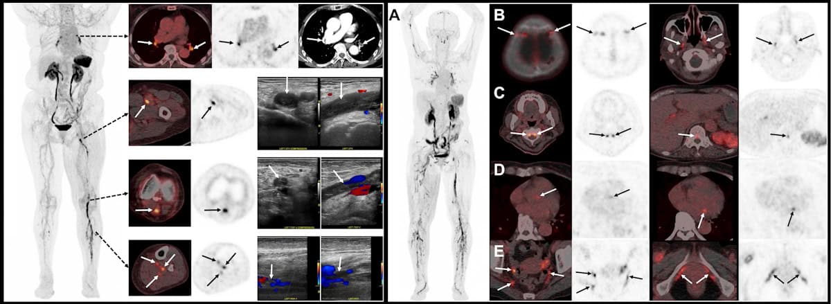
Image of the year Left: 18F-GP1 PET/CT images showing multiple blood clots in the deep veins of the left leg, plus several clots in the right calf and clots in both lungs. Right: 18F-GP1 PET/CT images showing widespread blood clots throughout the body. (Courtesy: S Han et al. Asan Medical Center, Seoul, Korea)
A PET radiotracer that can detect deep vein thrombosis (DVT) in the legs and clots that have travelled to the lungs has been chosen as the “Image of the Year” at the Society of Nuclear Medicine and Molecular Imaging (SNMMI) 2026 annual meeting. Developed by Sangwon Han and colleagues at the Asan Medical Center, University of Ulsan College of Medicine, in Korea, the novel tracer enables whole‑body imaging of blood clots (thrombi) in the legs and lungs in a single scan.
A DVT is a blood clot that forms in a deep vein, usually in the legs. It’s a common condition, with an incidence of roughly half that of all cancers, and it can lead to serious complications. Clots can break off and travel to the lungs, which could cause a potentially life‑threatening pulmonary embolism (a blockage in the artery supplying blood to the lungs). Early detection of DVT is therefore critical for determining the most appropriate treatment for each patient.
Currently, the standard imaging method for diagnosing DVT is venous ultrasonography (VUS). But while this works well for detecting clots in the thigh-to-knee region, whole-leg VUS requires skilled operators and advanced machines, takes longer, and has lower diagnostic sensitivity in the calf. In addition, conventional imaging techniques such as VUS and CT rely on indirect structural changes rather than directly visualizing the clot.
Aiming to enable faster and more efficient DVT diagnosis, Han and his research team are studying fluorinated GP1 (18F-GP1) a novel thrombus-targeted PET tracer. The tracer selectively binds to specific receptors on activated platelets (the cell fragments that cause blood to clots), allowing direct visualization of active thrombus formation.
“In our Phase 1 study, 18F-GP1 PET/CT showed 100% detection rate in 20 patients with confirmed DVT or pulmonary embolism,” Han told the SNMMI delegates. “But that study was limited by its small sample size and an absence of negative groups so specificity could not be assessed at that time.”
So in this latest work, Han and his team performed a phase 2, non-randomized study investigating the ability of 18F-GP1 PET/CT to identify acute lower-extremity DVT in 46 symptomatic patients. This included 22 patients with proximal DVT and 24 with none or distal DVT, as diagnosed using VUS.
The researchers acquired chest-to-feet PET/CT scans approximately 2 h after intravenous administration of 250 MBq of the radiotracer. The images were assessed by three blinded nuclear medicine physicians from different institutions, who assigned focal 18F-GP1 uptake higher than background activity as positive for thrombosis. They classified proximal DVT as clots involving the iliac (pelvic), femoral (thigh) and popliteal veins (behind the knee), and distal DVT as clots confined to the calf veins.
“Our primary objective was to assess the sensitivity and specificity of qualitive 18F-GP1 PET/CT interpretation for proximal DVT,” Han explained. “Secondary objectives included assessing the agreement between PET/CT and VUS for distal DVT, inter-reader reproducibility, exploring the detection of pulmonary embolism and assessing safety.”
When evaluated against VUS as a reference standard, 18F-GP1 PET/CT exhibited high diagnostic accuracy for detecting clots, demonstrating a sensitivity of 95% and a specificity of 92% for proximal DVT. “For distal DVT, both positive and negative agreement between PET/CT and VUS were strong,” added Han. “Inter-reader agreement was also excellent.”
The scans also identified concomitant pulmonary emboli in some patients, as confirmed by CT pulmonary angiography, illustrating the advantage of simultaneously assessing DVT and pulmonary embolism in a single scan. The researchers noted that the radiotracer was well tolerated, with no drug-related adverse events observed.
Speaking in the plenary session when his award was announced, Han shared a “striking image” recorded using 18F-GP1 PET/CT, which showed extensive blood clots, not only in the leg and lungs, but also in many unusual sites, including cranial and spinal vessels, cardiac valves, and vessels in the pelvic region. “This image clearly shows the remarkability ability of fluorinated GP1 to visualize thrombi throughout the body,” he explained.
“We believe this represents an important step towards thrombus-specific imaging,” Han concluded. “The potential of GP1 PET can expand beyond DVTs to many other thrombotic diseases such as embolic stroke or other cardiovascular diseases.”
The SNMMI Image of the Year is the society’s highest award, and the most anticipated, given out in recognition of an image that’s truly cutting-edge and representative of the future of nuclear medicine. This year’s winning image was chosen from nearly 1500 abstracts submitted for the meeting.
“It is truly a great honour to receive the Image of the Year award,” said Han.
In a significant development within the realm of nuclear medicine and molecular imaging, Dr. Jason S. Lewis, PhD, FSNMMI, has been appointed as the Vice President-Elect of the Society of Nuclear Medicine and Molecular Imaging (SNMMI) for the 2026-27 term. His appointment was officially announced at the SNMMI’s 2026 Annual Meeting held in Los Angeles, marking a pivotal moment for the society as it continues to expand its influence in advancing nuclear medicine and molecular imaging sciences. Dr. Lewis’s career, distinguished by a profound commitment to both scientific innovation and educational leadership, positions him uniquely to shape the future trajectory of the society.
Dr. Lewis holds prestigious roles as the Emily Tow Chair in Oncology at Memorial Sloan Kettering Cancer Center (MSK) and deputy director of the Sloan Kettering Institute in New York. His extensive background embodies a fusion of rigorous scientific inquiry and clinical application, a dual focus that reflects the integral nature of molecular imaging and nuclear medicine in bridging laboratory discoveries with patient-centered therapies. At SNMMI, he aims to leverage this integration by elevating basic science visibility, thereby fostering a symbiotic relationship between foundational research and its clinical deployment.
A central theme in Dr. Lewis’s vision as Vice President-Elect involves enhancing the educational landscape within SNMMI. He asserts that the society must strengthen its role as an educational powerhouse, catering to members across all career stages. By enriching educational content and fostering interdisciplinary collaboration, particularly between basic scientists and clinical investigators, SNMMI can solidify its reputation as an incubator for innovative research methodologies and translational sciences that define tomorrow’s standards of precision medicine.
Dr. Lewis’s commitment extends to nurturing early-career scientists, a group vital for sustaining the momentum of innovation in nuclear medicine. He intends to create novel forums and opportunities tailored specifically for these emerging investigators, emphasizing mentorship, scientific exchange, and active participation in society initiatives. This approach ensures a dynamic generational handoff that preserves and amplifies the society’s mission to push the boundaries of molecular imaging technology and radiopharmaceutical sciences.
Tracing Dr. Lewis’s academic journey underscores the depth of his expertise. He obtained his Bachelor and Master of Science degrees in chemistry from the University of Essex, followed by a doctorate in biochemistry from the University of Kent. His postdoctoral research at Washington University School of Medicine in St. Louis laid the foundation for a distinguished academic trajectory, culminating in his faculty appointment and subsequent transition to MSK, where he has consistently contributed to cutting-edge oncology imaging research.
Within SNMMI, Dr. Lewis’s involvement has been multifaceted. His tenure as secretary and treasurer over the past four years coincided with a period of strategic growth and policy development within the society. He is currently the chair of the SNMMI Task Force on Policy and Review Alignment and the SNMMI Committee on Finance. His membership spans committees encompassing radiopharmaceuticals, awards, and the Clinical Trials Network Research Committee, reflecting his broad influence and leadership across scientific, financial, and clinical dimensions of the society.
Dr. Lewis’s editorial contributions also shape the scientific discourse in nuclear medicine, notably through his role as an associate editor of The Journal of Nuclear Medicine since 2016. This position enables him to guide the dissemination of high-impact research, ensuring rigorous peer review and fostering a scholarly milieu that champions innovative molecular imaging modalities, including positron emission tomography (PET) and theranostics.
His leadership credentials extend internationally, having served as president of the World Molecular Imaging Society (WMIS) and the Society of Radiopharmaceutical Sciences (SRS). These roles highlight his global influence and dedication to the advancement of molecular imaging sciences on a worldwide scale. Such leadership roles complement his recognition as a fellow in several esteemed organizations, including the American Association for the Advancement of Science, the Royal Society of Chemistry, the American Institute for Medical and Biological Engineering, and notably, the National Academy of Inventors.
Dr. Lewis’s scientific achievements have garnered numerous prestigious awards, underscoring his contributions to the nuclear medicine field. These accolades include the Paul C. Aebersold Award, the Michael J. Welch Award, and the Dr. Saul Hertz Lifetime Achievement Award from SNMMI, the ACS Glenn T. Seaborg Award for Nuclear Chemistry, and the WMIS Gold Medal for Lifetime Achievement. Such recognition attests to his innovative research and leadership in developing radiopharmaceuticals and molecular imaging technologies that have clinical impact.
The 2026-27 SNMMI leadership cohort also includes Heather Jacene, MD, as president, and Gary Ulaner, MD, PhD, FSNMMI, FACNM, as president-elect, all of whom share a commitment to advancing scientific discovery and clinical excellence in molecular imaging. The technologist section leadership similarly reflects leadership aimed at advancing clinical practice and technological innovation in nuclear medicine and molecular imaging.
At the core of SNMMI’s mission is the dedication to advancing nuclear medicine, molecular imaging, and theranostics—fields that underpin precision medicine by tailoring diagnostic and therapeutic approaches to individual patient profiles. Under the guidance of leaders like Dr. Lewis, SNMMI is poised to continue driving forward innovations that integrate diagnostic imaging with targeted treatment, enhancing clinical outcomes and expanding the possibilities of personalized medicine.
Dr. Lewis’s appointment heralds a new era at SNMMI, emphasizing synergy between molecular imaging’s scientific foundations and its clinical applications. His strategic focus on education, collaboration, and early-career engagement promises to keep the society at the forefront of medical innovation. As molecular imaging technologies evolve rapidly, the role of guiding institutions and visionary leaders becomes paramount in translating scientific breakthroughs into meaningful clinical benefits.
Driven by a mission to expand the frontiers of molecular imaging and nuclear medicine, Dr. Lewis and the SNMMI will play pivotal roles in orchestrating scientific discourse, policy development, and educational excellence. This trajectory will not only invigorate research communities but also ensure that innovations continue to translate into enhanced patient care paradigms, defining the future landscape of precision oncology and beyond.
Subject of Research: Nuclear Medicine and Molecular Imaging
Article Title: Jason S. Lewis, PhD, FSNMMI, Named Vice President-Elect of the Society of Nuclear Medicine and Molecular Imaging
In a significant development within the realm of nuclear medicine and molecular imaging, Dr. Gary Ulaner has been appointed as the president-elect of the Society of Nuclear Medicine and Molecular Imaging (SNMMI). This appointment, announced during the SNMMI 2026 Annual Meeting held from May 30 to June 2 in Los Angeles, highlights the growing importance and transformative potential of nuclear medicine in contemporary healthcare. Dr. Ulaner’s expertise and leadership are poised to drive forward innovative research and clinical applications that could redefine patient care, particularly in oncology and molecular diagnostics.
Dr. Ulaner currently holds the James & Pamela Muzzy Endowed Chair of Molecular Imaging and Therapy at the Hoag Family Cancer Institute and serves as a Professor of Radiology and Translational Genomics at the University of Southern California. His multifaceted roles underscore a career dedicated to the integration of molecular imaging technologies and translational research, aligning with the broader goals of personalized medicine and precision oncology. His background exemplifies the merger of academic rigor and clinical application crucial for advancing this rapidly evolving field.
Nuclear medicine, a specialty focused on the use of radioactive substances in diagnosis and therapy, stands at the forefront of precision medicine innovation. The role of the president-elect extends beyond administrative leadership; it includes championing initiatives that fortify research infrastructures, expand educational platforms, and secure funding to nurture the next generation of radiochemistry and nuclear physics professionals. Dr. Ulaner’s vision emphasizes a holistic advancement, where technological innovation dovetails with workforce development and interdisciplinary collaboration.
Dr. Ulaner’s academic foundation was established at Stanford University School of Medicine, where he earned both his MD and PhD in Cancer Biology. His post-doctoral training involved rigorous residencies in Nuclear Medicine and Diagnostic Radiology at the University of Southern California. This robust training has empowered him with a unique perspective that bridges molecular imaging technology, radiopharmaceutical development, and clinical oncology, driving impactful translational research.
Before his current tenure at Hoag Family Cancer Institute, Dr. Ulaner was an Associate Member on a tenure track at Memorial Sloan Kettering Cancer Center—a leading institution in cancer research and treatment. At MSK, he developed significant academic and clinical roles that contributed to the institution’s pioneering work in PET imaging and molecular diagnostics. His professional credentials are further reinforced by certifications from the American Board of Radiology and the American Board of Nuclear Medicine, underscoring his specialized expertise.
Within the SNMMI, Dr. Ulaner has been an active and influential member, occupying vital leadership positions such as director at large on the board of directors, president of the PET Center of Excellence, and chair of the Mars Shot Campaign—a bold initiative aimed at advancing nuclear medicine research. His multifaceted involvement signals his commitment to driving SNMMI’s strategic objectives, including the formulation of standards, educational outreach, and advocacy for nuclear medicine’s value in clinical practice.
His scholarly contributions are substantial, with over 190 journal articles and more than 300 invited presentations. Dr. Ulaner has contributed to seminal guidelines such as SNMMI’s Appropriate Use Criteria for Fluoroestradiol PET, setting standards that influence clinical decision-making globally. His editorial roles and authorship of textbooks like “Fundamentals of Oncologic PET/CT” demonstrate his dedication to disseminating knowledge and fostering an educated workforce proficient in advanced imaging modalities.
The Mars Shot Campaign, under Dr. Ulaner’s leadership, exemplifies a visionary approach to accelerating research and innovation within nuclear medicine. This initiative targets critical translational gaps, funding high-impact projects that aim to develop novel radiopharmaceuticals and imaging technologies with the potential to revolutionize diagnostic accuracy and therapeutic efficacy. Such efforts are crucial in overcoming existing limitations related to imaging biomarkers and personalized treatment monitoring.
Dr. Ulaner’s dedication to education and training extends beyond research innovation. He actively advocates for expanding educational opportunities for nuclear medicine professionals—technologists, clinicians, physicists, and radiochemists—recognizing the interdisciplinary nature of the field. This approach is vital for sustaining a skilled workforce capable of navigating the complexities of molecular imaging and theranostics, transforming patient outcomes in oncology and other disease domains.
Throughout his career, Dr. Ulaner has garnered numerous accolades, including the Susan G. Komen Career Catalyst Award and the Department of Defense Breakthrough Award. His recognition as a Distinguished Investigator by the Academy for Radiology & Biomedical Imaging Research and as a healthcare visionary highlights both his scientific contributions and leadership qualities. Such honors reflect his role as a catalyst for innovation at the interface of cancer biology, imaging science, and clinical oncology.
The SNMMI’s election of new officers alongside Dr. Ulaner—Heather Jacene, MD as president and Jason S. Lewis, PhD as vice president-elect—illustrates a leadership cohort poised to navigate the next frontier of nuclear medicine. Their collective expertise underscores the society’s commitment to fostering cutting-edge research, expanding educational horizons, and enhancing policy frameworks to elevate the role of molecular imaging in modern medicine.
As president-elect, Dr. Ulaner’s agenda will involve steering the SNMMI to harness the full potential of nuclear medicine and molecular imaging technologies. These advancements promise not only to enhance the early detection and characterization of malignancies but also to optimize individualized therapy through theranostics—combining targeted diagnostics with personalized treatment regimens. This paradigm shift aligns closely with contemporary trends aimed at achieving superior patient outcomes through precision health strategies.
The Society of Nuclear Medicine and Molecular Imaging remains at the vanguard of scientific and medical innovation, dedicated to advancing nuclear medicine, molecular imaging, and theranostics worldwide. Dr. Ulaner’s ascension to the role of president-elect represents a pivotal moment in reinforcing the society’s mission. His leadership is expected to invigorate research efforts, expand educational initiatives, and advocate for policies that solidify the critical role of molecular imaging in the healthcare continuum, driving transformative advances for patients globally.
Subject of Research:
Nuclear medicine, molecular imaging, and theranostics with a focus on oncologic PET/CT and translational genomics in cancer care.
Article Title:
Gary Ulaner, MD, PhD, Named President-Elect of the Society of Nuclear Medicine and Molecular Imaging, Heralding New Era in Molecular Imaging and Theranostics
Los Angeles—In a distinguished ceremony at the Society of Nuclear Medicine and Molecular Imaging (SNMMI) 2026 Annual Meeting, six eminent professionals were inducted as new SNMMI Fellows, an accolade that honors exceptional contributions to nuclear medicine and molecular imaging. Since its inception in 2016, the SNMMI Fellowship has become one of the most prestigious recognitions awarded to members who have demonstrated extraordinary dedication to advancing the field through service, innovation, education, and clinical excellence.
The SNMMI Fellowship reflects a rigorous selection process that emphasizes not only distinguished volunteer service to the society but also outstanding achievement in scientific discovery, educational impact, or clinical practice. These criteria ensure that the honorees represent the pinnacle of expertise and leadership, fostering the ongoing evolution of nuclear medicine and molecular imaging techniques that are central to modern precision medicine.
One of the newly inducted Fellows, Dr. Gholam Reza Berenji, currently directs nuclear cardiology at the VA Greater Los Angeles Healthcare System. His academic role as an adjunct professor at the University of Victoria in Canada underscores his commitment to fostering interdisciplinary knowledge transfer. Dr. Berenji’s involvement in multiple SNMMI councils, including the Academic and Cardiovascular Councils and specialized centers of excellence, positions him at the forefront of facilitating cutting-edge research and practice integration in cardiovascular molecular imaging modalities.
Dr. Mehdi Djekidel, another inductee, serves as associate professor of radiology at the Zucker School of Medicine at Hofstra University and practices diagnostic radiology and nuclear medicine at Northwell Health. His leadership roles within the Theranostics Leadership Group and other critical committees highlight his active participation in the development and oversight of radiopharmaceutical therapies and brain imaging initiatives, contributing significantly to the refinement of neuroimaging and personalized treatment paradigms.
In Washington, D.C., Dr. Giuseppe Esposito presides as chief of nuclear medicine at Medstar Georgetown University Hospital and co-directs nuclear medicine services at Medstar Medical Group Radiology. His stewardship on the SNMMI Board of Directors and as chair of the Scientific Program and Education Committee reflects his dedication to advancing scientific education and orchestrating high-impact sessions at annual meetings that disseminate the latest research breakthroughs and clinical protocols widely across the nuclear medicine community.
Distinguished for his contributions to oncologic imaging, Dr. Homer Macapinlac holds the James E. Anderson Distinguished Professorship of Nuclear Medicine at the University of Texas MD Anderson Cancer Center. His longstanding leadership within the SNMMI PET Center of Excellence, including serving as its president, underscores his pivotal role in promoting positron emission tomography applications in cancer diagnostics and therapy management, fostering innovations that enhance tumor detection sensitivity and treatment monitoring.
Professor John Prior, based at Lausanne University Hospital in Switzerland, is renowned for his expertise in nuclear medicine and molecular imaging, where he heads the related department. His multifaceted contributions as a society leader, educator, and prolific speaker at SNMMI conferences have significantly influenced the international scientific discourse, particularly emphasizing molecular imaging’s capacity to revolutionize disease detection and therapeutic strategies on a global scale.
Recognizing the importance of patient advocacy in advancing nuclear medicine, Josh Mailman was honored as an Honorary Fellow. An internationally respected advocate for neuroendocrine tumor patients, Mailman’s pivotal role as the inaugural chair of SNMMI’s Patient Advocacy Advisory Board exemplifies his efforts to bridge the gap between patient communities and medical practitioners, ensuring that patient narratives inform therapeutic innovation and regulatory policies alike.
The 2026 Fellowship also acknowledged the career of Dr. Libero (Lou) Marzella, a former director at the FDA Division of Imaging and Radiation Medicine. Dr. Marzella’s contributions have been instrumental in shaping regulatory frameworks that govern PET radiopharmaceutical drug development. His expertise has not only guided policy in the United States but has also fostered international collaborations that streamline PET imaging agent approval, proving vital for translational research and clinical trial success worldwide.
The upcoming SNMMI president for 2025-26, Dr. Jean-Luc Urbain, will receive Fellowship status after his term, recognizing his extensive leadership across multiple domains within the society. Dr. Urbain’s commitment to international collaboration and educational outreach continues to drive innovation by integrating research, clinical application, and global partnerships, enabling nuclear medicine to address challenges in personalized diagnostics and tailored therapies comprehensively.
Throughout these recognitions, SNMMI reiterates its mission to promote nuclear medicine and molecular imaging as indispensable tools in precision medicine. These imaging techniques exploit radiopharmaceuticals to visualize and measure biological processes at the molecular and cellular levels, providing unparalleled insights into disease mechanisms while facilitating the tailored treatment of conditions ranging from cardiac disorders to complex malignancies.
The integration of theranostics—where diagnostic imaging and therapeutic delivery are fused—represents a paradigm shift in patient care, enabling clinicians to predict, monitor, and optimize treatments based on individualized biological data. The honored Fellows’ varied expertise across PET, radiopharmaceutical therapy, and clinical oncology underscores the dynamic and interdisciplinary evolution of this field.
The SNMMI’s emphasis on Fellow recognition not only celebrates individual excellence but also highlights the collaborative and translational efforts necessary to push the boundaries of nuclear medicine. By fostering a vibrant community of innovators, educators, and advocates, SNMMI ensures that molecular imaging continues to impact patient outcomes profoundly, influencing future healthcare practices globally.
The 2026 Annual Meeting itself, a cornerstone event for the nuclear medicine community, provides an invaluable platform for sharing advancements, debating challenges, and forging partnerships that accelerate scientific discovery. The induction of these Fellows symbolizes the ongoing quest for excellence and the relentless pursuit to harness molecular insights for groundbreaking clinical applications.
As the SNMMI Fellowship cohort grows, the society reinforces its commitment to recognizing those who enhance the knowledge base, clinical capabilities, and patient-centered focus of the nuclear medicine and molecular imaging fields. This prestigious designation serves as an inspiration to both emerging and established professionals dedicated to improving diagnostics and therapies through cutting-edge science.
While modern radiotherapy techniques provide high-precision cancer treatment, cure rates for some advanced cancers have plateaued, with five-year local control rates often remaining as low as 50–60%. Recently, researchers have hypothesized that this clinical resistance may be primarily driven by tumour heterogeneity.
Positron emission tomography (PET) is the gold standard imaging technique for non-invasively mapping biological processes in the body – and could help define tumour regions that may be more resistant to the effects of radiation. Yet conventional scanners remain “monochromatic”, limited to imaging a single radiotracer per session. This physical limitation means that radiotherapy plans are often based on a “one-size-fits-all” dose model that assumes uniform radioresistance across the entire tumour volume.
Multiplexed PET (mPET) is an emerging innovation that offers a significant enhancement by utilizing radiotracers that emit both positrons and gamma photons to detect multiple biological signals simultaneously. The technique holds promise for enabling biologically individualized radiotherapy, allowing for more personalized treatment plans tailored to the unique needs of each patient’s tumour.
Principles of PET
Positron emission tomography (PET) is a widely used functional imaging technique that enables the visualization of metabolic processes within the body. PET imaging relies on electron–positron annihilation, in which gamma-ray photons are emitted when a radiotracer (a pharmaceutical tagged with a positron-emitting isotope, most commonly 18F-fluorodeoxyglucose (18F-FDG)) administered to the patient undergoes beta decay and emits a positron from its nucleus.
This energetic positron travels a short distance (typically less than 1 mm) through tissue until it encounters an electron in the body. Upon collision, the positron and electron annihilate, converting their mass into energy and releasing two 511 keV gamma photons, emitted approximately 180° apart to conserve momentum. These gamma photons are detected by scintillation crystals in the PET scanner, which convert the photon energy into light. This light is then captured by photomultiplier tubes (PMTs) or silicon avalanche photodiodes (Si-APDs) for precise photon event detection.
The fundamental detection mechanism in PET is coincidence detection, which relies on the arrival of the two photons at opposite sides of the detector ring within a very short time window (typically 6-12 ns). Each coincidence event defines a line-of-response (LOR), which connects the two specific points where the photons strike the detectors. By recording these coincidence events from multiple angles, the system reconstructs a detailed image of the radiotracer’s distribution within the body, allowing for the visualization of physiological processes.
Although PET provides excellent sensitivity for visualizing metabolic activity, conventional single-tracer PET is limited to only one biological process per scan. Since all positron-emitting isotopes produce identical 511 keV photons, standard scanners cannot differentiate between multiple radiotracers based on energy alone. This presents a significant challenge for modern clinical oncology, where tumours exhibit inherent heterogeneity. Different regions within a single tumour often have markedly distinct characteristics, such as variations in oxygenation and vascularization (the network of blood vessels developed by a tumour), which directly influence their radiosensitivity.
For example, hypoxic regions (which lack oxygen) within tumours can increase radiation resistance by up to threefold. While a single radiotracer like FDG can identify metabolically active regions, it does not capture hypoxic, radioresistant areas or variations in clonogenic cell density. This limitation forces radiotherapy to rely on a uniform approach, which often fails to address the complexities of tumour biology.
Sequential imaging with different radiotracers provides more insight into tumour biology but is clinically suboptimal, due to increased radiation burden from multiple accompanying CT scans (used for anatomical registration with the PET images) and higher costs. A method to simultaneously track multiple biological processes in a single scan is needed to fully capture the dynamic nature of tumour biology.
The physical principles of multiplexed PET
In standard PET scans, photons produced by positron–electron annihilation are detected when they arrive simultaneously at opposite sides of a detector ring, defining the LOR. Multiplexed PET builds upon these principles. With dual-tracer PET, however, the detection process becomes more complex due to the need to separate the photon signals from different radiotracers.
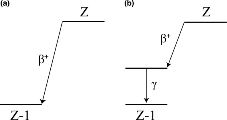
To achieve this separation, mPET exploits positron-gamma emitters such as 124I, for instance, which in addition to emitting positrons, emit an additional prompt gamma photon following the positron decay. Such isotopes decay to an excited state of the daughter nucleus, followed by near-instantaneous emission of a de-excitation gamma photon. This additional photon enables the detection of triple coincidence events, providing more biological information in a single scan.
Decay schemes (a) A positron-emitting isotope undergoes beta decay and transitions directly to the ground state of a daughter nucleus. (b) A positron–gamma emitter, after transition to an excited state of the daughter nucleus through beta decay, emits a prompt gamma photon, which de-excites the daughter nucleus to the ground state. (Courtesy: T Fukuchi et al. Med. Phys. 10.1002/mp.12149)
Using a triple-emitting radiotracer in combination with a pure positron emitter enables mPET scanners to achieve effective signal separation by utilizing an expanded energy window (350–700 keV, for example), which enables capture of both the 511 keV annihilation pairs and the higher-energy prompt gamma photons.
These data are then sorted into two streams: the primary dataset, which includes all detected LORs from both isotopes, and a smaller, tagged dataset containing only the triple coincidences. These triple events are identified via a specific timing selection rule, ensuring that the time difference between the prompt gamma detection and the average detection time of the annihilation photons falls within a narrow coincidence window, typically around 4.5 ns.
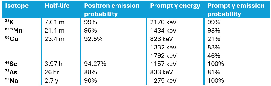
Triple emitters Examples of positron-emitting isotopes that also emit prompt gamma radiation, providing multiple signals for advanced imaging techniques. (Courtesy: adapted from Phys. Med. Biol. 10.1088/0031-9155/56/14/020)
To reconstruct the separate radiotracer activity distributions, specialized image reconstruction strategies can be used to address the noise and artefacts inherent in basic subtraction methods. One approach is LOR sorting, which compares line integrals from the initial reconstruction to determine the likelihood that a specific LOR corresponds to one of the two isotopes. Furthermore, triple events can be reconstructed using V-shaped LORs, combining two probable LORs from a triple event into a single geometric unit to more accurately approximate the radioactive origin.
This process requires a spatially variant normalization factor that corrects for the camera’s varying efficiency in detecting prompt gammas across the field-of-view, as certain areas may be shadowed by the scanner geometry. Accurate reconstruction must also account for single-photon attenuation correction for the prompt gamma as it travels through the body.
By generating distinct datasets within a single scan, this method provides perfectly co-registered functional maps, allowing clinicians to simultaneously characterize multiple biological processes within a tumour in a single imaging session.
Towards personalized radiotherapy
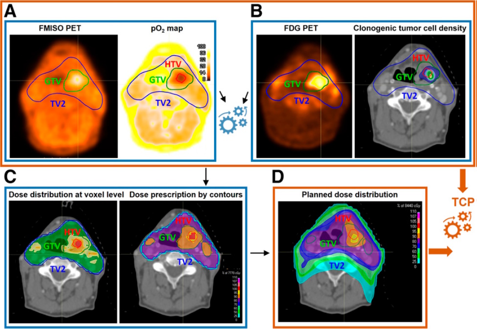
The introduction of mPET facilitates the transition towards biologically individualized radiotherapy, by delivering perfectly co-registered functional maps in a single imaging session. One promising application is the treatment of head-and-neck squamous cell carcinoma, where the radiotracers 18F-FDG and 18F-FMISO have been used to map clonogenic cell density and hypoxia-related radioresistance, respectively.
Biologically individualized radiotherapy (A) 18F-FMISO PET is used to generate oxygen distribution maps at the voxel level. (B) 18F-FDG PET provides insights into the distribution of clonogenic tumour cells. These datasets were then utilized to define dose prescriptions for different tumour regions (C). The final planned dose distribution (D) was based on these contours and used to predict the tumour control probability. (Courtesy: Lazzeroni et al. 2025 Journal of Nuclear Medicine)
Using radiobiological modelling, radiotracer uptake is converted into voxel-level cellularity maps via linear functions and oxygen partial pressure (pO2) maps via nonlinear sigmoid functions. These biomarkers inform “dose-painting” strategies that strategically escalate radiation to radioresistant areas, such as the hypoxic target volume, while maintaining safe limits for adjacent organs-at-risk. Modelling indicates this synergistic approach could increase tumour control probability from the clinical standard of 60% to a projected 90% or higher.
Researchers have also validated the feasibility of mPET in melanoma mouse models. Here, mPET successfully separated the signals of the triple-emitter 124I-trametinib (targeting proliferation) and 18F-FDG (targeting metabolism). This preclinical trial confirmed that mPET’s ability to separate dual isotopes offers a more detailed and timely assessment of tumour biology.
Future outlook
The clinical translation of mPET represents a significant potential advancement over traditional sequential PET scanning, providing an inherently quicker, cheaper and safer approach. By acquiring dual functional maps simultaneously, the second CT scan required in sequential procedures is no longer needed, roughly halving the patient’s cumulative radiation exposure.
Furthermore, mPET offers the advantage of shorter study duration, as both radiotracers are imaged simultaneously, eliminating the need to wait for the first to decay or wash out before injecting the second. This operational efficiency not only enhances patient compliance but also reduces total costs by minimizing scanner time and overheads. Crucially, mPET is highly viable for near-term implementation as it requires no modifications to existing hardware or acquisition software, when using standard clinical systems, such as the Siemens Biograph mCT, for example.
Despite these advantages, the primary technical pitfall remains the low statistics of the tagged “triples” dataset, which typically represents only a small fraction of total events. This statistical scarcity can introduce significant noise and “shadow” crosstalk artefacts into reconstructed images, potentially affecting quantitative accuracy. To mitigate this, ongoing research into bilateral guided filters and specialized V-shaped LOR algorithms is essential.
In addition, while the physics is compatible with current hardware, many clinical software packages still lack built-in capability for simultaneous multi-energy window acquisition or automated triple-coincidence tagging. This requires the development of manual workarounds that must be standardized for hospital use.
In the next five to 10 years, as the field moves from discovery into prospective interventional trials, the integration of machine learning for multi-parametric analysis will likely refine signal separation and tumour characterization. Looking further ahead, simultaneous imaging is not necessarily limited to two radiotracers: by utilizing multiple positron–gamma emitters and detecting their unique prompt gamma energies, mPET could evolve into “several-colour” imaging, capable or tracking three or more biological processes at once.
Ultimately, if upcoming trials confirm that predicted gains in tumour control probability translate into actual long-term patient survival, mPET may revolutionize oncology by enabling the first truly biologically individualized radiotherapy.