❌

Normal view

Siloton secures £8.4m to speed development of portable retinal imaging system

16 October 2026 at 14:30

UK medical technology start-up Siloton has been awarded an £8.4 million contract to accelerate the development of its portable eye imaging system. Based on optical coherence tomography (OCT), the company’s GiraffeOCT device generates detailed retinal images for detection and monitoring of a range of treatable eye diseases.

OCT is a non-invasive optical technique that creates high-resolution, depth-resolved images of biological tissue. While OCT is the clinical gold standard for diagnosing retinal diseases, existing systems are large and expensive and typically only available in hospitals and optometry clinics. By using photonic chip technology to shrink the bulky optical components onto a single coin-sized chip, Siloton is developing an easy-to-use, handheld binocular-style system.

The contract, from Innovate UK, will massively accelerate development of both the in-clinic GiraffeOCT system and the at-home version, which will have built-in automation and guidance so that users can reliably image themselves. “The contract will reduce the time to market for the medical device by one to two years,” says Alasdair Price, CEO of Siloton. “It will also allow us to grow the company, bringing in new skills around manufacturing and regulation, both of which are critical to the next stage of the business.”

Continuing clinical trials

In 2024, Siloton was the first commercial organization to demonstrate sub-surface imaging of a human retina, using its first-generation Akepa OCT photonic chip. The following year, the company trialled its device with patients for the first time, imaging their retinas to identify the presence and progression of age-related macular degeneration (AMD). Testing continued as the system developed, with a second cohort of patients imaged earlier this year.

In addition, the Project Ginan consortium in Australia has purchased a GiraffeOCT research system and independently evaluated its retinal imaging performance with a group of healthy volunteers.

Over the course of the two-year Innovate UK contract, Siloton will work with Sunderland Specialist Eye Hospital in the UK to perform clinical trials of the in-clinic device, giving patients access to the technology during its development. “These trials will be based around the prototype for the full medical device, providing additional post-processing and improved ergonomics compared to our research-use only product line,” Price explains.

The funding will also support the development and internal testing of Siloton’s second-generation CheetahOCT system, a research-use device that will measure different layers of the retina, expanding the number of conditions that it can detect. CheetahOCT incorporates the company’s second‑generation BlueJay chip – the same architecture used in the system that Siloton is building for astronauts – and will be available from early 2027.

“The image performance will be significantly improved, increasing the number of biomarkers that it can be used to measure. This means you will be able see more of the eye and in finer detail,” Price tells Physics World. “CheetahOCT will also be much more compact, as we continue to miniaturize other parts of the system that fall outside the photonic chip.”

The post Siloton secures £8.4m to speed development of portable retinal imaging system appeared first on Physics World.
Miniature microprobe enables internal imaging of smaller blood vessels than ever before

23 September 2026 at 09:00

Cardiovascular disease is the leading cause of death globally, with atherosclerotic disease – caused by the build-up of fatty plaques in the arteries – responsible for around three-quarters of these deaths. Modern intravascular imaging allows us to see these fatty plaque build-ups in detailed cross sections, revolutionizing atherosclerosis diagnosis and treatment planning. A research team in China is pushing this imaging technique to smaller blood vessels and higher resolution than ever before. Their approach could even enable imaging of the cerebrovasculature – the network of tiny vessels within the brain.

Intravascular optical coherence tomography (IV-OCT) uses near-infrared light emitted and received at the end of a catheter to image the walls and texture of the blood vessel. It works by comparing the wavelength and phase offset of reflected light from tissues with reference light reflected from a mirror, with 360° sensor rotation generated by electromagnetic motors.

The two most common designs of OCT catheters are proximal (where rotational force is generated outside of the body) and distal (with rotational force generated by a small motor inside the catheter). However, proximal systems suffer from friction-induced rotational distortion in smaller vessels, while distal scans are blocked from full 360° vison by wire artefacts.

Size is a major limitation in IV-OCT imaging. The catheters traditionally used are around 2 mm in diameter and perform optimally in vessels of 10 mm in diameter – medium/large arteries in the body. This diameter is limited by the size of electromagnetic motors needed to induce the sensor rotation and the wiring of these motors.

In this latest work, the researchers – led by Dawei Wu at Nanjing University of Aeronautics and Astronautics and Rui Liu at Nanjing University Medical School – show that it is possible to make a tiny 0.55 mm diameter probe, optimal for imaging 2 mm vessels, whilst retaining 360° field-of-view. The piezoelectric microprobe achieves this small size, and resolves both the rotational distortion and limited field-of-view difficulties, by exploiting a functionally different miniature drive mechanism.

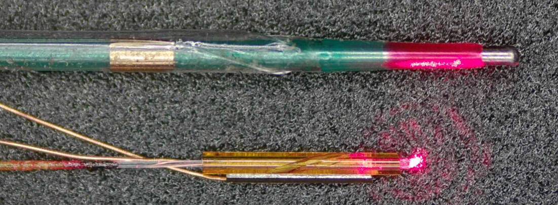
A piezoelectric micro probe and a commercial balloon dilatation catheter
Dimension comparison The piezoelectric micro-optical probe (bottom) and a commercial balloon dilatation catheter (top). (Courtesy: Dawei Wu, Nanjing University of Aeronautics and Astronautics)

The microprobe uses a single-phase AC circuit and piezoelectric crystal to vibrate a glass tube. A 10° groove in the glass translates this longitudinal vibration into torsional vibration, thus generating elliptical motion of the lens in a similar way to a crank and slider. When AC voltage is applied, the piezoelectric crystal expands and contracts and the lens rotates, thereby producing the required optical scanning.

Device testing

First author Boquan Wang and colleagues first tested whether their probe reduces rotational distortion by imaging metal tubes arranged around a small, curved vessel. The probe was able to scan the tubes at 50 revolutions per second while maintaining angular deviation of just 1° – a significant improvement compared with the 9° found in traditional proximal IV-OCT catheters. They note that this test also demonstrated the probe’s full 360° field-of-view and that the single-phase voltage required to rotate the lens meant that the wiring was minimal.

The researchers confirmed the probe’s ability to navigate and image small vessels via successful tests on leaf microveins, a vascular stent and ex vivo pig vessels. In an important milestone in assessing the suitability of the probe for imaging the cerebrovascular system, the probe successfully traversed through a full-scale human vascular model to reach the middle cerebral artery.

Finally, to verify the efficacy of the probe in identifying pathological lesions, the team compared images of human plaques acquired by the probe with histological analyses. The OCT and histology findings agreed well in identifying locations of plaque rupture sites and regions high in collagen fibres.

Why does this matter?

By putting a novel miniature rotational motor directly inside the probe and powering it with a single-phase AC circuit, the team created a probe that’s smaller than traditional IV-OCT probes and eliminates rotational torsion artefacts while achieving full 360° imaging.

Importantly, the improved catheter navigation could allow access to high-resolution images of the structure of arterial walls in smaller, more curved vessels than is currently possible clinically. This opens the door to assessment of plaque pathology and planning of stent positions in the heart–brain system.

“Although the probe would need more development and testing to be used clinically, intravascular OCT could one day give physicians a much closer look at what is happening inside the small arteries of the brain,” says Wu in a press statement.

The researchers describe the microprobe in Biomedical Optics Express.

The post Miniature microprobe enables internal imaging of smaller blood vessels than ever before appeared first on Physics World.

Fast 3D imaging reveals how seizures move through the brain

16 September 2026 at 09:30

Neuroscientists are debating a seemingly simple question: which way does a seizure propagate in the brain? Answering such a question could advance our understanding of epilepsy, but requires the development of cutting-edge microscopy techniques that are far from simple. Researchers from the University of Georgia have now developed an improved light-sheet microscope with the ability to perform fast 3D imaging of zebrafish seizures.

Reporting their findings in Biomedical Optics Express, the researchers found that seizures originated in the hindbrain and propagated forward (anteriorly). This corroborates a previous study examining 3D imaging of zebrafish seizures, but contradicts two earlier studies of 2D imaging (reported in Frontiers in Neural Circuits and eNeuro) that observed propagation in the reverse direction.

“I think the discrepancies about the direction of seizure propagation point to the need for more imaging,” says Peter Kner, an engineering professor who led the research efforts at the University of Georgia. “I believe we still don’t know how probabilistic the behaviour is and what factors are important.”

Adding a third dimension

The microscope developed by Kner and his team uses a so-called light sheet — a thin pancake of light that selectively illuminates a single 2D plane of a sample. This technique strongly reduces background noise from other planes that are not illuminated. Fluorescence light from the sample plane passes through a series of lenses, orthogonal to the light sheet, which reimage the sample onto a camera.

“There have been several papers analysing seizure events in zebrafish by looking at 2D imaging. So, the motivation here was to extend that work to 3D imaging”, explains Kner. Compared with other microscopy techniques, such as confocal microscopy, Kner’s team wanted to develop a solution for simple, fast and large field-of-view imaging in all three dimensions.

Light-sheet microscopy works inherently in 2D. To extend it to 3D, the researchers dynamically swept the light sheet through different planes of their sample. To keep the illuminated plane in focus at all times, they synchronously shifted the imaging system’s focal plane using an electrically tunable lens (ETL), which has a focal length that changes with applied current.

While this sounds straightforward in theory, in practice, adjusting the ETL introduces distortions or optical aberrations that degrade image quality. The researchers compensated for wavefront distortions using a deformable mirror whose shape was carefully calibrated at each axial plane imaged.

A wider view of seizures

Using predetermined settings for the deformable mirror, the team imaged volumes of 499 x 499 x 148 µm at a rate of four volumes per second. They also reported a fivefold increase in the area over which imaging remained near-diffraction-limited compared with uncorrected microscope images. These achievements allowed them to image zebrafish seizure propagation in real time under their microscope.

“This work adds pragmatic utility to 3D light-sheet microscopy for fast volumetric dynamics,” says Sixian You, an electrical engineering professor at the Massachusetts Institute of Technology who was not involved in this research. While deformable mirrors have been used to correct ETL aberrations before, she emphasizes that the advance comes from pre-calibration of the corrections; this is what makes the fast, wide field-of-view imaging possible. “I expect the same approach will transfer readily to other scanning modalities that stand to benefit from tunable lenses,” she adds.

After imaging the zebrafish continuously over a 2.5 min period, the researchers found that seizures propagated from the back of the brain to the front, and that the seizures subsided over tens of seconds. The findings add one more data point towards elucidating the statistics of seizure propagation which, if better understood, could aid epilepsy treatments in the future.

Kner’s team hopes to extend this work by next exploring different strains of zebrafish.

The post Fast 3D imaging reveals how seizures move through the brain appeared first on Physics World.

Structured light could deliver a new method for diagnosing eye disease

20 July 2026 at 15:00

By controlling a type of polarized light widely used in quantum optics, researchers have transformed subtle optical patterns in our peripheral vision into vibrant, easily perceptible shapes. Led by Dusan Sarenac at the University of Waterloo, Canada, the team showed for the first time that the “Boehm’s brushes” perceived by participants can be altered by adjusting the topology of spin-orbit coupled light. Their results, reported in Proceedings of the National Academy of Sciences, could lead to promising new techniques for checking our eye health.

For the most part, human eyes are incapable of detecting polarization in visible light. But in some rare cases, structures inside the eye itself can scatter light in different ways depending on its polarization, creating subtle yet perceivable “entoptic” patterns. Perhaps the best-known examples of these are “Haidinger’s brushes”: hourglass-shaped patterns that can form in our central vision, but only when viewing linearly polarized light at blue wavelengths.

In 1940, Swiss biophysicist Gundo Boehm discovered another example: a two-lobed, bowtie-shaped entoptic pattern that forms when viewing linearly polarized light in the peripheral vision as it scatters from subcellular structures in the inner retina. Unlike Haidinger’s brushes, this lesser-known effect is driven by angular variations in scattering strength relative to the polarization direction.

But as Sarenac explains, these ethereal shapes are more than just a curiosity. “Because retinal disease may alter a person’s ability to perceive the pattern, researchers have long considered whether Boehm’s brushes could serve as a biomarker of retinal health,” he says. “However, their weak visibility has limited practical use.”

Whereas previous studies simply asked participants to passively observe the effect, Sarenac’s team considered what would happen if the light’s polarization was constantly varied. To explore this possibility, the researchers created a beam of spin-orbit coupled light, in which polarization is linked to the light’s orbital angular momentum – a property describing how the wavefront twists as the beam travels – so that the polarization direction rotates steadily across the beam.

In quantum optics, these structured waveforms are already being widely explored for their possibilities in imaging, communication and information processing. But for the first time, Sarenac’s team considered how our eyes could actually perceive this light. “We engineered the spatial polarization of the light to match the symmetry of the retinal scattering response,” Sarenac describes. “This allowed many weak local contributions to reinforce one another.”

The researchers then projected this structured light through a ring-shaped aperture, targeting specific regions in the periphery of participants’ retinas, which could also be adjusted to target varying distances from the centre of vision. Using an automated test, they measured how much contrast participants needed to reliably distinguish a Boehm’s brush from the background at these varying distances.

Based on the subject’s responses, Sarenac’s team determined that the use of spin-orbit coupled light not only made Boehm’s brushes appear far more vivid to the participants, it also transformed the bowtie shape into a multi-lobed pattern, with the number of lobes varying depending on the topology of the light’s twisting polarization.

“Across 11 participants, detection thresholds improved exponentially away from the centre of vision, and the pattern became robustly visible at approximately one degree of retinal angular distance,” Sarenac describes. These results reveal a fascinating link between human vision and quantum mechanics, which the team hope could be harnessed in practical healthcare. With further studies involving patients with retinal disease, they now envisage eye tests where retinal conditions could be diagnosed in people who can’t clearly perceive vibrant, multi-lobed Boehm’s brushes in their peripheral vision.

“In the longer term, these patterns may provide a non-invasive functional probe of retinal integrity because their shape and visibility can be measured across different retinal locations,” Sarenac says.

The post Structured light could deliver a new method for diagnosing eye disease appeared first on Physics World.

Wearable pacemaker uses ultrasound to control heart rhythm

2 July 2026 at 09:00
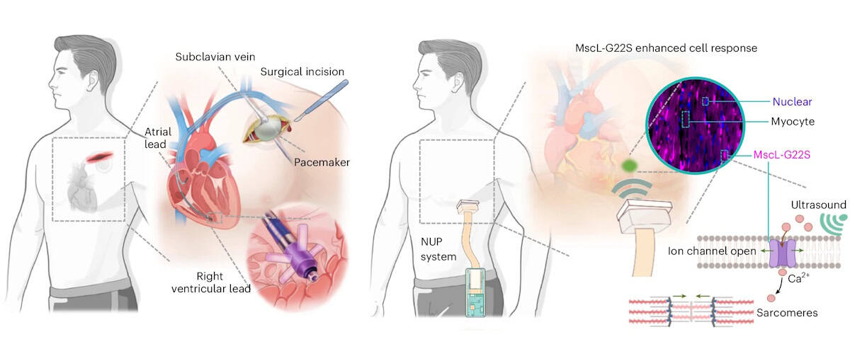
Illustrations showing traditional and sonogenetic cardiac pacing
Traditional versus sonogenetic pacing Left: schematic illustration of the steps for traditional surgical implantation of a cardiac pacemaker. Right: operational workflow of the non-invasive ultrasound pacemaker with sonogenetics. Created in BioRender. (Courtesy: C Gong et al. Nat. Biomed. Eng. 10.1038/s41551-026-01673-z 2026, Springer Nature)

Cardiac arrhythmia – a disorder in which the heart beats too fast, too slowly or irregularly – affects millions of people worldwide. Currently, arrhythmias are treated using pacemakers to regulate the heart’s rhythm, but traditional pacemakers require invasive surgical implantation and intracardiac leads that risk infection and tissue injury. As an alternative, a research collaboration headed up at Massachusetts Institute of Technology (MIT) and the University of Southern California (USC) has developed a non-invasive pacemaker that’s worn like a sticker on the chest and stimulates the heart using ultrasound.

“Ultrasound offers a unique combination of deep tissue penetration, spatial focusing and non-invasive delivery,” explains first author Chen Gong from USC. Previous approaches for ultrasound-based cardiac pacing, however, lacked reliability or required the use of microbubbles. In this new study, Gong and collaborators employed a technique called sonogenetics, in which cardiac cells are genetically altered to increase their sensitivity to ultrasound. Once exposed to ultrasound waves, the engineered cells trigger the opening of ion channels that let in calcium, which signals the cells to squeeze and beat.

“Our central motivation was to address the limitations of conventional pacemakers, while preserving precise control of cardiac rhythm,” says Gong.

The team first examined whether ultrasound could modulate cardiac cells in vitro, using sonogenetically engineered human heart muscle cells. Ultrasound stimulation caused almost three-quarters of the engineered cells to beat in synchrony with the ultrasound waves. In contrast, unaltered cells did not exhibit such behaviour. Importantly, ultrasound exposure did not impact the cells’ expression of biomarkers for inflammation, myocardial injury or oxidative stress, demonstrating the safety of this approach.

The non-invasive ultrasound pacemaker

The researchers designed a sonogenetics-based non-invasive ultrasound pacemaker (NUP) capable of real-time imaging and steerable stimulation, describing the device in Nature Biomedical Engineering. The postage stamp-sized device, which sticks to the skin via a bioadhesive interface, incorporates a 64-channel phased-array transducer, as well as data acquisition, wireless transmission and power modules.

To assess whether this prototype NUP could achieve non-invasive cardiac pacing in sonogenetically engineered rats, they attached the device onto the rats’ chests and applied ultrasound stimulation through the chest wall to the heart.

At an acoustic pressure of 2 MPa and a 40 ms pulse duration, the NUP increased the rats’ heart rates from 240 to 360–540 bpm, with a higher pulse repetition frequency leading to increased heart rate. Upon stopping the ultrasound stimulation, the heart returned to its natural rhythm. In control rats without the genetic modification, NUP could not control the heart rate effectively.

The team demonstrated that the NUP could electrically steer the ultrasound beam focus with a spatial precision of less than 1 mm, enabling targeting of different regions of the heart to induce chamber-specific pacing. NUP also successfully treated cardiac arrhythmias induced in the engineered rats, returning their heart rates to normal levels. In contrast, ultrasound stimulation of control rats did not effectively treat their arrythmias.

“A major objective of this work was to move beyond proof-of-concept stimulation and demonstrate a wearable system that could realistically support daily use,” explains Qifa Zhou, group leader at USC.  “The current prototype integrates imaging, stimulation, wireless communication and battery-powered operation into a wearable format.”

Clinical potential

For future clinical use, the NUP device needs to automatically detect the wearer’s heart rate, locate heart chambers and deliver stimuli. To achieve this, the researchers used a cloud-based, artificial intelligence (AI)-powered imaging feedback loop that determines the heart rate and stimulation coordinates, and steers the ultrasound beam to target sites.

To test its feasibility for human-scale applications, they assessed the acoustic energy penetration of the device. Using simulations and experiments on a pig heart beneath layers of tissue, they confirmed that, even after tissue attenuation, NUP could deliver sufficient ultrasound pressure (approximately 2 MPa) to a human heart to enable stimulation at clinically relevant pacemaker sites.

The team also assessed the biosafety of the NUP approach, finding that the delivered ultrasound energy remained below approved safety limits and generated minimal thermal effects during and after stimulation. They also confirmed that the sonogenetic engineering induced no off-target effects, immune responses or pathological changes in the rats.

“In the long term, we are optimistic about applying sonogenetics in humans,” explains co-author Gengxi Lu from MIT. “Sonogenetics is different from gene editing – it does not aim to rewrite a patient’s DNA sequence, but instead enables target cells to temporarily or controllably express ultrasound-responsive proteins. At the same time, extensive clinical validation will still be needed to establish safety and long-term effectiveness.”

The team is now focusing on clinical translation and validation of the NUP technology, which includes improving gene delivery strategies, validating the system in larger animal models, and developing closed-loop control approaches that combine real-time physiological sensing with adaptive stimulation. There’s also much engineering work to be done, such as device miniaturization, and improving battery life, skin coupling and motion robustness.

“For cardiac pacing, we envisage that the final goal of NUP technology is to be a permanent alternative to a long-term implanted pacemaker,” Xuanhe Zhao from MIT tells Physics World. “More broadly, we are interested in expanding ultrasound-enabled bioelectronic medicine beyond cardiac pacing toward other organs and therapeutic applications where non-invasive, spatially precise modulation could have clinical impact.”

The post Wearable pacemaker uses ultrasound to control heart rhythm appeared first on Physics World.

Laser-based rotation of floating samples enables high-resolution 3D microscopy

18 June 2026 at 10:00

A new contact-free way of rotating delicate samples, such as biological cells, undergoing three-dimensional (3D) optical microscopy increases the image resolution and could reveal more details about microscale structures and processes.

Current optical microscopes provide extremely good image resolution in a single plane, but since depth information is often lacking, samples are routinely rotated and imaged at multiple different angles to create a 3D picture. Because rotating via mechanical microtools such as tiny grippers, needles or pipettes can damage samples, contact-free approaches such as optical tweezers, magnetic manipulation or optothermal forces are often used instead. However, these methods rely on the sample being birefringent, magnetic, light-absorbing or specially shaped.

By contrast, the laser-based technique developed by a team led by Moritz Kreysing and Fan Nan from the Institute of Biological and Chemical Systems at the Karlsruher Institut für Technologie (KIT) in Germany can rotate and spin floating microscopic objects no matter what their shape or material properties.

Their method involves focusing a 1455 nm infrared laser beam on a small region of a highly viscous liquid – initially at 25°C – in which the sample will be suspended. This gently heats that region of the liquid by a few degrees. But rather than focusing on one spot, an acousto-optic deflector steers the beam rapidly along a carefully designed two-dimensional (2D) pattern.

“Because the viscosity of the liquid changes with temperature, this moving heat pattern generates tiny fluid flows, known as thermoviscous flows. By choosing the scan geometry and timing correctly, these flows become three-dimensional and helical, like microscopic corkscrew-shaped currents,” explain Kreysing and Nan. Once a small object is floated freely in these 3D helical flows, it can be rotated, spun, transported or stabilized solely via software-defined scan patterns, with different symmetries controlling the movement of the surrounding liquid.

The KIT technique allows stacks of high-resolution images focused at different depths to be acquired along multiple different angles by re-orienting the sample, a feature that “can help overcome the anisotropic resolution of conventional 3D microscopy”, explain the researchers. Using a high-viscosity liquid to suspend the samples not only enables kinematic stop-and-go actuation, it also suppresses Brownian motion (the jiggling around of tiny particles in fluids), which otherwise decreases image resolution and makes the study of cellular transport processes tricky.

Since previous methods of laser-driving fluid flows could only create motion in one plane, the researchers found it “particularly exciting” when they first observed their laser scanning in a 2D plane generating 3D flow fields. “Another key moment was the observation of opto-hydrodynamic focusing. The particles did not just rotate; their spiral motion converged toward a stable height and position. This was significant because it meant that the optofluid could stabilize the sample, rather than requiring mechanical confinement or complex feedback control.”

As detailed in their recent Light: Science & Applications paper, the KIT team demonstrated rotation and spinning of various samples including stained biological cells, nano-printed micro-tiles and perfectly spherical beads. “We see immediate potential in advanced optical microscopy, especially multi-view imaging of suspended cells, cell clusters, organelles, microstructures or soft materials,” Kreysing and Nan tell Physics World.

Since the basic hardware components of their system will be familiar to many optics and microscopy labs, they are hoping for early adoption by specialized research groups. But to optimize the technique for biological applications, they will now investigate the use of different liquids and operating conditions “that better preserve the viability of live mammalian cells while still providing sufficient stability for precise rotation and imaging”.

The researchers suggest that future areas of application could include microfluidics, microrobotics, colloidal assembly, materials science and 3D microfabrication. As such, they also want to develop more advanced scan patterns that can generate curved, programmable or multi-axis helical flows. “This could allow more complex 3D manipulation, such as controlled orientation of irregular objects, coordinated actuation of multiple particles, or integration with microfabrication workflows,” they explain.

The post Laser-based rotation of floating samples enables high-resolution 3D microscopy appeared first on Physics World.

Abnormal snoring sounds help diagnose common sleep disorder

8 June 2026 at 16:00

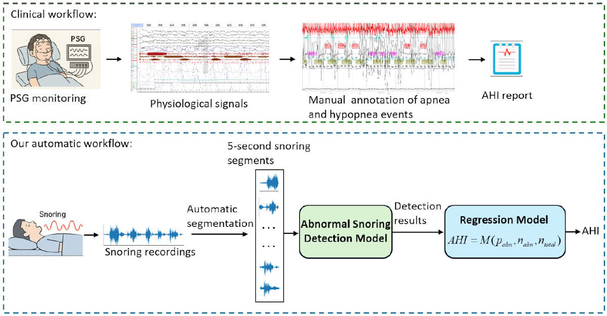
Obstructive sleep apnoea (OSA) is a sleep disorder in which the throat muscles relax and block the airway during sleep, causing pauses in breathing. If left untreated, OSA can lead to serious health problems. A research team led by Mingjiang Wang at the Harbin Institute of Technology, Shenzhen in China, in collaboration with Huizhou University, has now come up with a new method for detecting OSA – based on recordings of the subject snoring.

“OSA is a common yet underdiagnosed sleep disorder,” explains first author Heng Li. “The clinical standard for diagnosis is polysomnography [PSG], which is expensive, time-consuming and unsuitable for large-scale or home-based screening.”

PSG collects multiple physiological signals via various sensors that patients wear while asleep, potentially disrupting their sleep patterns. “Therefore, we aim to develop a non-contact detection method using snoring, which is a typical symptom of OSA and can be recorded contactlessly using a microphone,” says Li.

The acoustic patterns of snoring, produced by the vibrations of upper airway, contain markers that reflect airway narrowing and collapse. However, developing a reliable snoring-based detection model is challenging, as labelled snoring datasets are scarce and snoring sounds vary greatly between individuals.

In this study, published in Physiological Measurement, the research team uses a pretrained audio model called Wav2vec 2.0 to address these challenges. By transferring acoustic knowledge learned from large-scale unlabelled speech and audio data, the model removes the need for labelled snoring datasets.

Wav2vec 2.0 was originally designed for speech recognition and is computationally intensive, making it unsuitable for routine OSA monitoring. Thus the team adapted it for snoring sounds, removing layers more relevant to speech recognition while retaining acoustic features relevant to snoring pathology, aiming to lower the computational cost.

“We do not use the pretrained model directly,” Li explains. “Instead, we prune higher speech-related Transformer layers and fuse low- and mid-level representations, so that the model emphasizes snoring-related acoustic information.”

Constructing a snoring dataset

In collaboration with the Shenzhen People’s Hospital, the team collected sleep recordings from 100 individuals with apnoea-hypopnea indices (AHI) ranging from 2.4 to 68.3 events/hour. AHI – defined as the average number of apnoea (where breathing stops completely) and hypopnoea (shallow breathing) events per hour of sleep – is used to classify OSA from normal (AHI of below 5) through to severe (30 or above).

All subjects slept in hospital wards and were also monitored with a PSG device during sleeping. Sleep physicians then used the PSG data to annotate the audio recordings with apnoea and hypopnea events. Each recording was divided into 5-s audio segments and assigned as either abnormal, if they included apnoea/hypopnoea events, or normal.

Illustration of methods for detecting abnormal snoring
Abnormal snoring detection Upper panel: the clinical workflow using PSG and physiological signals to determine AHI. Lower panel: the proposed automatic workflow, which estimates AHI directly from snoring recordings using segment-level abnormal snoring detection and regression models. (Courtesy: Physiol. Meas. 10.1088/1361-6579/ae6f6a)

To evaluate their framework’s ability to detect abnormal snoring, the researchers compared it against five other pretrained audio models. They first ran a subject-dependent evaluation, in which random audio segments from all subjects were used for training, validation and testing. All models performed well – as expected when the training and test data come from the same subjects – but the adapted Wav2vec 2.0 model achieved the highest accuracy and sensitivity.

The team then performed a subject-independent evaluation, which better reflects a real-world scenario, with each subject’s data randomly assigned to the training, validation or test set. Here, the existing methods exhibited a reduced accuracy of between 65% and 71%, while the new model retained an accuracy of 73.95%.

“In subject-independent testing, this adapted model achieved better performance than the compared baseline models, suggesting improved generalization to unseen subjects,” says Li. The team’s model was also significantly more computationally efficient, an important factor for OSA screening in home-based or resource-limited scenarios.

AHI estimation

Finally, the researchers designed a snoring-based AHI estimation framework, using their detection model to identify abnormal snoring segments and calculating a ratio-based statistic – the number of abnormal segments divided by the total number of segments – to quantify OSA severity. They then used four common linear regression models to estimate AHI for individual subjects.

All models demonstrated reasonable accuracy and good correlation between predicted and PSG-derived AHI values. The mean absolute error was roughly 11 events/h, which means that the estimated AHI may deviate from the true value by 11 events per hour of sleep and could (if near a threshold between categories) result in an incorrect severity diagnosis. However, while the current model is not yet precise enough for clinical diagnosis, it could prove ideal as a potential tool for home-based OSA screening or pre-screening.

The researchers are now investigating how to more effectively apply pretrained audio models to clinical snoring analysis. “We are focused on making these models lighter and more suitable for home-based screening, while validating their effectiveness across large, multicentre datasets,” Yun Lu, one of the corresponding authors from Huizhou University, tells Physics World. “Additionally, we plan to improve the accuracy of AHI estimation by integrating snoring-based predictions with additional non-contact physiological information.”

The post Abnormal snoring sounds help diagnose common sleep disorder appeared first on Physics World.

Molecular spin sensor takes the temperature of cancer cells

29 May 2026 at 16:45

Researchers in Japan have succeeded in measuring the temperature inside living cells with high precision using a new class of biocompatible quantum nanosensor – something that has been difficult to do until now even. If improved, the nanosensor could be used to characterize a wide range of biological phenomena and so help in disease diagnosis, they say.

Recent years have seen the advent of a new generation of nanoscale quantum sensors that can detect the tiny magnetic fields of biological systems. Some of these sensors rely on photons and others on electrons or spin defects – typically diamond specially engineered with nitrogen–vacancy (NV) defects. This material is made by removing two carbon atoms from the diamond lattice and replacing one with a nitrogen atom. The other “hole” is left empty, thereby creating a vacancy or defect. The spin state of the defect is influenced by the local magnetic field that can be “read out” from the way it fluoresces.

While a powerful tool, and biocompatible, this type of quantum sensor does suffer from certain limits. For one, it can be structurally inhomogeneous, which affects how it detects temperature and other physical or chemical parameters inside biological cells.

A more homogenous structure

Even though the new molecular quantum nanosensor (MoQN) works in the same way as these conventional devices, it does not suffer from this problem, explain Nobuhiro Yanai of the University of Tokyo and Hitoshi Ishiwata of the National Institutes for Quantum Science and Technology (QST), who led this research effort. This is because it has a more homogenous structure and does not contain any defects. Instead, it is made by embedding molecular spin qubits, in this case fabricated from pentacene, in nanocrystals of para-terphenyl. This design makes the structure uniform on a molecular scale and preserves the quantum coherence of the spin qubits. It is then coated with Pluronic F127, which is a biocompatible surfactant.

By detecting the spin direction of the “excited triplet state” of the pentacene qubits using a technique known as optically detected magnetic resonance (OMDR), the researchers can precisely determine the temperature of the qubits’ surroundings from the OMDR peak position. When they tested their method inside the cytoplasm of cancer cells in vivo, they found that the intracellular temperature was consistently higher than the surrounding medium.

Yanai says he embarked on this study after reading about the work of Sam Bayliss’ group at the UK’s University of Glasgow, and Ashok Ajoy’s group at the University of California, Berkeley in the US on OMDR in pentacene-doped para-terphenyl crystals. He says he immediately got the idea that nanocrystals of this material could be used for quantum sensing inside cells. This was because his group had already developed such nanocrystals for a different purpose in previous research.

Ensuring biocompatibility

“I then spoke with Hitoshi Ishiwata, who is an expert in quantum sensing using NV centres,” he recalls. “While many molecular qubits have been developed to date, there had been no examples demonstrating their sensing ability within living cells.”

The project required materials science expertise, he tells Physics World, and in particular, finding out how to reduce the material to the nanoscale and ensuring it was biocompatible.

“We already knew that nanodiamonds are good quantum sensors for temperature measurements, but I had noticed a practical limitation: their ODMR spectra often vary significantly from particle to particle,” he says. “This spectral dispersion can introduce errors, especially when trying to perform precise measurements at the single-particle level.”

Replacing hydrogen with deuterium

The researchers thought they had overcome this problem during the first run of their experiments because they found that different particles showed identical OMDR spectra. However, their joy quickly waned when they observed that the spectra were still broadened by hyperfine interactions between the pentacene-doped para-terphenyl molecules’ electron spins and hydrogen nuclear spins.

To improve the spectral resolution, Ishiwata says he suggested chemically modifying the molecule by replacing the hydrogen in it with deuterium. And the technique worked: “the hyperfine broadening was strongly suppressed, allowing us to determine the OMDR spectra much more precisely.”

These findings, which are detailed in Science Advances, show that MoQNs are a chemically versatile platform for quantum sensing in living cells and that they can operate directly inside them while maintaining the precision needed for absolute thermometry, he says. Their appeal also lies in in the fact that their structures can be easily modified.

It will not all be plain sailing, however, adds Yanai. MoQNs cannot yet target specific organelles within cells, so endowing them with this targeting capability is an important future challenge. “What is more, their size has been limited to around 200 nm so far, so creating smaller MoQN particles will be crucial,” he says.

The post Molecular spin sensor takes the temperature of cancer cells appeared first on Physics World.

❌