Biofilms are complex microbial communities that adhere to a surface and encase themselves in a viscous matrix they produce. This structure protects bacteria from antibiotics and the immune system far more effectively than if they lived in a free-floating state, making biofilms one of the most common causes of hard-to-treat chronic infections—such as respiratory infections in people with cystic fibrosis and chronic obstructive pulmonary disease (COPD), chronic wounds, or infections associated with catheters and prosthetic devices. Added to this is the global rise in antibiotic resistance, which makes it increasingly urgent to have tools capable of predicting which treatment will work in each case.
The ocean's aphotic zone—the dark, high-pressure region below the surface, where sunlight cannot reach—is home to a vast wealth of microbial life. Yet scientists know relatively little about the genetic makeup, growth and function of many of these enigmatic organisms.
The ocean's aphotic zone—the dark, high-pressure region below the surface, where sunlight cannot reach—is home to a vast wealth of microbial life. Yet scientists know relatively little about the genetic makeup, growth and function of many of these enigmatic organisms.
Researchers at Queen Mary University of London are closer to understanding tendinopathy, a common condition that can cause persistent pain and impaired movement in people and many animals.
In recent years, there has been a lot of excitement about the potential for studying human diseases in what are called "organoids." These small patches of tissue, formed using stem cells, appear to produce many of the same cell types and at least some of the structures normally formed in actual organs, and thus can provide a better model for diseases that rely on the sometimes complex interactions among multiple specialized cell types that are a feature of the human body.
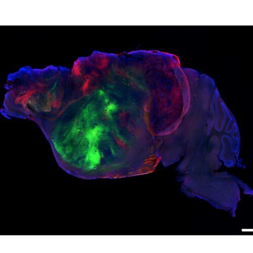
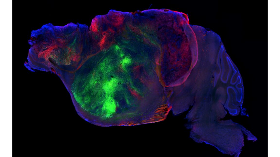
But even the most sophisticated organoids lack a lot of the features of a real human body. This is especially true for brain organoids, which don't form any of the connections with specialized brain structures needed to behave "normally." On Wednesday, a research group at Stanford University described a possible way to study brain organoids in a somewhat more natural context: They genetically wiped out a large portion of the mouse brain and replaced it with human brain organoid cells.
The replacements
Organoids, because they adopt a three-dimensional tissue structure and consist of various specialized cells, provide a much better model for an intact tissue than simply having a bunch of disassociated cells lying flat on a culture dish. But they still have a lot of limitations—they're not hooked up to a circulatory system that allows the liver to process chemicals the organoid produces and don't have immune cells moving through them, to give just a couple of examples.
When you think of viruses, you probably think of flu-like symptoms and quarantines. Humankind has suffered from viral diseases ranging from smallpox to COVID-19, hantavirus and Ebola since the dawn of recorded history.
Ancient ferns may have helped keep Triassic Europe burning by rapidly regrowing after fires and supplying fresh fuel for the next blaze. Around 201 million years ago, Earth was rocked by the end-Triassic mass extinction. The crisis has been linked to enormous volcanic activity associated with the breakup of Pangea. Those eruptions released huge quantities [...]
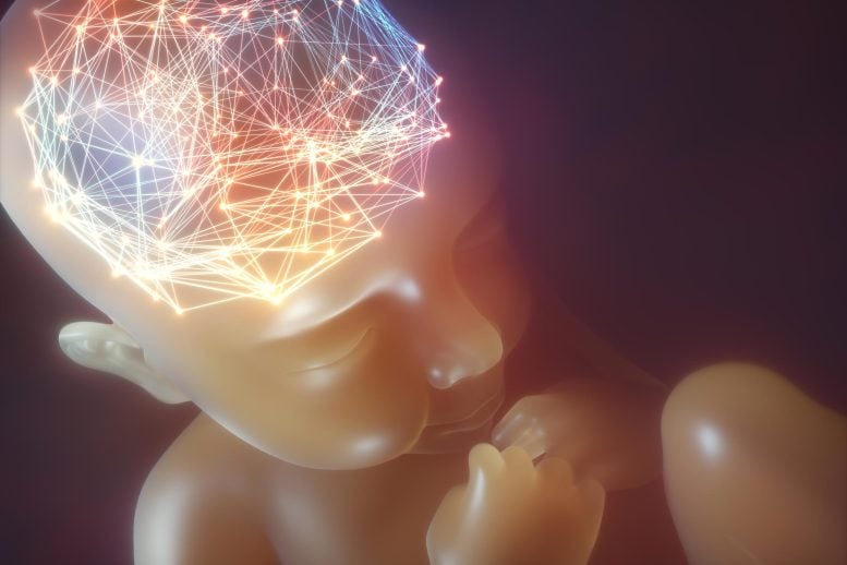
Scientists have uncovered two surprising forces that help guide how the human brain forms before birth. Before birth, the human brain is shaped in large part by an unusual class of stem cells known as radial glia. These cells help determine which brain cells are produced, when they appear, and how the cerebral cortex, the [...]
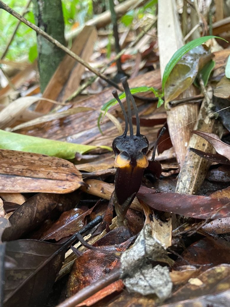
Researchers have identified a new plant species in western Thailand whose unusual appearance inspired the dramatic name Thismia daemona, or the “devil flower.” A nearly black flower with horn-like projections and flashes of reddish orange has been discovered beneath leaf litter in a mountain forest in western Thailand. The plant cannot photosynthesize, spends most of [...]
The mammalian gut microbiome teems with a delicate balance of bacteria and the viruses that keep them in check, called bacteriophages, or phages. Each bacterial species usually has its own set of phage partners.
The mammalian gut microbiome teems with a delicate balance of bacteria and the viruses that keep them in check, called bacteriophages, or phages. Each bacterial species usually has its own set of phage partners.
Despite major strides in education and conservation efforts, rabies remains a serious public health issue. The Center for Disease Control (CDC) estimates around 90 percent of cases occur in wildlife, but their consequences still reverberate across ecosystems and routinely endanger humans. Combating the viral disease costs the United States over $300 million every year, but the hefty price tag is justified. Contracting one of the oldest known diseases invariably causes severe symptoms like abnormal sensations, violent movements, hydrophobia, and loss of consciousness. Rabies is fatal in virtually every documented case, and still kills around 59,000 people around the world each year.
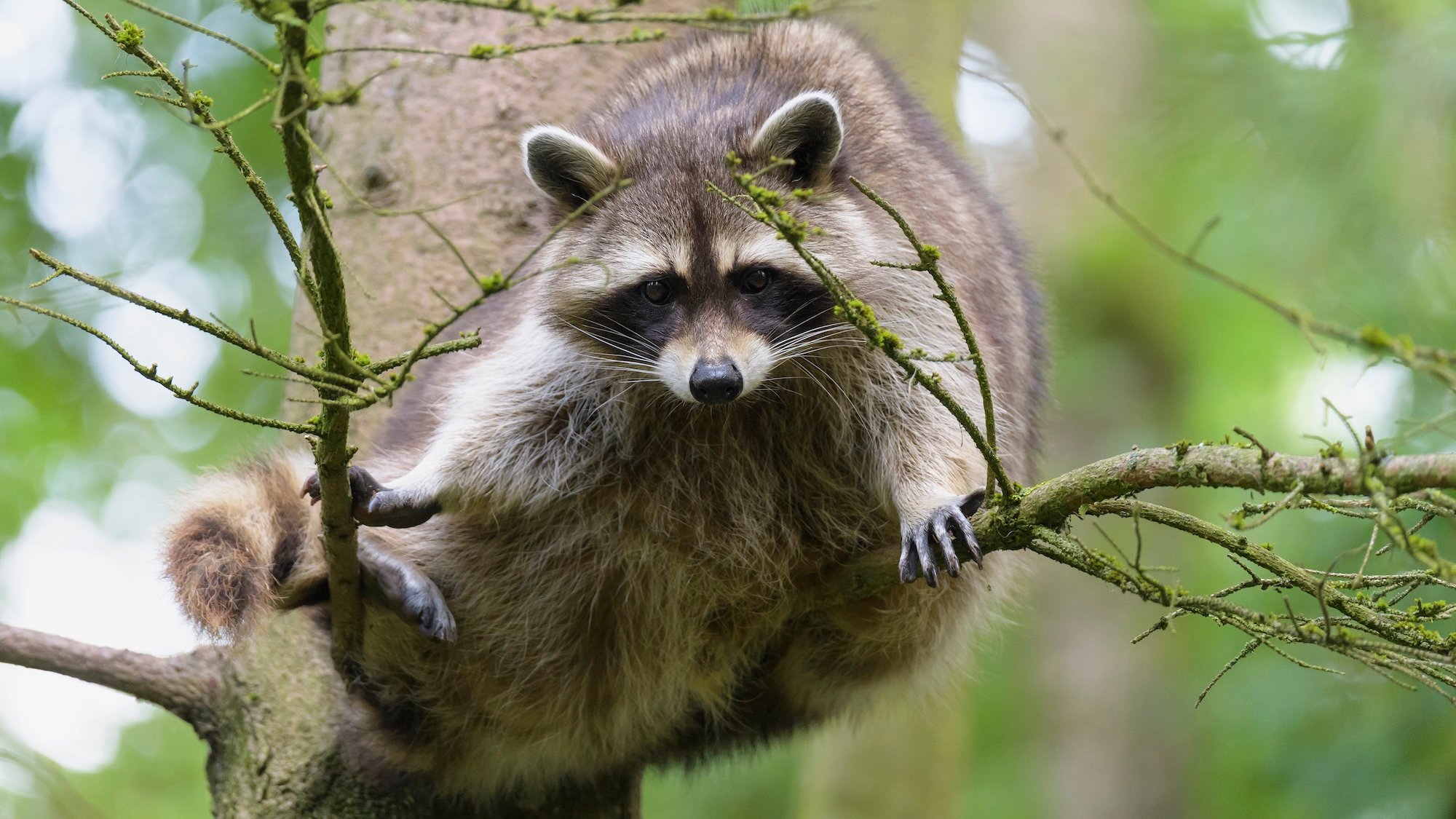
Once primarily associated with dogs, rabies has since spread to wildlife such as raccoons, skunks, bats, and foxes—making the issue much more difficult to track and control. Since 1995, the United States Department of Agriculture (USDA) has overseen a National Rabies Management Program in cooperation with state and federal departments, university researchers, and other organizations. After over 30 years of work, one of the most preferred mitigation methods is regularly placing oral rabies vaccine (ORV) bait in strategically selected wildlife areas. The easiest way to spread millions of bait blocks across thousands of square miles? Airdrops.
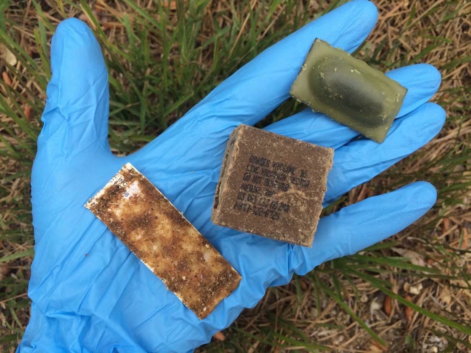
The bait isn’t toxic, but the USDA recommends using gloves to handle it. Credit: USDA
Next month, the USDA will once again begin tossing fishmeal-coated ORV packets from planes throughout the Southeastern states of Virginia, North Carolina, Tennessee, Georgia, and Alabama. These tiny edibles are specifically crafted to target the most prominent rabies variants seen in raccoons, coyotes, and gray foxes.
“The goal is to prevent westward spread of raccoon rabies by creating a continuous vaccine barrier that stays in front of rabies occurrence on the landscape,” a USDA spokesperson recently told FOX Carolina News. The representative added that although the distribution’s scope doesn’t currently include South Carolina, they are “very close to establishing an ORV area in South Carolina counties bordering North Carolina and Georgia in the next few years.”
If all goes as planned, the National Rabies Management Program’s geographic scope will shrink over time as the disease is regionally eliminated. Until then, anyone who accidentally comes into contact with an ORV bait brick shouldn’t worry about contracting the disease. The vaccine inside each brick is proven safe for over 60 animal species, including cats and dogs. If you happen to locate one in a residential area, the USDA recommends to either simply leave it alone, or use gloves to move it to a more remote location for its intended recipients. Always wash your hands thoroughly after handling ORV bait—and try to ignore the pungent odor of fishmeal.
Authors: Elisa Rigosi, Researcher in Sensory Biology, Department of Biology, Lund University and David O’Carroll, Professor of Sensory Biology, Department of Biology, Lund University
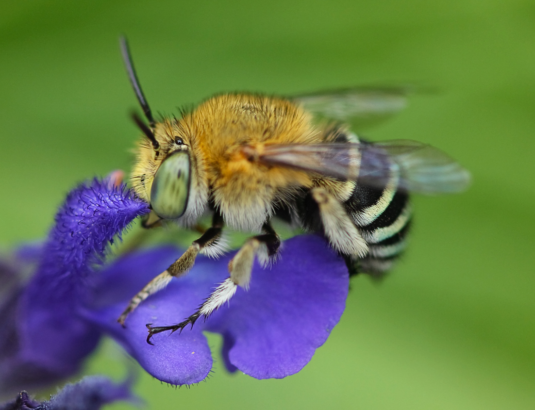
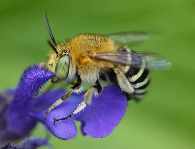
An Australian blue-banded bee: different species have evolved very different solutions to the challenge of seeing their world. Photo: Louise Docker / CC BY 2.0, Wikimedia Commons
One bee has long been regarded as a benchmark for visual performance: the honeybee drone.
While the honeybee lives in massive colonies, most bee species are solitary so must do everything themselves: find food, locate mates, provision their nests. In terms of vision, several of the solitary species we researched outperformed honeybee workers in a number of ways, including the sharpness of images formed by their eyes.
Understanding how these different evolutionary solutions work is important for more than just understanding bee biology. It could provide ideas for nature-inspired technologies, from compact visual sensors to autonomous robots.
How bees’ eyes have adapted
With more than 20,000 species worldwide, bees show extraordinary diversity in lifestyle, body size and ecological role. Some live in open environments; others forage in dense vegetation or in dim light at dawn, dusk and even at night.
Such remarkable diversity prompted us to ask the question: how have bees’ eyes adapted to such different lifestyles and visual demands?
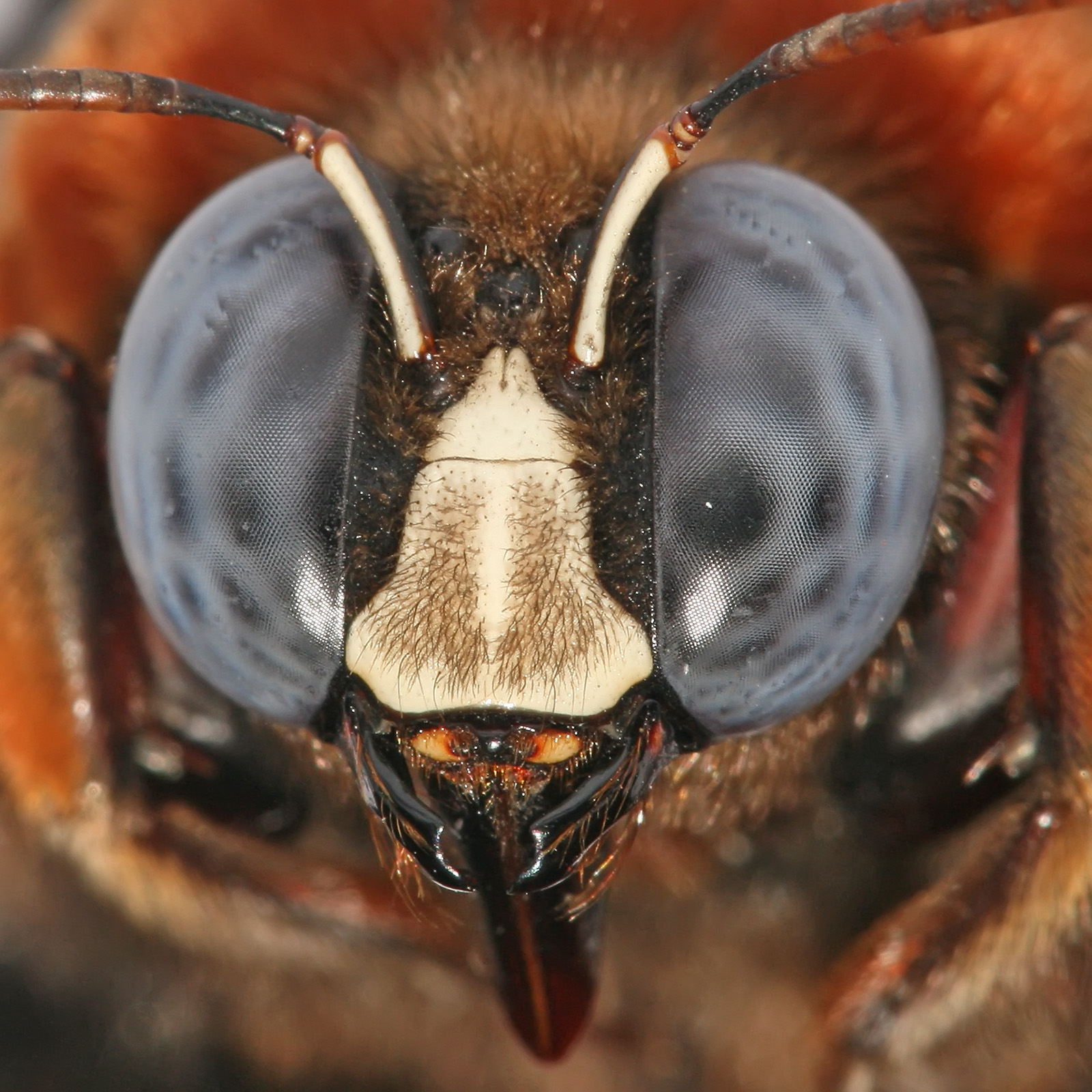
Bees see the world through compound eyes made up of thousands of tiny lenses. These odd-looking structures are their main tools to navigate, locate flowers and identify important landmarks.
Our new study of some daytime-active bee species shows that even bees with similar-looking eyes can achieve impressive visual performance in surprisingly different ways.
Male wool carder bees (Anthidium manicatum), for example, spend much of their day patrolling territories and chasing rivals or potential mates through the air. This bee pushes visual sharpness to the limit with its exceptionally small photoreceptors that minimise optical blur. In contrast, honeybee drones have evolved large lenses that maximise sensitivity.
Video: David O’Carroll/Lund University.
The Australian blue-banded bee (Amegilla) manages to detect small objects and see sharp images just as well without either of these extreme strategies. Instead, it achieves similar visual performance using a different combination of optical design and sampling.
From the outside, the eyes of these bees all look very similar. But when we looked more closely, we found the size of their visual sensors (photoreceptors) differed greatly.
These light-sensitive structures are a bit like the pixels in a smartphone camera. They sit behind the bees’ tiny eye lenses, converting light into electrical signals. Larger pixels capture more light, improving sensitivity, while smaller ones can help preserve image sharpness.
The challenge – for smartphone designers and evolution alike – is balancing these competing demands.
Something unexpected
Looking at anatomy tells us how the eye is built, but not how well it performs. To answer this question, we recorded directly from individual photoreceptors in living bees.
We used glass microelectrodes with tips just a few tens of nanometers wide – thousands of times thinner than a human hair. That allowed us, for the first time in these species, to measure the tiny electrical signals that single cells produce when the bees were shown tiny objects on a computer display.
Our results revealed something unexpected. The diminutive Australian blue-banded bee lacks the enormous eyes of a honeybee drone, yet achieved a similar level of sensitivity in its photoreceptors.
At first glance, this seems paradoxical. How can bee eyes built so differently perform so similarly?
The answer is that evolution is not working towards a single ideal design. Different species have arrived at similar visual performance through different combinations of optical design, photoreceptor structure and visual sampling.
Our findings show why it is important to look beyond a handful of familiar species. By studying a wider range of bees, we can better understand how different senses have evolved, leading to a variety of adaptive solutions.
This is true not only for vision but for other traits, including how bees respond to environmental stresses such as heat and pesticide exposure.
Unique evolutionary innovations
Through our research, we are just beginning to appreciate the remarkably different ways of seeing the world that bees have evolved.
Different bee species have evolved different solutions to the challenges of seeing their world. For example, the Australian blue-banded bee is a buzz pollinator, able to vibrate flowers to release pollen in a way that honeybees cannot. This makes it a potentially valuable pollinator of crops such as tomatoes in Australia, where bumblebees are not naturally present.
Such diverse evolutionary solutions may also prove useful to engineers. Designers of cameras, robots and autonomous vehicles face many of the same challenges as bees: how to gather enough visual information while keeping sensors small, efficient and affordable.
Unfortunately, as bee diversity declines worldwide, there is a risk that we lose not only species, but the unique evolutionary innovations that have taken millions of years to develop.
Today’s chatbots aren’t likely to help lone actors invent a deadly pathogen, experts say. But newer models, trained in biology, may need stronger safeguards.
Ancient teeth suggest humans were using psychoactive betel nuts 25,000 years ago, pushing the history of habitual drug use deep into the Ice Age. Archaeologists from Griffith University have found evidence suggesting that humans were using mind-altering substances far earlier than previously believed. The research team, led by Professor Adam Brumm of Griffith’s Australian Research [...]

 Login