❌

Reading view

Hands On: Sharge's Disk Pro 2 Ultra is a compact, magnetic DIY SSD dock with active cooling, 80W passthrough charging, and HDMI 2.1 that supports up to 8TB drives

With its built-in USB / HDMI hub and a small fan to keep temperatures in check, Sharge's Disk Pro SSD is one of the more interesting (and generally useful) external storage drives in recent memory, easily earning a spot on our Best External SSD list. And the company is following that success up with a more DIY-focused Disk Pro 2, which lets you install your own M.2 drive (supporting 2230, 2242, and 2280 sizes and capacities up to 8TB), while adding greater fan flexibility, media card slots, and an HDMI port that's capable of more bandwidth – depending on the model you choose.

Sharge is offering the Disk Pro 2 in two varieties. There's the Lite model, with HDMI 2.0 (4K@60 Hz) and a 5W minimum operating power, and the Disk Pro Ultra (which we have in for testing) with HDMI 2.1 (4K@144 Hz or 8K@30 Hz), a lower 1.5W minimum power draw, and a faster card reader. Below is the full list of differences, according to the product page. The company even lists the internal chips, some of which you can see through its transparent top.

Disk Pro Ultra

Disk Pro Lite

HDMI

HDMI 2.1, 4K@144 Hz, 8K@30 Hz

HDMI 2.0, 4K@60 Hz

Minimum Operating power

1.5W

5W

Chips

VL822, VL605, RTL9210, SM2705

GVS2201S, VL822, GL3231, RTL9201

SD speed

Read 180 MB/s, Write 120 MB/s

Read 100 MB/s, Write 90 MB/s

Sharge Disk Pro 2 Ultra

(Image credit: Tom's Hardware)

The Ultra model is likely to appeal more to professionals and gamers, offering faster card transfers and higher monitor refresh rates. But the main limitation here is the overall bandwidth, as both models are capped at a 10 Gbps interface, just like the original Disk Pro. That makes either model better as kind of a travel-friendly Swiss Army hub than something you'd want to use as your primary hub / dock all the time. Because any modern M.2 drive can saturate all available throughput by itself, before you connect any video or USB devices. But there's no denying the utility of carrying around a pocket-friendly SSD that also lets you connect to a monitor or TV, plug in a couple extra peripherals, and empty your SD cards without an extra hub, adapters, or cables.

Sharge Disk Pro 2 Ultra

(Image credit: Tom's Hardware)

At 3.65 x 2.33 x 0.531 inches (92.6 × 59.2 × 13.5 mm) without the cable, the Disk Pro 2 is slightly thicker and longer than the original Disk Pro, but still smaller than some standalone drives. Again, with its size and transparent top, it reminds me of an old-school cassette tape.

Sharge Disk Pro 2 Ultra

(Image credit: Tom's Hardware)

One thing that has definitely improved over the original Disk Pro is that Sharge has made the USB-C cable removable. This is good in part because you don't have to worry about the cable breaking over years of use. But also, particularly if you're going to use the drive's hub capabilities, you might want something longer than the included cable's sub-nine-inch length. And when necessary, you might want to plug the drive into a USB-A port. So having the option of using different cables is a big plus.

The cable is also designed to function as a lanyard, which can be handy. But it's held in place solely by the USB-C connection, so I wouldn't trust it for much more than hanging the hub up somewhere at home, so you know where it is when you need it.

Sharge Disk Pro 2 Ultra

(Image credit: Tom's Hardware)

In terms of ports, starting from the bottom edge, you get the main USB-C port, capable of passing through up to 80W of power, a full-size HDMI port (2.1 on the Ultra model we're testing or 2.0 on the lite model), a USB 3.0 Type-A port, and a 10 Gbps USB-C port that can handle up to 100W of power input (and pass through up to 80W for charging a laptop or other device). The previous Disk Pro included a second USB-A 2.0 port here as well, but it's been ditched in favor of the removable USB-C cable, which I think is well worth the sacrifice.

Sharge Disk Pro 2 Ultra

(Image credit: Tom's Hardware)

On the opposite (top) edge, Sharge has added an SD card slot and a microSD card slot. These are capabilities the original Disk Pro didn't have, which make the newer model a lot more versatile for content creators. Just remember the Ultra model offers up faster read and write speeds for the card slots.

Sharge Disk Pro 2 Ultra

(Image credit: Tom's Hardware)

Around back, there's not a lot to see save for the fan intake, but the back is also magnetic, letting you snap the drive onto smartphones that support MagSafe devices or Qi2 magnetic peripherals — it's just a physical mount, though, not receiving power. I've also found this feature handy for sticking the drive to a refrigerator or other iron surface so I always know where it is when I need it.

Sharge Disk Pro 2 Ultra

(Image credit: Tom's Hardware)

It's also worth pointing out that the fan in the Disk Pro 2 is smaller than on the Disk Pro. But there are larger cutouts for air intake on the sides for when the hub is lying on a table or desk, and the Pro offers a three-mode switch (off, on, and turbo), whereas the original's switch had two settings (auto and on). I appreciate the active cooling, and it will certainly be helpful if you're using most of the hub's ports and the SSD at the same time. But if you're mostly using the Disk Pro 2 solely for basic, bursty storage tasks, the 10 Gbps interface and an efficient SSD aren't likely to get warm enough to require active cooling. Of course, that will heavily depend on which drive you install in the enclosure, as well.

Testing with Lexar's 710 SSD

Sharge sent along its Disk Pro 2 Ultra with a Lexar 710 M.2 SSD (a PCIe 4.0 drive that's capable of about 5x the bandwidth of the hub itself), and installing it is about as straightforward as you'd expect. Use the small included screwdriver to remove a tiny screw on the side, and the SSD cover lifts off. Loosen the drive anchor post, install the SSD, tighten the post, put the cover back on, and reinstall the cover anchor screw on the side.

You can, of course, install your SSD of choice, and below the 10 Gbps interface cap, your performance will vary by drive. Still, since the company sent along the Lexar drive, we ran a couple of our external storage benchmarks, and the Disk Pro 2 Ultra performed well for an SSD in this class, just like the original Disk Pro.

Trace Testing - PCMark 10 Storage Benchmark

PCMark 10 is a trace-based benchmark that uses a wide-ranging set of real-world traces from popular applications and everyday tasks to measure the performance of storage devices.

Sharge Disk Pro 2 Ultra

(Image credit: Tom's Hardware)

The Sharge Disk Pro 2 Ultra does exceptionally well here, beating everything save for Corsair's USB4-based EX400U. Impressively, the 10 Gbps drive even beat the 10 Gbps X10 Pro on this test, proving available bandwidth isn't everything when it comes to drive responsiveness.

Sharge Disk Pro 2 Ultra

(Image credit: Tom's Hardware)

In this real-world bulk transfer test, Sharge's Disk Pro 2 Ultra lands in a comfortable fourth place, behind 40 Gbps and 20 Gbps drives, and one spot behind the 10 Gbps Crucial X9 Pro drive on reads. But on writes, Sharge's enclosure, running the Lexar SSD, again beats everything in this chart save for Corsair's USB4 drive.

Synthetic Testing CrystalDiskMark

CrystalDiskMark (CDM) is a free and easy-to-run storage benchmarking tool that SSD companies commonly use to assign product performance specifications. It gives us insight into how each device handles different file sizes. We run this test at its default settings

Sharge Disk Pro 2 Ultra

(Image credit: Tom's Hardware)

Lastly, in this best-case-scenario synthetic test, the Disk Pro 2 lands in fifth place when it comes to reads, but fourth place on writes.

Again, the Disk Pro 2's performance on benchmarks will depend largely on what drive you put inside it. But it's clear that the SSD enclosure is capable of performing well, despite its 10 Gbps bandwidth limitations.

Pricing and Bottom Line

Sharge Disk Pro 2 Ultra

(Image credit: Tom's Hardware)

If all you really care about is dropping in a drive and moving some files around, there are plenty of cheap 10 Gbps enclosures, often on sale for $20 or less. But if you travel a lot with tech and like the idea of a drive enclosure that includes a data and video hub with PD power passthrough, the Disk Pro 2 offers a lot to like, with the Ultra version we tested capable of handling video output bandwidth as high as 8K at 30 Hz (or 4K at 144 Hz). And the inclusion of card reader slots makes the enclosure even more useful for creators on the go.

It's far from cheap, at $109 for the Ultra version we tested or $79 for the Lite version, which is still capable of 4K video at 60 Hz, but won't play as well with phones, due to a higher minimum power draw. But Sharge's devices often go on sale, and when this was written, the Late model was selling for $66.51 and $80.91, directly from the company. This is technically a pre-order price, as the enclosures are expected to start shipping on October 15th.

There are also enclosure / hub combos on Amazon and Newegg that promise similar functionality for less. But the ones I saw have generally poor reviews, are larger than the Disk Pro 2, and none of them look as pleasing or are as feature-packed as Sharge's design, with its transparent cover, active cooling, and MagSafe-compatible back. For those looking for portable connectivity and up to 8TB of storage in one device on the go, the Sharge Disk Pro 2 is in a class of its own, and easily earns a spot on our best SSD and hard drive enclosure list.

  •  

How heat and cooling methods alter basalt fiber concrete’s mechanical behavior

When a tunnel catches fire, temperatures inside the concrete lining can soar to 800 or 900 degrees Celsius, and the emergency response almost always involves dousing the structure with water. A new study suggests that this familiar firefighting image—flames followed by a torrent of cooling water—may be doing far more damage to the concrete than engineers have typically accounted for. Researchers investigating basalt fiber-reinforced concrete have found that how a fire-damaged structure is cooled can be as important as how hot it got in the first place, with water quenching consistently inflicting greater strength loss than slow, natural cooling across every temperature they tested.

The research, published in Case Studies in Construction Materials, systematically examined how heating temperature, cooling regime, fiber content, and impact loading combine to determine whether concrete survives a fire with its load-bearing capacity intact. The team, led by Yang Liu and colleagues including Delin Li, Jinyue Dong, Yingda Zhang, Xianliang Zhou, and Qiong Zhou, chose basalt fiber-reinforced concrete, or BFRC, as their test material because of the fiber’s unusual combination of properties. Basalt fibers, drawn from volcanic rock, offer high tensile strength, thermal stability up to melting points between 1350 and 1450 degrees Celsius, and a cost profile far more attractive than premium alternatives such as carbon fiber. This makes them an appealing candidate for tunnel linings, where fire resistance and economy must be balanced.

The experimental design was exhaustive. The researchers prepared a C35-grade concrete mix—the grade commonly recommended for tunnel design—using ordinary Portland cement, medium sand, and continuously graded gravel between 5 and 10 millimeters. Into this base mix they incorporated chopped basalt fibers, 12 millimeters long with a monofilament diameter of 17 micrometers, at volume fractions of 0, 0.1, and 0.2 percent. The fibers were added in small batches during dry mixing to prevent agglomeration, and the fresh concrete was cast into cylindrical specimens measuring 50 millimeters in diameter and 25 millimeters in height, then cured under standard conditions at 20 degrees Celsius and 95 percent relative humidity.

The thermal assault began in a high-temperature electric oven capable of reaching 1200 degrees Celsius. Specimens were heated at a controlled rate of 5 degrees Celsius per minute to target temperatures of 200, 400, 600, and 800 degrees Celsius, then held at temperature for a full hour to ensure the heat penetrated the interior. Half the specimens were then removed and allowed to cool naturally in ambient air, mimicking a structure left to cool on its own after a fire. The other half were plunged immediately into a tank of tap water, replicating the thermal shock of firefighting operations. After cooling, the water-immersed specimens were conditioned in air for seven days to return them to a natural moisture state before testing.

The dynamic mechanical properties were then probed using a split Hopkinson pressure bar, the workhorse instrument for measuring how materials behave under the extreme strain rates produced by impacts and blasts. The apparatus used bars 75 millimeters in diameter, made of high-strength steel with an elastic modulus of 210 gigapascals, with a striker bar propelled by compressed gas striking an incident bar that transmitted stress waves through the specimen. Three impact velocities—3.5, 4.4, and 5.2 meters per second—were tested, achieved by varying the operating gas pressure, with three specimens per condition. A thin pulse shaper was pasted at the impact end of the incident bar to smooth out high-frequency oscillations, and stress equilibrium between the two faces of each specimen was verified from strain-gauge signals, confirming the reliability of the data. Fragments from each impact were sieved through meshes ranging from 4.75 millimeters down to 75 micrometers so that a fractal dimension could be calculated, providing a quantitative measure of how thoroughly each specimen shattered.

The visual evidence of thermal damage followed a familiar but telling progression. At 200 degrees Celsius, the concrete surfaces turned bluish-gray, with no cracking. By 400 degrees, the color shifted to yellowish-gray, still structurally sound. At 600 degrees, fine cracks appeared along with slight surface peeling, and by 800 degrees the specimens showed severe cracking, spalling, and edge breakage. Water-cooled specimens generally exhibited more pronounced surface damage than their naturally cooled counterparts, and at 800 degrees the concave shape at the start of the stress-strain curve—evidence of pre-existing microcracks being closed under compression—was especially marked in the water-quenched samples.

The strength data told a stark quantitative story. For naturally cooled specimens containing 0.2 percent basalt fiber, the dynamic peak stress at an impact velocity of 3.5 meters per second fell from 51.1 megapascals at room temperature to 18.6 megapascals at 800 degrees—a reduction of 63.6 percent. Water cooling made matters worse across the board: at the same impact velocity, the reductions reached 23.9 percent at just 200 degrees and climbed to 64.9 percent at 800 degrees. Averaged across the temperature range, each 100-degree increment cost naturally cooled specimens roughly 6.7 to 8.0 percent of their strength, while water-cooled specimens lost about 7.5 to 8.1 percent per 100 degrees. The gap between the two cooling regimes widened at higher impact velocities: at 5.2 meters per second and 800 degrees, the water-cooled specimens suffered 6.4 percent greater strength loss than the naturally cooled ones, whereas at the lowest velocity the difference was a mere 1.3 percent. The explanation lies in competing timescales—at slow loading rates, cracks have time to propagate and dissipate energy, blunting the effect of water-induced damage, but at high rates the loading outpaces the material’s energy dissipation, and the microcracks created by thermal shock propagate catastrophically.

Against this backdrop of degradation, the basalt fibers demonstrated genuine protective power. At every temperature from 200 to 800 degrees, specimens with 0.2 percent fiber content outperformed both plain concrete and the 0.1 percent mix. At 400 degrees and an impact velocity of 3.5 meters per second, the naturally cooled fiber-reinforced specimens reached peak stresses of 40.1 megapascals, some 16.9 percent above plain concrete, while at 200 degrees under water cooling the 0.2 percent mix achieved 38.8 megapascals—a remarkable 32.4 percent improvement over unreinforced concrete. The mechanism, the authors explain, is architectural: randomly distributed fibers form a three-dimensional network that binds tightly with the cementitious matrix, reduces internal pores and cracks, and bridges microcracks before they can coalesce into fracture surfaces. But this protection has a ceiling. Above roughly 600 degrees, the fibers begin to degrade and lose their reinforcing capability, and the fiber-matrix interface—already weakened by thermal mismatch between fiber and paste—becomes a liability during water quenching.

Fractal analysis of the impact debris added another layer of insight. The fractal dimension of the fragments rose steadily with temperature, from around 1.82 at 200 degrees to above 2.3 at 800 degrees under the highest loading rate, indicating progressively finer and more complex fragmentation. Water-cooled specimens consistently shattered into finer pieces than naturally cooled ones, particularly at 600 and 800 degrees, confirming that the steep thermal gradients during quenching seed dense populations of microcracks that later explode into fracture networks under impact.

Microstructural investigation grounded these observations in chemistry and physics. X-ray diffraction of specimens heated to 800 degrees showed the disappearance of calcium hydroxide peaks and weakened calcium carbonate signals, evidence that these phases had thermally decomposed—reactions that release vapor, increase porosity, and weaken the matrix. Scanning electron microscopy revealed the deterioration in vivid detail: an unheated specimen showed a dense microstructure with abundant calcium-silicate-hydrate gel and a compact interfacial transition zone around each aggregate, while specimens heated to 800 degrees contained no detectable C-S-H gel at all, their structure riddled with pores and cracks and hosting instead a dicalcium silicate-like phase. Comparing cooling regimes at 400 degrees, the naturally cooled sample remained relatively dense and rich in hydration products, whereas its water-quenched twin displayed obvious cracks at the interfacial transition zone and a loosened surrounding matrix—the fingerprint of thermal shock stress racing through the cooling material.

The practical implications reach well beyond the laboratory. Tunnel linings that survive a fire are routinely assessed and, if necessary, repaired, but this study shows that the assessment itself must account for the cooling history. A lining quenched by firefighting water may be significantly weaker than its peak temperature alone would suggest, particularly if it subsequently faces dynamic loads from traffic vibration, rockfall impacts, or secondary events. Conversely, the results give engineers a positive tool: specifying basalt fiber reinforcement at around 0.2 percent by volume can meaningfully bolster the impact resistance of fire-exposed concrete below 600 degrees, at a modest material cost. The authors frame their work as a theoretical foundation for post-fire safety assessment of tunnel linings and for the wider engineering adoption of basalt fiber-reinforced concrete—a reminder that in structural fire engineering, what happens in the minutes after the flames matter almost as much as the fire itself.

Subject of Research: Dynamic mechanical properties and deterioration mechanisms of basalt fiber-reinforced concrete after exposure to high temperatures under natural cooling and water cooling, investigated using split Hopkinson pressure bar testing, XRD, and SEM analysis.

Article Title: Dynamic mechanical properties of basalt fiber-reinforced concrete after exposure to high temperature and different cooling methods

Article References: Liu, Y., Li, D., Dong, J., Zhang, Y., Zhou, X., & Zhou, Q. (2026). Dynamic mechanical properties of basalt fiber-reinforced concrete after exposure to high temperature and different cooling methods. Case Studies in Construction Materials, 25, Article e06505. https://doi.org/10.1016/j.cscm.2026.e06505

Image Credits: AI Generated

DOI: 10.1016/j.cscm.2026.e06505

Keywords: basalt fiber-reinforced concrete, tunnel fire, high temperature exposure, water cooling, thermal shock, split Hopkinson pressure bar, dynamic compressive strength, fractal dimension, interfacial transition zone, XRD, SEM, microstructural deterioration

  •  

3D imaging techniques reveal hidden damage in soybean seeds

Scientists in Brazil have demonstrated that a combination of three non-destructive imaging techniques, visible-spectrum (RGB) photography, X-ray radiography, and three-dimensional magnetic resonance imaging (MRI), can detect and characterize hidden damage in soybean seeds that conventional quality tests frequently miss. The study, published in Smart Agricultural Technology, comes at a critical moment: Brazil is the world’s largest soybean producer and exporter, with an estimated harvest of 166 million tons for the 2024/2025 growing season, and seed quality is increasingly under threat from the very climate extremes that help drive the country’s agricultural productivity.

The research, led by Rafael Mateus Alves and colleagues at institutions including the Seed Technology Center of Embrapa Soybean and the University of São Paulo, was funded by the São Paulo Research Foundation (FAPESP). Its central premise is deceptively simple: because seed quality is largely determined in the field, before harvest even begins, researchers and seed companies need tools that can look inside a seed without killing it. Destructive assays such as the tetrazolium viability test, which stains metabolically active tissue red, are the industry standard, but they end the life of the sample. That is a particular problem for breeding programs, where each seed represents a unique genetic combination that must survive analysis if it is to be planted.

The seed itself is a small miracle of engineering. A protective coat, derived from the ovule integuments and marked by a scar called the hilum, regulates water uptake and shields the embryo within. The embryo contains the plumule, which gives rise to the plant’s first true leaves; the apical meristem, precursor of the aerial plant; and the hypocotyl–radicle axis, which breaks through the soil and forms the primary root. Flanking this axis are the cotyledons, storage organs packed with macromolecules that are metabolized and shipped to the growing axis during germination. Damage to any of these structures, from insects, fungi, drought, heat, or rough handling during harvest, can silently reduce a seed lot’s vigor, the capacity for rapid, uniform germination that underpins successful crop establishment.

To interrogate this architecture non-invasively, the team assembled ten representative seeds from production fields, commercial lots, and storage samples, each embodying a distinct damage category: stink bug injury, purple seed stain caused by fungi, green seeds from incomplete maturation, wrinkled seeds from heat and drought stress, weathering-related coat ruptures, severe structural failure, mechanical harvest damage, and internal galleries bored by the cigarette beetle (Lasioderma serricorne) during storage. Every seed was imaged sequentially with each of the three modalities and then subjected to the tetrazolium test as a qualitative reference standard.

The technical pipeline was meticulously standardized. For visible-light imaging, seeds were mounted on acetate sheets and photographed with a Leica DMLB microscope equipped with a DFC 310FX digital camera, oriented in four standardized positions with the hilum consistently to the right. X-ray radiographs were acquired on a Faxitron MX-20 DC-12 system operating at 34 kilovolts, with a nine-second exposure and an object-to-source distance of 11.4 centimeters; uniform contrast settings were applied to all images to make damaged tissues directly comparable. The MRI work used a horizontal 2 Tesla superconducting magnet coupled to a Bruker Avance III spectrometer operating at 85.24 megahertz for proton nuclei. Before scanning, seeds were conditioned to raise their moisture content from roughly 8–10 percent (wet basis) by incubation on moistened germination paper for three hours at 25 °C, a step that ensured sufficient hydrogen signal for imaging. A three-dimensional FLASH pulse sequence, with a 3-millisecond echo time and 75-millisecond repetition time, collected data on a 128×128×128 matrix over a 12.8-millimeter field of view, yielding isotropic voxels of 100 micrometers per side. Datasets were zero-padded to 256³ before Fourier transformation and rendered in three dimensions using ImageJ with the 3Dscript plugin.

The resulting image galleries are striking. On seeds punctured by stink bugs, the dominant pest of Brazilian soybean fields, RGB images revealed darkened patches on the cotyledons and near the plumule, the visible legacy of insect feeding that lacerates tissue and injects yeasts that degrade it from within. X-ray images flagged zones of reduced radiation absorption in the same darkened regions, while MRI cross-sections exposed the depth of tissue destruction with unusual clarity, precisely because degraded tissue no longer absorbs water and therefore produces no magnetic resonance signal. The tetrazolium test confirmed that these zones lacked respiratory activity, meaning they were metabolically dead.

Not every symptom tells the same story, however, and the multimodal approach proved especially powerful at distinguishing superficial from structural damage. Seeds with purple seed stain, caused by Cercospora fungi that secrete the toxin cercosporin, showed internal structures that appeared entirely normal in both X-ray and MRI. Yet the tetrazolium test produced an unusually intense red coloration at the stained coat, indicating that fungal activity had compromised the seed coat’s ability to regulate water uptake, allowing the staining solution to penetrate more deeply into the embryo. Similarly, green seeds, which retain chlorophyll because heat and drought interrupted maturation, showed no structural anomalies under X-ray or MRI, but their membranes proved unusually permeable to the tetrazolium solution, hinting at the physiological deterioration typical of immature tissue.

Where MRI truly distinguished itself was in detecting damage invisible to every other method. One seed that appeared flawless in visible light and radiographically normal in X-ray images revealed, in its three-dimensional MRI reconstruction, internal ruptures running through the central cotyledons, the likely consequence of rapid, uneven drying that forces differential cellular expansion and eventual tissue collapse. In mechanically damaged seeds, the X-ray images showed a crack in the hypocotyl–radicle axis that the camera could not see, but MRI went further, revealing the full extent of the fissure and the displacement of the axis relative to the cotyledons. The team also documented a dramatic structural failure nicknamed the “popcorn effect,” in which alternating wetting and rapid drying cycles tear the seed coat and force the cotyledons apart, a phenomenon increasingly reported in Brazilian seed lots as late-season weather grows more erratic.

To move beyond qualitative observation, the researchers extracted grayscale histograms from each X-ray image, normalizing pixel frequencies across the full 0–255 intensity range. Two statistical descriptors, mean gray level and Shannon entropy, were then compared across damage categories using one-way ANOVA followed by Tukey’s honest significance test. The analysis yielded clear separation: undamaged seeds showed the narrowest distributions with peaks at high grayscale values, while beetle-infested seeds and wrinkled seeds displayed the broadest distributions with heavy weight in the lower gray ranges, reflecting hollowed-out tissues and sparse, deformed structures. Mean gray level differed significantly across categories, peaking in undamaged seeds and bottoming out in wrinkled ones, while Shannon entropy, a measure of histogram complexity, was highest in beetle-damaged seeds and lowest in healthy ones. Because each damage category produced a characteristic radiographic fingerprint, the authors suggest these histogram metrics could support automated, quantitative screening in commercial seed laboratories.

Each modality, the authors stress, occupies a distinct niche. RGB imaging is fast, cheap, and ideal for surface screening but blind to anything beneath the coat. X-ray radiography, already a mainstay of seed testing labs, reveals internal voids, malformations, and cavities rapidly and non-destructively, though it cannot report on metabolic viability and demands careful standardization. MRI, with its superb soft-tissue contrast and genuine three-dimensional resolution, excels at detecting subtle physiological and structural alterations, latent mechanical injury, hydration-induced ruptures, and moisture redistribution, but its cost and acquisition time currently confine it to research settings. The tetrazolium test remains valuable as a reference but is destructive and subject to interpreter bias.

The authors argue the techniques should be viewed as complementary rather than competing, and they sketch a future in which RGB and X-ray screening handle high-throughput commercial quality control while MRI resolves ambiguous or high-stakes cases, such as breeding lines and seed physiology studies. Advances in imaging hardware, automation, and artificial intelligence-based analysis, they note, could eventually fuse these modalities into scalable phenotyping workflows. For now, the message is clear: in an era when a single season of heat, drought, and hungry insects can undermine an entire seed lot, the ability to see damage in all three dimensions, without sacrificing the seed itself, may prove as transformative for agriculture as it has been for medicine.

Subject of Research: Non-destructive multimodal imaging (RGB, X-ray, and MRI) for detecting abiotic- and biotic-stress-induced damage in soybean seeds

Article Title: When damage goes deep: a multi-technique approach including 3D for soybean seed damage assessment

Article References: Alves, R. M., Teixeira, J. M., Marassi, A. G., Henning, F. A., Umburanas, R. C., Gomes-Junior, F. G., & Tannús, A. (2026). When damage goes deep: a multi-technique approach including 3D for soybean seed damage assessment. Smart Agricultural Technology, 15, Article 102523. https://doi.org/10.1016/j.atech.2026.102523

Image Credits: AI Generated

DOI: 10.1016/j.atech.2026.102523

Keywords: soybean seeds, seed quality, MRI, X-ray imaging, RGB imaging, seed vigor, multimodal imaging, stink bug damage, tetrazolium test, non-destructive testing, seed coat rupture, cigarette beetle

  •  

Extreme weather reshapes how soils store and release carbon

Beneath every step we take, soils hold one of the planet’s largest reservoirs of carbon, a vast, slow-moving bank of organic matter that quietly regulates how much of the world’s carbon stays locked away and how much escapes into the atmosphere as greenhouse gas. Now a sweeping new review warns that climate-driven extremes, droughts, floods, and wildfires, are rewriting the rules of that subterranean economy in ways that conventional models and land-management practices have not yet caught up with. The analysis, led by environmental scientist Nanthi Bolan of the University of Western Australia and colleagues, including a corresponding authorship by Bolan himself, synthesizes a substantial body of global evidence on how extreme weather reshapes carbon inputs to soil, microbial processing, erosion, and the sequestration of carbon that underpins soil health, water retention, nutrient cycling, and agricultural productivity.

The work, published in the journal Carbon Research, is notable for the scale of its underlying evidence base. On 14 January 2026, the authors searched Google Scholar, PubMed, and Web of Science using terms spanning climate change, extreme weather, soil carbon, microbial activity, decomposition, erosion, dissolved organic matter, and leaching. The initial sweep returned 14,218 records; after removing duplicates and screening for eligibility, 3,754 articles remained for bibliometric and qualitative analysis. From those, 187 publications were selected for detailed synthesis based on relevance, methodological rigor, and the substantive quality of their findings. Keyword co-occurrence patterns were mapped with VOSviewer, a bibliometric tool that reveals the intellectual architecture of a research field. The result is a critical review grounded in structured literature screening rather than new experimental data, but its synthesis offers one of the most comprehensive maps yet of how extremes rearrange the soil carbon cycle.

The review’s central finding is that no two extremes act alike. Drought, the most widespread disturbance, operates largely through the plant side of the equation. When rainfall fails, photosynthesis slows, plant biomass production falls, and the total flux of carbon entering soils through roots, residues, and litter declines. There is a nuance here: under moderate water stress, plants may temporarily shift a greater proportion of their fixed carbon belowground, investing in roots to chase moisture. But prolonged drought overwhelms that compensatory response. Microbial biomass shrinks, litter decomposition stalls, and the living engine of the soil carbon cycle idles. Then comes the deceptive twist: when rain finally returns, rewetting triggers a rapid pulse of carbon dioxide as starved soil microbes seize upon the accumulated substrates left behind by months of suspended activity. That post-drought CO₂ flush, the review emphasizes, can erase much of the apparent “savings” that accumulated during the dry period. Across the assembled evidence, repeated and multi-year drought was consistently associated with declining soil organic carbon, with grasslands and arable croplands showing particularly pronounced losses, ecosystems that happen to anchor much of the world’s food production.

Flooding tells the opposite story, but it ends in many of the same places. Water saturation chokes off oxygen diffusion through soil pores, forcing microbial metabolism into anaerobic pathways. That shift changes everything downstream. Prolonged waterlogging can drive the chemical reduction of iron oxyhydroxides, minerals that in oxygenated conditions act as microscopic glues binding organic carbon to soil particles. When those minerals dissolve, the carbon they held is released as dissolved organic carbon, which can then be stripped away by erosion and leaching and exported to rivers, lakes, and ultimately the ocean. The review stresses, however, that flooding is not unambiguously a carbon loss story. Wetlands and other periodically inundated ecosystems can accumulate and retain enormous carbon stocks precisely because water constrains decomposition. Whether a flood event releases carbon or banks it depends on the interplay between plant inputs, oxygen availability, and hydrological transport, a delicate balance that shifting rainfall regimes are actively destabilizing.

Wildfire is the most dramatic and immediately visible of the three forces, and its effects cascade on multiple timescales. Combustion directly strips vegetation, litter, and soil organic matter, transferring carbon to the atmosphere in hours. But fire also rearranges the physical and biological landscape of the soil in subtler ways: it can increase erosion by removing protective ground cover, alter soil wettability in ways that change hydrological behavior, restructure microbial communities, and expose previously protected carbon, once shielded inside aggregates or mineral associations, to fresh decomposition. There is a paradoxical bright spot. Pyrogenic carbon, the charcoal-like residue of incomplete combustion, is chemically recalcitrant and can persist in soils for centuries or millennia, functioning as a long-term carbon sink. Whether a fire leaves a landscape as a net carbon source or stabilizes some of its carbon in pyrogenic form depends on fire severity, soil moisture, fuel load, ecosystem type, and the trajectory of post-fire recovery. The review is clear that this balance cannot be predicted from fire alone; it is contingent, site by site, on conditions that vary widely across the globe.

Perhaps the most consequential theme running through the synthesis is that carbon responses cannot be inferred from the type of extreme event alone. Land use, soil mineralogy, hydrology, salinity, vegetation composition, microbial community makeup, and the timing of events within a season all determine whether carbon ends up stored, mobilized, or released. Iron and aluminum oxyhydroxides and clay minerals, for instance, provide mineral protection that can shield organic matter even under stress, while sandy or low-clay soils offer little such refuge. Managed croplands respond differently from native grasslands; saline coastal soils behave differently from acidic forest soils. And compound events, drought followed by wildfire, or alternating drought and flooding in rapid succession, may produce effects that are qualitatively different from those of isolated disturbances, as when a dried landscape burns more intensely, or when post-fire soils, stripped of vegetation, wash away in the first heavy rain. The review argues that the climate science and soil science communities have largely studied these disturbances one at a time, while the real world increasingly delivers them in combination.

The implications for climate mitigation and adaptation are substantial. Soil organic carbon is frequently invoked in national climate pledges and voluntary carbon markets as a bankable sink, yet the review suggests that the security of that bank depends heavily on how often and how severely extremes strike. If multi-year droughts steadily drain carbon from grasslands and croplands, or if fires repeatedly reset accumulation cycles, the effective permanence of soil carbon storage becomes far more fragile than many accounting frameworks assume. Conversely, protecting and rebuilding soil carbon delivers benefits well beyond climate: it improves water retention in drought-prone regions, supports nutrient cycling, stabilizes soil structure against erosion, and sustains the microbial diversity on which fertility depends. Carbon stewardship, in other words, is not separable from food security or ecosystem resilience; it is the same problem viewed from different angles.

The authors are candid about the limits of the current evidence base. The article does not report newly generated datasets and does not present a dedicated limitations section, and its conclusions rest on a qualitative synthesis of selected publications. Consequently, they call for improved and more consistent evidence across soil types, land uses, and both terrestrial and aquatic environments, and across multiple temporal scales, from the instantaneous CO₂ pulse after rewetting to the multi-decadal trajectory of carbon storage. Standardizing measurements of carbon pools, microbial functions, and event intensity, they argue, would make results comparable across ecosystems and enable more confident generalization, something the field currently struggles to achieve given heterogeneous methods and site-specific reporting.

Looking forward, the review sketches a research agenda that is as technologically ambitious as it is mechanistically focused. The authors recommend integrating high-resolution remote sensing with ground observations, advanced spectroscopy, molecular analyses, and integrated modeling to track carbon as it moves through soils and into waterways. They single out several priorities: characterizing microbial genomic and functional diversity as it responds to stress, quantifying carbon stoichiometry, probing the interactions between organic matter and iron and aluminum oxyhydroxides, understanding clay-mineral protection mechanisms, tracking long-term storage outcomes, and systematically studying the sequencing of compound extreme events. Each of these threads addresses a gap in the mechanistic chain that links a weather event in the atmosphere to a molecule of carbon, stabilized or released, in the soil.

What emerges from the review is a picture of the soil carbon cycle not as a stable background process but as a dynamic, contested system whose behavior under climate extremes is far less predictable than once assumed. The same storm that floods a rice paddy and exports carbon to a river delta may, a few hundred kilometers away, end a drought and briefly green a grassland, with opposite consequences for the atmosphere. As extremes grow more frequent and more compound, the review makes the case that understanding, and safeguarding, the carbon held beneath our feet will require science that is as interconnected and as restless as the weather itself.

Keywords

soil organic carbon, extreme weather, drought, flooding, wildfire, carbon sequestration, microbial activity, dissolved organic carbon, erosion, pyrogenic carbon, climate change, soil health

Subject of Research: Impacts of climate change-induced extreme weather events (drought, flooding, wildfire) on carbon dynamics in soil

Article Title: Impacts of climate change-induced extreme weather events on carbon dynamics in soil

Article References: Bolan, N., Messiga, A. J., Sharma, S., Mukherjee, S., Bolan, S., Salehin, S. M. U., Rupngam, T., Rajan, N., Zhang, T., Yang, Y., Jagadesh, M., & Siddique, K. H. M. (2026). Impacts of climate change-induced extreme weather events on carbon dynamics in soil. Carbon Research, 5(1), Article 58. https://doi.org/10.1007/s44246-026-00300-5

Image Credits: AI Generated

DOI: 10.1007/s44246-026-00300-5

Keywords: adaptation of land management practices to climate extremes, challenges to conventional soil carbon models, Climate change impacts on soil carbon storage, effects of droughts and floods on soil microbial activity, global evidence on soil carbon dynamics, implications for greenhouse gas emissions, influence of extreme weather on nutrient cycling and water retention, resilience of soil carbon reservoirs to climate, review of climate-driven soil carbon processes, soil erosion and carbon loss due to extreme weather, soil health and agricultural productivity under climate stress, wildfire influence on soil organic matter

  •  

The Trial of Romeo Oscar review – Ayckbourn goes back to the future with android accused of murder

Stephen Joseph theatre, Scarborough
The playwright explores our relationship with technology, asks if robots have souls and even questions the value of art in this sci-fi courtroom drama

For play number 92, Alan Ayckbourn returns to his preoccupation of exploring where we as a species are heading through a science-fiction lens. He goes back to the future. In The Trial of Romeo Oscar, the playwright famed for his domestic comedies looks to the year 2194 when humans have spread about the galaxy and androids live alongside us. One of them, RSDO83, is on trial for the murder of a teenage girl in his charge.

This is Ayckbourn philosophising about the future of humanity as we sit at an obvious inflection point, asking what this moment in our relationship with technology means and where it will lead us. The questions running through the play are manifold, complex and echo those asked by sci-fi writers for decades: can robots hurt humans? Do they have souls? He is also questioning the value of art and even its purpose.

Continue reading...

© Photograph: Tony Bartholomew

© Photograph: Tony Bartholomew

© Photograph: Tony Bartholomew

  •  

A Landslide in Italy Just Exposed a 2,500-Year-Old Tomb From a People Who Traded With the Greeks

On the slope below the cemetery of Sant’Elpidio a Mare, in Italy’s Le Marche region, recent heavy rains loosened the hillside enough to expose something no one was looking for: a rounded ceramic vessel, decoration still visible, sitting almost intact in the displaced earth. It looked, at first, like debris. It turned out to be […]

  •  

iOS 27 Battery and Storage: The Numbers Apple Did Not Publish Before Monday

Apple has published three performance claims for iOS 27, each with the device it was tested on. It has published nothing about what the update does to battery life, nothing about how large the download is, and nothing about how much free space it needs. Those are the three things people actually search for on […]

The post iOS 27 Battery and Storage: The Numbers Apple Did Not Publish Before Monday appeared first on The Mac Observer.

  •  

Instagram agora permite adicionar ao perfil posts em que você foi marcado

O Instagram anunciou que agora será possível adicionar as publicações em que você foi marcado ao grid principal do seu perfil.

Com a novidade, é possível adicionar uma publicação com uma marcação ao seu perfil tanto a partir de uma notificação recebida por mensagem direta quando você é marcado quanto por meio da aba de Marcações. Também é possível remover a publicação do seu perfil a qualquer momento, sem excluir a sua marcação da postagem original.

Foto marcada no Grid do Instagram
Divulgação/Instagram

Filtros de IA nos Stories

Agora, conforme relatou o site TudoCelular.com, a Meta liberou para os usuários brasileiros do Instagram novos filtros desenvolvidos com o modelo de inteligência artificial (IA) recentemente lançado pela empresa, o Muse Image, para os Stories.

Divulgação/Meta

Há efeitos que emulam cabines fotográficas, a estética de câmeras digitais dos anos 2000 ou transformam as pessoas em bonecos de crochê ou papel, dentre outras possibilidades. Também é possível modificar os efeitos existentes ou criar suas próprias versões.

  •  

Promising to Turn Someone Into a Sausage Roll is Not a Genuine ‘Death Threat’

Genuine death threats against politicians are no joke. But that doesn't mean every joke – such as threatening to turn someone into a sausage roll – should be taken as an actual death threat, says Steven Tucker.

The post Promising to Turn Someone Into a Sausage Roll is Not a Genuine ‘Death Threat’ appeared first on The Daily Sceptic.

  •  

★ Face swap com IA: como a tecnologia está mudando a edição de fotos e vídeos

Já imaginou assistir a uma cena de filme em que um rosto muda completamente e, pouco depois, ver algo parecido viralizar nas redes sociais? O que antes parecia reservado ao cinema hoje está ao alcance de qualquer pessoa.

Com ferramentas de inteligência artificial, trocar rostos deixou de ser uma tarefa complexa de edição. Em poucos passos, uma foto comum pode ganhar um novo contexto, permitindo transformar ideias criativas em imagens e vídeos compartilháveis.

Face swap

Como o face swap com IA está mudando a edição de fotos e vídeos

Durante muito tempo, fazer uma troca de rosto convincente exigia softwares de edição, conhecimento técnico e bastante trabalho manual. Era necessário recortar imagens, ajustar proporções, combinar cores e corrigir imperfeições para obter um resultado natural.

Com a IA, boa parte dessas etapas pode ser automatizada. Os modelos conseguem identificar rostos, analisar características visuais e adaptar uma face à posição, iluminação e expressão da imagem original.

Essa evolução também mudou a produção de conteúdo. Uma ferramenta de troca de rostos como o VidMage permite experimentar diferentes ideias sem passar horas em um editor tradicional, tornando o face swap mais acessível para criadores e usuários comuns.

O que é possível criar com face swap hoje

O face swap atual vai muito além de simplesmente colocar um rosto em outra fotografia. A combinação entre IA e edição visual permite explorar diferentes estilos, personagens e situações, seja para entretenimento, redes sociais ou expressão criativa.

Algumas possibilidades incluem:

Face swap

Estilos criativos

Uma selfie pode ganhar uma nova identidade visual ao ser inserida em uma pintura clássica, uma cena de ficção científica, um universo de fantasia ou uma temática sazonal. É uma forma simples de experimentar diferentes estilos sem precisar criar a imagem do zero.

Memes para redes sociais

O face swap também pode transformar modelos conhecidos de memes em conteúdos personalizados. Colocar o próprio rosto em uma situação divertida pode gerar imagens rápidas para compartilhar com amigos e seguidores.

Personagens fictícios

Outra possibilidade é assumir o papel de personagens fictícios, como cavaleiros, exploradores espaciais ou heróis de histórias em quadrinhos. Esse tipo de edição pode ser útil para narrativas visuais, fantasias e avatares.

Correção e edição de fotos

Nem todo uso precisa ser apenas criativo. O face swap pode ajudar a corrigir uma expressão, substituir uma foto em que alguém piscou ou combinar diferentes registros para obter uma imagem mais interessante, sem exigir experiência em edição.

Da criação ao resultado: como escolher um software de face swap

Com tantas ferramentas disponíveis, vale observar mais do que apenas a qualidade do efeito. Uma boa solução deve equilibrar facilidade de uso, controle criativo e segurança, especialmente para quem pretende trabalhar regularmente com fotos e vídeos.

Ao escolher um software, vale considerar:

  • Facilidade de uso: uma interface simples permite criar resultados sem conhecimentos avançados de edição.
  • Qualidade do resultado: rostos, expressões e proporções devem ser integrados de maneira natural.
  • Variedade de recursos: suporte a diferentes formatos e possibilidades de edição amplia os usos da ferramenta.
  • Privacidade: é importante entender como as imagens enviadas são processadas e armazenadas.
  • Compatibilidade: ferramentas disponíveis tanto online quanto em desktop oferecem mais flexibilidade para diferentes fluxos de trabalho.

Nesse sentido, o VidMage para troca de rostos no Mac é uma opção voltada para usuários que preferem realizar esse tipo de edição diretamente no computador. Ter uma versão para desktop também pode ser interessante para quem trabalha com um volume maior de imagens e busca mais praticidade no processo.

Face swap

Privacidade, segurança e controle sobre suas imagens

As imagens devem ser usadas apenas com a autorização das pessoas retratadas. O face swap não deve ser usado para enganar, prejudicar ou constranger terceiros. Também é importante respeitar os direitos de imagem e evitar conteúdos que possam causar confusão sobre a identidade de alguém.

A privacidade é outro ponto importante. Segundo a política do VidMage, fotos, vídeos e informações de características faciais usados no processamento não são armazenados nem compartilhados com terceiros. Os arquivos enviados para os serviços online são excluídos automaticamente dos servidores em até duas horas.

Além disso, o VidMage informa que os dados dos usuários, incluindo fotos e vídeos enviados, não são utilizados para treinar modelos de IA. Nos aplicativos para desktop, os arquivos permanecem armazenados localmente no dispositivo, oferecendo ao usuário maior controle sobre seus próprios dados.

O futuro do face swap na criação de conteúdo

O face swap com IA está tornando a edição visual mais rápida e acessível. O que antes dependia de ferramentas complexas agora pode ser explorado por qualquer pessoa interessada em experimentar novas ideias com fotos e vídeos.

À medida que os modelos de IA evoluem, a tecnologia tende a oferecer resultados mais naturais e maior controle sobre as criações. O desafio será encontrar o equilíbrio entre liberdade criativa, qualidade e uso responsável, para que o face swap continue sendo uma ferramenta de expressão e não apenas um efeito visual.

  •  

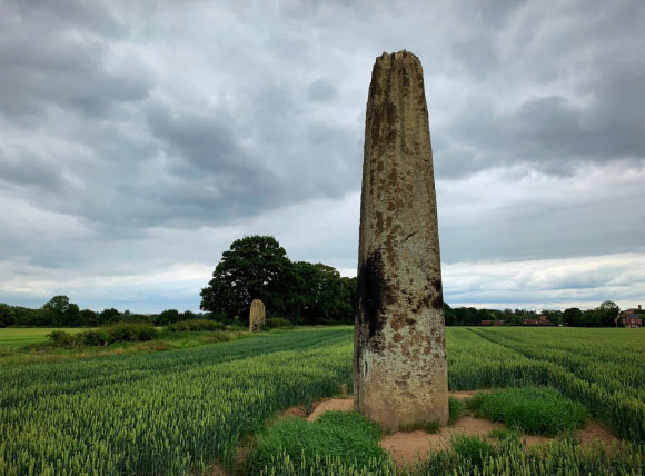
Mysterious Origins of Britain’s 4,000-Year-Old ‘Devils’ Arrows’ Stone Monument Have Finally Been Solved

A Curtin University-led research team has successfully identified the mysterious origin of several 4,000-year-old 25-ton stones, which ancient architects used to construct Britain’s iconic Devil’s Arrows, as Birmingham Rocks in North Yorkshire, over 11 miles (18 kilometers) from the monument’s location.

The researcher behind the first-ever geological source identification of the ancient stone monument said the quarry’s distance and the work required to move such massive stones suggest the ancient architects intentionally selected the material used to construct Devil’s Arrows.

Unraveling the Mysterious Origin of the 4,000-Year-Old Devil’s Arrows

Announcing the discovery, the research team, which included scientists from the University of York, UK, noted that the famous stone pillars stand up to seven meters (22 feet) tall. The team also said the site is unusual because such alignments are rare in lowland settings. Still, researchers have struggled to locate the mysterious origin of the massive stone pillars used to create the over 4,000-year-old monument.

The Curtin-led team noted that previous studies determined that the stones used in Devil’s Arrows were transported to the site during the Late Neolithic or Early Bronze Age. This range means the site could have been constructed as early as 2000 BC.

Lead author Dr Anthony Clarke, from Curtin’s School of Earth and Planetary Sciences, noted the significant challenges with moving the stones over such difficult terrain.

“Each stone weighs more than 25 tonnes, so moving them would have required incredible planning, coordination and engineering skill,” the researcher explained.

Dr. Clarke also highlighted the value this effort suggests these ancient Britons “placed real importance” on the material used in the monument’s construction and the landscape itself.

“It wasn’t just about convenience – the location the stones came from likely held meaning for them,” the researcher explained.

Low Impact Sampling Method Confirms Ancient Stone’s ‘Fingerprints’

To unravel the ancient site’s mysterious origin, the researchers placed tiny pieces of low-adhesion tape on the stones and then removed them. The team said this “low impact” sampling process collected several tiny mineral grains on the tape without measurable damage to the ancient monuments.

According to the statement announcing their solution to the Devil’s Arrows origin mystery, the team performed an analysis of the “fingerprints” of the selected grains. The results matched material from Brimham Rocks.

Critically, the mystery’s solution also addressed the possibility that glacial movement or other natural processes moved the stones over the needed distance. Instead, the team said their results demonstrated that “the stones were intentionally transported by people.”

“Our research shows these stones weren’t simply taken from the nearest available source – they were deliberately chosen from a challenging landscape 18 kilometers away,” Dr Clarke said.

Clarke also said that the results eliminate the nearer site of Plimpton Rocks, which “had previously been identified as the probable source of the stones.”

“Our findings instead point to Brimham Rocks, showing that prehistoric builders transported these enormous stones significantly further than thought,” he explained.

Solving Similar Stone Monument Origin Mysteries

After discovering the mysterious origin of the 4,000-year-old pillars, Dr Clarke said the team will apply its low-impact sampling method to similar stone monument origin mysteries. The team also hopes to conduct future “targeted excavations” at the Devil’s Arrows site and the newly identified source.

Dr. Clarke said that analysis might help explain how these ancient architects moved the massive stone pillars several miles over rugged terrain over 4,000 years ago. The research said the work could also unravel “how these extraordinary monuments were constructed.”

Co-author Dr Jim Leary from the University of York echoed Dr Clarke’s sentiments, while also suggesting that the team’s findings ‘reshaped’ how researchers understand the art of prehistoric monument-building.

“These results show that ancient communities were making deliberate and meaningful choices about where their stones came from,” Dr Leary explained.

The York researcher said their solution to the Devil’s Arrows origin mystery also “highlights the importance of landscape and shared belief systems, as well as the remarkable effort involved in transporting and raising stones of this size.”

The study, “Deliberate Prehistoric Sourcing of the Devil’s Arrows, Britain’s Tallest Stone Row,” was published in the Proceedings of the Royal Society. 

Christopher Plain has spent the last six years as Associate News Editor and Head Science Reporter at The Debrief. Follow and connect with him on X, learn about his novels at plainfiction.com, or email him at christopher@thedebrief.org.

  •  

Integrated Chromosomal and Bioinformatics Analysis Reveals Genetic Causes of Recurrent Pregnancy Loss

Recurrent pregnancy loss affects an estimated one in a hundred couples trying to build a family, yet in a striking proportion of cases no cause is ever identified, leaving patients and clinicians alike without answers. A new study from researchers at the Institute of Medical Sciences, Banaras Hindu University in Varanasi, India, published in the journal Biochemical Genetics, offers a detailed picture of the genetic abnormalities lurking in lost pregnancies and points to a two-tier testing strategy that could substantially improve diagnostic yield. Combining rapid aneuploidy screening with high-resolution chromosomal microarray analysis and a layer of computational biology, the team has mapped not only which chromosomes go wrong in recurrent miscarriage but also which biological pathways those errors may disrupt.

The research team, led by Shivani Mishra and corresponding author Royana Singh, recruited 125 fetal specimens from pregnancies that ended in loss, ultimately analyzing 118 samples after applying predefined exclusion criteria designed to guard against contamination and degraded tissue. The specimens, which included products of conception and fetal tissues such as skin, muscle, and cartilage obtained during autopsy, underwent DNA extraction and quantitative fluorescent PCR, a technique that amplifies short tandem repeat markers scattered across chromosomes 13, 18, 21, X, and Y. By measuring the relative quantity of fluorescent signals at these markers, QF-PCR can detect the most common whole-chromosome imbalances, the trisomies and monosomies that account for the majority of chromosomally abnormal miscarriages, often within a day or two of sample receipt.

The results were sobering but clinically rich. Of the 118 samples analyzed, QF-PCR identified aneuploidy in 36 cases, a detection rate of 30.5 percent. Twenty of these were monosomies, in which an entire chromosome is missing from the fetal genome, and sixteen were trisomies, in which an extra copy is present. The remaining 82 samples appeared normal by this rapid screen. That nearly one in three lost pregnancies carried a whole-chromosome imbalance confirms the central role of aneuploidy in early pregnancy failure, and it also carries a practical message for genetic counseling: when an aneuploidy is identified as the cause of a loss, the recurrence risk for future pregnancies is generally low, information that can meaningfully reduce anxiety for couples facing the aftermath of repeated loss.

Beyond the raw detection numbers, the study teased out clinical associations that matter for patient management. Maternal age showed a statistically significant relationship with the presence of chromosomal abnormalities, echoing decades of reproductive biology linking the aging oocyte to meiotic segregation errors. More intriguing was the finding of a weak but statistically significant negative correlation between gestational age and aneuploidy risk, with a correlation coefficient of −0.238 and a p-value of 0.008855. In other words, the earlier the loss, the more likely it was to be chromosomally abnormal, a pattern consistent with the idea that grossly imbalanced embryos tend to fail early in development, while losses later in gestation are more often driven by other mechanisms, including immunological, endocrine, thrombotic, and anatomical factors.

But QF-PCR has a well-known blind spot. It interrogates a limited set of chromosomes and cannot see submicroscopic copy number variations, the deletions and duplications of genomic segments too small to alter chromosome number but large enough to remove or duplicate genes. To peer beneath that resolution floor, the team selected a subset of 30 samples, stratified by their QF-PCR outcomes, and subjected them to array comparative genomic hybridization. In aCGH, differentially labeled fetal and reference DNA are co-hybridized to a microarray carrying hundreds of thousands of immobilized genomic probes, and the fluorescence ratio at each probe reveals whether the corresponding region is deleted or duplicated in the fetal genome. This technique can detect imbalances ranging from roughly 100 kilobases upward, an order of magnitude finer than conventional karyotyping.

The aCGH analysis proved its worth. Within the 30-sample cohort, the researchers identified clinically relevant copy number variations spanning the spectrum of pathogenicity, including pathogenic and likely pathogenic variants as well as variants of uncertain significance, the latter category reflecting regions whose dosage sensitivity has not yet been firmly established. Each variant was interpreted according to the joint guidelines of the American College of Medical Genetics and the Clinical Genome Resource, the standard framework that weighs factors such as gene content, inheritance pattern, and overlap with established disease loci before assigning a clinical classification.

Among the genes caught in deleted or duplicated segments were several with compelling biological narratives. CFHR3, a member of the complement factor H-related gene family, participates in the regulation of the complement cascade, the arm of innate immunity whose dysregulation has been implicated in inflammatory damage at the maternal-fetal interface. TNFRSF4 encodes a tumor necrosis factor receptor superfamily member with roles in immune cell signaling and tolerance. UGT2B17 belongs to the uridine diphosphate glucuronosyltransferase family, a set of metabolic detoxification enzymes whose variation has previously been linked to recurrent miscarriage, possibly through altered clearance of environmental and endogenous compounds. CD24 and MSR1 are involved in immune signaling and macrophage scavenger function, respectively, while the pregnancy-specific glycoprotein gene family, or PSG genes, is expressed almost exclusively in the placenta and is thought to modulate maternal immune tolerance of the fetus.

To move from gene lists to mechanism, the authors turned the implicated genes loose on a battery of bioinformatics tools. Using FunRich and WebGestalt for functional annotation, KEGG for pathway enrichment, and STRING for protein-protein interaction network construction, the team asked what biological themes united the disrupted loci. The answer converged on a coherent set of processes: immune-inflammatory signaling, endocrine regulation, lipid metabolism, extracellular matrix remodeling, and placental development. The involvement of TNF receptor signaling, MAPK cascades, and interleukin-1-related pathways emerged from the network analyses, weaving the copy number variations into the same immunological tapestry that independent studies have implicated in unexplained recurrent loss, including work on maternal B cell signaling and natural killer cell-mediated immunopathology.

The authors are careful, appropriately so, to frame the pathway findings as exploratory. The cohort of 30 microarray samples is modest, and several of the variants of uncertain significance may prove benign as genomic databases mature. The statistical associations between individual CNVs and miscarriage phenotypes will require validation in larger, well-powered cohorts with matched controls before they can inform clinical decision-making. Still, the architectural message of the study is clear: sequential testing with QF-PCR followed by aCGH captures abnormalities that neither method detects alone, and it does so at a fraction of the cost and turnaround time of broader approaches such as whole-genome sequencing.

That message lands at a moment of ferment in reproductive genetics. European Society of Human Reproduction and Embryology guidelines define recurrent pregnancy loss as two or more consecutive losses, and professional societies have been wrestling with the optimal diagnostic workup of lost pregnancy tissue for years. Previous studies comparing karyotyping with molecular methods have shown that conventional cytogenetics suffers from culture failure and maternal cell contamination, problems that DNA-based methods largely sidestep. The Banaras Hindu University team’s approach, anchored in a real clinical diagnostic laboratory serving a population with limited access to advanced genomic testing, demonstrates that a tiered molecular protocol is feasible and productive outside of well-funded Western centers.

For couples enduring the grief of repeated loss, the study’s findings translate into a concrete recommendation worth discussing with a genetic counselor: comprehensive cytogenetic analysis of lost pregnancy tissue is not merely an academic exercise. Identifying an aneuploidy or a pathogenic copy number variant can close the diagnostic loop, refine recurrence risk estimates, and in some cases guide the management of subsequent pregnancies, including targeted prenatal or preimplantation genetic testing. Conversely, a normal molecular karyotype redirects the clinical search toward the non-genetic contributors, from antiphospholipid syndrome to uterine anomalies.

The work also adds to a growing map of the genomic landscape of pregnancy failure, in which the PSG gene cluster on chromosome 19, the complement factor H-related locus, and immune receptor genes recur across independent cohorts. As genome-wide datasets accumulate and functional studies test the dosage sensitivity of these loci, variants of uncertain significance will gradually be reclassified, and the exploratory pathway signals reported here may harden into causal mechanisms. Until then, the study stands as both a practical diagnostic template and a hypothesis-generating catalog of the fragile genetic architecture underlying one of medicine’s most quietly devastating problems.

Subject of Research: Genetic risk factors in recurrent pregnancy loss, identified through combined QF-PCR and aCGH chromosomal analysis with bioinformatics pathway enrichment analysis of fetal specimens

Article Title: Decoding Genetic Risk Factors in Recurrent Pregnancy Loss: An Integrative Chromosomal and Bioinformatics Approach

Article References: Mishra, S., Ashish, A., Rai, S., Yadav, A. K., & Singh, R. (2026). Decoding Genetic Risk Factors in Recurrent Pregnancy Loss: An Integrative Chromosomal and Bioinformatics Approach. Biochemical Genetics. https://doi.org/10.1007/s10528-026-11424-z

Image Credits: AI Generated

DOI: 10.1007/s10528-026-11424-z

Keywords: recurrent pregnancy loss, QF-PCR, aCGH microarray, copy number variations, aneuploidy, gene ontology, chromosomal abnormalities, placental development, immune-inflammatory pathways, bioinformatics

  •  

How systemic inflammation drives muscle dysfunction in COPD exercise

A sweeping new narrative review published in the Journal of Translational Medicine is pulling back the curtain on one of the most overlooked aspects of chronic obstructive pulmonary disease: the devastating toll the disease takes on the body’s muscles, and the crucial — but complicated — role that exercise plays in fighting back. Led by Domenico Di Raimondo and colleagues at the University of Palermo, the review synthesizes decades of mechanistic research into how COPD reshapes both limb and respiratory muscles, how systemic inflammation may or may not be driving that damage, and why rehabilitation remains the most powerful non-drug therapy available despite nagging scientific uncertainties.

COPD has long been framed as a lung disease, defined by airway obstruction, chronic bronchitis, and emphysematous destruction of lung tissue. But clinicians have increasingly recognized that its most disabling features extend far beyond the chest. Patients with COPD frequently suffer from profound muscle wasting, particularly in the quadriceps, and this skeletal muscle dysfunction has emerged as a clinically relevant systemic manifestation of the disease. It contributes directly to the breathlessness that defines daily life for patients, reduces exercise tolerance, erodes quality of life, and — perhaps most tellingly — predicts mortality. A patient’s ability to stand from a chair or climb a flight of stairs, the review argues, may reveal as much about their prognosis as any spirometry reading.

The muscle changes in COPD are not simply the result of disuse, though physical inactivity certainly plays a role. Under the microscope, the peripheral muscles of these patients tell a story of deep biological remodeling. Muscle fibers shift in type, favoring fast-twitch, glycolytic fibers over the oxidative, fatigue-resistant fibers that sustain everyday activity. The oxidative capacity of the muscle falls, undermining the tissue’s ability to generate adenosine triphosphate efficiently and causing lactic acid to accumulate early during exertion — a process that patients experience as overwhelming leg fatigue long before their lungs reach their limits. Capillarization is impaired, reducing the delivery of oxygen and nutrients to working fibers. Mitochondria, the cellular power plants, show structural and functional dysfunction. Oxidative stress, driven by an excess of reactive oxygen species, damages proteins and membranes. And catabolic pathways, including the ubiquitin-proteasome system, are activated, actively breaking down muscle protein faster than the body can rebuild it.

One of the review’s central arguments is that not all muscle in COPD suffers equally, or in the same way. The quadriceps and other limb muscles typically show the destructive changes described above, but the diaphragm follows a different trajectory. The diaphragm in COPD patients is caught in a double bind: it is subject to intrinsic remodeling driven by chronic overload, yet it also works under a severe mechanical disadvantage caused by lung hyperinflation. As the lungs overinflate, the diaphragm flattens, shortens, and loses its mechanical leverage, making every breath more costly. In response, the diaphragm adapts in ways that differ from limb muscles — sometimes showing adaptive, rather than purely destructive, features as it struggles to meet the relentless demands of breathing through compromised lungs. This distinction between limb and respiratory muscle biology, the authors emphasize, matters enormously for understanding why patients with severe airflow obstruction can still have relatively preserved diaphragm function even as their legs waste away.

If the muscle pathology is the crime scene, systemic inflammation has long stood as the prime suspect. Elevated levels of inflammatory mediators such as tumor necrosis factor-alpha, interleukin-6, and C-reactive protein are commonly detected in COPD patients, and these signaling molecules are known to promote muscle protein breakdown, suppress anabolic signaling, and activate catabolic cascades. The review details the molecular routes by which inflammation can sabotage muscle: activation of nuclear factor-kappa B, engagement of the ubiquitin-proteasome system, interference with insulin-like growth factor-1 signaling, and disruption of insulin receptor substrate pathways that normally support muscle growth and glucose uptake. Yet the authors are refreshingly candid about the limits of this story. The true source of systemic inflammation in COPD is still debated — is it the lungs themselves, comorbid conditions, altered gut microbiota, or something else? — and, crucially, the causal contribution of systemic inflammation to muscle loss and dysfunction in COPD patients remains incompletely proven. Inflammation may be an amplifier rather than the sole architect of muscle decline.

Indeed, the review paints muscle wasting in COPD as a multifactorial process in which inflammation is only one ingredient. Ageing naturally erodes muscle mass and regenerative capacity. Multimorbidity — the coexistence of heart failure, chronic kidney disease, and other chronic conditions — compounds the burden. Malnutrition and inadequate caloric intake starve muscle tissue of the building blocks it needs. Hypoxemia, the low blood oxygen characteristic of advanced disease, impairs mitochondrial function and pushes muscle fibers toward glycolytic metabolism. Corticosteroid exposure, a frequent feature of COPD treatment during exacerbations, can further promote protein breakdown and impair anabolic signaling, a process partly mediated by enzymes such as 11 beta-hydroxysteroid dehydrogenase 1 that amplify glucocorticoid activity within muscle tissue. And physical inactivity, both a cause and a consequence of muscle decline, completes a vicious circle in which weakness breeds inactivity, which in turn breeds further weakness. Acute exacerbations of COPD accelerate this cycle dramatically, with each hospitalization potentially stripping away months of hard-won muscle mass and function.

Against this grim mechanistic backdrop, the review delivers its most clinically significant message: exercise training works. Pulmonary rehabilitation, built around structured exercise programs, is firmly supported as a cornerstone of non-pharmacological COPD management. The evidence base shows that exercise training improves muscle function, increases exercise capacity, alleviates symptoms, and restores functional autonomy — the ability of patients to manage their own daily lives. The mechanisms behind these gains are well understood from exercise physiology. Endurance and resistance training stimulate mitochondrial biogenesis through pathways involving peroxisome proliferator-activated receptor-gamma coactivator 1-alpha, the master regulator of oxidative metabolism. Training restores capillary density, shifts muscle fiber types back toward oxidative phenotypes, strengthens the antioxidant defense system against reactive oxygen species, and tips the balance between protein synthesis and breakdown back toward growth. Resistance training in particular offers a direct counterweight to catabolic signaling, mechanically stimulating muscle protein synthesis even in the presence of inflammatory stress.

But the review does not let exercise off the hook without scrutiny. One of its most provocative observations concerns inflammation. If exercise is so beneficial, and if inflammation is a key driver of muscle damage, one might expect training to measurably reduce circulating inflammatory biomarkers such as C-reactive protein, tumor necrosis factor-alpha, or interleukin-6. Yet the current evidence, the authors find, does not consistently demonstrate such a parallel reduction after training programs. Patients get stronger and more functional, but their blood inflammatory profiles often remain stubbornly unchanged. This disconnect raises a fundamental question about how exercise exerts its benefits in COPD: are the improvements driven by chronic adaptations within the cardiac, respiratory, and muscular systems themselves — local remodeling of muscle fibers, capillaries, and mitochondria — or do they also depend on systemic anti-inflammatory effects that clinical trials have so far failed to detect reliably?

The authors frame the resolution of this question as the field’s main unresolved challenge. Beyond the inflammation puzzle, the review identifies a set of pressing practical questions that clinicians and researchers have yet to answer definitively. What is the optimal training modality — endurance, resistance, interval training, or some combination? What intensity and duration produce the greatest and most durable muscle outcomes? And how should rehabilitation be tailored to the phenotype of the individual patient, given that a cachectic, hypoxemic patient with severe hyperinflation may need a fundamentally different program from a breathless but well-nourished patient with preserved muscle mass? The notion of phenotype-specific rehabilitation — matching the training prescription to the patient’s muscle biology, nutritional status, and inflammatory profile — emerges as a promising but still untested frontier.

The review also situates these findings within the broader translational landscape. Many of the molecular pathways it describes — the ubiquitin-proteasome system, the NOD-like receptor pyrin domain-containing protein 3 inflammasome, myostatin-related signaling, and the interplay between hypoxia-inducible factor 1 alpha and mitochondrial function — are potential drug targets. But the authors’ implicit conclusion is that no pharmacological shortcut will soon replace the accumulated, adaptive power of regular physical training. Muscle is plastic tissue, and the review suggests that the therapeutic leverage lies precisely in that plasticity, harnessed through carefully dosed and progressive exercise.

What makes the review timely is its honesty. Rather than inflating the anti-inflammatory credentials of exercise or overselling any single mechanism, it maps the terrain as it actually stands: muscle dysfunction in COPD is real, multifactorial, and deadly serious; exercise is genuinely effective; and the causal chain linking inflammation to muscle loss remains, frustratingly, an open question. For the millions of patients living with COPD worldwide, the practical takeaway is clear. Rehabilitation is not an optional add-on to inhaler therapy — it is a biological intervention that works at the level of the muscle fiber, the mitochondrion, and the capillary, even when the exact systemic chemistry of its benefits remains to be fully explained.

For researchers, the review is a call to arms. Future studies must disentangle the contribution of inflammation from the many other forces eroding muscle in COPD, identify biomarkers that predict who will respond best to which training modality, and design trials that test phenotype-matched rehabilitation strategies rather than one-size-fits-all programs. Until then, the message for clinical practice stands on remarkably firm ground: in COPD, what the lungs cannot fix, the muscles — given the right training — may yet do.

Subject of Research: Mechanisms linking COPD to skeletal and respiratory muscle dysfunction, systemic inflammation, and the effects of exercise-based rehabilitation

Article Title: Muscle dysfunction, systemic inflammation and exercise in COPD – a narrative review

Article References: Di Raimondo, D., Siragusa, P., Pirera, E., Romito, G., Calcullo, D., Scaglione, S., Daidone, M., Ferrantelli, S., Siscaro, G., & Tuttolomondo, A. (2026). Muscle dysfunction, systemic inflammation and exercise in COPD – a narrative review. Journal of Translational Medicine. https://doi.org/10.1186/s12967-026-08961-x

Image Credits: AI Generated

DOI: 10.1186/s12967-026-08961-x

Keywords: Chronic obstructive pulmonary disease, Muscle dysfunction, Systemic inflammation, Exercise, Rehabilitation, Quadriceps, Diaphragm, Cachexia, Mitochondrial dysfunction, Oxidative stress, Hyperinflation, Pulmonary rehabilitation

  •  

Advanced oxidation process reduces micropollutant toxicity in wastewater for agricultural reuse

Researchers in Brazil have demonstrated that a hybrid advanced oxidation process can transform pharmaceutical-laden wastewater into an effluent that is dramatically safer for plants, earthworms, onion root cells, and beneficial microorganisms, offering a rigorous ecotoxicological case for reusing treated sewage in agriculture. The study, published in Environmental Science and Pollution Research, addresses a long-standing blind spot in water treatment: proving that a chemically “clean” effluent is also biologically compatible.

Conventional wastewater treatment plants were never designed to strip out recalcitrant organic micropollutants such as pharmaceuticals, and trace amounts routinely slip through into receiving waters. Advanced oxidation processes (AOPs) attack these stubborn compounds by generating highly reactive free radicals that fragment contaminant molecules. But chemical degradation alone does not guarantee safety. As contaminants break down, they spawn transformation products (TPs) that can, in some cases, be as toxic as—or more toxic than—the parent compounds. The research team, led by Lucas Gustavo da Costa and Alam Gustavo Trovó of the Federal University of Uberlândia, together with collaborators at the Oswaldo Cruz Institute, set out to answer a deceptively simple question: does measurable chemical removal of micropollutants actually translate into reduced biological harm?

The centerpiece of their work is the H₂O₂/S₂O₈²⁻/UVC process, a hybrid system that combines hydrogen peroxide and persulfate with short-wave ultraviolet C radiation. UVC photons cleave both oxidants simultaneously, generating hydroxyl radicals (HO•) and sulfate radicals (SO₄•⁻) in the same reaction volume. This dual-radical strategy outperforms systems relying on either oxidant alone, or on peroxymonosulfate (HSO₅⁻), which is costlier and demands more energy for activation. The hybrid route also resists interference from inorganic species commonly found in real effluents, making it an economically attractive candidate for deployment at full-scale treatment plants. The process had already been chemically optimized in the team’s earlier work using multivariate mixture design and rotatable central composite design experiments; the new study is its first integrated biological stress test.

The experiments used real municipal effluent from a wastewater treatment plant in Uberlândia, Minas Gerais, sampled after the plant’s final tertiary treatment stage of coagulation-flocculation with ferric chloride and flotation. The effluent was enriched with three pharmaceuticals representing different therapeutic classes: colchicine (COL, an antimitotic agent), nitazoxanide (NTZ, an antiparasitic), and sulfamethoxazole (SMX, a widely detected antibiotic), each at 325 nmol L⁻¹—equivalent to 130, 100, and 82 micrograms per liter, respectively. That concentration was deliberately chosen as high enough for direct HPLC–DAD analysis without preconcentration, yet low enough to be environmentally representative. Treatment was carried out in an amber glass reactor irradiated by two 8-watt UVC mercury lamps emitting at 254 nm, with a measured irradiance of 4.9 W m⁻². Residual oxidants were neutralized with sodium thiosulfate before any biological testing, ensuring that observed effects could not be attributed to leftover peroxide chemistry.

The ecotoxicological battery spanned multiple trophic levels and levels of biological organization. Oxidative stress was assessed in the earthworm Eisenia andrei by measuring malondialdehyde (MDA), a marker of lipid peroxidation, and protein carbonylation, an irreversible oxidative modification of proteins. Phytotoxicity was evaluated using lettuce (Lactuca sativa) seeds and a growth index combining germination rate and root elongation. Cytotoxicity and genotoxicity were quantified in onion (Allium cepa) root meristems through the mitotic index and the frequency of chromosomal and nuclear aberrations—micronuclei, chromosome breaks, stickiness, bridges, and nuclear buds—across 5,000 cells per sample. Finally, growth inhibition was tested in two environmentally relevant microbes: Azospirillum brasilense, a plant growth-promoting bacterium used in soybean cultivation, and Saccharomyces cerevisiae, a yeast that persists in soils and fermentative niches.

The untreated enriched effluent told a worrying story. It exhibited substantial genotoxicity in onion cells—21%, approaching the 24% seen with the positive control, methyl methanesulfonate—and severe phytotoxicity, with lettuce growth indices far below the 80% threshold that signals absence of toxicity. Even the unspiked effluent itself suppressed lettuce growth, achieving a growth index of only 47%, evidence that bioactive compounds survive conventional treatment. In earthworms, individual aqueous solutions of NTZ and SMX raised MDA levels by 37.2% and 23.5% respectively, while the three-compound mixture increased lipid peroxidation by 38.0%, pointing to additive or synergistic oxidative stress mechanisms involving reactive oxygen species and Fenton-type chemistry within cells.

The picture changed decisively after oxidation. Following 10 minutes of UVC-driven treatment—the point at which 80% chemical degradation had been achieved, matching the minimum removal target proposed in the European Union’s COM(2022)541 directive—genotoxicity fell to 10%, and after 20 minutes, corresponding to the limit of quantification for the parent compounds, it dropped further to 6%, a statistically significant reduction. The plant growth index climbed above 80%, crossing from toxic territory into biocompatibility. The mitotic index in onion cells remained statistically indistinguishable from the deionized-water control (around 39–40% versus 40%), demonstrating that the process generated no cytotoxic byproducts capable of arresting cell division. And crucially for agricultural applications, neither the treated effluent nor its transformation products inhibited growth of A. brasilense or S. cerevisiae.

The mechanistic details are instructive. In the untreated enriched effluent, micronuclei—membrane-bound DNA fragments expelled from the nucleus during flawed cell division—were among the most frequent aberrations, indicating clastogenic and aneugenic damage. Colchicine exposure predictably produced C-metaphase accumulation, a signature of its tubulin-binding, spindle-disrupting mechanism first described in Allium nearly a century ago. SMX depressed the mitotic index and induced chromosomal damage, consistent with prior findings in Vicia faba. NTZ, whose genotoxicity had never before been reported, produced chromosome breaks, stickiness, and nuclear buds. After oxidation, the overall aberration burden fell sharply, particularly micronuclei and stickiness, although a few bridges and polyploid cells persisted even at 20 minutes—a reminder that some transformation products or resistant residuals may linger.

The microbial results came with a subtlety. Reduced A. brasilense growth after treatment initially looked like a red flag, but the team attributes it to depletion of assimilable organic matter, which serves as radical scavenger during oxidation and as a nutrient source afterward—not to toxicity. The negative control likewise grew slowly. This distinction matters because AOPs are designed to mineralize organic carbon, and a nutrient-poorer medium should not be misread as a more toxic one. For S. cerevisiae, no inhibition occurred at any stage of treatment, although NTZ alone significantly depressed yeast optical density, reinforcing the compound’s cell-cycle interference potential even at nanomolar-scale exposures.

The study’s broader significance lies in its methodological stance. By adopting an “effect-driven approach,” the team evaluated the toxicity of whole reaction mixtures without needing to isolate and identify individual transformation products—an impractical task given that many degradation byproducts are not commercially available. Their findings align with a growing consensus that treatment efficacy must be judged not merely by parent-compound removal but by the nature and reactivity of the resulting transformation products. Biochemical biomarkers such as MDA and protein carbonylation detected sublethal disturbances that mortality-based endpoints would have missed entirely, providing early warnings of cellular distress at environmentally relevant concentrations.

One caveat deserves attention: while lipid peroxidation declined after treatment, protein carbonylation rose slightly—1.05-fold above control—suggesting some transformation products may still provoke protein oxidation even as lipid damage subsides. The authors flag this discrepancy and call for future identification of the specific TPs responsible, alongside expanded organism panels, longer-term exposure studies, and pilot-scale validation across different environmental matrices.

The regulatory context sharpens the urgency. The EU’s proposed urban wastewater treatment directive mandates at least 80% removal of specified organic micropollutants and microbiological control for agricultural reuse, yet it conspicuously omits eco-compatibility assessment of the treated water itself. This study supplies exactly that missing dimension, showing that a process satisfying the chemical benchmark also delivers measurable biological benefit—lower genotoxicity, restored plant growth, intact cell division, and unharmed beneficial microbes. As water scarcity intensifies globally and reuse becomes less optional, the Brazilian team’s integrated bioassay framework offers a template for ensuring that the water farmers irrigate with is not merely chemically compliant, but genuinely ecologically safe.

Subject of Research: Mitigation of micropollutant toxicity in treated wastewater using the H₂O₂/S₂O₈²⁻/UVC advanced oxidation process, assessed through integrated ecotoxicological bioassays for potential agricultural reuse.

Article Title: Mitigation of micropollutant toxicity in treated wastewater using the H2O2/S2O82−/UVC process: An ecotoxicological perspective for agricultural reuse

Article References: da Costa, L. G., dos Santos, G. M., Marson, E. O., de Lima, M. G. F., de Souza Bessa, M. A., Scarafiz, G., Junior, S. F. S., Saggioro, E. M., de Carvalho, S. R., de Siqueira Ferreira, A., Nilin, J., Neto, W. B., & Trovó, A. G. (2026). Mitigation of micropollutant toxicity in treated wastewater using the H2O2/S2O82−/UVC process: An ecotoxicological perspective for agricultural reuse. Environmental Science and Pollution Research. https://doi.org/10.1007/s11356-026-38218-5

Image Credits: AI Generated

DOI: 10.1007/s11356-026-38218-5

Keywords: Advanced oxidation process, Micropollutants, Wastewater reuse, Ecotoxicity, Genotoxicity, Phytotoxicity, Oxidative stress, Hydroxyl radicals, Sulfate radicals, UVC treatment, Agricultural irrigation, Transformation products

  •  

A Bronze Age Tribe in Poland Was Already Eating a Cereal Brought from China before Tutankhamun was even born

Around 1590 BCE — more than 800 years before Rome was founded, and before Tutankhamun was even born — a small number of people in what is now southeastern Poland started eating a cereal that had never grown in their fields. It wasn’t a local crop, and it didn’t belong to any plant known elsewhere […]

  •  

Forget Beer: Ice Age Hunters Were Getting High 12,000 Years Earlier — With Betel Nuts

Two skeletons from the Indonesian island of Sulawesi had something strange going on in their mouths. Not cavities. Not the usual wear from a tough diet. Deep, rounded grooves cut into the teeth — a pattern no archaeologist had documented before, and one nobody could explain. The answer, published in Science Advances, pushes the known […]

  •  

Imagem de Referência da Apple: recurso dos iPhones 18 Pro comprova a autenticidade de fotos

Com o lançamento dos iPhones 18 Pro/18 Pro Max, a Apple oficializou e deu mais detalhes sobre um novo recurso opcional — o qual havia dado as caras na quinta beta do iOS 27 — que ajuda os usuários a comprovar que uma foto não foi alterada após ter sido tirada.

Chamado Imagem de Referência da Apple (Apple Reference Image), o recurso utiliza um novo sensor na câmera principal que assina os dados do sensor em nível de pixel, no momento da captura. Quando uma foto é tirada em um novo modo chamado Referência (Reference), esses dados são enviados para o Private Cloud Compute, que os processa para criar o que a Apple chama de “imagem de referência inalterável” (algo como um “negativo digital”).

Agora, você pode provar a autenticidade de uma foto tirada no iPhone. A assinatura segura dos dados da imagem no nível do pixel acontece no momento da foto, registrando tudo que o sensor da câmera viu.

As imagens de referência aparecem no aplicativo Fotos (Photos) ao lado da foto correspondente, permitindo que os usuários comparem visualmente as duas para verificar se houve alguma edição. A Apple também está disponibilizando APIs 1Application programming interfaces, ou interfaces de programação de aplicações. no iOS 27, iPadOS 27 e macOS Golden Gate 27 para que aplicativos de terceiros possam exibir essas imagens de referência.

Divulgação/Apple

A Apple apresentou o recurso como parte de uma iniciativa mais ampla voltada para a autenticidade de imagens, complementando os metadados de imagem já existentes e o futuro suporte ao padrão SynthID, que ajudará a identificar imagens geradas ou editadas com IA.

Entretanto, o novo recurso não estará disponível — a princípio — na China, devido a exigências regulatórias. Na União Europeia, a captura de imagens com o Apple Reference Image também não estará disponível já no lançamento dos novos iPhones; no entanto, usuários que estão rodando o iOS 27, iPadOS 27 e macOS 27 ainda poderão processar e visualizar imagens de referência.

Notas de rodapé

  • 1
    Application programming interfaces, ou interfaces de programação de aplicações.

  •  

iPhone 18 Pro and iPhone 18 Pro Max

Apple (video, MacRumors, Hacker News, Slashdot):

Apple today announced iPhone 18 Pro and iPhone 18 Pro Max, delivering the most advanced pro camera system in an iPhone, and huge leaps in battery life and performance. The new 48MP Fusion Main camera, now with variable aperture, brings creative control that wasn’t possible before, and new Pro controls let users customize their experience in the Camera app. Both models feature a smaller and even more useful Dynamic Island, as well as A20 Pro and a next-generation vapor chamber that together deliver the highest sustained performance in iPhone history.

[…]

Pro controls is a new customizable experience in the Camera app for iPhone 18 Pro and iPhone 18 Pro Max that makes it easier than ever for users to access their most used settings and introduces new options for manual control.

[…]

When a photo is taken in the new Reference mode, the camera captures signed sensor data that Private Cloud Compute develops into an unalterable reference image.

[…]

The upgraded Dynamic Island can now show three Live Activities at once[…]

[…]

eSIM-only models deliver up to 36 hours of video playback on iPhone 18 Pro and up to 45 hours of video playback on iPhone 18 Pro Max.

[…]

iPhone 18 Pro and iPhone 18 Pro Max will be available in 256GB, 512GB, 1TB, and 2TB storage capacities. Available in burgundy, glacier, silver, and black, iPhone 18 Pro starts at $1,199 or $49.95 per month, and iPhone 18 Pro Max starts at $1,299 or $54.12 per month for 24 months.

What I’m most happy to see is the ability to increase the depth of field. I don’t understand what Apple Reference Image is doing that it needs to be uploaded/offloaded to Private Cloud Compute.

Previously:

Update (2026-09-11): Hartley Charlton:

Pro Controls is built around the variable aperture on the new Main camera. Six laser-cut blades move smoothly between four aperture settings, and the Camera app can adjust aperture automatically based on depth of field and lighting, or users can take manual control themselves for what Apple calls “truly unique shots that weren’t possible before” on an iPhone.

Pro Controls works across both photo and video capture. It also introduces Smart Focus Tracking, which uses on-device AI models to lock onto a subject and maintain focus even if that subject briefly leaves the frame and comes back into view.

  •  

Britain’s Tallest Standing Stones Traced to Their Geological Source

The Devil’s Arrows, the three towering gritstone megaliths on the western outskirts of Boroughbridge, Yorkshire, the United Kingdom. Image credit: Curtin University.

Scientists from Curtin University and the University of York have pinpointed the origin of the Devil’s Arrows, the three towering gritstone megaliths that have stood on the edge of Boroughbridge, Yorkshire, for roughly 4,000 years, ending a long-running archaeological debate.

The post Britain’s Tallest Standing Stones Traced to Their Geological Source appeared first on Sci.News: Breaking Science News.

  •  

Apple apresenta o iPhone Duo, seu primeiro dobrável

Depois de anos de especulações, a Apple enfim revelou o iPhone Duo, a sua estreia oficial no segmento dos smartphones com tela dobrável.

O formato já é conhecido por quem acompanhou os rumores e viu as mudanças recentes entre os dobráveis de outras empresas, como a HUAWEI e a Samsung: o aparelho conta com uma tela externa (Ceramic Shield 2) de 5,4 polegadas e uma tela interna de 7,6″, seguindo um formato parecido com o de um passaporte. Ambas também têm exatamente a mesma proporção, e o acabamento fosco (nano-texture) está presente na tela interna.

Divulgação/Apple

Quando aberto, o iPhone Duo torna-se o iPhone mais fino já vendido pela Apple, com 5,2mm. O mecanismo de dobradiça também foi outro ponto particularmente trabalhado pela companhia (e o possível motivo de tanta demora para esse lançamento), sendo composto por mais de 100 componentes — a tampa da dobradiça, por sua vez, é impressa em 3D.

Divulgação/Apple

Outro elemento que vale ser mencionado é a volta do Touch ID nos iPhones, já que a finura do aparelho não comporta os sensores do Face ID. A biometria digital está localizada na lateral direita do aparelho.

Também estão agora nessa lateral os principais controles em nível de software do iOS 27, incluindo a barra de status (que ganhou um novo design mais compacto), as barras de ferramentas dos apps e até mesmo o Dock, que agora fica na vertical. Todos os principais apps do sistema foram adaptados para o novo formato de modo a aproveitar o maior espaço disponível, além de permitirem que você use o smartphone com ele parcialmente fechado.

Divulgação/Apple
Divulgação/Apple

O modo Em Espera (Stand By) é compatível com ambos os displays, transformando o iPhone Duo em um verdadeiro porta-retrato interativo ou em um relógio digital, só para citar alguns exemplos. A Apple também introduziu novos mostradores para esse recurso focados nos apps Calendário (Calendar) e Tempo (Weather).

Divulgação/Apple
Divulgação/Apple

Assim como no iPad, é possível colocar dois apps no modo Split View ao usar a tela interna e, em breve (provavelmente depois de um update), usar uma Apple Pencil (USB-C) para fazer ilustrações e anotações em apps suportados.

Divulgação/Apple

O iPhone Duo vem equipado com o A20 Pro, chip com uma litografia de 2 nanômetros, possuindo 6 núcleos destinados a CPU 1Central processing unit, ou unidade central de processamento. (2 de desempenho e 4 de eficiência) e uma GPU 2Graphics processing unit, ou unidade de processamento gráfico. de 7 núcleos, além de um Display Engine novo para “empurrar” as duas telas. Com tudo isso, a Apple promete uma performance sustentada até 35% maior em comparação com o iPhone 17 Pro.

O novíssimo modem C2 também está presente no primeiro dobrável da Apple, oferecendo conexões até 50% mais rápidas e conectividade 5G mmWave (apenas nos Estados Unidos). Ele também está mais eficiente do ponto de vista energético, consumindo 15% menos energia em comparação com o modem C1X. Responsável por possibilidade conexões Wi-Fi 7, Bluetooth 6 e Thread, o chip N1 também deu as caras nesse smartphone.

O dobrável conta com duas câmeras frontais, uma para cada tela do aparelho; mais tradicional, a câmera frontal da tela externa (Center Stage) fica no canto superior direito do dispositivo, servindo também como uma Ilha Dinâmica (Dynamic Island), enquanto a câmera frontal da tela interna (FaceTime camera) está um pouco mais ao centro e, vejam só, fica posicionada sob o display, aparecendo apenas quando acionada — seja para capturar fotos/vídeos ou para exibir Atividades ao Vivo (Live Activities).

Divulgação/Apple

Já no conjunto de câmeras traseiras, localizado no módulo de câmera horizontal em formato de pílula, temos a câmera Fusion principal de 48 megapixels — a qual conta com shutter lag zero e também pode simular uma lente telefoto de 2x — e uma lente Fusion ultra-angular de também 48MP — mesmíssima lente dos iPhones 18 Pro. E, por conta da tela externa, usuários podem tirar selfies facilmente com essas lentes, que também são capazes de capturar fotos automaticamente graças ao recurso Smart Take, que detecta poses.

Divulgação/Apple

Além de novas formas de navegar pelo sistema, as duas telas também desbloqueiam maneiras inéditas de capturar imagens. De acordo com a Apple, a FaceTime camera permite realizar chamadas de vídeo com o iPhone Duo completamente aberto ou com ele parcialmente fechado — enquanto fica apoiado numa mesa, por exemplo. O iPhone Duo também pode exibir a imagem de uma pessoa durante uma chamada de vídeo na tela externa, permitindo que outras pessoas participem da conversa sem precisar compartilhar uma câmera com o dono do aparelho.

Mesmo com uma espessura reduzida, teremos uma das maiores baterias já utilizadas pela Apple em um iPhone, capaz de reproduzir até 31 horas ininterruptas de vídeo com a tela interna e até 44 horas com o display de fora. Em uso alternado, o iPhone Duo pode durar até 24 horas com uma única carga, de modo que apenas 15 minutos na tomada (via cabo) são suficientes para garantir 50% de carga. Carregadores MagSafe e Qi2 também são suportados.

Em termos de durabilidade, o iPhone Duo vem com a certificação IP68 contra água e poeira, além de contar, como dito, com a tecnologia Ceramic Shield 2 na tela externa. A primeira geração do Ceramic Shield também está presente nesse aparelho na forma do vidro traseiro.

Preço e disponibilidade

O iPhone Duo contará com opções de 256GB, 512GB, 1TB e 2TB de armazenamento, com o valor inicial de US$2.000 nos Estados Unidos e podendo chegar a US$3.200. Ele pode ser adquirido nas cores branco-estrela (star white) e céu noturno (night sky).

No Brasil e em Portugal, o dobrável sairá pelos preços iniciais de R$22.000 e 2.400€, respectivamente.

Nos EUA, no Brasil e em Portugal, a pré-venda começará no dia 16 de outubro (uma sexta-feira). O lançamento geral ocorrerá no dia 23/10 (outra sexta) em todas essas localidades.


Apoio: Google Workspace

Logo do Google Workspace

Junte-se a milhões de empresas que usam as versões premium de apps como Gmail, Agenda, Drive e Meet. Com o Gemini integrado ao Google Workspace, você escreve, resume e organiza em segundos — e tudo continua funcionando de qualquer lugar, com apps completos e intuitivos para iPhone, iPad e Mac. Comece hoje mesmo com 20% de desconto nos 6 primeiros meses!

NOTA DE TRANSPARÊNCIA: O MacMagazine recebe uma pequena comissão sobre vendas concluídas por meio de links deste post, mas você, como consumidor, não paga nada mais pelos produtos/serviços usando os nossos links de afiliado.

Notas de rodapé

  • 1
    Central processing unit, ou unidade central de processamento.
  • 2
    Graphics processing unit, ou unidade de processamento gráfico.

  •  
❌