❌

Reading view

Wait Times for Sentinel Node Biopsy Impact Melanoma Outcomes

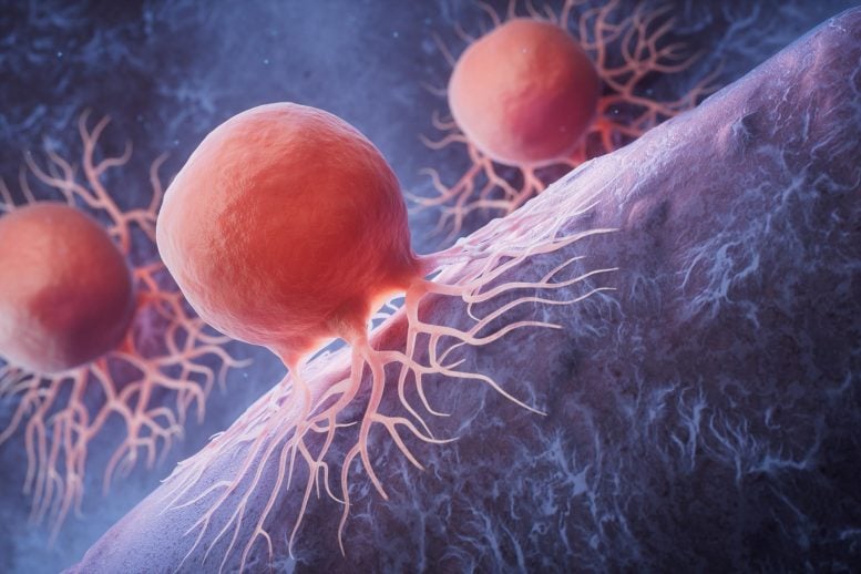
In the ever-evolving landscape of oncology, the management of melanoma remains a critical field of study due to the aggressive nature of this skin cancer and its potential for rapid progression. A groundbreaking new study published in the British Journal of Cancer on June 16, 2026, has shed light on one of the pivotal components in melanoma staging and treatment: the timing of sentinel lymph node biopsy (SLNB). This research provides the most comprehensive analysis to date on how wait times for SLNB affect disease progression, the burden of micrometastatic tumors, and overall survival outcomes in an era marked by advanced treatment modalities.

Sentinel lymph node biopsy is a standard procedure employed to determine the extent of melanoma spread beyond the primary tumor site. By pinpointing the first lymph node (or nodes) to which cancer cells are likely to migrate, clinicians can more accurately stage the disease and tailor treatment regimens accordingly. However, discrepancies in wait times for this critical diagnostic intervention have historically been reported, raising concern about potential impacts on patient prognosis. The new investigation by Breeze et al. systematically explores the implications of these delays using state-of-the-art patient cohorts and rigorous statistical modeling.

The researchers embarked on an extensive retrospective cohort study, leveraging a dataset of melanoma patients from multiple high-volume cancer centers. The timeline from initial biopsy-confirmed melanoma diagnosis to the performance of SLNB was meticulously documented, allowing for stratification of patients based on the duration of delay. Through this stratification, the authors correlated wait times with key clinical endpoints, such as progression of melanoma stage, micrometastatic tumor burden within sentinel nodes, and long-term survival rates, thus providing a granular understanding of the temporal impact on melanoma dynamics.

A central revelation from the study was a statistically significant correlation between prolonged wait times for sentinel node biopsy and increased micrometastatic tumor burden. This finding indicates that delays may facilitate the proliferation of melanoma cells within the nodal basin, effectively allowing the disease to advance silently. This finding is of particular clinical relevance because increased tumor burden within sentinel nodes has been associated with worse prognostic outcomes and may necessitate more aggressive adjuvant therapies.

Delving deeper, the study analyzed the heterogeneity of micrometastatic tumor volume and revealed that patients who underwent SLNB within an optimal window exhibited minimal tumor load, while those subjected to protracted wait times showed proliferative nodal involvement, often translating to higher nodal staging. This advancement of disease within the nodal system provides a pathway for further dissemination to distant sites, thereby complicating therapeutic strategies and reducing survivorship.

Beyond tumor burden, the research also explored the relationship between SLNB wait times and survival outcomes. Utilizing survival analysis techniques such as Kaplan-Meier curves and Cox proportional hazards modeling, the authors demonstrated a clear survival disadvantage for patients experiencing delays beyond four to six weeks. These data underscore the crucial time sensitivity of sentinel node biopsy in the management paradigm of melanoma and advocate for minimizing procedural wait times as a standard of care.

In the context of modern melanoma therapies, including immune checkpoint inhibitors and targeted molecular agents, the findings gain additional weight. Early and accurate staging facilitated by timely SLNB allows for better patient selection for these advanced treatments, which have revolutionized outcomes but are most efficacious when implemented in a well-defined disease context. Delays that permit tumor progression can diminish the window of therapeutic opportunity, underscoring a need for systemic healthcare improvements.

Importantly, the study also aligns with emerging biomolecular data suggesting that during the waiting period, micrometastatic melanoma cells may undergo phenotypic transformations that enhance invasive potential and resistance to apoptosis. These cellular alterations contribute to a more aggressive disease profile and argue for clinical urgency in timely diagnosis and staging. Thus, the research integrates molecular oncology perspectives with clinical outcomes, presenting a holistic view of melanoma pathophysiology.

Healthcare system factors contributing to wait times were also examined, revealing that logistical inefficiencies, resource limitations, and prioritization protocols significantly influence patient experiences. The authors advocate for policy interventions to optimize scheduling processes and resource allocation to reduce unnecessary delays. This approach resonates with ongoing quality improvement initiatives aiming to enhance cancer care delivery at institutional and national levels.

The implications of these findings extend into the realm of patient counseling and shared decision-making. Oncologists and surgical teams can leverage this evidence to stress the importance of expeditious sentinel node biopsy and provide realistic prognostic expectations based on timing. This transparency enhances patient engagement and may improve adherence to follow-up and treatment plans.

From a research standpoint, this study paves the way for prospective trials to evaluate interventions aimed at reducing SLNB wait times and their impact on clinical outcomes. Additionally, it highlights the need for the development of predictive biomarkers that could identify patients at greatest risk from biopsy delays, enabling prioritized scheduling or alternative staging strategies.

Another salient point is the potential economic impact of optimizing SLNB wait times. By mitigating disease progression, fewer patients may require extensive treatments or experience complications associated with advanced melanoma. This reduction in downstream healthcare costs reinforces the value proposition of investing in timely diagnostic services, aligning clinical benefit with economic sustainability.

The study also prompts a re-examination of current clinical guidelines regarding melanoma management. While many protocols recommend SLNB within a certain timeframe, empirical data to justify these benchmarks have been limited. Breeze et al.’s work provides a robust evidence base that can refine these recommendations and harmonize practices internationally.

Moreover, the integration of advanced imaging techniques and diagnostic pathways to streamline SLNB scheduling is an area ripe for innovation inspired by these findings. Combining molecular profiling with precise imaging could improve risk stratification and expedite treatment timelines, a critical step toward personalized melanoma care.

Finally, the study’s emphasis on the “modern treatment era” context ensures that conclusions are relevant to current practice, reflecting the transformations brought by immunotherapy and targeted treatments. This temporal relevance makes the findings particularly urgent for contemporary oncology clinics striving to maximize patient outcomes in a highly competitive therapeutic landscape.

In summary, the meticulous investigation by Breeze and colleagues elucidates the profound impact of sentinel lymph node biopsy timing on melanoma progression, tumor burden, and survival. It sends a clear message to clinicians, healthcare systems, and policymakers: reducing delays in sentinel node assessment is not merely a procedural target but a critical determinant of melanoma prognosis. As melanoma incidence continues to rise globally, these insights will be instrumental in shaping strategies that improve survival while optimizing healthcare resources.

The study signals a paradigm shift by firmly establishing temporal aspects of melanoma staging as a modifiable factor influencing disease biology and clinical outcomes. This research underscores the urgent need for concerted action to reduce procedural delays, integrate multidisciplinary care, and adapt guidelines to embrace new evidence. The future of melanoma management hinges on such data-driven optimizations that align molecular insights with clinical pragmatism, ultimately altering the trajectory of this complex disease.


Subject of Research: The impact of wait times for sentinel lymph node biopsy on melanoma disease progression, micrometastatic tumor burden, and survival outcomes.

Article Title: Implications of wait times for sentinel node biopsy on melanoma disease progression, micrometastatic tumour burden and survival outcomes in the modern treatment era.

Article References:
Breeze, S.O., Heaton, M.J., Snelling, A.P. et al. Implications of wait times for sentinel node biopsy on melanoma disease progression, micrometastatic tumour burden and survival outcomes in the modern treatment era. Br J Cancer (2026). https://doi.org/10.1038/s41416-026-03497-9

Image Credits: AI Generated

DOI: 16 June 2026
  •  

Outdoor Air Pollution Linked to Ovarian, Endometrial Cancer

In an ambitious new study published in the Journal of Exposure Science and Environmental Epidemiology, researchers report compelling evidence linking long-term exposure to outdoor air pollution with an increased risk of ovarian and endometrial cancers. This groundbreaking investigation harnesses a large prospective cohort to unravel the intricate relationship between environmental pollutants and gynecologic malignancies, offering a critical perspective on public health implications that extend far beyond respiratory diseases.

Air pollution has long been recognized as a major contributor to cardiovascular and respiratory conditions, but its potential carcinogenic effects on reproductive organs have remained underexplored. The study conducted by Ammons, Fisher, Madrigal, and colleagues seeks to fill this gap by focusing on two of the most common gynecologic cancers – ovarian and endometrial cancer – and evaluating the impact of chronic exposure to ambient air pollutants. By analyzing data from thousands of participants over an extended period, the researchers embarked on an epidemiologic journey to quantify risks associated with particulate matter and other airborne toxins.

The cohort employed in this study includes a diverse population sampled across multiple geographic regions, allowing researchers to capture variations in pollution levels and their corresponding cancer incidences. Utilizing cutting-edge exposure assessment techniques, the team mapped pollutant concentrations at individual residences with unprecedented accuracy. These methods integrated satellite data, ground-level monitors, and atmospheric modeling to deliver robust, spatially resolved exposure metrics. Such advances surpass prior limitations inherent in ecological and self-reported exposure assessments, lending unprecedented credibility to the findings.

Delving into the biological plausibility of how air pollution could influence the pathogenesis of ovarian and endometrial cancers, the researchers propose several mechanistic pathways. Persistent exposure to fine particulate matter (PM2.5) and polycyclic aromatic hydrocarbons (PAHs) may induce systemic inflammation, oxidative stress, and hormonal dysregulation. These factors could disrupt the delicate microenvironment of reproductive tissues, promoting DNA damage, aberrant cell signaling, and ultimately malignant transformation. The findings echo mounting laboratory evidence that airborne pollutants can act as endocrine disruptors, complicating hormonal balance crucial for gynecologic health.

Statistical analyses revealed that the risk for endometrial and ovarian cancers escalates in tandem with pollutant concentration levels. Notably, women residing in urban areas with higher vehicular emissions and industrial activities exhibited significantly greater cancer risks compared to those in less polluted settings. After adjusting for known confounders such as age, BMI, smoking status, and reproductive history, the association remained robust, underscoring the independent contribution of chronic air pollution exposure to cancer pathogenesis.

Importantly, this research highlights a disproportionately higher vulnerability among subgroups, including postmenopausal women and those with pre-existing metabolic disorders. These populations may experience exacerbated inflammatory responses or impaired detoxification pathways, amplifying carcinogenic potential. Such differential susceptibility underscores the urgency for targeted interventions and personalized public health strategies to mitigate the undue burden of pollution-induced cancers.

Beyond its epidemiological insights, the study calls attention to the glaring disparities in environmental exposures driven by socioeconomic and racial factors. Marginalized communities situated near highways, factories, and waste sites confront a compounded risk landscape for gynecologic cancers due to cumulative environmental and social stressors. Addressing these inequities demands integrated policy responses encompassing urban planning, emission regulation, and healthcare accessibility.

From a preventive medicine standpoint, these findings compel a re-evaluation of current cancer risk models and screening guidelines to incorporate environmental exposure metrics. Recognizing air pollution as a modifiable risk factor opens avenues for novel primary prevention strategies including community-level emission reductions, green space expansion, and personal behavior modifications. Public awareness campaigns and healthcare provider education must integrate these environmental determinants to holistically address cancer prevention.

Moreover, the study’s prospective cohort design strengthens the causal inference between pollution and cancer development, circumventing biases typical of retrospective analyses. Continuous monitoring and follow-up of the cohort will enable future research to delineate critical exposure windows, dose-response relationships, and potential synergistic effects with other carcinogens. Such longitudinal data is vital for refining risk assessments and tailoring interventions.

The multidisciplinary collaboration driving this research showcases the essential convergence of epidemiology, environmental science, oncology, and biostatistics in addressing complex health challenges. It underscores the power of leveraging big data and sophisticated modeling to uncover subtle yet impactful health hazards lurking within everyday environments. The authors advocate for sustained funding and cross-sector partnerships to expand these investigations globally, given the universal prevalence of air pollution.

As air quality deteriorates worldwide amidst urbanization and industrial growth, this study serves as a timely warning of the hidden costs borne by women’s health. It challenges policymakers, scientists, and clinicians alike to rethink environmental determinants as integral components of cancer etiology and control. The evidence presented marks a significant stride toward recognizing air pollution not only as a respiratory threat but also as a stealthy contributor to oncologic morbidity.

Crucially, these findings could galvanize international efforts to adhere to stricter air quality standards and accelerate transitions to clean energy sources. Reducing exposure to harmful pollutants would yield multifaceted benefits including diminishing cancer risks, improving cardiovascular health, and enhancing overall quality of life. The research invites a paradigm shift wherein environmental stewardship aligns squarely with cancer prevention goals.

In conclusion, the pioneering work by Ammons and colleagues elucidates how invisible toxins permeating the air we breathe insidiously influence the development of ovarian and endometrial cancers. Their rigorous approach and comprehensive analyses illuminate a previously underrecognized dimension of cancer risk, compelling a broad re-examination of environmental health policies and clinical practices. As the global burden of cancer continues to rise, addressing modifiable environmental exposures emerges as an indispensable frontier in safeguarding women’s health for generations to come.


Subject of Research: Long-term outdoor air pollution exposure and its association with ovarian and endometrial cancer risk.

Article Title: Long-term outdoor air pollution and risk of ovarian and endometrial cancers in a large prospective cohort.

Article References:
Ammons, S., Fisher, J.A., Madrigal, J.M. et al. Long-term outdoor air pollution and risk of ovarian and endometrial cancers in a large prospective cohort. J Expo Sci Environ Epidemiol (2026). https://doi.org/10.1038/s41370-026-00901-7

Image Credits: AI Generated

DOI: 10.1038/s41370-026-00901-7 (15 June 2026)

  •  

LiON: A Fluorescent Probe for Monitoring Iron and Oxygen Dynamics in Single Cells

A groundbreaking fluorescent reporter enabling real-time visualization of biologically active iron and oxygen at the single-cell level inside living organisms has been developed by researchers at the Institute of Science Tokyo. This revolutionary tool, named LiON (labile iron and oxygen notifier), ushers in a new era for cellular biology by offering unprecedented insight into the dynamic interplay and distribution of these essential elements within living tissues. Published in the esteemed journal Cell Reports Methods, this innovation promises to transform the study of fundamental physiological processes and complex diseases, from oncology and liver pathology to neurodegenerative disorders and aging.

Iron and oxygen are indispensable to cellular function, acting as core contributors to energy production, metabolic regulation, and DNA synthesis. Despite their vital roles, maintaining the delicate balance and spatial distribution of labile ferrous iron and reactive oxygen species remains critical, as disruptions often lead to cellular damage and disease. Historically, the challenge has been the absence of imaging tools capable of monitoring these elements within living cells at sufficient resolution, particularly differentiating biologically active forms from inert reservoirs.

Traditional methods of detecting iron, such as histochemical staining or mass spectrometry, require fixed tissue samples, offering only static snapshots devoid of temporal context. Although advanced clinical imaging modalities like magnetic resonance imaging enable in vivo iron mapping, they lack cellular resolution and fail to discriminate between active and stored iron pools. This limitation impairs the ability to study cellular heterogeneity and microenvironmental influences critical to disease pathogenesis.

To bridge this gap, the team led by Professor Toshiro Moroishi and doctoral researcher Ayato Maeda ingeniously exploited the iron- and oxygen-sensitive regulatory domain of FBXL5, a protein known to undergo degradation in low-iron and low-oxygen conditions. By genetically fusing this domain to a red fluorescent protein while coupling it with a stable green fluorescent protein, they engineered a ratiometric biosensor. Variations in red-to-green fluorescence ratios directly reflect intracellular labile iron and oxygen levels, enabling precise, quantitative assessments at single-cell resolution in living tissues.

Generating transgenic mice expressing LiON, the investigators revealed remarkable gradients and heterogeneity in iron and oxygen distribution across various organs and within homogeneous cell populations. For instance, they identified spatially distinct iron accumulation proximal to portal veins in the liver, highlighting areas susceptible to oxidative stress and metabolic dysfunction. These nuanced observations suggest complex regulatory mechanisms governing iron homeostasis that could not previously be elucidated in vivo.

Beyond fundamental biology, the capacity to monitor labile iron and oxygen in living systems has profound clinical implications. Iron dysregulation and hypoxia are hallmarks of cancer progression, neurodegeneration, and age-related tissue decline. The ability to dynamically track these parameters within specific cell types allows for the investigation of pathophysiological processes at an unprecedented scale, facilitating the development and evaluation of targeted therapies.

Importantly, the modular genetic design of LiON enables conditional activation in defined tissues or cell populations using well-established genetic tools. This versatility empowers researchers to tailor investigations according to disease models or biological questions, drastically expanding the utility of this fluorescent reporter across scientific disciplines. It also paves the way for high-throughput screening approaches and longitudinal studies previously unattainable.

Technically, the LiON sensor leverages competitive degradation pathways mediated by FBXL5’s hemerythrin-like domain, which senses iron and oxygen availability to regulate protein stability. When iron and oxygen are sufficient, FBXL5 remains stable, maintaining the red fluorescence. Under conditions of deficiency, FBXL5 is degraded, reducing red fluorescence and shifting the fluorescence ratio. By normalizing against stable green fluorescence, this ratiometric approach cancels out confounding factors such as expression levels or photobleaching, ensuring robust quantification.

Further development of LiON may enable integration with multiphoton or super-resolution microscopy, amplifying the ability to investigate cellular metabolism and microenvironmental niches in unprecedented detail. Moreover, its application in live imaging models can accelerate translational research by offering real-time insights into therapeutic interventions and disease progression, a vital step toward precision medicine.

The researchers emphasize that understanding heterogeneity in iron and oxygen management at the single-cell level will clarify diverse cellular responses within tissues, facilitating the identification of vulnerable or resistant cell populations. This information is invaluable for unraveling mechanisms underpinning complex diseases, optimizing drug delivery, and minimizing off-target effects.

As the first fluorescent reporter capable of in vivo, single-cell resolution imaging of labile iron and oxygen, LiON represents a transformative leap forward. The integration of this tool into mainstream biomedical research has the potential to catalyze breakthroughs spanning from fundamental cell biology to clinical therapeutics, establishing a new paradigm in the visualization and understanding of elemental biology.

In the words of Professor Moroishi, “LiON opens a window into cellular microenvironments that were previously invisible, enabling us to observe and quantify critical biochemical landscapes that dictate health and disease. We anticipate that this technology will become an indispensable asset across many branches of life science research.”

With the establishment of the Institute of Science Tokyo, a visionary new institution formed through the union of Tokyo Medical and Dental University and Tokyo Institute of Technology, this breakthrough exemplifies a commitment to pioneering innovative science aimed at enhancing human well-being. The convergence of multidisciplinary expertise and cutting-edge technology exemplified by LiON underscores the promise of collaborative research to address some of the most pressing biomedical challenges of our time.


Subject of Research: Animals
Article Title: In vivo visualization of bioactive iron and oxygen using LiON, the labile iron and oxygen notifier
News Publication Date: 8-May-2026
Web References: DOI: 10.1016/j.crmeth.2026.101431
Image Credits: Institute of Science Tokyo
Keywords: Life sciences, Cell biology, Fluorescent proteins, Biomolecules, Biochemistry, Iron, Oxygen, Human health, Diseases and disorders, Neurodegenerative diseases, Cancer research, Clinical medicine

  •  

Unveiling Cancer’s Secret Pathway to Escape

In the ongoing battle against prostate cancer, one of the most formidable obstacles researchers and clinicians face is the cancer cells’ remarkable ability to develop resistance to treatments. These malignant cells employ sophisticated adaptive mechanisms to survive the onslaught of therapeutic agents, rendering many promising drugs less effective over time. A groundbreaking study led by Dr. Noel Warfel and his team at the MUSC Hollings Cancer Center has uncovered a hitherto unrecognized pathway that explains why certain protein-targeting drugs falter, offering fresh hope for more potent and durable therapies. Published in the latest issue of Cancer Letters, this research not only elucidates a novel survival mechanism in prostate cancer cells but also proposes an innovative therapeutic strategy to circumvent drug resistance.

At the heart of this discovery lies PIM1, a serine/threonine kinase well-known for its role in promoting prostate tumor growth, survival, and resistance to conventional therapies. Despite the development of various PIM1 inhibitors aimed at curbing its kinase activity, clinical success has been elusive, particularly in patients with solid tumors. The study probes the inadequacies of these conventional inhibitors and shifts the focus towards understanding the multifaceted biology of PIM1. Dr. Warfel’s work reveals that simply inhibiting PIM1’s enzymatic function does not fully neutralize its cancer-supporting properties, as the protein wields influence beyond its traditional kinase signaling.

Classically, kinase inhibitors designed to target PIM1 have been intended to block its enzymatic activity—effectively halting the phosphorylation events that drive tumor progression. However, Warfel’s team discovered that these drugs paradoxically cause an accumulation of PIM1 protein within cancer cells. Rather than being degraded, the surplus protein lingers and continues to facilitate cancer cell survival through kinase-independent mechanisms. This phenomenon results in a paradoxical biological double-edged sword: while inhibiting the enzyme’s catalytic function, the drugs inadvertently empower cancer cells with a fresh lifeline to resist death.

Key to this newly uncovered survival mechanism is the interaction between PIM1 and another protein known as HMGB1, a chromatin-binding factor usually confined to the nucleus. HMGB1 has a pivotal role in orchestrating cellular responses to DNA damage, but when PIM1 protein is abundant, these two form a complex that relocates HMGB1 from the nucleus to the cytoplasm. Once in the cytoplasm, HMGB1 ignites autophagy—a cellular recycling process that allows cancer cells to eliminate dysfunctional organelles, particularly damaged mitochondria.

Damaged mitochondria are notorious sources of reactive oxygen species and oxidative stress, conditions that can precipitate cell death. By facilitating the clearance of these harmful mitochondria, the PIM1-HMGB1 axis effectively lowers oxidative stress, bestowing cancer cells with a remarkable resilience against therapies designed to induce lethal damage. This mitophagy-driven defense mechanism enables prostate cancer cells to survive treatment regimens that would otherwise be effective, thus revealing a sophisticated layer of therapeutic evasion.

The implications of these findings are profound. They underscore a fundamental flaw in the current approach to drug design for kinase targets: the assumption that merely inhibiting the catalytic activity of a protein suffices to halt its oncogenic functions. Dr. Warfel emphasizes that the presence of the PIM1 protein itself—irrespective of its enzymatic activity—can sustain drug resistance, signaling a need for therapies that eliminate the protein entirely rather than merely neutralizing its kinase function.

In response to this challenge, the research team previously engineered a novel class of molecules known as proteolysis-targeting chimeras (PROTACs), specifically designed to induce the degradation of the PIM1 protein. Their lead compound, PIMTAC, capitalizes on the cell’s own proteasomal machinery to selectively tag and destroy PIM proteins, rather than simply inhibiting their kinase activity. Laboratory experiments and mouse model studies demonstrate that PIMTAC significantly enhances cancer cell death by increasing oxidative stress and disrupting the HMGB1-mediated survival pathway, outperforming conventional PIM1 inhibitors.

PIMTAC’s capacity to degrade PIM1 addresses both the signaling-dependent and -independent functions of the protein, offering a more comprehensive treatment strategy. By eliminating the kinase-independent survival effects, this approach holds promise for overcoming the persistent issue of drug resistance that hampers the efficacy of current therapies. The data suggest that this novel method could extend beyond prostate cancer to other malignancies where PIM proteins contribute to disease progression, including breast, lung, and various hematologic cancers.

While the development of PIMTAC represents a significant advance, the research remains in its preclinical phase. Challenges such as optimizing systemic delivery of the relatively large PROTAC molecule and improving its tumor-targeting specificity need to be addressed before clinical trials can commence. However, the insights gleaned from these studies reaffirm the importance of in-depth biological exploration of cancer targets, even those that have been the focus of research for many years.

This work also reflects a broader paradigm shift in oncology drug development. Increasing recognition of non-catalytic roles played by kinases and other oncogenic proteins suggests a future where protein degradation technologies might supersede traditional enzyme inhibition. Dr. Warfel envisions a landscape in which cancer therapeutics not only disable protein functions but remove the underlying protein itself, thereby dismantling multiple cancer-supportive mechanisms simultaneously.

Ultimately, this study epitomizes the continuous innovation and relentless inquiry needed to outsmart cancer’s adaptability. By uncovering a concealed survival pathway and offering a way to dismantle it, researchers add a crucial weapon to the anticancer arsenal. For patients battling advanced prostate cancer, particularly those facing the frustrations of treatment resistance, such advances kindle hope for more effective, durable therapies that can translate to improved outcomes and prolonged survival.

The journey from laboratory breakthrough to clinical application involves numerous hurdles, but endeavors like Dr. Warfel’s offer a compelling blueprint for future cancer research. Exploring the nuanced biology of proteins like PIM1 not only deepens scientific understanding but also fuels the creation of revolutionary treatments with the potential to save lives. This study stands as a testament to the power of reexamining established targets with fresh eyes and cutting-edge techniques, underscoring the importance of basic and translational research in reshaping cancer therapy.

As the medical community continues to explore the complexities of tumor biology, the integration of protein-targeting strategies such as PROTACs will likely play an instrumental role in overcoming therapeutic resistance. The PIM1-HMGB1 interaction and its influence on mitophagy highlight how intricate and multifaceted cancer cell survival mechanisms can be. Future investigations will undoubtedly build upon this foundational work, expanding the horizon of possibilities for precise, effective, and personalized cancer treatment modalities.


Subject of Research: Cells

Article Title: Kinase-independent signaling by PIM1 promotes drug resistance by increasing mitophagy and reducing oxidative stress

News Publication Date: 27-May-2026

Web References:

References: DOI: 10.1016/j.canlet.2026.218611

Image Credits: Medical University of South Carolina, Photo by Clif Rhodes

Keywords: Kinase inhibitors, Prostate cancer, Autophagy

  •  

New Study Reveals Unexpected Way To Destroy Pancreatic Cancer Cells

Biological Cancer Cells RenderingA new study suggests that overstimulating key cancer-growth pathways, rather than shutting them down, could offer a novel way to target pancreatic tumors driven by KRAS mutations. New research points to a possible way to fight pancreatic cancer by turning the disease’s own growth signals against it. The research, published in the journal Aging, focused [...]
  •  

Programmable mRNA 3′UTR Restores MHC-I, Fights Prostate Cancer

In a groundbreaking study poised to redefine cancer immunotherapy paradigms, researchers have unveiled a programmable RNA engineering platform that restores immune recognition in notoriously immune-resistant tumors such as prostate cancer. This innovative approach, centered on precise manipulation of mRNA alternative polyadenylation (APA), revives the expression of major histocompatibility complex class I (MHC-I) molecules — the pivotal sentinels of immune surveillance routinely downregulated in these tumors. The findings, detailed in Nature Biomedical Engineering, shed light on an elusive molecular mechanism of immune evasion and herald a new frontier in translational cancer immunology.

Prostate cancer and analogous immune-cold tumors have long bedeviled clinicians by resisting immune checkpoint inhibitor therapies, a frontline strategy in modern oncology. A primary culprit in this resistance is the diminished presentation of tumor-specific antigens on MHC-I molecules, molecules intrinsically responsible for flagging abnormal cells to cytotoxic CD8 T cells. Without adequate MHC-I display, these malignant cells evade immune detection, rendering checkpoint blockade largely ineffective. Despite the clinical significance, no therapies have yet emerged that selectively restore MHC-I expression, underscoring a critical unmet medical need.

The investigative team addressed this gap by engineering the 3′UTR CRISPR/dCas13 system (3′UTRCES), an advanced RNA-editing platform designed to modulate mRNA 3′ untranslated region (3′UTR) dynamics in vivo. Unlike conventional gene editing tools that alter DNA sequences, 3′UTRCES uniquely targets RNA transcripts’ polyadenylation sites to manipulate APA patterns — a post-transcriptional regulatory mechanism influencing mRNA stability, localization, and translation. Leveraging a catalytically inactive Cas13 (dCas13) fused to programmable effectors, this system reprograms 3′UTR architecture to reinstate normal gene expression profiles critical for immune function.

Central to their discovery is the identification of tumor-specific 3′UTR shortening in the E3 ubiquitin ligase adaptor SPSB1, a molecular player hitherto not linked to immune modulation. Through sophisticated analyses, the researchers demonstrated that aberrant SPSB1 3′UTR truncations promote enhanced ubiquitination and subsequent degradation of MHC-I molecules. This selective downregulation disrupts the immune system’s ability to recognize and eliminate malignant cells without affecting programmed death-ligand 1 (PD-L1), an immune checkpoint protein. These insights decode a novel mechanism by which tumors suppress antigen presentation, bypassing immune control.

Armed with this mechanistic understanding, the team deployed lipid nanoparticle (LNP) delivery of the 3′UTRCES constructs directly into syngeneic mouse models harboring prostate tumors. This therapeutic intervention reversed the aberrant SPSB1 3′UTR shortening, effectively restoring MHC-I expression on tumor cells. The restored antigen presentation reinvigorated the infiltration of CD8 cytotoxic T lymphocytes, amplifying antitumor immune responses and sensitizing tumors to subsequent immune checkpoint therapies. This combinatorial strategy outperformed existing modalities, offering a tangible path to overcome immune resistance.

The implications of this study extend beyond prostate cancer, representing a conceptual shift in targeting post-transcriptional gene regulation to modulate tumor immunity. By precisely tailoring RNA isoforms through 3′UTRCES, it becomes feasible to rectify pathological gene expression patterns without permanent genomic alterations, minimizing off-target effects and paving the way for tunable, reversible therapies. This technological stride potentially positions LNP-3′UTRCES as a versatile platform for broad-spectrum cancer immunotherapy.

Delving deeper into the molecular underpinnings, the research highlights how alternative polyadenylation — once considered a subtle nuance of gene regulation — plays an outsized role in immune evasion. APA events that truncate key regulatory 3′UTRs can rewire protein expression profiles, impacting ubiquitin-mediated degradation pathways. In SPSB1’s case, 3′UTR shortening shields MHC-I from stable expression, underscoring the delicate interplay between RNA processing and immune visibility. These revelations chart a new investigative avenue for uncovering APA-driven oncogenic mechanisms.

The study harnessed state-of-the-art transcriptomic and proteomic methodologies to validate the APA signatures and subsequent functional outcomes. Single-cell RNA sequencing and cross-linking immunoprecipitation unveiled the precise RNA-protein interactions modulated by 3′UTR alterations. Meanwhile, in vivo tumor growth assays meticulously quantified the therapeutic efficacy of 3′UTRCES-mediated intervention. The robust experimental design marries molecular biology with immuno-oncology, reinforcing the translational promise of RNA engineering.

Beyond prostate cancer, the researchers posit that similar APA-driven immune escape mechanisms may underpin resistance in other malignancies characterized as immune-cold or immunologically excluded. Tumor heterogeneity presents obstacles, yet the adaptability of the 3′UTRCES platform offers a pathway to customize RNA editing interventions tailored to tumor-specific APA landscapes. This precision medicine approach aligns with emerging oncology paradigms striving to unify genetic and epigenetic therapeutic targeting.

Crucially, this work confronts a longstanding conceptual bottleneck — the absence of clinically viable modalities to restore MHC-I expression selectively. Immune checkpoint inhibitors, though transformative, rely heavily on intact antigen presentation machinery to elicit durable responses. By restoring the tumor’s antigen-presenting capacity, 3′UTRCES synergizes with immune checkpoint blockade, reinvigorating exhausted cytotoxic T cells and promoting tumor clearance. This paradigm underscores the importance of targeting upstream immune suppression pathways in cancer.

The LNP delivery mechanism employed is equally noteworthy, showcasing advances in nanomedicine that enable precise nucleic acid transport to tumor sites. Lipid nanoparticles offer biocompatibility, customizable surface chemistry, and efficient endosomal escape — attributes critical for clinical translation. As the field gravitates toward RNA-based therapeutics post-COVID-19, leveraging these delivery systems in oncology sets a precedent for future innovations combining synthetic biology and immunotherapy.

Looking ahead, the scalability and safety of 3′UTRCES-mediated therapies warrant thorough exploration through preclinical and clinical studies. Immunogenicity of RNA-guided effectors, off-target RNA interactions, and durability of therapeutic responses remain important considerations. Nevertheless, the current findings provide a compelling proof of concept, invigorating translational research efforts to combat immune escape mechanisms effectively.

This pioneering research not only reframes our understanding of tumor immune evasion but also exemplifies how RNA technology can be harnessed to unlock therapeutic opportunities in recalcitrant cancers. By transforming APA from a molecular signature of immune suppression into a therapeutic target, the study opens vistas for next-generation cancer treatments that restore immune competence at the RNA level. This convergence of molecular biology, immunotherapy, and RNA engineering heralds a new dawn in oncology innovation.

In summary, the programmable editing of mRNA 3′UTRs via the 3′UTRCES platform revitalizes MHC-I expression in prostate tumors, counteracting immune suppression driven by SPSB1-mediated degradation pathways. This novel mechanistic insight, coupled with effective lipid nanoparticle delivery and demonstrated synergy with immune checkpoint inhibitors, marks a transformative advance in overcoming tumor immune resistance. The study exemplifies how precision RNA engineering can recalibrate tumor immunogenicity and redefine therapeutic potentials in cancer treatment.

As research continues to unravel the complexities of RNA-mediated regulation of immune visibility in cancer, the integration of programmable RNA tools like 3′UTRCES with established immunotherapies holds immense promise. These synergistic strategies may soon accelerate the development of personalized cancer therapies designed to circumvent immune escape and foster more durable clinical remissions. The future of oncology may indeed lie at the interface of RNA engineering and immunomodulation.

This landmark work not only elucidates a previously unrecognized molecular mechanism of immune evasion but also delivers a versatile, programmable therapeutic platform for precise post-transcriptional gene regulation in cancer. Its translational implications inspire optimism for expanding the repertoire of RNA-based interventions that can be tailored to diverse tumor contexts, ultimately enhancing patient outcomes and transforming cancer immunotherapy at its molecular roots.


Subject of Research: Programmable RNA engineering to restore MHC-I expression and overcome immune evasion in prostate cancer.

Article Title: Programmable mRNA 3′UTR engineering restores MHC-I and overcomes immune evasion in prostate cancer.

Article References:
Huang, F., Yuan, F., Li, K. et al. Programmable mRNA 3′UTR engineering restores MHC-I and overcomes immune evasion in prostate cancer. Nat. Biomed. Eng (2026). https://doi.org/10.1038/s41551-026-01720-9

Image Credits: AI Generated

DOI: https://doi.org/10.1038/s41551-026-01720-9

  •  

Quinolinic Acid Boosts Prostate Cancer Therapy Response

In the ongoing battle against prostate cancer, researchers have uncovered a biochemical pathway that promises to revolutionize treatment strategies. A recent study by Zhang, H., Feng, T., Lv, M., and colleagues, published in Cell Death Discovery, reveals how quinolinic acid, a metabolite derived from the enzyme HAAO, significantly influences androgen receptor (AR) signaling via an FDPS-dependent mechanism. This discovery not only deepens our understanding of prostate cancer biology but also highlights a novel avenue for enhancing the efficacy of combination therapies.

Prostate cancer remains one of the most prevalent malignancies affecting men worldwide, with androgen receptor signaling playing a pivotal role in disease progression and therapy resistance. Androgen deprivation therapies (ADT) have long been the frontline treatment; however, many patients eventually develop castration-resistant prostate cancer (CRPC), underscoring the urgent need for new molecular targets and therapeutic strategies. The study conducted by Zhang and colleagues brings to light the critical interplay between metabolites in the kynurenine pathway and AR signaling, potentially offering new hope for overcoming therapeutic resistance.

Central to their findings is quinolinic acid (QA), a metabolite produced downstream of 3-hydroxyanthranilic acid oxygenase (HAAO) activity within the kynurenine pathway of tryptophan metabolism. Quinolinic acid has traditionally been studied for its neurotoxic properties, but this research innovatively identifies QA as a key modulator within prostate cancer cells. The team demonstrated that HAAO expression is markedly elevated in prostate cancer tissues, correlating with advanced disease stages and poorer prognosis.

Mechanistically, quinolinic acid appears to fuel FDPS (farnesyl diphosphate synthase)-dependent androgen receptor signaling. FDPS is a crucial enzyme in the mevalonate pathway, responsible for the synthesis of isoprenoids, which are vital for protein prenylation and cellular proliferation. The researchers showed that elevated QA enhances FDPS activity, leading to increased AR signaling activity. This hyperactivation of AR signaling amplifies cancer cell growth and survival, facilitating resistance to standard therapies.

What makes this discovery particularly compelling is the demonstration that modulating levels of quinolinic acid can sensitize prostate cancer cells to combination therapeutic approaches. The study revealed that inhibiting HAAO or reducing quinolinic acid production diminishes FDPS-dependent AR signaling, thereby suppressing tumor cell proliferation and improving the effectiveness of anti-androgen drugs. These findings suggest a promising therapeutic window where targeting metabolic pathways can synergize with hormonal therapies to overcome resistance.

The implications of these results extend beyond a simple metabolic snapshot. They connect two major biological systems—the kynurenine pathway and the mevalonate pathway—each with independent significance in cancer biology but now linked through quinolinic acid’s regulatory effects. This metabolic crosstalk introduces a paradigm shift in how metabolic intermediates can be exploited to influence oncogenic signaling cascades.

Moreover, the study utilized a combination of advanced molecular techniques including transcriptomics, metabolomics, and functional assays in both in vitro and in vivo models. These comprehensive approaches validated the critical role of HAAO-derived quinolinic acid and its impact on FDPS expression and activity. Importantly, patient-derived xenografts and clinical data underscored the translational relevance of the findings, suggesting readiness for clinical exploration.

Therapeutically, the prospect of combining HAAO inhibitors or agents targeting quinolinic acid synthesis with established anti-androgen therapies such as enzalutamide marks a significant advancement. This combination strategy could potentially prevent or delay the onset of treatment resistance, a major hurdle in managing advanced prostate cancer. Additionally, the study hints at the broader possibility of targeting metabolic vulnerabilities in other androgen-driven cancers.

From a mechanistic perspective, the identification of quinolinic acid as an oncometabolite challenges previous notions relegating it to neurotoxicity roles. It invites further exploration of how metabolic byproducts can act as signaling molecules within the tumor microenvironment, influencing cancer cell behavior and treatment outcomes. This insight opens doors for novel biomarkers that can predict response to therapy and guide personalized medicine approaches.

Furthermore, the study paves the way to investigate whether similar metabolic dependencies exist in other malignancies where steroid hormone receptor signaling is critical. If so, the HAAO-FDPS-AR axis may become a universal target across multiple cancer types, broadening the scope of therapeutic interventions beyond prostate cancer alone.

On the molecular signaling axis, the team discovered that quinolinic acid’s impact on FDPS promotes AR nuclear translocation and transcriptional activity, leading to enhanced expression of AR target genes that drive proliferation and survival pathways. This detailed mechanistic insight provides concrete targets for pharmacological intervention and biomarker development.

This groundbreaking study also raises important questions about the metabolic plasticity of cancer cells and their ability to adapt biosynthetic pathways to sustain oncogenic signaling. Understanding how cancer cells leverage metabolites like quinolinic acid could illuminate resistance mechanisms and identify vulnerabilities exploitable by combination therapies.

In conclusion, the elucidation of HAAO-derived quinolinic acid as a central actor fueling FDPS-dependent androgen receptor signaling heralds a novel metabolic axis driving prostate cancer progression and therapy resistance. By bridging metabolic biochemistry and signal transduction, the research sets the stage for innovative treatment paradigms that combine metabolic inhibition with hormonal therapy, potentially transforming patient outcomes in prostate cancer and beyond.

Zhang and colleagues’ work exemplifies the power of integrative omics and translational research in unraveling complex cancer biology, inviting the scientific community to rethink how metabolic intermediates can be harnessed in the fight against malignancy. As research advances, the HAAO-quinolinic acid-FDPS-AR signaling axis may well become a cornerstone in precision oncology strategies targeting prostate cancer metabolism and hormone receptor signaling.


Subject of Research: Prostate cancer metabolism and androgen receptor signaling modulation by HAAO-derived quinolinic acid.

Article Title: HAAO-derived quinolinic acid fuels FDPS-dependent AR signaling and sensitizes prostate cancer to combination therapy.

Article References:
Zhang, H., Feng, T., Lv, M. et al. HAAO‑derived quinolinic acid fuels FDPS‑dependent AR signaling and sensitizes prostate cancer to combination therapy. Cell Death Discov. (2026). https://doi.org/10.1038/s41420-026-03203-x

Image Credits: AI Generated

DOI: https://doi.org/10.1038/s41420-026-03203-x

  •  

Face à une menace mondiale : ce médicament contre le cancer pourrait devenir une arme contre les bactéries résistantes

Un médicament anticancéreux visant notamment à traiter le cancer de la vessie s’est révélé très efficace pour tuer des bactéries résistantes aux antibiotiques à l’origine de pneumonies sévères.  

  •  

Hidden Heart Clues May Predict Cancer Nearly Two Decades Earlier

Most people think of heart disease and cancer as completely different illnesses. One affects the heart and blood vessels, while the other involves abnormal cell growth that can spread throughout the body. Yet researchers are increasingly discovering that the two conditions may be connected in ways that were not understood before. A new study led […]

The post Hidden Heart Clues May Predict Cancer Nearly Two Decades Earlier appeared first on Knowridge Science Report.

  •  

Processed Meat and Colon Cancer: What You Need to Know

Colorectal cancer is one of the most common cancers around the world, yet many people still know very little about the everyday habits that may increase their risk. This cancer affects the colon and rectum, which are parts of the large intestine. The large intestine plays an important role in digestion by absorbing water and […]

The post Processed Meat and Colon Cancer: What You Need to Know appeared first on Knowridge Science Report.

  •  

Popular Weight-Loss Drugs Like Ozempic Linked to Lower Breast Cancer Risk

Female Semaglutide Injection Pen in StomachOzempic and similar weight-loss drugs were linked to a striking 30% reduction in breast cancer risk in a study of more than 110,000 women. Popular weight-loss and diabetes medications such as Ozempic, Wegovy, Mounjaro, and Zepbound may offer an unexpected benefit beyond helping people lose weight. A large new study found that women taking these [...]
  •  

Surprising Cancer-Like Changes in the Brain May Drive Alzheimer’s Disease

As people age, changes slowly build up inside their cells. Every day, our cells divide, repair damage, and respond to the environment around them. During these processes, small mistakes can appear in DNA. These mistakes, known as mutations, become more common over time. Most of them are harmless, but some can affect how cells behave. […]

The post Surprising Cancer-Like Changes in the Brain May Drive Alzheimer’s Disease appeared first on Knowridge Science Report.

  •  

Study Finds Surprising Link Between Cats and Cancer in Human

A major international study has revealed a surprising connection between cancer in cats and cancer in humans. Scientists have discovered that many cancers found in pet cats share important genetic features with human cancers. The finding could help researchers better understand how cancer develops and may lead to improved treatments for both animals and people […]

The post Study Finds Surprising Link Between Cats and Cancer in Human appeared first on Knowridge Science Report.

  •  

Could Your Heart Reveal Cancer Risk Years Before Diagnosis?

Doctors have long treated heart disease and cancer as two separate health problems. Heart specialists focus on the cardiovascular system, while cancer specialists focus on tumors and abnormal cell growth. However, scientists are increasingly finding that these two major diseases may be more closely connected than previously believed. A new study led by researchers at […]

The post Could Your Heart Reveal Cancer Risk Years Before Diagnosis? appeared first on Knowridge Science Report.

  •  

150 000 personnes suivies durant 30 ans. Verdict ? Le combo « cardio + muscu », c’est le top pour la longévité

Et si le secret pour vivre plus longtemps ne se jouait ni sur le tapis de course ni sous la barre d'haltères, mais dans l'association des deux ? En suivant plus de 150 000 personnes pendant 30 ans, des chercheurs ont identifié la combinaison sportive la plus efficace pour réduire le risque de...

  •  

The Breast Cancer ‘Breakthrough’ is a Story of Overtreatment Not a Triumph of Testing

The breast cancer 'breakthrough' is a story of overtreatment, not a triumph of testing, says Dr Clare Craig. The truth is, the genetic test failed, but it's being rolled out anyway.

The post The Breast Cancer ‘Breakthrough’ is a Story of Overtreatment Not a Triumph of Testing appeared first on The Daily Sceptic.

  •  

Could Weight-Loss Injections Help Fight Cancer?

The rise of weight-loss injections has transformed obesity treatment in a way few experts predicted just a few years ago. Medications such as Wegovy, Ozempic, Zepbound, and Mounjaro have helped many people lose substantial amounts of weight when diet and exercise alone were not enough. Now, researchers have uncovered evidence suggesting that these medicines may […]

The post Could Weight-Loss Injections Help Fight Cancer? appeared first on Knowridge Science Report.

  •  

New Cancer Treatments Bring Hope but Also Hidden Dangers

Modern cancer treatment is entering a new era. Scientists are developing medicines that are far more precise than traditional chemotherapy, offering patients new hope and better outcomes. Among the most promising innovations are antibody-drug conjugates, a type of treatment that has rapidly become an important part of cancer care. These therapies are designed to act […]

The post New Cancer Treatments Bring Hope but Also Hidden Dangers appeared first on Knowridge Science Report.

  •  

Could a ‘Healthy’ Fat Help Pancreatic Cancer Grow?

For many years, people have been told that some fats are healthier than others. Olive oil, for example, is often praised as a heart-friendly food and is a key part of the Mediterranean diet. Because of this reputation, many people assume that foods rich in olive oil are beneficial for overall health. However, a new […]

The post Could a ‘Healthy’ Fat Help Pancreatic Cancer Grow? appeared first on Knowridge Science Report.

  •  
❌