❌

Normal view

The bacteria behind gum disease may also harm your heart valves, early research suggests

20 July 2026 at 21:15

A common culprit behind severe gum disease may also stiffen and narrow the heart's aortic valve, preliminary research suggests. This may help explain links between gum problems and poor heart health.

Scientists have long linked severe gum disease to inflammation throughout the body and a higher risk of heart problems, including coronary artery disease, which can raise the risk of heart attack. Studies have also found that treating gum disease can improve blood-vessel health.

Now, researchers suggest that Porphyromonas gingivalis, one of the main bacteria involved in severe gum disease, may also contribute to calcific aortic valve disease (CAVD), a condition in which calcium builds up in the heart's aortic valve. The findings were presented in July at a meeting of the American Heart Association in Boston and have not been peer-reviewed yet.

Dr. Elena Aikawa, a professor of medicine at Harvard Medical School who was not involved in the study, said the findings "fit well with our current understanding of calcific aortic valve disease as an active, inflammation-driven disease rather than simply wear and tear of the valve."

Periodontal disease is likely just one contributor among many, rather than the sole cause of CAVD, she added. But the study does identify a biologically plausible pathway linking oral health to heart health that could open new opportunities for prevention and treatment.

How mouth bacteria harm the heart

CAVD affects millions of people worldwide, and there are no approved drugs that slow or stop its progression. As the disease worsens, people may develop fatigue, shortness of breath and chest pain, and in severe cases, they often require valve-replacement surgery.

Meanwhile, prior to the new research, it was known that bacteria and inflammatory signals from the mouth can sometimes enter the bloodstream and affect distant tissues, said Richard Lamont, chair of oral immunology and infectious diseases at the University of Louisville School of Dentistry, who was not involved in the study. Over time, these effects can accrue and gradually reshape how the immune system behaves, he said.

An illustration of a human heart

In CAVD, calcium builds up in the aortic valve, which connects the left side of the heart to the large artery that carries oxygen-rich blood to the body. (Image credit: Noctiluxx via Getty Images)

P. gingivalis is known to be particularly destructive to gum tissue, causing lots of inflammation, and it's been tied to cardiovascular disease in the past. To see if the bacterium might be directly impacting heart health, researchers at Fuwai Hospital in Beijing looked for P. gingivalis in aortic valve tissue removed during valve-replacement surgery. They compared this diseased tissue to valves that weren't calcified.

The calcified valves contained much higher levels of P. gingivalis DNA and proteins made by the bacterium. "P. gingivalis was about 30 times more abundant in calcified valves than in non-calcified valves," study first author Dr. Chenyang Li, a doctoral candidate at Fuwai Hospital, told Live Science in an email.

To test whether the bacteria might directly contribute to valve damage, Li and colleagues injected live P. gingivalis into the bloodstreams of healthy lab mice. The bacteria reached the animals' aortic valves, triggering inflammation, calcium buildup and narrowing of the valve.

"We were surprised to see valve calcification even in mice without high cholesterol," Li said. "This suggests P. gingivalis may directly contribute to valve disease, beyond traditional cardiovascular risk factors."

A comparison group of mice was given antibiotics before the injection and had less bacteria in their aortic valves and slower disease progression. Meanwhile, mice injected with dead P. gingivalis bacteria did not show any changes in their aortic valves.

The researchers traced the changes in the mice's hearts to interleukin-1 beta, a signalling molecule that normally helps the body fight infections. Exposure to P. gingivalis ramped up the production of this molecule, and in turn, the immune signal switched on genes that pushed healthy valve cells to behave more like bone-building cells, which is a hallmark of CAVD. This switch causes the cells to lay down calcium in the surrounding valve tissue.

When the scientists blocked interleukin-1 beta in some mice, the animals developed much less valve calcification even when the bacteria reached the valve. Lamont said that the mouse experiments strengthen the case that there's a biological mechanism directly linking gum and heart diseases. However, he cautioned that mice have very different oral microbiomes than humans, making it difficult to know whether the exact same process occurs in people.

That said, the team also saw the same pattern in human valve cells grown in the lab. Exposure to P. gingivalis increased inflammation and calcium buildup among the cells, while blocking interleukin-1 beta blunted those effects.

Both Aikawa and Lamont said more research is needed to confirm the relationship in humans. The findings give researchers a new pathway to explore for future treatments, raising questions of whether targeting inflammatory signals like interleukin-1 beta could help slow the progression of this widespread heart disease.

This article is for informational purposes only and is not meant to offer medical advice.

What do you know about the body's hardest-working muscle? Find out with our heart quiz!

Can Ozempic prevent or treat cancer? It's way too soon to say, an expert cautions

19 July 2026 at 20:00

In the weeks around the 2026 annual meeting of the American Society of Clinical Oncology, my phone kept buzzing with alerts about GLP-1 drugs and cancer. The headlines were everywhere β€” from NPR and The Washington Post to Substack and heated exchanges on social media β€” all circling the same claim: Ozempic might lower the risk of cancer.

Behind those headlines is a real wave of studies, involving millions of patients. I'm a physician and clinical epidemiologist, and my team and I design and interpret these same kinds of studies that test what widely used drugs actually do.

The excitement around the idea that GLP-1 drugs can prevent cancer is running well ahead of the evidence. Not wrong, necessarily. Just not yet earned.

Cancer is among the hardest things to study, because it is not one disease but more than a hundred. Breast cancer, lung cancer and blood cancers are not variations of the same disease; each has its own distinct biology and its own mix of genetic, environmental and behavioral risks.

A single drug never gets one verdict on how it affects cancer. It gets a hundred, raising the risk of some, lowering it for others and leaving most untouched.

The early cancer concerns

Before the current buzz that GLP-1 drugs could reduce cancer risk, the worry ran in the opposite direction: that they might raise cancer risk.

Researchers were originally concerned about thyroid cancer. Studies in rodents found that Ozempic caused thyroid C cell tumors, which is why U.S. regulators added a black box warning in June 2026 to the drug that advises against its use among those with a personal or family history of related conditions.

But rodents are not people. Human thyroid C cells are less sensitive to GLP-1 drugs compared to rodent C cells because they have far fewer GLP-1 receptors. Long-term studies in monkeys did not show the same abnormal thyroid cell growth seen in rodents.

A 2025 analysis of data from 93 clinical trials found no clear link between taking certain GLP-1 drugs and thyroid cancer. European regulators reached the same conclusion in 2023 after reviewing available research on GLP-1s.

Pancreatic cancer was another concern. Here, too, researchers found no consistent increased risk from taking GLP-1 drugs in a 2025 analysis of data from 62 studies.

The reassurance is real but provisional. These drugs are young, and cancer can take decades to surface.

The cancer story turns on its head

GLP-1 drugs, once scrutinized for possibly causing cancer, are now being discussed as drugs that might prevent or even treat cancer.

A 2024 study of more than 1.6 million people with Type 2 diabetes found lower rates of 10 out of 13 obesity-related cancers in people treated with a GLP-1 drug compared to insulin.

A 2025 study of about 87,000 adults found that overall cancer rates among those who started a GLP-1 drug were roughly 17% lower, with the clearest reductions in endometrial and ovarian cancers as well as a type of brain cancer called meningioma. The risk of kidney cancer was 38% higher among patients taking GLP-1 drugs, though larger studies are needed to confirm its significance.

A 2026 study of more than 110,000 women undergoing breast imaging found a roughly 30% lower risk of breast cancer among those who use GLP-1 drugs compared to those who do not.

Researchers are also studying how GLP-1 drugs affect a patient's chances of surviving cancer. Among more than 6,800 colon cancer patients in a 2024 study, around 16% of the 103 on a GLP-1 drug died within five years compared to 37% of those who were not taking one. A study presented at the 2026 American Society of Clinical Oncology reported a 34% lower risk of death across six cancer types for those taking GLP-1 drugs.

Why the evidence is so easy to misread

While these findings are exciting, studies on the effects of GLP-1 drugs on cancer risk have three features that make them especially easy to misread.

The first is healthy user bias. People who start a GLP-1 drug tend to be healthier and wealthier than those who do not. A person with obesity who has insurance, sees doctors regularly and can afford Ozempic is far more likely to start the drug than someone with the same height and weight who lacks those advantages.

Healthy user bias is very difficult to scrub out of observational studies, which compare people as they are treated in real life rather than assigning the drug at random. The very advantages that made it possible to receive a GLP-1 prescription, not the drug itself, may be what lowered a patient's cancer risk.

The second is the choice of the comparison drug. To understand the effect of a drug, it has to be measured against something else. What it's measured against will shape the results of the study. The 2024 study reported that patients taking GLP-1 drugs saw large reductions in cancer risk compared to those taking insulin. But comparing patients on GLP-1 drugs with patients taking the diabetes drug metformin showed no clear reduction in cancer risk.

Insulin is reserved for patients with more advanced diabetes, a condition that is itself a risk factor for cancer. In these studies, patients on insulin start out with a higher risk of cancer. Measured against a group at higher risk of developing cancer, almost anything can look protective. The apparent benefit came from the comparison drug, not the GLP-1 drug.

The third is timing. It is not so much another source of bias as a clue that other biases may be at work. Cancer can take decades to develop, yet most GLP-1 studies follow people for only a handful of years. In some studies, the apparent cancer benefits of GLP-1 drugs show almost immediately. But prevention doesn't work that quickly. A drug that truly lowered the risk of developing cancer would show its effect gradually as fewer tumors surface over time, not within the first few months of taking a drug.

When cancer risk appears to drop almost as soon as a patient starts treatment, the speed is not a magical triumph of GLP-1 drugs. Rather, in my view, it is the tell: People who start these drugs were already at lower cancer risk, and the GLP-1 drugs are getting credit for that preexisting advantage.

Moreover, almost all of this research comes from a handful of high-income countries, even as uptake of these drugs is increasing globally and the burden of cancer has disproportionately grown in lower-income countries. The idea that GLP-1s may reduce cancer risk for much of the world is being inferred from data generated almost entirely in a few rich ones.

What do randomized trials show?

The cleanest way around these study biases is randomized trials, which by design make comparison groups alike from the start.

Available clinical trials tell a quieter story than the headlines. Two 2025 meta-analyses, which pool data from multiple studies β€” one covering 50 trials and a second covering 48 β€” found little evidence that GLP-1 drugs either raise or lower cancer risk.

a person holds a GLP-1 injector

There are many GLP-1 brands available for consumers. (Image credit: Iuliia Burmistrova via Getty Images)

The findings of these meta-analyses are generally more reliable than those of observational studies because their total amount of data increases their statistical power. Despite their strengths, these analyses inherit the limitations of the trials they include. Most of the trials in these 2025 studies had a short follow-up of a year or two and recorded too few cancer cases to settle the question.

Randomized clinical trials designed to answer the question of whether GLP-1 drugs affect cancer risk would need to enroll tens of thousands of people and follow them for many years. Short of that, the next best evidence will come from observational studies designed to emulate randomized trials.

Until then, the idea that GLP-1 drugs lower cancer risk is a hypothesis worth testing but not a conclusion to act on.

The bottom line

Based on the available evidence, the clearest conclusion is the reassuring one: GLP-1 drugs do not appear to raise overall cancer risk. However, more ambitious claims that GLP-1s actively prevent cancer or improve survival after diagnosis remain unproven.

Suppose GLP-1s do have an effect on cancer risk. Researchers still would not know where that effect comes from: weight loss itself, broader improvement in metabolic function, or a more direct effect on inflammation, the immune system or tumors.

The surest thing I can say is also the least satisfying: It's still too early. These drugs are far younger than the cancers they are being credited with preventing.

This edited article is republished from The Conversation under a Creative Commons license. Read the original article.

Cells from your mother likely infiltrated your brain in the womb, and they could survive for decades, study reveals

19 July 2026 at 12:00

Researchers have discovered that children's brains can contain cells with their mother's DNA and that these cells can persist for decades.

The findings, which were posted to the preprint database bioRxiv June 10 but have not been peer-reviewed yet, are part of a growing body of work showing that a mother and fetus exchange cells during pregnancy β€ͺ—‬ a phenomenon known as "microchimerism." Previously, scientists had found that a mother's brain harbors cells with her children's DNA.

The findings are important for several reasons, said Amy Boddy, co-director of the Microchimerism, Human Health and Evolution Project at the University of California, Santa Barbara, who was not involved in the study. Past work mostly found evidence of maternal microchimerism in infancy, and in blood samples, she told Live Science in an email. "What's exciting here is that it's tissue, not blood; it's real human data, not an animal model; and the methods are cutting-edge."

More broadly, the work reinforces the idea that microchimerism is "a normal process of mammalian biology," Boddy said.

Hunting down maternal cells in the brain

Before this study, there was sparse evidence for maternal microchimeric cells in brains, mostly because it is hard for researchers to get samples of human brain tissue and DNA from both parents and their children.

To overcome this challenge, a team led by Sami Kanaan, a staff scientist at the Fred Hutchinson Cancer Center in Seattle, analyzed brain tissue that had been surgically removed from dozens of children with severe epilepsy as part of their treatment. The patients ranged in age from 28 days to 19 years at the time of their surgery, and their mothers provided DNA samples through cheek swabs.

Kanaan's team used a tool called quantitative PCR to identify and count maternal cells hiding among millions of cells in the children's brains.

Out of 37 mother-child pairs, 26 children β€ͺ—‬ 70% β€ͺ—‬ had their mother's cells in their brains. These maternal cells were distributed across multiple regions of the brain, including the frontal, temporal and parietal lobes, which sit on the brain's outer surface, and the hippocampus, which is buried deep inside. Each sample had, on average, about 2.2 maternal cells per 100,000, though one sample from the hippocampus had 459 maternal cells per 100,000 and 11 children had no evidence of maternal DNA in their brains.

That prevalence is probably underestimated, Boddy said, "due to the limits of detecting rare cells at low frequency with this method."

Being a firstborn child seemed to increase the odds of having maternal cells in the brain. Of the children who carried their mother's cells, 14 were firstborns and 12 were later-born. In contrast, among the children who didn't have these cells, only one was a firstborn and 10 were later-born.

Using a technique called single nucleus RNA sequencing, which reveals what a cell is doing by showing which genes within the cells are switched on, the researchers found that the maternal cells had transformed into several types of brain cells. Likely, these cells were originally leukocytes and stem cells and had been transferred to the fetus via the placenta or during pregnancy or breastfeeding.

The transformed cells included neurons; oligodendrocytes, which produce the protective sheath around neurons; astrocytes, which support many brain functions and help fuel neurons; microglia, the brain's immune cells; and endothelial cells, which line blood vessels.

"Being able to use single-nucleus RNA sequencing to identify what type of cells the maternal microchimeric cells actually are is amazing," Boddy said. "We've been so limited in understanding the function of these cells, and papers like this, with these methods, are getting us closer."

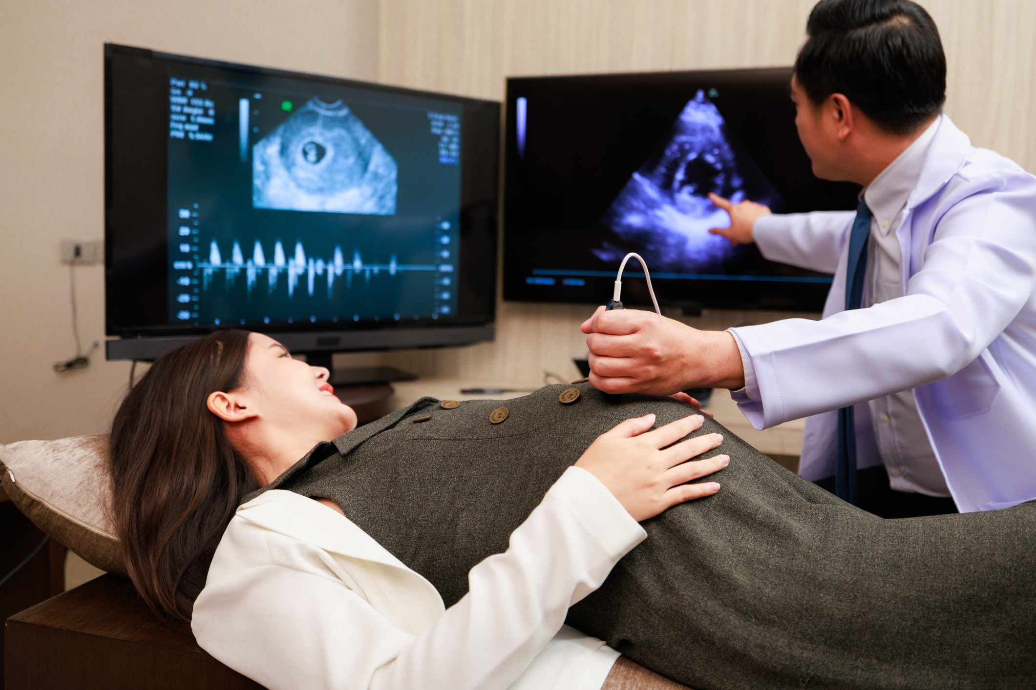
A woman lays on a table with a man giving her an ultrasound

During pregnancy, the mother and fetus exchange cells β€” a phenomenon called "microchimerism." (Image credit: Rhenizara S via Getty Images)

Checking healthy brains

To see whether these findings also applied to people without epilepsy, the researchers examined brain autopsy data from 29 individuals with no known neurodevelopmental conditions, ranging in age from 22 weeks of gestation to 40 years. They also analyzed brain tissue from three men in their late 80s and early 90s without any known brain conditions originally collected as part of an Alzheimer's disease study.

In total, the researchers found foreign cells in 25 of 32 people β€” about 78% β€” including in the brain of a man in his 90s.

The researchers suspect these foreign cells are maternal, but they couldn't confirm it because they didn't have DNA from the mothers. It is possible that the foreign cells came from a twin; an older sibling; a past pregnancy, miscarriage or abortion (in females); or, rarely, from a maternal grandmother, the team wrote in their paper.

As in the epilepsy patients, these cells had matured into several types of working brain cells. In younger brains, they most often became a specific kind of neuron, a layer 2/3 neuron, while in older brains, maternal cells were more likely to be microglia.

Given that these microchimeric cells take on very different roles, it would be interesting to know whether that diversity reflects their origins β€ͺ—‬ for example, whether they came from a mother, an older biological sibling or a maternal grandmother, said Dr. Sing Sing Way, a microchimerism researcher at Cincinnati Children's Hospital Medical Center who was not involved in the study.

The number of maternal cells declined with age, but they never completely disappeared.

Boddy said the findings mostly raise new questions. "Do we maybe need microchimeric cells to 'help out'?" she asked. "Is diversity of cells in the brain important, or is it just a byproduct of being a placental mammal?”

Because microchimeric cells appear to be common, Boddy suspects they "are doing an important job in the brain, so understanding their function could be very important for understanding healthy brain development."

By using new analytical tools to pinpoint microchimeric cells, the study "pushes the boundaries" of previous research, but future studies would benefit from larger and more uniform datasets, Way said. That would mean obtaining more brain biopsies, analyzing more cells from each sample, collecting specimens at different ages, and sampling similar brain regions across individuals to better compare the results.

This article is for informational purposes only and is not meant to offer medical advice.

Science word of the day: Neuroplasticity

18 July 2026 at 09:00

Science word of the day: Neuroplasticity

Pronunciation: Nur-roh-pla-STISS'-ih-tee

What it means: Neuroplasticity is the ability of the nervous system to adapt to the environment or to injuries by forming new connections and modifying and strengthening existing connections. Neuroplasticity can happen after an injury, such as a stroke or trauma to the brain, but it also occurs during ordinary learning. Just by reading this definition, you may be making new, sustainable brain connections.

How to use it in a sentence: Scientists used to think neuroplasticity was an ability only the the young had, but researchers are increasingly learning that aging brains can adapt throughout the lifespan, much to everyone's relief.

Can you crack our science word of the day puzzle, Chain Word?

'RNA can do things which we have never seen before': New study challenges assumptions about what RNA was up to at the dawn of life

17 July 2026 at 18:20

Scientists long thought that when RNA kick-started life on Earth 4 billion years ago, it could form only small, simple structures. But new research shows that naturally occurring RNA molecules can also adopt large, sophisticated geometries, like filaments and cages. Now, scientists wonder whether the structures were present at life's beginning.

According to an idea known as the RNA world hypothesis, RNA-based life-forms preceded modern ones that use DNA and protein. RNA, a molecular cousin of DNA, still plays roles in modern cells but does not serve as the primary genetic material. By comparison, primordial species used RNA to store genetic information and to catalyze reactions as stand-in enzymes.

Proteins eventually dominated as enzymes, perhaps because they can fold into more diverse figures than RNA can. That's because proteins are composed of 20 types of subunits, called amino acids, each with a unique structure, whereas RNA is composed of only four subunits, called nucleotides, that all adopt similar shapes.

Scientists originally thought that only proteins were varied enough to assemble into large structures, but a new paper has demonstrated that RNA β€” though more limited in its variety β€” also has the capacity to form these large configurations. The research was posted to the preprint server bioRxiv July 1 and has not been peer-reviewed yet.

"We show RNA can do things which we have never seen before," said study co-author Lin Huang, an RNA biologist at Sun Yat-Sen University in China. "It suggests that at the origin of life RNA could assemble into all kinds of shapes," he told Live Science.

Huang and his colleagues had hypothesized that RNA molecules could link together if they possessed sequences that fold into "kissing stem loops." This occurs when an RNA strand folds over on itself, forming a structure that resembles a loop in a shoelace. If loops from different RNAs bond together, or "kiss," the molecules could link up and form larger complexes, the researchers proposed.

After sifting through a bevy of RNA sequences, the researchers found a family of RNA molecules encoded by bacteriophages β€ͺ—‬ viruses that infect bacteria β€ͺ—‬ that form these loops. They purified several of these RNA molecules in the lab, allowed them to assemble in a dish, and then captured their structures using cryo-electron microscopy.

They discovered that some of the RNA molecules formed long filaments. These resembled protein-based filaments such as the cellular cytoskeleton, a scaffold that participates in many functions, including shaping and moving the cell.

Other RNA molecules assembled into cages as large as common viruses. Some of these took the form of an icosahedron β€” a 3D shape that is built from 20 equilateral triangles and resembles a soccer ball. Many viruses, including herpesviruses, package their genome into protein-based icosahedra called capsids. This raises a question: Could RNA-based capsids have packaged genomes in the RNA world?

A colorful illustration of an RNA molecule folded

RNA structures assemble into icosahedra as large as protein-based virus capsids. (Image credit: Lin Huang)

This work demonstrates that RNA had the capacity to assemble into these elaborate structures during the RNA world, Huang said, but that doesn't prove it actually happened.

"I definitely think that environmental parameters are a question," Anna Medvegy, an evolutionary biologist at EΓΆtvΓΆs LorΓ‘nd University in Hungary, told Live Science in an email. "Can these structures form in the environment in which the hypothetical RNA World existed?" said Medvegy, who was not involved in the new work.

If scientists could recreate these environmental conditions at the dawn of life, such as high temperatures and low pH, and still observe that these structures take shape, that would strengthen the theory that they could have been present in the RNA world, she said.

Although the RNA cages and filaments were large, Huang's team generated them using only short RNA strands, each no longer than 200 subunits. Medvegy said long RNAs are susceptible to breaking, so if short strands can assemble into these structures, that provides more promise that these multi-tiered molecules could have formed in the RNA world.

Another question is whether these elaborate RNA complexes currently assemble inside the bacteriophage-infected bacteria from which they were derived. So far, Huang's team has only seen these structures form in a lab dish, so they need to determine if factors inside bacteria, such as proteins, would either disrupt or enable their formation inside cells.

Beyond providing insight into life's beginnings, these RNA cages could have potential applications in biotechnology, Huang thinks. Efforts are underway to use DNA folded into "DNA origami" to deliver drugs into cells, and Huang thinks DNA's older cousin, RNA, could one day play a similar role in medicine.

'Explosive diarrhea' parasite infections in 5 states linked to Taco Bell lettuce; other cases still under investigation

17 July 2026 at 16:48

U.S. health officials have identified shredded iceberg lettuce served at Taco Bell as one source of contamination behind an outbreak of parasitic infections affecting over 1,000 people.

The outbreak has been caused by the protozoan parasite Cyclospora cayetanensis, which can enter the body when someone consumes food or water contaminated with the parasite. Since May 1, there have been 1,644 confirmed cases across Indiana, Kentucky, Ohio, West Virginia and Michigan, which is currently the hardest-hit state.

(Health officials are also investigating additional cases of Cyclospora infection in the U.S. that have yet to be linked to this specific lettuce-related outbreak. Those other potential cases remain under investigation and total over 5,100, per the Centers of Disease Control and Prevention (CDC).)

In the five-state outbreak, there have been 94 hospitalizations and no deaths to date.

Through an investigation, health officials found that all 1,644 of the sickened people with confirmed cases of Cyclospora reported eating Taco Bell in Indiana, Kentucky, Michigan, Ohio or West Virginia before becoming ill. Interviews with the affected individuals suggested that shredded lettuce might be a common source of exposure.

The Food and Drug Administration (FDA) then investigated the source of the lettuce, identifying a single supplier of iceberg lettuce from Mexico that was used by Taco Bell locations where the people ate before becoming ill. News reports have named California-based Taylor Farms as the supplier.

"FDA is working directly with the identified supplier to determine if potentially contaminated shredded iceberg lettuce remains on the market," an FDA notice states. "As part of this investigation, FDA and state partners have initiated collection of product samples for testing and analysis."

The investigation is ongoing, and additional sources of contamination or affected restaurants could possibly be identified later, the FDA noted. In the meantime, Taco Bell has said it will stop using lettuce from the implicated supplier.

"Taco Bell is working to stop use of all lettuce implicated by this investigation. Not all Taco Bell locations in these states received implicated product," the FDA statement says.

Currently, the CDC is warning consumers not to eat shredded iceberg lettuce served at Taco Bell locations in Indiana, Kentucky, Michigan, Ohio or West Virginia.

Case counts reported by individual states may differ from the FDA's and CDC's counts, because states may include both probable and confirmed cases. The FDA and CDC will update their totals once they can confirm additional cases reported to them.

Symptoms of Cyclospora infection typically emerge two to 14 days after a person consumes the parasite. These symptoms include watery diarrhea, fatigue, cramping, bloating, gas and loss of appetite, and without treatment, they can last between a few days to a month or more. The infection is treated with an antibiotic, which blocks the parasite's ability to multiply.

This article is for informational purposes only and is not meant to offer medical advice.

❌