❌

Normal view

AI Reads Tea Leaves’ Light Signatures to Measure Frost Damage

11 October 2026 at 09:12

Frost is one of the most punishing adversaries of tea cultivation. A single cold snap can scorch tender leaves, derail an entire season’s harvest, and degrade the delicate chemistry that gives tea its value. For growers and breeders, however, judging how badly a field has been damaged has long been a stubbornly subjective exercise: visual ratings lump continuous injury into crude categories, and laboratory assays destroy the very tissue they measure. A new study published in Plant Methods by Zhengtong He of Shihezi University, Zhiwei Chen of the Tea Research Institute of Shandong Academy of Agricultural Sciences, and colleagues offers a way out, pairing hyperspectral imaging with a purpose-built deep learning model to read frost damage directly from the way leaves reflect light.

The core problem the team confronted is that frost injury is not a binary event but a continuum. Leaves exposed to sub-zero temperatures suffer a cascade of overlapping stresses: photosynthetic pigments degrade, the machinery of photosynthesis falters, and cell membranes and walls break down. Any single measurement captures only a sliver of that cascade. To capture the whole picture, the researchers collected hyperspectral data together with eight key physicochemical indicators from tea plants growing across natural frost damage gradients in the field, rather than in the artificial conditions of a growth chamber. From these multidimensional physiological responses they constructed a composite measure, the tea cold stress response index, or TCSRI, which expresses frost damage severity as a continuous value instead of a coarse visual class.

Building TCSRI is a meaningful advance in itself. Traditional frost assessment often relies on ordinal scales, where an observer assigns a score of mild, moderate, or severe. Such scales throw away information and make it difficult to track recovery or compare cultivars with fine precision. By integrating several physiological dimensions into one index, the team created a target variable that reflects the plant’s actual internal state. The challenge then became predicting that index, and the individual chemical indicators behind it, from spectral data alone, without touching or destroying a single leaf.

Hyperspectral sensing is well suited to this task in principle. Across hundreds of narrow wavelength bands, plant leaves encode a wealth of information: chlorophyll absorption in the visible range, water and structural features in the near infrared. But the data are high-dimensional, with far more spectral channels than samples, a regime that invites overfitting, especially in field studies where collecting ground-truth measurements is laborious. Conventional chemometric approaches, from partial least squares regression to standard machine learning pipelines, often struggle to separate genuine physiological signals from noise, sensor drift, and the confounding effects of leaf surface properties and illumination variation.

The researchers’ answer is a model they call STAMPformer, short for spectral-prior-guided multi-view attention pooling. Its architecture reflects a deliberate design philosophy: rather than letting a neural network rediscover everything from scratch, the model is steered by what spectroscopists already know about how plants respond to stress. At its front end, a parameter-free attention convolution extraction module jointly models both the raw reflectance spectra and their first-order derivatives. Derivative spectra are a classic spectral prior: they accentuate subtle local features such as absorption peaks and shoulders that are easily drowned out in raw reflectance, while suppressing broad baseline drift and noise. By processing both views together, the module sharpens the local spectral signatures that carry physiological meaning.

On top of this extraction stage sits the second innovation, a multi-view attention pooling module. Deep networks processing spectral sequences produce long streams of features, but not all wavelengths matter equally for a given prediction. The pooling module adaptively aggregates sequence features drawn from different feature subspaces, learning to amplify the responses that are most diagnostic of cold stress and to down-weight the rest. This attention mechanism acts like a learned spotlight, and because it operates across multiple subspaces, it can capture complementary patterns that a single attention head might miss. The result is a compact representation tailored to the prediction task at hand, whether that is estimating a specific chemical indicator or the composite TCSRI.

The performance numbers are striking. Across all nine prediction tasks, spanning the eight physicochemical indicators and the TCSRI itself, STAMPformer achieved coefficients of determination above 0.87 and ratios of performance to deviation above 2.80, thresholds generally regarded as indicating reliable quantitative prediction. In other words, from a leaf’s reflectance fingerprint alone, the model could reconstruct the plant’s internal frost-stress state with an accuracy that approaches laboratory measurement, while remaining entirely non-destructive. The team did not stop at headline metrics. Ablation experiments systematically removed each proposed module to confirm that both the attention convolution extraction and the multi-view pooling contributed measurably to the gains, and paired statistical tests across multiple random seeds ensured the improvements were not artifacts of a lucky initialization.

Interpretability was a further priority, and here the study delivers something rarer than raw accuracy. Applying SHAP analysis, a technique that attributes each prediction to the contributions of individual input features, the researchers identified the wavelengths the model relied on most heavily. These key spectral regions correspond to physiologically meaningful processes: pigment degradation, photosynthetic impairment, and the disruption of cellular structure. That alignment between machine attention and plant biology matters on two fronts. It gives spectroscopists confidence that the model has learned genuine stress signatures rather than spurious correlations, and it hands plant physiologists a data-driven map of which parts of the spectrum track which facets of cold injury, potentially guiding future sensor design and breeding assays.

Crucially, the model also proved itself on data it had never seen. Independent external validation, using samples not involved in training, demonstrated that STAMPformer’s predictive ability generalizes beyond its original dataset, addressing one of the most common failure modes of machine learning in agricultural sensing. For a field where models are often tuned and tested on the same plots, this external check is an important credibility marker, suggesting the framework could be deployed across different farms, seasons, and frost events rather than remaining a laboratory curiosity.

The practical implications reach well beyond tea. An accurate, non-destructive, continuous measure of cold damage opens the door to rapid post-frost field surveys, letting growers quantify losses within hours instead of waiting for symptoms to develop or sending samples to a lab. For breeders screening for cold-tolerant germplasm, a spectral readout of TCSRI could accelerate the evaluation of hundreds of candidate lines without destructive sampling. And in the emerging vision of the smart tea garden, where drones and handheld spectrometers roam the rows, a model like STAMPformer could feed real-time frost maps into irrigation, shading, and recovery-management decisions. The authors frame their work as supporting frost monitoring, cold-tolerant germplasm evaluation, and intelligent garden management, and the architecture’s reliance on general spectral priors rather than tea-specific tricks suggests the approach could be adapted to other crops facing abiotic stress. As climate variability makes late-season frosts increasingly erratic, tools that turn light reflected from a leaf into a precise physiological diagnosis may become as essential to the tea garden as the thermometer.

Subject of Research: Hyperspectral deep learning prediction of continuous frost damage severity in tea plants

Article Title: STAMPformer: spectral-prior-guided multi-view attention learning for hyperspectral prediction of continuous frost damage severity in field-grown tea plants

Article References: He, Z., Chen, Z., Ding, Z., Wang, Z., Zhang, F., Zhao, Y., & Dong, C. (2026). STAMPformer: spectral-prior-guided multi-view attention learning for hyperspectral prediction of continuous frost damage severity in field-grown tea plants. Plant Methods. https://doi.org/10.1186/s13007-026-01597-4

Image Credits: AI Generated

DOI: 10.1186/s13007-026-01597-4

Keywords: tea plant, frost damage, hyperspectral imaging, deep learning, attention mechanism, cold stress index, non-destructive prediction, SHAP analysis, precision agriculture, spectral priors, plant physiology, smart farming


Why Metal Ions Make Fluorescent Probes Glow or Go Dark: New Design Rules Emerge

11 October 2026 at 09:08

Metal ions are everywhere in biology and the environment, and telling them apart quickly and cheaply has long been a challenge for chemists. A new open-access review published in Discover Chemistry by M. S. Ajay Kumar, Pradeep Bhaskar, and B. Anindita of Presidency University in Bengaluru takes a fresh look at one of the most versatile tools in the detection toolbox: organic fluorescent probes. Rather than simply cataloguing probes and their detection limits, the review argues that the real key to building better sensors lies in understanding why different metal ions produce fundamentally different fluorescence responses, from brilliant turn-on emission to complete quenching.

The stakes are high. Essential ions such as copper, zinc, and iron participate in enzymatic catalysis, cellular signaling, redox regulation, and gene expression, but when their balance is disrupted they can contribute to oxidative stress, metabolic disorders, and neurodegenerative diseases. Toxic heavy metals such as mercury and lead offer no biological benefit at all and are hazardous even at trace levels because of their persistence, bioaccumulation, and long biological half-lives. Conventional analytical techniques like atomic absorption spectroscopy and inductively coupled plasma mass spectrometry deliver excellent accuracy, but they demand expensive instruments, trained personnel, and complex sample preparation, making them poorly suited for real-time or on-site detection. Fluorescent probes, which convert metal-ligand interactions into measurable optical signals, promise something different: fast, simple, and biologically compatible sensing.

The central insight of the review is that probe performance depends not only on the fluorophore but on the compatibility between the target ion and the sensing mechanism. Paramagnetic ions such as Cu2+ and Fe3+ usually promote fluorescence quenching through non-radiative decay, intersystem crossing, and charge-transfer pathways. Diamagnetic Zn2+, by contrast, more readily supports turn-on emission through suppression of photoinduced electron transfer, known as PET, and chelation-induced rigidification of the molecular framework. Soft heavy metals such as Hg2+ and Pb2+ occupy a middle ground: they bind strongly to carefully selected donor atoms, yet their heavy-atom character can trigger spin-orbit coupling that quenches emission, so designers must balance strong binding against signal reliability.

The authors walk through the major photophysical mechanisms one by one. PET-based probes contain an electron-rich donor unit that quenches the excited fluorophore in the free state; when a metal ion coordinates to the donor, the electron-transfer pathway is blocked and fluorescence returns. This OFF-ON switching works beautifully for closed-shell d10 ions like Zn2+ but often fails for paramagnetic targets, where suppressing PET merely opens the door to stronger quenching routes such as ligand-to-metal charge transfer. Intramolecular charge transfer, or ICT, systems integrate the binding site directly into the conjugated framework, producing wavelength shifts that enable ratiometric sensing, a more reliable readout than simple intensity changes because it reduces dependence on probe concentration and excitation conditions. Twisted intramolecular charge transfer adds a conformational dimension: metal coordination can restrict torsional motion, suppressing non-emissive twisted states and boosting emission.

Chelation-enhanced fluorescence and its counterpart, chelation-enhanced quenching, are presented not as separate mechanisms but as opposite outcomes of the same coordination event. When metal binding locks a flexible ligand into a rigid, planar geometry, non-radiative decay is suppressed and brightness increases; when the bound ion instead introduces intersystem crossing or electron transfer, the same binding event extinguishes the signal. Excited-state intramolecular proton transfer, or ESIPT, offers another elegant route: photoexcitation drives a proton from a donor group to a nearby acceptor, generating a keto tautomer with a much larger Stokes shift. Metal ions can either disrupt the hydrogen bond required for this process or stabilize it, giving designers a tunable switch between enol and keto emission bands.

The review also highlights through-bond energy transfer, an emerging mechanism in which excitation energy moves along a covalently linked donor-acceptor backbone rather than through space as in FRET. Because TBET does not require strict spectral overlap or precise donor-acceptor distances, it can generate large pseudo-Stokes shifts and support ratiometric, multi-channel detection. A representative Hg2+-responsive cassette shows weak emission in the OFF state but strong acceptor fluorescence once metal binding enhances electronic communication between the chromophores. The authors caution, however, that TBET systems demand precise molecular architecture and often struggle with aggregation-induced quenching in water.

Computational chemistry receives a notably critical treatment. Density functional theory and time-dependent DFT are now standard in probe studies, but the authors argue their value is frequently squandered on decorative HOMO-LUMO diagrams. Used properly, ground-state DFT can confirm binding sites, coordination geometry, and structural rearrangements such as rigidification or ring opening, while TD-DFT can assign whether an observed transition is locally excited, charge-transfer, or ligand-to-metal in character. The review stresses that a smaller HOMO-LUMO gap alone proves nothing; orbital localization, charge-transfer direction, and experimental fluorescence output must all be correlated. Method choice matters too: hybrid functionals like B3LYP suit organic fluorophores, while effective core potential basis sets such as LANL2DZ are needed for heavy metals, and long-range corrected functionals like CAM-B3LYP or omega-B97XD better describe charge-transfer states.

Metal-by-metal, the review distills design lessons from dozens of representative probes. For Cu2+, quenching dominates because of its paramagnetic d9 configuration, with peptide-based probes reaching detection limits as low as 1.5 nanomolar, while rhodamine ring-opening and ratiometric platforms offer turn-on and quantitative alternatives. Iron sensing is even harder because Fe3+ is a hard, highly charged, paramagnetic d5 ion that promotes ligand-to-metal charge transfer, so most probes are turn-off or colorimetric. Zn2+ is the friendliest target: ratiometric and turn-on probes routinely achieve low-nanomolar detection in seconds, though discriminating Zn2+ from its fellow d10 ion Cd2+ remains the field’s stubborn challenge. For Hg2+ and Pb2+, the standout performers combine aggregation-induced emission, near-infrared output, and ratiometric readout, with some systems reporting detection limits down to the picomolar range in water, food, and living cells.

The application landscape is shifting rapidly from bench-top demonstrations to real-world deployment. Recent probes have been validated in tap water, groundwater, river water, milk, soil, fruit juice, and plant tissues, with recovery values typically between 90 and 102 percent. Smartphone-assisted detection, 3D-printed sensing devices, and paper test strips are turning molecular recognition into field-ready tools. In bioimaging, ratiometric probes have tracked intracellular Cu2+ in HeLa cells, an AIE probe with a 130-nanometer Stokes shift visualized Hg2+ in A549 cells and in the roots of rice plants, and a peptide-based self-assembling system detected Pb2+ in groundwater and living cells below 10 nanomolar. Nitrogen and sulfur co-doped carbon dots have even combined metal sensing with logic-gate behavior in a single platform.

Significant hurdles remain before fluorescent probes fulfill their promise. Absolute selectivity in complex matrices is still elusive, because borderline and soft ions such as Pb2+, Hg2+, and Cd2+ exhibit overlapping coordination behavior. Quantitative accuracy inside cells is compromised by probe distribution, intracellular heterogeneity, and interference from endogenous biomolecules like glutathione. Many probes lack reversibility, subcellular targeting, or the long-term stability needed for continuous monitoring. The review’s closing message is clear and likely to shape the next generation of research: successful probes must be mechanism-guided rather than structure-driven, matching the electronic personality of each metal ion with the right signal-transduction pathway, and validated not under ideal laboratory conditions but in the messy, competitive environments where metal ions actually matter.

Subject of Research: Organic fluorescent probes for detecting metal ions through mechanism-guided photophysical design and bioimaging

Article Title: Organic fluorescent metal ion probes for understanding design rules photophysical mechanisms and bioimaging applications

Article References: Ajay Kumar, M. S., Bhaskar, P., & Anindita, B. (2026). Organic fluorescent metal ion probes for understanding design rules photophysical mechanisms and bioimaging applications. Discover Chemistry, 3(1), Article 472. https://doi.org/10.1007/s44371-026-00857-5

Image Credits: AI Generated

DOI: 10.1007/s44371-026-00857-5

Keywords: fluorescent probes, metal ion sensing, photoinduced electron transfer, intramolecular charge transfer, ESIPT, chelation-enhanced fluorescence, aggregation-induced emission, DFT calculations, zinc detection, mercury detection, lead detection, bioimaging

Scientists have identified a 'protein signature' in blood that could predict healthy aging

9 October 2026 at 16:00

Two people born on the exact same day can age in totally different ways ‪—‬ one in relative health and the other with chronic disease. Now, scientists have found a key difference in the immune systems of people who are more prone to age-related conditions, and it could help explain why some people remain healthier longer.

In young people, the immune system contains many naive T cells, a type of white blood cell. Over time, exposure to pathogens and vaccinations transforms some of these T cells into memory cells, which can respond quickly to defend the body against germs.

These memory cells make different types of characteristic proteins, called granzymes, which kill infected cells. They come in different "flavors," including granzyme K and granzyme B.

In a new study, published Friday (Oct. 9) in the journal Immunity, scientists found that individuals with more memory cells that make granzyme K than cells that make granzyme B tend to have healthier aging.

In the future, the balance of cells making granzyme K and granzyme B in a person's blood could be used as a test to determine "whether you're on the right trajectory of aging or not," study co-author Maxim Artyomov, an immunologist at Washington University in St. Louis, told Live Science.

Hunting for a signature of healthy aging

In earlier research, the same team found that, in old lab mice, granzyme K-making cells accumulated in several tissues and were linked to inflammation. They also observed the same increase in granzyme K cells in blood samples from older adults.

In the new study, they uncovered another piece of the puzzle: a dynamic with granzyme B-producing cells.

They began by analyzing immune cells sampled from over 2,600 people ages 18 to 97, looking at the balance of granzyme K- and granzyme B-making cells. They found that people with a lower ratio of granzyme K to granzyme B cell populations were more likely to have autoimmune diseases or other chronic immune conditions.

In a second analysis, they looked at both immune cells and blood-borne proteins sampled from 329 people, using the data to pinpoint a "protein signature" linked to an abundance of granzyme B-making cells.

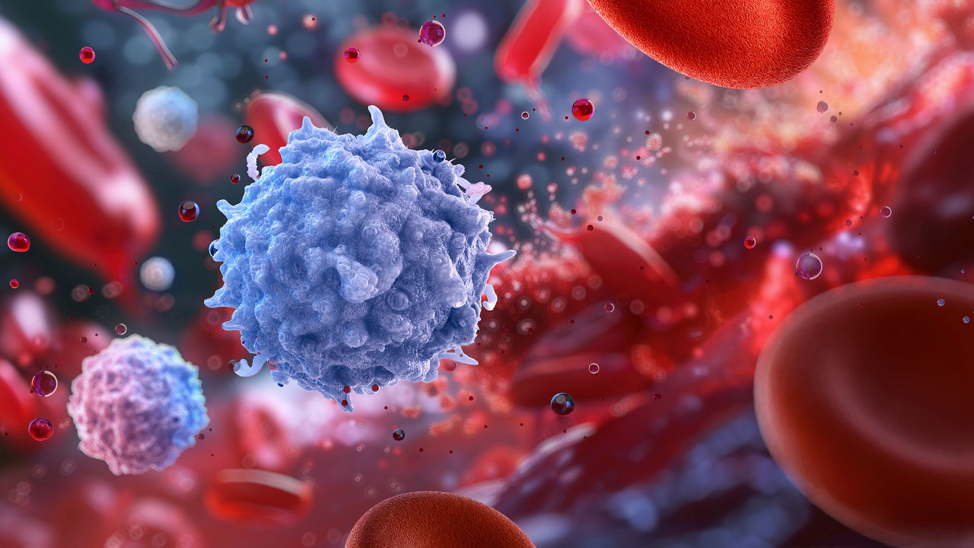
An illustration of a spiky purple sphere in the midst of blood cells

The balance of cells that make different granzymes may be important to how the immune system functions with age. (Image credit: RUSLANAS BARANAUSKAS/SCIENCE PHOTO LIBRARY via Getty Images)

They went on to hunt for this protein signature in around 48,000 participants who'd provided data to the UK Biobank, a large database of health data from U.K.-based adults. People who were predicted to have higher levels of granzyme B-making cells based on their blood proteins had worse health outcomes in the following 17 years, with a higher risk of death and of conditions like diabetes, high blood pressure, and liver and kidney disease.

The findings might point to an aspect of immune aging that wasn't previously understood.

"I think that's really exciting," Claire Gustafson, an immunologist at the Allen Institute, said of the results. "It highlights there's potentially some novel biology that we haven't really observed before," Gustafson, who was not involved in the research, told Live Science.

However, while there's a strong association between the balance of these cell populations and disease risks, Artyomov emphasized that the study does not prove that this ratio directly causes the poor health outcomes. Further studies would be needed to understand why having more granzyme B cells is linked with poorer health.

"This observation might be just a consequence, not a cause of the disease," Artyomov noted.

It's also important to note that granzyme K-making cells in mice are not the same as granzyme K-making cells in humans. In mice, they represent a more mature, specialized stage of immune cell development, whereas in humans, granzyme B cells better fit that description, he told Live Science.

That points to a key way that immunity in mice and humans is different, which was "underappreciated" before, Gustafson said. So when we're comparing these cells in mice and humans, "we're not looking at apples to apples," she said. Scientists now specifically "need to look more into humans, as it's not reflected in animal models."

In people, immune cells' features can also vary across diverse human populations, Gustafson added. We need more data across different populations to get a "true understanding of universal immune health," she said. The K-to-B ratio may be a "novel" clue that can help scientists understand healthy aging, but more data is needed to unpack why and how that ratio is linked to healthy or unhealthy aging.

"This is kind of like the first step to be able to get there," she told Live Science.

This article is for informational purposes only and is not meant to offer medical advice.

Would you trust AI to calculate your true 'biological age'?

8 October 2026 at 17:09

Scientists have recently harnessed artificial intelligence (AI) to build "aging clocks" that aim to estimate a person's true biological age.

While a person's chronological age reflects the number of years they've been alive, their biological age is intended to capture how well their bodily systems are functioning. The idea is that people with older biological ages have a higher risk of age-related diseases ‪—‬ such as cancer, heart disease or dementia ‪—‬ while younger biological ages come with less risk. Some aging clocks even aim to predict people's impending risk of death.

Many aging clocks look at epigenetic markers, meaning chemical tags that bind DNA and change how genes activate, while others measure features such as the pace and pitch of people's voices. But some scientists say we don't know how to use these aging clocks yet, arguing that the tools are useful for studying aging at a population level but not for guiding individual health decisions.

Others posit that the data the clocks are based on is too noisy, and thus, there's potential for drawing faulty conclusions about what they're measuring. They say, for example, that we don't know if the clocks are measuring the downstream effects of aging or its underlying causes. And in any case, we don't currently have any treatments to "wind back" the clocks.

What do you think? Would you use these AI tools to measure your biological age? Let us know in the poll below, and explain your reasoning and additional thoughts about the technology in the comments.

'Zombie cells' may drive aging, and scientists are devising new ways to get rid of them

7 October 2026 at 11:00

Emerging treatments aim to reverse or stall signs of aging in the body and thus cut the risk of age-related diseases. One of those signs is an accumulation of "zombie cells" — damaged cells that spew harmful substances rather than dying off and getting cleared away by the immune system.

Some scientists are designing treatments aimed at killing those zombies directly. But what if we instead helped the body's cleanup crew get rid of them?

"I think rejuvenating the immune system could rectify a lot of problems," said Roel de Maeyer, an immunologist at the University of Oxford. "We know hospitals are full of older people suffering from infections that younger people don't need to go to [the] hospital for."

The aim of these rejuvenating therapies would be to help people live healthier for longer, he told Live Science.

How do cells turn into zombies?

The immune system constantly gets rid of old, dysfunctional and dead cells to keep tissues healthy, but that cleanup gets less effective with age.

When cells die, they display proteins on their surfaces that act as "eat me" signals for immune cells, called macrophages, to engulf and destroy them. However, some cells refuse to die and instead become senescent. These zombified cells can lurk in tissues for years, releasing chemicals that drive inflammation and damage.

Senescent cells can form due to DNA damage from oxidative stress, radiation, or even normal cell division. Cells lose bits of DNA each time they make copies of themselves, and they have intricate mechanisms to decide when it becomes a "bit too dangerous to keep dividing," de Maeyer said. If a cell can no longer divide, it usually undergoes a process called programmed cell death or becomes senescent, he said.

Senescence can be beneficial in some contexts; it helps heal wounds, for example. But an accumulation of senescent cells is linked to damaging inflammation and impaired organ function.

"If there are many senescent cells in a tissue, that's usually a bad sign," de Maeyer said. The number of senescent cells goes up with age and is linked to many age-related conditions, such as heart disease and dementia.

Slaying zombies

Studies in mice have shown that using drugs to kill senescent cells can reverse inflammation and some signs of cellular aging. These drugs, called senolytics, force senescent cells to enter programmed cell death, thus reducing their numbers, said Jure Povsin, a biochemist at the Max Planck Institute of Biochemistry in Germany. This ultimately reduces inflammation and improves tissue function, he told Live Science.

But while senolytics can help rid the body of zombie cells, it's not clear why the immune system fails to eliminate senescent cells in the first place. To find answers, scientists began looking at macrophages, the cells tasked with devouring dying cells.

Research pointed to a receptor on macrophages called EP2. One study, published in July in the journal Science, found that old macrophages have a greater number of these receptors and cannot eat senescent cells effectively.

Genetically deleting the EP2 receptor in lab mice restored the macrophages' function and reduced the number of senescent cells. Compared with normal mice of the same age, the genetically tweaked mice showed less cognitive decline, muscle loss, cardiac dysfunction and systemic inflammation.

On some of these metrics, such as the ability to remember familiar objects, the modified mice were comparable to younger mice.

Gobbling up cells requires a ton of energy.

Dr. Katrin Andreasson, a neurologist at Stanford University

A problem with energy

Why is the EP2 receptor at the root of this problem? Scientists found a clue in a huge study of older adults.

A molecule called PGE2 plugs into the EP2 receptor. It's a type of prostaglandin — a molecule that helps cells communicate during stress and injury and contributes to fever, pain and swelling. Drugs such as ibuprofen reduce inflammation by blocking prostaglandin production.

In 2001, a large study uncovered a connection between these prostaglandin-blocking drugs and Alzheimer's disease: People ages 55 and older who routinely took the drugs for at least two years had a lower risk of developing Alzheimer's than nonusers did. The study could not prove that the drugs directly prevented the disease, but it added to growing evidence that inflammation related to EP2 activity plays a role in Alzheimer's.

This caught the attention of Dr. Katrin Andreasson, a neurologist at Stanford University and co-author of the recent Science study.

"I thought, 'Wow, this is interesting,'" she told Live Science. "I wondered if there's a connection."

Andreasson and colleagues had an important breakthrough in 2021: They found that aging macrophages had higher levels of the EP2 receptor and that increased EP2 activity disrupted how the cells used their energy. Instead of burning glucose to produce energy like young macrophages do, these cells stashed it away.

As a result, the cells could not do their job properly, as "gobbling up cells requires a ton of energy," Andreasson said. Blocking the EP2 receptor restored normal energy use in the macrophages. In old mice, it also led to reduced inflammation and better scores on cognitive tests, compared with mice in which EP2 activity remained high.

Making senescent cells vulnerable

In their recent study, Andreasson's team uncovered another consequence of aging: Old mice accumulated many senescent neutrophils, a type of short-lived immune cell that acts as the body's first line of defense against germs. Neutrophils normally die within days and are then cleared away, but with age, they become more prone to senescence and accumulate.

"I think that's kind of an odd concept because they're [usually] very short-lived cells," said de Maeyer, who was not involved in the Science study. "Neutrophils are inherently quite inflammatory, so wanting to clear them from tissues is very valuable."

Old mice have too much EP2 activity in their macrophages, and that means they can't get rid of these neutrophils, Andreasson's team found. That may be why deleting EP2 seems to have anti-aging effects in multiple organs, they concluded.

But increased EP2 activity is just one side of the story; some studies suggest that zombie cells actively suppress macrophages' ability to eat them. They do this by using a tricky protein called CD47.

CD47 is a "don't eat me" signal that stops macrophages from destroying cells, Povsin said. "It signals that the cell is healthy, alive and doesn't need to be eaten."

However, that signal gets amplified in senescent cells, Max Planck researchers showed in a 2023 study. That means that, when macrophages interact with senescent cells, macrophages' function becomes impaired. Afterward, they even lose the ability to engulf dying, nonsenescent cells. This allows a ton of cellular debris to accumulate.

Microscope images show red and blue stained macrophages engulf senescent cells (fluorescent green)

Top row: Macrophages (stained blue and red) incubated with young, proliferating cells will engulf dead cells and debris (green) when they encounter it. By contrast, macrophages grown in culture with senescent cells will not engulf dead cells, allowing cellular trash to accumulate (bottom row). This is because the aging cells release a strong "don't eat me" signal. (Image credit: Adapted from Schloesser et al, J. Cell Biology, 2022.)

So, rather than just blocking EP2, it could also be beneficial to reduce that "don't eat me" signal in senescent cells, this research hints. But CD47 is found on all living cells, so scientists can't just deplete its numbers across the body.

A specific enzyme modifies CD47 on senescent cells, and in the 2023 study, blocking the enzyme helped spare macrophages from the effects of CD47. Therefore, blocking the enzyme could be a more selective approach to weaken the "don't eat me" signal on senescent cells, Povsin said.

A long road to treatments for people

Translating these ideas into anti-aging treatments for humans would be a major success in the science of aging, but experts say there are significant problems to overcome.

Although blocking EP2 had consistent results in lab mice, the rodents are genetically identical and bred in controlled environments. Humans are inherently more variable in their biology and the environmental factors they're exposed to, introducing confounding factors that would need to be addressed, de Maeyer said.

Additionally, in clinical trials, EP2-blocking drugs have so far been explored as cancer treatments, but they are "underexplored" in the context of aging, he added.

Drugs targeting CD47 are in an even earlier stage of development. In the 2023 research from Max Planck, the team used both human and mouse macrophages in their experiments, but the approach has not been tested as an anti-aging treatment in living mice or humans yet.

In 2020, de Maeyer's team used a different strategy to rejuvenate macrophages in older adults. They blocked a protein called p38, whose activity was high in the macrophages of older adults. Blocking the protein helped macrophages better recognize and eat dying cells in specific tests.

In that study, the team gave adults ages 65 and up an experimental drug called losmapimod to block p38. Although it helped restore their immune cell function, it was not suitable for long-term use because it caused liver problems in later trials, de Maeyer said.

In their mouse studies, Andreasson's team was able to block EP2 without causing any negative side effects. But she cautioned that we "really need to drill down on the research and confirm all this stuff in humans."

"The next logical step," she said, "would be to figure out how to inhibit this receptor in a safe way."

This article is for informational purposes only and is not meant to offer medical advice.

Help us improve Live Science Pro: We're always trying to make our content better. Leave us feedback about Pro here.

Why Your Immune System Weakens With Age Is More Complicated Than Scientists Thought

15 September 2026 at 20:06
White Blood Cell Lymphocyte Immune SystemA new study reveals how aging changes the immune system in complex ways and identifies potential targets for healthier aging. Why does immunity weaken with age, and does that decline happen the same way throughout the body? Researchers at USC approached that question by looking across the body rather than focusing on a single organ. [...]

Dust Deal with It

15 September 2026 at 17:00

zodiacal light

Editor’s Note: Astrobites is a graduate-student-run organization that digests astrophysical literature for undergraduate students. As part of the partnership between the AAS and astrobites, we occasionally repost astrobites content here at AAS Nova. We hope you enjoy this post from astrobites; the original can be viewed at astrobites.org.

Title: The Exozodi Spectral Effect: Residual Habitable Zone Dust May Bias exoEarth Characterization
Authors: Miles H. Currie et al.
First Author’s Institution: NASA Goddard Space Flight Center
Status: Published in AJ

Figure 1: Zodiacal light as seen from Cerro Paranal. [ESO/Y. Beletsky; CC BY 4.0]

We have dust in our solar system — a huge cloud of it that permeates every corner. The dust itself is thought to be the product of many different processes including comet tails, collisions between asteroids, leftover debris from planetary formation, and gravitational collisions induced by planets. It’s small (on the order of 10–100 microns), made up of different silicates, and creates a noticeable triangle-shaped light that can be seen on the darkest nights (see Fig.1). This zodiacal dust is aptly named since the scattered light from the dust grains follows the line of the zodiac, or the ecliptic plane, across the sky. Its structure is quite complicated, with dust rings and trails and clumps near larger planetary bodies. For more information on the solar system’s zodiacal dust, you can read these bites here and here!

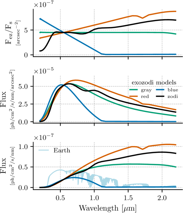
This zodiacal dust is not exclusive to the solar system. Other star systems, such as Tau Ceti and Vega, have been found to host similar clouds of interplanetary dust. This means planets imaged in these systems will also have emission from this dust mixed into the light that we observe from the planets. The Habitable Worlds Observatory (HWO) is a future space telescope mission under development by NASA. One of its primary science goals is to directly image 25 Earth-like planets in the habitable zones of nearby stars and look for signs of life (or biosignatures) in these planets’ atmospheres. Exozodiacal dust (aka exozodi) represents the biggest source of astrophysical noise that will need to be subtracted out from HWO observations of exoEarths. The authors of today’s article look at the potential impact of exozodi emission on the spectra of exoEarth atmospheres.

exozodiacal dust models

Figure 2: Different models of three “zodis” of dust observed at an inclination of 60 degrees plotted across HWO’s potential wavelength range. The gray scattering model is shown in green, the solar system’s model is shown in black, with the red and blue debris disk models shown in their respective colors. The top panel shows the flux contrast between the dust and the star. The middle panel shows the flux per area. The bottom panel shows the total flux captured with a 7.2-meter HWO with the thin blue line representing the spectrum of an Earth twin 10 parsecs (33 light-years) away for comparison. [Currie et al. 2026]

Show Me the Zodi

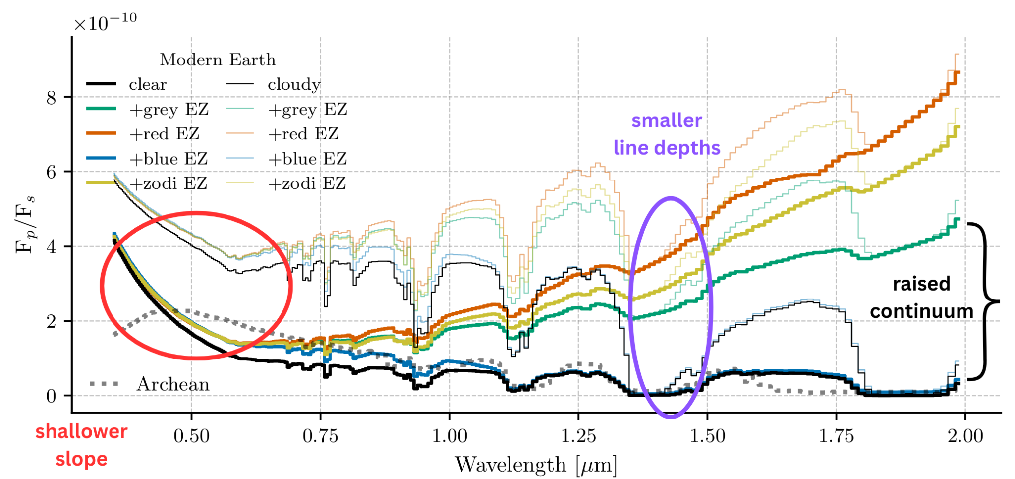
The authors first assume four different models of exozodi dust and how the dust’s emission changes with wavelength. The first model assumes a dust cloud that acts as a gray scatterer, meaning the particles scatter all wavelengths of light with no preference, and the second model’s dust acts just like the solar system’s does. However, it’s unknown whether the composition of exozodi will be the same as the solar system’s. Therefore, they adopt two more models of exozodi that act just like debris disks that have different compositions. One model is bluer in color because it is made up of smaller particles that preferentially scatter blue light, and the other model is redder in color because it is made up of bigger dust particles or different chemical compositions. Each of these models over HWO’s wavelength range can be seen in Figure 2. The authors then simulate a modern Earth atmosphere (both clear and 50% cloudy) as well as an Archean Earth atmosphere to understand how different exozodi models will affect observations of different exoplanet atmosphere conditions and compositions.

There’s Dust in My Spectra

In general, the effect that zodiacal dust has on the spectra of Earth-like planets can be broken down into two categories: its effect on the continuum and on the spectral features. The continuum is the object’s light emitted over all wavelengths and uninterrupted by spectral features. Spectral features are absorption and emission lines. These create either a dip or peak in the spectrum where atoms have either absorbed or emitted light at that particular wavelength. Lines at specific wavelengths tell us that a certain material is there, providing a list of possible chemicals that could be present in the planet’s atmosphere. For a brief summary of spectroscopy more generally, see here.

In the case of the continuum, exozodiacal emission raises the continuum, meaning the base flux of the planet is raised and absorption lines become more shallow. These effects can be seen in Figure 3, where the authors’ simulated spectra are plotted with and without zodiacal dust included. Not only is the continuum elevated for all models including zodiacal dust, notice how the dips become shallower at longer wavelengths. Because dust primarily radiates thermally, the red debris disk model nearly smooths out all dips beyond 1.25 microns! Molecules that absorb at these longer wavelengths, such as water or methane, may not be correctly identified with this type of exozodiacal emission.

simulated spectra of Earth-like planets with varying atmospheres and types of exozodiacal dust

Figure 3: Spectra of simulated modern-Earth atmospheres for clear (solid bright lines) and cloudy (solid dimmer lines) scenarios. The Archean Earth atmosphere is plotted in dotted gray. The redder exozodiacal spectrum has an elevated continuum and smoothed spectral features as opposed to the original, zodi-free spectrum plotted in black. [Currie et al. 2026, with annotations by Kaz Gary]

Additionally, the slope of the spectra between 0.25 and 0.75 microns (called the Rayleigh scattering slope) becomes shallower with zodiacal dust emission, which may bias our models toward lower-molecular-weight atmospheres. The slope at these wavelengths is dependent on the size distribution of molecules in the atmosphere, meaning it can tell us how much the average molecule in an atmosphere weighs.

From Figure 3, the effects of zodiacal dust are minimized with a bluer zodiacal dust model. This means systems with “bluer” zodiacal dust may be more optimal for HWO observations, as bluer dust does not scatter as much starlight as gray, solar system–like, or red dust. From their results, the authors recommend that the residual exozodi-to-planet flux ratio (in the V-band wavelengths) be kept to less than 10-2 in order to correctly understand the line depths (identify if a molecule is present or not) and 10-3 in order to accurately retrieve molecular abundances (understand how much of a molecule is present in the atmosphere). Dust is pesky enough here on Earth, but to find life on another planet like ours, we’ll dust have to deal with it.

Original astrobite edited by Anavi Uppal.

About the author, Kaz Gary:

I am a fourth-year PhD candidate at The Ohio State University with a passion for planets. My current work focuses on modeling exoplanet observations for the Habitable Worlds Observatory and understanding planetary atmospheres. Outside of research, I help develop planetarium shows and love all forms of science communication. In my free time, I enjoy playing tabletop RPGs, painting, watching terrible reality TV, and hanging out with my pet hedgehog.

The post Dust Deal with It appeared first on AAS Nova.

Could a Decades-Old Diabetes Drug Help the Body Age More Slowly?

14 September 2026 at 15:11
Metformin Diabetes Medicine TabletA familiar diabetes pill could hold the power to slow the biological forces that leave the body increasingly vulnerable to disease as it ages. What if a drug already prescribed to millions of people could do more than control blood sugar? For years, researchers have wondered whether metformin, one of the world’s most widely used [...]

Harnessing sunlight to fight bacteria

14 September 2026 at 12:00

Billions of people still suffer from hunger and food insecurity worldwide. Meanwhile, microbial contamination causes a third of global food waste annually, resulting in major economic losses and illnesses. Although antibiotics and pesticides help control contamination, their excessive use can promote bacterial resistance or leave toxic residues. 

One alternative uses materials that generate highly reactive forms of oxygen when exposed to light, called photocatalysts, to prolong food freshness. These molecules kill bacteria by damaging their cell components in a process called photodynamic inactivation or PDI. However, current PDI systems rely on energy-intensive lasers and long exposure times because the bacteria’s protective cell membranes block reactive oxygen molecules. As a result, the systems don’t kill bacteria effectively and risk overheating food, affecting its freshness and nutritional value.

Scientists at Zhejiang University in China thought that it would be possible to achieve a PDI system using accessible, free sunlight with the right photocatalyst. Moreover, they also wanted to integrate this photocatalyst into food packaging to combat microbes during storage. 

To make the photocatalyst, the researchers mixed water with a glue-like plant fiber that provides structural support, called pectin, and a chemical from plant seeds that supplies phosphorus, called phytic acid. Then they heated the mixture in a sealed reactor at 220ºC (428ºF) for 5 hours to form tiny structures composed of a carbon core surrounded by a phosphorus-rich shell called phosphorus-modified carbonized polymer dots or PCDs.

To incorporate the PCDs into film packaging, the researchers dissolved equal amounts of a plant fiber from konjac, called glucomannan, and another plant-derived molecule, called carboxymethylcellulose, in warm water to form a network of entangled molecular chains. Then, they added 1.5 milligrams of PCD per milliliter to this mixture, which linked the chains together like molecular fasteners. Finally, they added a moisture-binding chemical called glycerin to prevent brittleness. The researchers cast this mixture onto separate Petri dishes and dried them, forming thin, plastic-like PCD films.

To test the films, the researchers placed Escherichia coli bacteria between 2 sterilized film sheets and exposed the samples to either darkness or simulated sunlight for 20 minutes. They then transferred the microbes onto agar plates and incubated them for 15 hours, allowing surviving bacteria to form visible colonies. They compared the number of bacterial colonies with those of untreated samples to calculate the percentage of bacteria the films eliminated in each environment. 

Tests under darkness showed that the films eliminated about 60% of the bacteria. The researchers attributed this result to the positive electrical charge of PCDs, which attracts the bacteria’s negatively charged cell surfaces. The electrical attraction stresses their cell membranes, interfering with essential life processes. This result suggested that the films use a separate charge-based antibacterial mechanism apart from PDI, which requires light to work.

Under simulated sunlight, the films eliminated nearly 100% of the bacteria. The researchers identified 2 principal mechanisms causing this. First, the PCDs acted as tiny heaters that generated localized heat, which stretched holes in the bacterial membranes, allowing contents to leak out of their cells through a process called photoporation. Second, the PCDs generated reactive oxygen molecules that could then penetrate the bacteria. Once inside the cells, these molecules irreversibly damage the bacteria’s internal components and genetic material through PDI. 

The researchers also investigated whether PCD films could serve as food packaging by using them to wrap fresh strawberries. Compared with conventional nonbiodegradable polyethylene packaging where visible rotting occurred, the PCD films kept the strawberries fresh for more than 4 days by suppressing bacterial growth and regulating moisture levels. When the team tested whether the films were toxic, they found that the films are safe for humans and compatible for food applications. They also buried the films in soil for 24 days and found that they degraded by 78%, demonstrating biodegradability suitable for packaging fresh foods.

The team concluded that PCD films could be a biodegradable food packaging that combines plant-based materials with multiple antibacterial strategies. The films retained antibacterial activity in darkness and became more effective under sunlight through the combined effects of photoporation and PDI. These findings demonstrated that by combining multiple mechanisms into a single material, researchers can create multifunctional food packaging that remains effective across different storage conditions.

The post Harnessing sunlight to fight bacteria appeared first on Sciworthy.

Five-Year Disease Progression in Synuclein-Positive Sporadic Parkinson’s Disease

11 September 2026 at 12:06

Parkinson’s disease has long been a diagnosis of observation and inference. Doctors watched for tremor, rigidity, and slowness of movement, and only at autopsy could the telltale clumps of alpha-synuclein protein—the pathological signature of the disease—be confirmed inside the brain. That diagnostic fog may finally be lifting. A new five-year study drawing on the landmark Parkinson’s Progression Markers Initiative (PPMI) has followed patients whose disease was defined not by their symptoms alone, but by biology: a positive cerebrospinal fluid seed amplification assay, the test that detects misfolded alpha-synuclein circulating in the nervous system. The results, published in Annals of Clinical and Translational Neurology, offer one of the clearest longitudinal pictures yet of how biologically confirmed Parkinson’s disease actually progresses in the era of modern treatment—and the findings are already reshaping how scientists think about staging, enrollment in clinical trials, and the very definition of the disease.

The research team set out with two central questions. First, what happens clinically to patients who test positive for synuclein seeding in their spinal fluid over five years of careful observation? Second, does a patient’s baseline stage on a new biological staging system—the Neuronal Synuclein Disease Integrated Staging System, or NSD-ISS—predict how quickly they will cross meaningful clinical milestones? Both questions cut to the heart of a quiet revolution in neurology. For more than a century, Parkinson’s was classified by what patients looked like in the examination room. Now, thanks to validated biomarkers, researchers can classify it by what is happening at the molecular level, potentially years before disabling symptoms emerge.

The seed amplification assay, or SAA, is the technological engine behind this shift. The test exploits a peculiar property of misfolded alpha-synuclein: it acts as a template that recruits normal, healthy synuclein proteins and forces them to misfold as well, seeding the aggregates known as Lewy bodies that riddle the brains of Parkinson’s patients. In the laboratory, a tiny sample of cerebrospinal fluid is mixed with synthetic alpha-synuclein and monitored for hours. If pathological seeds are present, the reaction accelerates into a detectable fluorescence signal. The assay has been validated across multiple international cohorts and, critically, against postmortem brain tissue, giving neurologists a window into pathology they previously could only glimpse after death. A complementary technique—detecting phosphorylated alpha-synuclein in small skin biopsies—has added a second, less invasive line of biological evidence.

Armed with these tools, two research groups have proposed frameworks for redefining Parkinson’s disease biologically. The SynNeurGe criteria classify patients by the combined presence of pathological alpha-synuclein biomarkers, neuroimaging evidence of neurodegeneration, and disease-relevant genetic variants. The Neuronal Synuclein Disease criteria take a parallel approach, defining disease by the presence of pathological synuclein as measured by a validated biomarker, with or without evidence of dopaminergic dysfunction detected through dopamine transporter imaging. The integrated staging system then arranges these biological anchors along a seven-stage ladder: Stage 0 reserved for carriers of fully penetrant mutations in the SNCA gene; Stages 1A and 1B for people with synuclein pathology but no symptoms, depending on whether dopaminergic dysfunction is present; Stages 2A and 2B for those with subtle signs that stop short of functional impairment; and Stages 3 through 6 capturing progressively severe clinical disability.

PPMI, the international observational study launched in 2010, provided the ideal laboratory for testing whether this staging framework means anything in the real world. The researchers focused on the sporadic Parkinson’s cohort: participants diagnosed within two years of enrollment who had never taken dopaminergic medication, whose examinations showed cardinal motor features, and whose dopamine transporter scans confirmed the characteristic deficit in the striatum. Crucially, the team selected only those participants who met biological NSD criteria through a positive CSF seed amplification assay, recruited before 2020 to guarantee at least five years of follow-up. This design deliberately stripped away a longstanding source of noise in Parkinson’s research: the clinical heterogeneity that arises when a “Parkinson’s” diagnosis might actually encompass unrelated neurodegenerative processes that mimic the disease but follow entirely different biological courses.

Over the five-year observation window, participants underwent an unusually thorough annual workup. Motor and non-motor function was tracked with the Movement Disorders Society Unified Parkinson’s Disease Rating Scale across all four of its parts, alongside the Hoehn and Yahr staging scale and the Schwab and England activities of daily living score. Smell was measured with the University of Pennsylvania Smell Identification Test, autonomic function with the SCOPA-AUT, mood with the Geriatric Depression Scale, and REM sleep behavior disorder risk with a dedicated screening questionnaire. Cognition was assessed with the Montreal Cognitive Assessment and, from the study’s third year onward, formal clinician diagnoses of normal cognition, mild cognitive impairment, or dementia. Medication burden was quantified as levodopa equivalent daily dose, and dopamine transporter imaging was repeated at years one, two, and four, quantified both in the putamen—the region most affected in Parkinson’s—and across the striatum as a whole.

The broad message from the five-year trajectories is one of measurable, biologically anchored progression. Patients recruited as freshly diagnosed, biologically confirmed sporadic Parkinson’s patients showed the expected decline across motor scales and dopaminergic imaging, with the earlier PPMI analysis by Simuni and colleagues having already documented significant—though modest—correlation between worsening clinical scores and falling DAT binding over five years. What the new analysis adds is the biological filter: by restricting the cohort to synuclein-seeding-positive individuals, the study reduces the contamination from look-alike conditions that has historically muddied progression estimates. When a cohort is defined by its underlying pathology rather than its outward symptoms, the resulting disease course becomes a truer reflection of what alpha-synuclein itself does to the nervous system over time.

Perhaps the most consequential findings concern prediction. If the NSD-ISS staging system is to earn its place in research clinics and, eventually, in therapeutic trials, it must do more than organize patients neatly on a page—it must forecast what comes next. The study analyzed whether a patient’s baseline stage predicted survival and the time required to reach clinically meaningful disease milestones: crossing thresholds on the clinical rating scales, advancing in Hoehn and Yahr stage, slipping in daily living independence, or developing cognitive impairment. The logic is straightforward and powerful. A patient sitting at Stage 2B—with confirmed synuclein pathology, dopaminergic dysfunction, and subtle signs but no functional impairment—should, in theory, march down the staging ladder at a predictable pace. Demonstrating that baseline stage genuinely stratifies risk would give trial designers a rational tool for enrichment, allowing them to recruit patients at the stage where a candidate drug is most likely to show benefit.

That trial-design implication is not academic. Across neurodegenerative disease research, therapeutic development is pivoting decisively toward biomarker-defined enrollment. The bitter lessons of Alzheimer’s trials—where anti-amyloid therapies only proved effective once trials recruited based on biological confirmation rather than syndrome alone—have not been lost on the Parkinson’s community. Drugs targeting alpha-synuclein directly, whether through immunotherapy, aggregation inhibition, or other mechanisms, are entering trials that increasingly require positive seed amplification assays or other biological confirmation as a gate for entry. A validated staging system that predicts five-year trajectory would allow sponsors to select participants early enough in the disease process for neuroprotective strategies to matter, while reserving later-stage patients for symptomatic interventions. The five-year PPMI data provide exactly the kind of naturalistic benchmark that such enrichment strategies demand.

The study also marks a conceptual milestone: the description of Parkinson’s disease under contemporary management. Patients diagnosed today are treated differently than those diagnosed twenty years ago, and their disease course may differ as a result. Describing outcomes in a biologically defined, prospectively observed cohort establishes a modern baseline against which future disease-modifying therapies can be judged. When an experimental drug claims to slow progression, the comparison will be against trajectories like those documented here—precise, biomarker-anchored, and free of the diagnostic uncertainty that plagued earlier natural history studies. In that sense, the paper functions simultaneously as a clinical report and as a foundation stone for the next generation of Parkinson’s trials.

What emerges from five years of watching synuclein-positive patients is a disease that can now be seen, staged, and tracked before it fully announces itself. The combination of CSF seed amplification assays, dopamine transporter imaging, and structured clinical assessment has converted a syndrome defined in the examination room into a biological disease measurable in the laboratory. If the staging system validated in this cohort continues to predict who declines fastest and who reaches milestones soonest, neurologists may one day tell a newly diagnosed patient not only what they have, but with unprecedented confidence what lies ahead—and researchers may finally test neuroprotective drugs in the early biological window where they stand the best chance of changing the story. For a disease that has resisted precise definition since James Parkinson first described it in 1817, that is a transformation worth watching.

Subject of Research: People

Article Title: Five-Year Disease Progression in Synuclein Seeding Positive Sporadic Parkinson’s Disease

Article References: Gonzalez‐Latapi, P., Gochanour, C., Choi, S. H., Cho, H., Caspell‐Garcia, C., Coffey, C., Brumm, M., Lafontant, D.-E., Xiao, Y., Tropea, T., Seibyl, J., Tanner, C., Venuto, C. S., Kieburtz, K., Chahine, L. M., Poston, K. L., Siderowf, A., Marek, K., Simuni, T., & The Parkinson's Progression Markers Initiative (2026). Five‐Year Disease Progression in Synuclein Seeding Positive Sporadic Parkinson's Disease. Annals of Clinical and Translational Neurology, 13(9), 1791-1806. https://doi.org/10.1002/acn3.70323

Image Credits: AI Generated

DOI: 10.1002/acn3.70323

Keywords: Parkinson’s disease, alpha-synuclein, seed amplification assay, Neuronal Synuclein Disease, NSD-ISS staging, PPMI, biomarkers, dopamine transporter imaging, disease progression

3D imaging techniques reveal hidden damage in soybean seeds

11 September 2026 at 11:50

Scientists in Brazil have demonstrated that a combination of three non-destructive imaging techniques, visible-spectrum (RGB) photography, X-ray radiography, and three-dimensional magnetic resonance imaging (MRI), can detect and characterize hidden damage in soybean seeds that conventional quality tests frequently miss. The study, published in Smart Agricultural Technology, comes at a critical moment: Brazil is the world’s largest soybean producer and exporter, with an estimated harvest of 166 million tons for the 2024/2025 growing season, and seed quality is increasingly under threat from the very climate extremes that help drive the country’s agricultural productivity.

The research, led by Rafael Mateus Alves and colleagues at institutions including the Seed Technology Center of Embrapa Soybean and the University of São Paulo, was funded by the São Paulo Research Foundation (FAPESP). Its central premise is deceptively simple: because seed quality is largely determined in the field, before harvest even begins, researchers and seed companies need tools that can look inside a seed without killing it. Destructive assays such as the tetrazolium viability test, which stains metabolically active tissue red, are the industry standard, but they end the life of the sample. That is a particular problem for breeding programs, where each seed represents a unique genetic combination that must survive analysis if it is to be planted.

The seed itself is a small miracle of engineering. A protective coat, derived from the ovule integuments and marked by a scar called the hilum, regulates water uptake and shields the embryo within. The embryo contains the plumule, which gives rise to the plant’s first true leaves; the apical meristem, precursor of the aerial plant; and the hypocotyl–radicle axis, which breaks through the soil and forms the primary root. Flanking this axis are the cotyledons, storage organs packed with macromolecules that are metabolized and shipped to the growing axis during germination. Damage to any of these structures, from insects, fungi, drought, heat, or rough handling during harvest, can silently reduce a seed lot’s vigor, the capacity for rapid, uniform germination that underpins successful crop establishment.

To interrogate this architecture non-invasively, the team assembled ten representative seeds from production fields, commercial lots, and storage samples, each embodying a distinct damage category: stink bug injury, purple seed stain caused by fungi, green seeds from incomplete maturation, wrinkled seeds from heat and drought stress, weathering-related coat ruptures, severe structural failure, mechanical harvest damage, and internal galleries bored by the cigarette beetle (Lasioderma serricorne) during storage. Every seed was imaged sequentially with each of the three modalities and then subjected to the tetrazolium test as a qualitative reference standard.

The technical pipeline was meticulously standardized. For visible-light imaging, seeds were mounted on acetate sheets and photographed with a Leica DMLB microscope equipped with a DFC 310FX digital camera, oriented in four standardized positions with the hilum consistently to the right. X-ray radiographs were acquired on a Faxitron MX-20 DC-12 system operating at 34 kilovolts, with a nine-second exposure and an object-to-source distance of 11.4 centimeters; uniform contrast settings were applied to all images to make damaged tissues directly comparable. The MRI work used a horizontal 2 Tesla superconducting magnet coupled to a Bruker Avance III spectrometer operating at 85.24 megahertz for proton nuclei. Before scanning, seeds were conditioned to raise their moisture content from roughly 8–10 percent (wet basis) by incubation on moistened germination paper for three hours at 25 °C, a step that ensured sufficient hydrogen signal for imaging. A three-dimensional FLASH pulse sequence, with a 3-millisecond echo time and 75-millisecond repetition time, collected data on a 128×128×128 matrix over a 12.8-millimeter field of view, yielding isotropic voxels of 100 micrometers per side. Datasets were zero-padded to 256³ before Fourier transformation and rendered in three dimensions using ImageJ with the 3Dscript plugin.

The resulting image galleries are striking. On seeds punctured by stink bugs, the dominant pest of Brazilian soybean fields, RGB images revealed darkened patches on the cotyledons and near the plumule, the visible legacy of insect feeding that lacerates tissue and injects yeasts that degrade it from within. X-ray images flagged zones of reduced radiation absorption in the same darkened regions, while MRI cross-sections exposed the depth of tissue destruction with unusual clarity, precisely because degraded tissue no longer absorbs water and therefore produces no magnetic resonance signal. The tetrazolium test confirmed that these zones lacked respiratory activity, meaning they were metabolically dead.

Not every symptom tells the same story, however, and the multimodal approach proved especially powerful at distinguishing superficial from structural damage. Seeds with purple seed stain, caused by Cercospora fungi that secrete the toxin cercosporin, showed internal structures that appeared entirely normal in both X-ray and MRI. Yet the tetrazolium test produced an unusually intense red coloration at the stained coat, indicating that fungal activity had compromised the seed coat’s ability to regulate water uptake, allowing the staining solution to penetrate more deeply into the embryo. Similarly, green seeds, which retain chlorophyll because heat and drought interrupted maturation, showed no structural anomalies under X-ray or MRI, but their membranes proved unusually permeable to the tetrazolium solution, hinting at the physiological deterioration typical of immature tissue.

Where MRI truly distinguished itself was in detecting damage invisible to every other method. One seed that appeared flawless in visible light and radiographically normal in X-ray images revealed, in its three-dimensional MRI reconstruction, internal ruptures running through the central cotyledons, the likely consequence of rapid, uneven drying that forces differential cellular expansion and eventual tissue collapse. In mechanically damaged seeds, the X-ray images showed a crack in the hypocotyl–radicle axis that the camera could not see, but MRI went further, revealing the full extent of the fissure and the displacement of the axis relative to the cotyledons. The team also documented a dramatic structural failure nicknamed the “popcorn effect,” in which alternating wetting and rapid drying cycles tear the seed coat and force the cotyledons apart, a phenomenon increasingly reported in Brazilian seed lots as late-season weather grows more erratic.

To move beyond qualitative observation, the researchers extracted grayscale histograms from each X-ray image, normalizing pixel frequencies across the full 0–255 intensity range. Two statistical descriptors, mean gray level and Shannon entropy, were then compared across damage categories using one-way ANOVA followed by Tukey’s honest significance test. The analysis yielded clear separation: undamaged seeds showed the narrowest distributions with peaks at high grayscale values, while beetle-infested seeds and wrinkled seeds displayed the broadest distributions with heavy weight in the lower gray ranges, reflecting hollowed-out tissues and sparse, deformed structures. Mean gray level differed significantly across categories, peaking in undamaged seeds and bottoming out in wrinkled ones, while Shannon entropy, a measure of histogram complexity, was highest in beetle-damaged seeds and lowest in healthy ones. Because each damage category produced a characteristic radiographic fingerprint, the authors suggest these histogram metrics could support automated, quantitative screening in commercial seed laboratories.

Each modality, the authors stress, occupies a distinct niche. RGB imaging is fast, cheap, and ideal for surface screening but blind to anything beneath the coat. X-ray radiography, already a mainstay of seed testing labs, reveals internal voids, malformations, and cavities rapidly and non-destructively, though it cannot report on metabolic viability and demands careful standardization. MRI, with its superb soft-tissue contrast and genuine three-dimensional resolution, excels at detecting subtle physiological and structural alterations, latent mechanical injury, hydration-induced ruptures, and moisture redistribution, but its cost and acquisition time currently confine it to research settings. The tetrazolium test remains valuable as a reference but is destructive and subject to interpreter bias.

The authors argue the techniques should be viewed as complementary rather than competing, and they sketch a future in which RGB and X-ray screening handle high-throughput commercial quality control while MRI resolves ambiguous or high-stakes cases, such as breeding lines and seed physiology studies. Advances in imaging hardware, automation, and artificial intelligence-based analysis, they note, could eventually fuse these modalities into scalable phenotyping workflows. For now, the message is clear: in an era when a single season of heat, drought, and hungry insects can undermine an entire seed lot, the ability to see damage in all three dimensions, without sacrificing the seed itself, may prove as transformative for agriculture as it has been for medicine.

Subject of Research: Non-destructive multimodal imaging (RGB, X-ray, and MRI) for detecting abiotic- and biotic-stress-induced damage in soybean seeds

Article Title: When damage goes deep: a multi-technique approach including 3D for soybean seed damage assessment

Article References: Alves, R. M., Teixeira, J. M., Marassi, A. G., Henning, F. A., Umburanas, R. C., Gomes-Junior, F. G., & Tannús, A. (2026). When damage goes deep: a multi-technique approach including 3D for soybean seed damage assessment. Smart Agricultural Technology, 15, Article 102523. https://doi.org/10.1016/j.atech.2026.102523

Image Credits: AI Generated

DOI: 10.1016/j.atech.2026.102523

Keywords: soybean seeds, seed quality, MRI, X-ray imaging, RGB imaging, seed vigor, multimodal imaging, stink bug damage, tetrazolium test, non-destructive testing, seed coat rupture, cigarette beetle

Strength Training May Be the Secret to Healthy Aging

10 September 2026 at 14:30
Human Health Boost Strength LongevityStrength training helps protect independence in aging by preserving the muscle needed for daily movement, balance, and resilience. Healthy aging means staying independent, moving well, and continuing to take part in daily life as the years pass. For many people, the most meaningful signs of good health are practical ones: standing up from a chair [...]

Open-source software tracks wrist bone motion in 3D and 4D CT scans

9 September 2026 at 18:20

In a development that could reshape how scientists study the mechanics of human movement, a team of biomedical engineers and computer scientists has unveiled an open-source software tool capable of tracking the tiny, intricate bones of the wrist as they glide and rotate during motion. The new tool, called Hierarchical 3D Registration, or 3DH, is a module within the SlicerAutoscoperM extension of the widely used 3DSlicer computing environment. Described in a study published in BioMedical Engineering OnLine, the software was validated against previously established methods using both sequential three-dimensional computed tomography, or 3DCT, scans of living participants performing thumb tasks and dynamic four-dimensional CT, or 4DCT, images of cadaveric wrists moving through flexion and extension. The results demonstrate that the open-source approach can match the accuracy of independent, previously published methodologies while offering researchers everywhere a free and reproducible path to quantifying skeletal kinematics.

The wrist has long been one of the most challenging joints for biomechanists to study. It contains eight small carpal bones, including the scaphoid and lunate, along with the radius of the forearm and the metacarpal bones of the hand, all articulating in a tightly packed arrangement that allows the remarkable range of motion humans rely on every day. Understanding how these bones move relative to one another, a field of study known as arthrokinematics, is essential for unraveling the causes of wrist instability, osteoarthritis, ligament injuries and the long-term outcomes of surgical repairs. Yet measuring those movements in living, moving joints has historically required either invasive techniques, labor-intensive manual analysis or proprietary software that few laboratories can access or verify.

Quantitative tracking of skeletal structures in vivo, with both efficiency and high temporal resolution, matters to a wide range of questions in biomechanical research, and the authors of the new study argue that the field has been held back by a shortage of accessible, semi-automated registration tools. Image registration, the computational process of aligning one three-dimensional image volume to another, lies at the heart of motion tracking from CT data. When a bone moves between two scans, its position and orientation change, and a registration algorithm must find the transformation that best overlays the bone’s image from one time point onto the next. Done well, this yields precise measurements of rotation and translation. Done poorly or inconsistently, it introduces errors that can swamp the small movements of carpal bones, which may shift by only a few millimeters and a few degrees during normal function.

The new 3DH module addresses this problem with a hierarchical strategy for registering bones across sequences of image volumes. The researchers presented and evaluated the software using two distinct classes of data drawn from previous studies. The first consisted of sequential 3DCT datasets of human participants performing various thumb tasks, in which the radius, the first metacarpal and the trapezium were the target bones. The second consisted of dynamic 4DCT datasets capturing simulated flexion-extension motion in cadaveric wrists, providing time-resolved, three-dimensional image volumes of the scaphoid and lunate in motion. By testing the tool on both static sequential scans and true dynamic imaging, the team was able to demonstrate that 3DH is not limited to a single imaging paradigm but can serve as a unified tracking solution across modalities.

To determine whether the new open-source tool could be trusted, the investigators compared the arthrokinematics computed with 3DH against values that had been previously calculated using independent approaches for the same 3DCT and 4DCT datasets. The statistical framework for this comparison was the Bland-Altman analysis, a standard method for assessing agreement between two measurement techniques. Rather than simply correlating the two sets of results, Bland-Altman analysis quantifies the mean bias, the systematic difference between methods, and the 95 percent limits of agreement, the range within which most individual differences between the methods are expected to fall. This approach is particularly well suited to method-comparison studies because it reveals whether a new technique introduces systematic error and how widely individual measurements might deviate from those of the established gold standard.

The performance figures reported in the study are striking. For the 3DCT data, in which the radius, first metacarpal and trapezium were tracked, the mean bias for the helical angle, a single-axis description of three-dimensional rotation, was just minus 0.06 degrees, with 95 percent limits of agreement ranging between minus 1.87 degrees and 1.74 degrees. Translation measurements fared nearly as well: the mean bias was 0.12 millimeters, with limits of agreement spanning from minus 2.23 millimeters to 2.00 millimeters. In practical terms, the open-source tool reproduced the rotational and translational measurements of the earlier methods to within roughly two degrees and two millimeters at the extremes, differences small enough to preserve the biological signal in most wrist kinematics experiments.

The dynamic 4DCT results told a similar story for the carpal bones. Tracking the scaphoid and lunate through simulated flexion-extension, 3DH produced mean biases of the Euler angle rotations, which decompose three-dimensional rotation into three orthogonal components, of less than 0.77 degrees about any direction. The widest 95 percent limits of agreement ranged between minus 7.11 degrees and 8.65 degrees. For translations, mean biases were less than 0.5 millimeters in any direction, with the widest limits of agreement ranging between minus 2.36 millimeters and 1.43 millimeters. The wider limits of agreement for rotations in the dynamic data likely reflect the added challenges of time-resolved imaging, including motion blur and the smaller, more complexly shaped carpal bones, but the near-zero mean biases indicate that the tool does not systematically drift away from established values.

Crucially, all target bones were successfully tracked in both the 3DCT and 4DCT datasets, meaning the hierarchical registration algorithm converged on correct solutions in every case tested. This robustness matters because registration algorithms can occasionally fail, locking onto incorrect alignments or diverging entirely, and such failures typically demand time-consuming manual intervention. A semi-automated tool that reliably succeeds across diverse bones, motion tasks and imaging protocols removes a significant bottleneck. Manual bone tracking in CT volume sequences can take hours per bone per trial, and the labor multiplies rapidly in studies involving multiple participants, multiple bones and dozens of motion cycles. By automating this process within a validated framework, 3DH promises to accelerate the pace at which wrist biomechanics data can be collected and analyzed.

The choice to build the module inside 3DSlicer, and specifically within the SlicerAutoscoperM extension, is a deliberate strategic decision with implications well beyond the wrist. 3DSlicer is a free, open-source platform for medical image computing and visualization with a large international user base spanning research, clinical and educational communities. By embedding 3DH in this ecosystem, the developers have made the tool immediately available to any laboratory already working with 3DSlicer, complete with the platform’s established infrastructure for loading DICOM data, segmenting anatomical structures and visualizing results. The authors note that the ability to track bones from both 3DCT and 4DCT data sources, with agreement to prior methodologies, demonstrates that the 3DH approach is a capable tool that could promote collaboration and consistency of results through open-source software.

Consistency is a quietly urgent issue in biomechanics. When different laboratories use different proprietary tools with undisclosed algorithms, it becomes difficult to distinguish true biological differences between study populations from mere methodological discrepancies. Open-source registration changes that calculus: the code can be inspected, verified, extended and reused, and results from different institutions become directly comparable because they are produced by identical algorithms. This transparency also lowers the barrier to entry for research groups in settings that cannot afford commercial licenses, potentially broadening the global community capable of conducting rigorous skeletal kinematics research.

The study itself emerged from a collaboration spanning several institutions. The team included Joseph J. Crisco, Amy M. Morton and John D. Holtgrewe of the Department of Orthopedics at The Warren Alpert Medical School of Brown University and Rhode Island Hospital; Cesar Lopez, Andrew Thoreson and Kristin D. Zhao of the Assistive and Restorative Technology Laboratory and the Department of Physical Medicine and Rehabilitation at the Mayo Clinic in Rochester, Minnesota; and Jean-Christophe Fillion-Robin and Beatriz Paniagua of Kitware Inc. in Clifton Park, New York. The involvement of Kitware, a company known for its leadership in open-source scientific computing, underscores the blend of academic rigor and software engineering expertise required to produce a research tool robust enough for widespread distribution. Funding was provided by the National Institute of Arthritis and Musculoskeletal and Skin Diseases of the National Institutes of Health under award numbers AR059185, AR078924 and AR071338.

The implications of this work extend naturally to the future of musculoskeletal imaging. As newer CT technologies, including photon-counting detector systems, deliver ever-finer spatial resolution and as dynamic 4DCT becomes more widely available, the volume of image data available for kinematic analysis will grow dramatically. Tools like 3DH, which are fast, semi-automated and validated across modalities, will be essential for converting that imaging bounty into quantitative biomechanical knowledge. For patients with wrist disorders, the downstream promise is equally tangible: better measurements of how carpal bones actually move in living joints can inform more accurate diagnoses, improved surgical planning, more realistic computational models of joint loading and ultimately better-designed treatments for conditions ranging from scapholunate instability to thumb-base arthritis. With the tool now openly available in the 3DSlicer environment, researchers around the world can begin putting it to work.

Subject of Research: Open-source kinematic tracking of the small bones of the wrist in sequential 3DCT and dynamic 4DCT images using Hierarchical 3D Registration

Article Title: Kinematic tracking of the small bones of the wrist in sequential 3DCT and dynamic 4DCT volume images using open-source Hierarchical 3D Registration, a module within SlicerAutoscoperM

Article References: Crisco, J. J., Morton, A. M., Lopez, C., Thoreson, A., Zhao, K. D., Holtgrewe, J. D., Fillion-Robin, J.-C., & Paniagua, B. (2026). Kinematic tracking of the small bones of the wrist in sequential 3DCT and dynamic 4DCT volume images using open-source Hierarchical 3D Registration, a module within SlicerAutoscoperM. BioMedical Engineering OnLine. https://doi.org/10.1186/s12938-026-01601-x

Image Credits: AI Generated

DOI: 10.1186/s12938-026-01601-x

Keywords: Computed tomography, 3DCT, 4DCT, Bone tracking, 3DH, 3DSlicer, Image registration, Wrist kinematics, Arthrokinematics, Open-source software, SlicerAutoscoperM, Bland-Altman analysis

Denisovans in southwestern China reveal their ancient subsistence strategies

9 September 2026 at 18:03

The Denisovans, the enigmatic archaic human relatives first identified from a finger bone and a handful of teeth in a Siberian cave, have long been known almost exclusively through their DNA. Now, a landmark study published in Nature has pulled back the curtain on how these ancient hominins actually lived — what they ate, how they hunted, what tools they made, and the environments they braved — through an extraordinary assemblage of fossils and artefacts from Bianfu Cave in Yunnan Province, southwestern China. The findings paint the most detailed portrait to date of Denisovan biology, behaviour and ecology in eastern Asia, and suggest that these vanished people left a deep genetic and cultural imprint on later populations across Southeast Asia and Oceania.

The research team, led by Qingfeng Ruan, Hua Li and Song Xing and their colleagues, conducted a comprehensive analysis of hominin fossils and the associated archaeological remains recovered from Bianfu Cave. The hominin fossils themselves date to roughly 167,000 to 134,000 years ago, placing them firmly in the Middle Pleistocene. But the cultural sequence preserved in the cave’s sediments is far longer, stretching from approximately 190,000 years ago to about 70,000 years ago. That span — some 120,000 years of continuous or near-continuous occupation — makes Bianfu Cave one of the longest cultural records that can most parsimoniously be attributed to Denisovans anywhere in the world.

The Denisovan identity of the fossils was established through two independent and technically demanding lines of evidence. The first involved analysis of the enamel–dentine junction, the internal boundary between the enamel cap and the underlying dentine of the teeth. This three-dimensional morphological interface acts something like a fingerprint of evolutionary relationships, revealing diagnostic traits of the tooth’s internal architecture that are invisible from the outside but characteristic of particular hominin lineages. The second line of evidence came from palaeoproteomics, the recovery and identification of ancient proteins preserved within fossilized tissues. Because proteins are far more abundant and durable than DNA, they can survive in warm, humid environments like those of southern China, where genetic material degrades rapidly. Sequencing these ancient proteins allowed the researchers to place the Bianfu hominins on the Denisovan branch of the human family tree with confidence.

The fossil collection itself is remarkable. It includes four teeth that substantially expand the known range of dental variation within the Denisovan lineage — a critical contribution, since Denisovans were previously represented by a very small number of dental specimens, making it difficult to gauge how variable their teeth could be. The new fossils also include two cranial fragments and a radius, a forearm bone, providing rare glimpses of Denisovan skull and postcranial anatomy that were previously almost entirely unknown. Together, these remains transform Denisovans from a genome and a scatter of isolated teeth into a population with recognizable skeletal biology.

Beyond the fossils, the archaeological record from Bianfu Cave offers something equally precious: a window into Denisovan daily life. The researchers reconstructed the ancient environment using faunal remains and pollen preserved in the cave deposits. These records indicate that across Marine Isotope Stages 6 through 4 — a period encompassing major glacial–interglacial climatic swings between roughly 190,000 and 57,000 years ago — the landscape around the cave was dominated by coniferous forest or a mosaic of forest and steppe. This was a cool, variable environment that Denisovans exploited successfully across tens of thousands of years, demonstrating a resilience to climatic change that adds to growing evidence of their adaptability, from the high altitudes of the Tibetan Plateau to the subtropical latitudes of mainland Southeast Asia.

The subsistence evidence is particularly striking. Analysis of the animal bones from the cave shows that its inhabitants practised specialized hunting focused on medium- to large-bodied prey. This was not opportunistic scavenging but a sustained, targeted hunting strategy — one that would have required knowledge of animal behaviour, coordinated effort and effective weaponry. Specialized big-game hunting of this kind has often been associated with modern humans and late Neanderthals in western Eurasia, so finding it in a Denisovan context more than 150,000 years ago dramatically narrows the perceived behavioural gap between these archaic lineages and our own species.

Yet the technological strategy documented at Bianfu Cave is, at first glance, surprisingly simple. The stone tool assemblage is characterized by expedient core reduction and tool production — that is, the inhabitants struck flakes from cores as needed, used them, and discarded them, rather than investing in carefully shaped, curated tools that were carried, maintained and resharpened over time. Alongside this, the Denisovans of Bianfu Cave made pervasive use of unmodified bones, employing skeletal elements of their prey directly as tools without altering them beforehand.

Taken together, the researchers interpret this pattern as an adaptive system that prioritized the exploitation of what they call object affordance — the inherent functional possibilities already present in a raw material or object — over intensive tool manufacture. A suitably shaped bone fragment could serve as a hammer, a digging implement or a scraping tool with little or no modification; a sharp flake struck from a convenient core could cut and butcher without further refinement. In the rich, resource-diverse environments of subtropical southwestern China, where raw materials and animal resources were plentiful, such a flexible, low-investment strategy may have been not a sign of simplicity but a highly efficient way of life. It suggests that Denisovans measured success not by the sophistication of their toolkit but by how skilfully they read and exploited the opportunities their landscape provided.

This behavioural portrait gains added significance when set against the wider geography of Denisovan presence. Over the past decade and a half, Denisovans have been documented across a staggering range: from the original genomic discoveries in the Altai Mountains of Siberia, first reported in 2010, to the mandible from the Tibetan Plateau described in 2019, Denisovan DNA recovered from sediments at Baishiya Karst Cave in 2020, a molar from the Annamite Chain of northern Laos announced in 2022, a male Denisovan mandible from Pleistocene Taiwan reported in 2025, and proteomic identification of the 146,000-year-old Harbin cranium, also in 2025. Bianfu Cave now anchors this story firmly in southwestern China, a region long suspected to have been a Denisovan heartland and a critical corridor for hominin movements between mainland Asia and Southeast Asia.

The study’s authors argue that the region’s Denisovans point to a substantial genetic and cultural legacy in later human populations. Modern populations in Southeast Asia and Oceania carry the highest known proportions of Denisovan ancestry anywhere in the world — a legacy of interbreeding that occurred as modern humans moved through and beyond island Southeast Asia. The new evidence from Bianfu Cave suggests that the ways of life practised by Denisovans in this region — their hunting strategies, their flexible technological approaches, their hard-won knowledge of tropical and subtropical environments — may have been part of the inheritance, alongside their genes, that helped later human populations thrive as they spread across the diverse and challenging landscapes of Asia and the Pacific.

For palaeoanthropologists, the significance of the Bianfu Cave discoveries is difficult to overstate. Denisovans have been the least anatomically documented of all known hominin groups — a lineage defined first by DNA and only slowly fleshed out with fragmentary fossils. The new teeth, cranial fragments and radius from Yunnan, combined with an unmatched 120,000-year archaeological sequence, mean that researchers can finally study Denisovans not just as a genome or a set of molecular signatures but as a living, adapting population: hunters of large game, makers and users of expedient tools, and inhabitants of conifer forests that swung between glacial cold and interglacial warmth. As analysis of the Bianfu material continues, it promises to reshape our understanding of how archaic humans lived in eastern Asia and how deeply their story is woven into our own.

Subject of Research: Denisovan fossils and archaeological remains from Bianfu Cave, Yunnan Province, southwestern China, revealing Denisovan biology, technology, subsistence strategies and palaeoecology

Article Title: Denisovans from southwestern China and their subsistence strategies

Article References: Ruan, Q., Li, H., Xing, S., Zhao, K., Liu, J., Zanolli, C., Doyon, L., Delpiano, D., Peresani, M., d’Errico, F., Demeter, F., Hublin, J.-J., Xiao, P., Wang, Y., Jia, Z., Yang, Q., Fu, Q., Sun, Q., Zhao, Y., … Chen, F. (2026). Denisovans from southwestern China and their subsistence strategies. Nature. https://doi.org/10.1038/s41586-026-10997-4

Image Credits: AI Generated

DOI: 10.1038/s41586-026-10997-4

Keywords: Denisovans, Bianfu Cave, Yunnan, palaeoproteomics, enamel–dentine junction, Middle Pleistocene, subsistence strategies, expedient tool use, object affordance, specialized hunting, Marine Isotope Stages, Southeast Asia ancestry

Cardiac Breakthrough Could Offer Lifesaving Treatment to More Patients

8 September 2026 at 22:34
Healthy Human Heart AnatomyA major trial found that implantable cardioverter-defibrillators may benefit some patients younger than 70 with mild-to-moderate heart dysfunction and cardiac scarring. A more than decade-long randomized trial suggests that implantable cardioverter-defibrillators may benefit some patients who have heart scarring and only mild-to-moderate loss of heart function, a group that currently falls outside standard treatment guidelines. [...]

GOG brings back "big box" PC games, one printable template at a time

8 September 2026 at 22:23

Those who remember PC gaming in the days before Steam may have fond memories of the "big box" packaging that made those games stand out as shelf eye candy and sought-after in-home collectibles. Today, PC gaming platform GOG is hoping to reignite some of the nostalgia for those days by adding downloadable 3D models and printable, foldable high-res packaging scans for a handful of PC games on the platform.

Thus far, the "Printable Big Box" collection on GOG is limited to just five games: Armikrog, Descent, Hitman: Codename 47, Myst, and Star Trek: 25th Anniversary. Players browsing those games on the GOG store will now see "3D Boxes" listed among the included "Goodies" before purchase. Owners of those games on GOG can download a ZIP file containing a version of the box art as an animated 3D object file (in an easy-to-open GLB format) and a "flat printable template" in PDF, SVG, and Adobe Illustrator formats. Those downloads allow for what GOG calls "a proper papercraft project for anyone who misses shelves full of PC game boxes."

The 3D model animates to show the front flap opening. Credit: GOG

"Physical media doesn’t survive the way software can—boxes get lost, crushed, thrown out," GOG writes in today's announcement. "Scanning them, and letting you print your own, is preservation in a different shape: the same mission that keeps these games running on modern PCs, applied to the objects they used to come in."

Read full article

Comments

© GOG

Depression in Later Life May Be an Early Warning Sign of Alzheimer’s

8 September 2026 at 21:24
Brain Depression Neuroscience Two SidesDepression in later life may be an early warning from the brain, appearing years before Alzheimer’s begins to affect memory and thinking A new study in JNeurosci found that faster accumulation of tau, a protein closely tied to Alzheimer’s, was associated with worsening depressive symptoms in older adults who remained cognitively healthy. Teodora Markova of [...]

Aging doesn't look the same for everyone at the molecular level — it's 'individualized and context-dependent'

4 September 2026 at 16:13

People who are the exact same chronological age can undergo very different molecular changes as they grow older, a new study finds.

"Molecular aging is dynamic and unique to each person," said study co-author Julia El-Sayed Moustafa, a computational genomics researcher at King's College London. Even if you and your friend were born the same day, the activity of some of your genes might ramp up while the same genes in your friend dial down. The same is true for metabolites, which are small molecules produced during the body's chemical processes essential for health.

Genetics, environmental exposures and daily biological rhythms may help to explain some of those differences, the researchers say.

The findings, published Thursday (Sept. 3) in the journal Science, may complicate efforts to capture aging with a single metric. For example, "biological age" scores estimate the body's true cellular and physical health, judging how much "older" it is than your chronological age. Scientists have developed "aging clocks" that use molecular markers, such as chemical tags on DNA, to estimate biological age.

But this new study suggests that "biological aging is less like a fixed score and more like a changing set of molecular trajectories," said Raghav Sehgal, an associate research scientist at the Yale School of Medicine who was not involved in the new research. "No single number can fully capture the many systems that age at different rates," he told Live Science in an email.

More than a snapshot

In the study, researchers followed 335 women ages 32 to 80 from the long-running TwinsUK cohort for eight years. The cohort includes identical and fraternal twin pairs, along with detailed health and biological data gathered to investigate how genetics and environment can influence health.

Each woman had at least three clinic visits between 2009 and 2017, with a median of six years between their first and last visits. At each visit, researchers took blood samples and measured gene activity and levels of metabolites.

"Most aging studies take only a 'snapshot,' comparing different people at one point in time," El-Sayed Moustafa said. "In this study, we have multiple measurements, so we can see how gene activity and metabolite levels change over time within the same person."

Two women with curly hair smile at the camera.

The study pulled data from the TwinsUK cohort, which in total includes over 15,000 identical and fraternal twins from across the U.K. (This is a stock image.) (Image credit: MesquitaFMS via Getty Images)

Of more than 16,000 genes and 915 metabolites analyzed, 5,061 genes and 181 metabolites changed significantly over time. Of those genes, the vast majority — 5,036 — showed a consistent increase or decrease across the group. The same was true of 45 metabolites.

Many of these genes were involved in pathways linked to immune function, metabolism and age-related conditions, including heart disease and neurodegenerative disorders.

That said, "even when most people's genes or metabolites moved in one direction, we still found groups moving the opposite way," said study co-author Kerrin Small, a professor of genomics at King's College London. So even if, on average, certain genes' activity trended in the same direction, there were individuals who broke from the pattern.

Among 136 metabolites, for example, the levels changed from person to person. A metabolite that increased in one woman could fall in another, while some showed little change over time. One consistent pattern was that each woman's overall metabolite profile also became less similar to her earlier profile.

What might shape those changes?

Several factors appeared to shape these molecular changes. Identical twins tended to have more similar gene-expression patterns than fraternal twins did, suggesting that genetics plays a strong role. Different immune cell types also showed distinct patterns of gene activity with age, suggesting age-related changes in the immune system are not uniform.

The molecular signals also varied with the time of day and season in which the blood samples were collected. About a quarter of the genes and metabolites showed seasonal shifts, including changes linked to energy production and immune activity, while up to 40% of the metabolites varied with the body's 24-hour internal clock.

The researchers also found an unexpected decline in blood levels of per- and polyfluoroalkyl substances (PFAS), including PFOA and PFOS. Often called "forever chemicals," PFAS are synthetic compounds used in a variety of products, such as nonstick cookware and food packaging. Their decline in the study was likely due to restrictions on their use in the U.K., the study authors suspect. Blood levels of PFAS were also associated with changes in some genes and metabolites, although the study could not show that the chemicals themselves caused those changes.

The team found more than 100,000 associations between genes and metabolites, suggesting that changes in certain genes go hand-in-hand with changes in specific metabolites. This highlights how interconnected the body’s biological processes are and how complex aging really is.

"I would not call aging unpredictable, but it is certainly more individualized and context-dependent," Sehgal said. Understanding these individual trajectories could one day help researchers distinguish healthy aging from molecular changes linked to disease, but that application is still a long way off.

The study was observational, included only women and relied solely on blood samples, so larger and more diverse studies are still needed to see whether these patterns hold in broader populations. The researchers now plan to track molecular changes over about 15 years, up from eight in the current study. In the future, Sehgal suggested, aging assessments could combine broad, biological-age scores with more personalized measures of immune, metabolic, brain and cardiovascular aging.

MacVoices #26197: Road to Macstock – Pat Fauquet

By: Chuck
30 June 2026 at 13:52
Pat Fauquet is the next stop onThe Road to Macstock is a conversation about her session, “Pills, Processes, and Procedures,” explaining how Apple devices can help manage medications, appointments, research, and communication with doctors. Drawing from years of health challenges and tech experience, she highlights the Apple Watch, iPhone, iPad, and Mac as practical tools for personal advocacy, caregiving, and preparedness. This might be the most important session this year. 

💾

❌