❌

Normal view

A Hidden Sleep Change May Be Tied to Alzheimer’s Memory Loss

16 September 2026 at 12:06

A night’s sleep may seem like a quiet break from the day, but the brain remains busy. While we sleep, it sorts through new information, strengthens some memories and prepares us to learn again when we wake up. That nighttime work becomes especially important as people get older. Many older adults notice that the small […]

The post A Hidden Sleep Change May Be Tied to Alzheimer’s Memory Loss appeared first on Knowridge Science Report.

18-year-old dies of measles in PA from severe neurological complication

15 September 2026 at 19:58

A fourth person in Pennsylvania has died of measles, the state health department confirmed Tuesday. The latest death was in an unvaccinated 18-year-old resident of Mifflin County.

In the announcement, the state health department also confirmed a county coroner's report over the weekend of the death of an unvaccinated 40-year-old woman in Jefferson County. The state had previously reported two deaths in Lancaster County; both were infants—a newborn boy and a 6-week-old girl, both too young to be vaccinated.

According to the Mifflin County coroner, Andrea L. Alcalde, the 18-year-old's cause of death was determined to be acute disseminated encephalomyelitis (ADEM), which is a rare but known severe complication of measles. The condition is characterized by a rapidly progressing autoimmune disorder in which aberrant inflammation from an infection strips away the protective coverings of nerve cells in the brain and spinal cord. Before measles vaccines were widely used, ADEM was commonly associated with measles, though other viral infections can also spur its development.

Read full article

Comments

© Getty | Natalya Maisheva

Scientists Find a Potential “Root” of Alzheimer’s Disease

15 September 2026 at 18:56
Degenerative Neurological Disease Dementia BrainAlzheimer’s may begin to unravel the brain when a small group of neurons switches on a receptor it was never meant to use, triggering damage that spreads from synapses to memories. Alzheimer’s disease is often defined by amyloid plaques and tau tangles, but those hallmarks are part of a much wider breakdown. Neurons become overactive, [...]

Does estrogen protect against dementia? And if so, who should take it?

14 September 2026 at 10:00

In women's reproductive years, their brains are exposed to high levels of sex hormones, and their neurons may come to depend on those hormones to function optimally. But leading up to and after menopause, the ovaries produce far less estrogen and progesterone, raising concerns that this drop in hormones may contribute to women's dementia risk.

"We think that the loss of those hormones at menopause renders those neurons more vulnerable to age-related cellular decline and pathologies like Alzheimer's," said Karyn Frick, a neuroscientist and professor of psychological and brain sciences at the University of Wisconsin-Milwaukee.

So, if falling hormone levels are a risk factor for dementia, could replacing those hormones help stave off the disease?

While the idea sounds logical on its face, studies that have looked at hormone replacement therapy (HRT) for dementia prevention have had mixed results. There are early hints that restoring estrogen during early menopause may protect brain health, but ultimately, more gold-standard clinical trials are needed to know for sure, experts told Live Science.

Support for estrogen protecting the brain

The idea that estrogen could help ward off dementia is plausible. Women are more likely to develop Alzheimer's disease than men are, and scientists think the loss of estrogen during menopause may be partly to blame, said Jennifer Bruno, an instructor of psychiatry and behavioral sciences at Stanford University.

Lab and animal studies buttress the idea that estrogen could protect brain health.

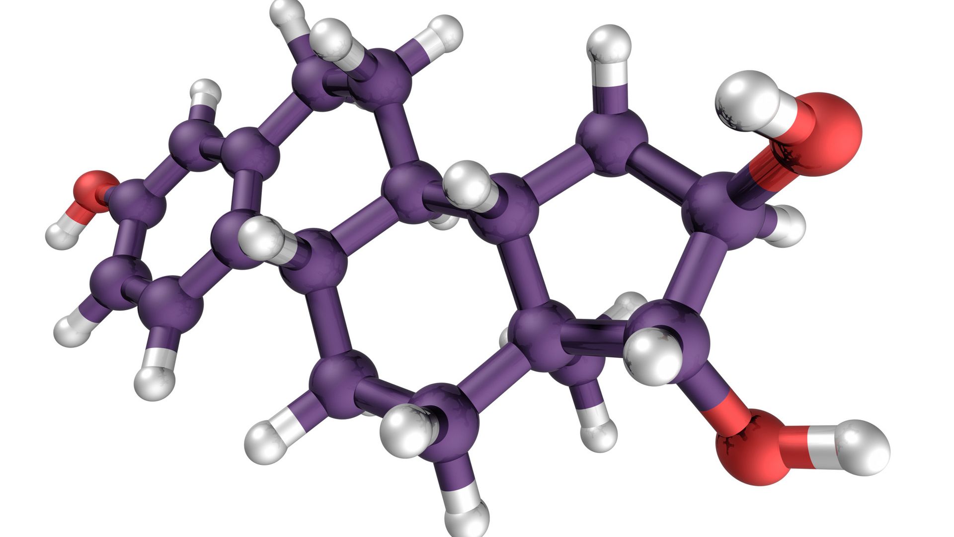
In the brain, estrogens act as neuromodulators, or chemical messengers that alter how neurons behave. Estrogen also helps neurons keep their shape. Estrogen loss in female mice triggers a drop in the number of dendritic spines ‪—‬ the small, bud-like bumps on neurons that receive chemical messages.

A molecule with purple and red bonds against a white background

An estrogen molecule. The body's levels of estrogen fluctuate in perimenopause, and they then dramatically decline and level off during and after menopause. (Image credit: theasis via Getty Images)

Researchers think that estrogen may help prevent the formation of the abnormal proteins — including one called amyloid beta — that accumulate, disrupt communication between neurons and ultimately kill cells in Alzheimer's disease. Amyloid beta is produced when a larger protein is cut the "wrong" way by an enzyme, Frick explained. Estrogen promotes the "normal" cutting of this protein, thus reducing amyloid beta, studies in lab mice show.

And estrogen may limit the buildup of a second Alzheimer's-related protein, called tau, by preventing the chemical processes that cause the tau proteins to clump together. This helps prevent the formation of tau tangles, a hallmark of Alzheimer's disease.

The hormone may also support brain health indirectly by improving sleep quality.

"It's well documented that menopausal women have difficulty sleeping, whether it's due to hot flashes or insomnia," Frick said. Estrogen can ease those nighttime hot flashes, thus helping menopausal women get more, less-fragmented sleep. Deep rest may help the brain clean out toxic proteins, while research shows sleep deficiency is linked to Alzheimer's disease.

Mixed results with estrogen-only HRT

HRT for menopause comes in two main forms: treatments that include only estrogen, and combined therapies with both estrogen and progesterone. The treatment has a complicated history.

HRT was popular in the 1990s but fell out of favor after results from the Women's Health Initiative study were published in 2002. That study identified various risks tied to both estrogen-only and combined HRT options, some of which have since been overturned by newer data.

That said, the Women's Health Initiative study found that estrogen-only HRT came with an increased risk of endometrial cancers, and later analyses have backed up this idea. That's why today, clinical guidelines recommend that women who have not gotten a hysterectomy take combined HRT, while estrogen-only options are reserved for those who have had their uterus removed. (The added progesterone counteracts estrogen's thickening effect on the uterine lining.)

For the subset of women who take estrogen-only HRT, observational data published in August showed that this form of the treatment was linked to a reduced risk of dementia. The study, published in the journal Neurology, found that women who took estrogen-only HRT were less likely to show Alzheimer's-related changes in the brain than women who didn't take any HRT.

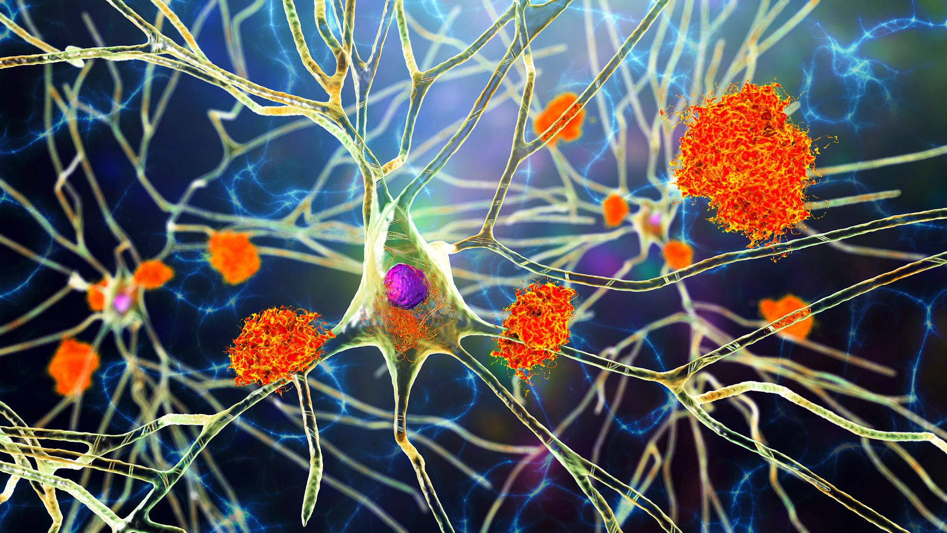
An illustration stringy neurons with orange cells on them.

Laboratory data suggest that estrogen may help prevent the formation and accumulation of abnormal proteins that are related to Alzheimer's. (Image credit: KATERYNA KON/SCIENCE PHOTO LIBRARY via Getty Images)

Estrogen-only HRT was also tied to better clinical outcomes: Treated women were less likely to be diagnosed with dementia, were less likely to show clinical and functional cognitive decline, and had better scores on memory tests, on average, compared with untreated women. These findings drew on two large, separate groups of older women, reporting data from 21,400 people in total, but they didn't include data on when each woman started HRT.

Whereas earlier studies of HRT and dementia risk relied on doctors' ratings of patients' symptoms, the Neurology study measured levels of Alzheimer's-related proteins in the brain after death. That's a more "definitive" marker of disease, said Bruno, a co-author of the study. However, the research was observational, so it can't say whether estrogen-only HRT directly causes the better brain outcomes the team observed, she stressed.

Although the Neurology study's findings are promising, they don't necessarily match the results of other studies of estrogen-only HRT and dementia. For example, a slightly larger observational study, published in 2023, found that estrogen-only HRT was associated with an increased dementia rate.

In that research, most of the women took a type of estrogen called estradiol, while Bruno's study included a greater variety of estrogens. That may have been the difference in the two studies' results, but teasing that out would require further study. Meanwhile, the conflicting findings muddy the waters of what estrogen-only HRT can do for brain health.

Does timing matter?

One big finding from the Women's Health Initiative study was that starting HRT — either estrogen-only or combined — after age 65 was associated with worse cognitive function and increased dementia risk, said Yuko Hara, director of aging and Alzheimer's prevention at the Alzheimer's Drug Discovery Foundation.

The same may not be true for people who start HRT earlier. For context, menopause starts around age 51 to 52, on average, and it's preceded by perimenopause, a transitional period when hormone levels begin to drop, typically around the mid-40s.

An observational study published in August in the journal Alzheimer's & Dementia found that starting HRT between ages 46 and 56 was linked to a reduced risk of dementia. That study did not differentiate between estrogen-only and combined treatment; it lumped all of the data together.

A close up of a person putting a patch on their arm.

Patches (pictured) are one form of systemic HRT. They deliver hormones through the skin and into the bloodstream. (Image credit: SVPhilon via Getty Images)

The protective link was especially strong in three groups: women with "surgical menopause," triggered by the removal of one or both ovaries; carriers of a certain variant of a gene called APOE that raises Alzheimer's risk; and women who had a relatively short time between their first period and menopause.

A 2024 meta-analysis of randomized controlled trials also found that HRT had a cognitive benefit for women with surgical menopause; that finding was tied mostly to estrogen-only therapies.

However, outside of that specific group, the idea of early HRT protecting brain health is far from settled.

Who might benefit, if anyone?

When it comes to using hormone replacement to protect brain health, the evidence isn't yet strong enough to recommend any kind of HRT as a way to protect the brain in all women, Hara said. Women who had one or both of their ovaries surgically removed are the exception to this rule.

"This is a distinct clinical population because the procedure can cause an abrupt, early loss of estrogen, rather than the gradual hormonal decline that occurs with natural menopause," Hara said.

For most women who have gone through natural menopause, "there is not enough evidence to recommend [HRT] specifically for preventing cognitive decline or dementia," she said.

HRT is not currently prescribed to prevent dementia — it is prescribed to ease menopause symptoms, such as hot flashes, night sweats and sleep disturbances. To settle the science on HRT and the brain, what's really needed is a well-designed clinical trial that follows women from perimenopause to postmenopause, Bruno said. That would reveal the effects of HRT on brain health and dementia risk more reliably than the studies done so far.

This article is for informational purposes only and is not meant to offer medical advice.

Help us improve Live Science Pro: We're always trying to make our content better. Leave us feedback about Pro here.

AI trained to 'think' like human pathologists may be better at spotting cancer

12 September 2026 at 13:00

Artificial intelligence (AI) algorithms that hunt for cancer may do a better job when they approach the analysis as if they were a human pathologist, a new study suggests.

Many AI systems analyze preselected regions of a tissue sample, or they split a whole pathology slide into patches of a fixed size. By contrast, a pathologist searches more dynamically, panning across the tissue, zooming in and out, and pausing over areas that raise red flags. A whole slide can contain billions of pixels, while the evidence of cancer may occupy only a tiny patch.

Study co-author Zhi Huang, an assistant professor of pathology and laboratory medicine at the University of Pennsylvania, compared the process to a search-and-rescue helicopter. "You don't start by inspecting one square meter of ground," Huang told Live Science. You scan the landscape first and then swoop in for a closer look.

In the new study, published in July in the journal Nature, Huang and colleagues demonstrated that cancer-detecting AI might work better when it takes this humanized approach.

Training AI to hunt for cancer

AI algorithms called vision language models (VLMs) struggle with the first step that Huang described — that initial, cursory scan. That's in part because many pathology AI systems learn from what pathologists leave behind at the end of that search: a labeled image pointing out where the cancer is or an official diagnosis.

Instead, the researchers trained their new AI on pathologists' search behavior. They called this approach to training "Pathology-CoT," short for "chain of thought." It turns observable actions, including where pathologists move around and zoom in on an image, into training data.

To collect the data, the team created a tool that recorded how pathologists moved around a slide and changed magnification. The raw logs, gathered from eight pathologists, were messy, as a given pathologist might drift across a slide, overshoot their intended region of focus or fiddle with magnification to adjust it to their liking.

To clean up the data, the researchers filtered out those incidental movements, focusing on moments that appeared to represent deliberate attention, such as lingering over one view or making a sustained pan. Then, they compared those regions with eye-tracking data to check that the software was capturing where pathologists were actually looking.

For each region a pathologist inspected, the VLM also drafted a short rationale explaining why the region was worth examining and what features were visible; human pathologists could then accept, edit, or reject the rationale, creating additional training data for the AI. In one example, the AI flagged a portion of a slide as potentially metastatic and suggested zooming in to look for atypical cells. Other inspected regions were flagged as healthy tissue.

Ultimately, the researchers used this training method to build a new tool called Pathology-o3. It scans a slide at low resolution, uses a model trained on pathologists' behavior to choose regions worth a closer look, then sends higher-resolution views of those regions to a VLM for analysis.

A close up of a microscope lens looking at a glass slide on the microscope mount.

Pathology-CoT trains algorithms to scan over a whole slide and then return to regions of interest for a closer look. (Image credit: Universal Images Group via Getty Images)

Putting it to the test

Huang said the goal of the new study was not to show that Pathology-o3 worked better than specialized AI models that are specifically built to detect specific types of cancer; those models are often trained disease by disease. Rather, the researchers wanted to see whether their new training approach could help a general-purpose AI navigate a pathology slide more effectively.

They compared Pathology-o3 to other general-use AI systems, such as OpenAI's o3, and asked the algorithms to examine slides containing lymph node tissue. These slides were collected from colorectal cancer cases and some contained metastatic cancer, which human pathologists had already labeled.

The algorithm correctly identified slides that were positive for cancer 100% of the time. However, of the slides it identified as positive, 15.5% were actually negative. By comparison, OpenAI o3 correctly identified slides that were positive for cancer 87.5% of the time. Of the slides it identified as positive, 53.3% were actually negative.

The researchers designed Pathology-o3 to err on the side of flagging something for another look, rather than potentially missing cancer. That might help to explain the rate of false positives, Huang said.

Whether that rate of false alarms is acceptable depends on how Pathology-o3 is used, said Mohammad Asadi, a data scientist at Stanford University who was not involved in the research. It's not precise enough for the AI to diagnose patients on its own, but it could still be useful for a system to point a human toward regions of a slide that are worth double-checking. It may be an advantage that the approach shows the pathologist a specific region to inspect rather than declaring an entire slide suspicious, he said.

The researchers tried repeating the test on an independent dataset that the algorithms hadn't seen before to see how well it worked on unfamiliar slides. Pathology-o3 correctly identified slides that were positive for cancer 97.6% of the time. Of the slides it identified as positive, 37.1% were actually negative. The finding is an example of how AI performance can change when the data source changes, even when the medical task stays the same.

Asadi explained this result suggests the system can still work with slides from a different source. But that result does not yet show that using this tool would make pathologists more accurate or efficient in practice.

Can it help pathologists?

The researchers applied their training approach to several existing VLMs, finding that the models' performance consistently improved after the training. That suggests that the navigation data from pathologists was useful across settings, Asadi said.

For Huang, that is the most important result. "The takeaway isn't our system," he said. "It's that the missing ingredient has been sitting in hospitals this whole time."

The study did not compare Pathology-o3 directly with human pathologists, but the researchers said that wasn't their aim.

"The right question isn't whether it beats a pathologist," Huang said. "It's whether a pathologist working with it catches more [cancer cases] and works faster." The current study did not address the latter question, either, but the team's next experiment is designed to test pathologists on the same cases with and without Pathology-o3, measuring what they catch and how long they take to do so.

The system's most plausible use is as a prescreening tool, Asadi said, but he stressed that the research has not yet shown that doctors who use it become faster or more accurate. Asadi wants an even tougher test: trials conducted across multiple hospitals that measure not just accuracy and speed but also pathologists' workloads. He wants the trails to assess the burden of false alarms from the AI algorithms and whether doctors recognize when the AI is wrong.

Importantly, cancer diagnoses can require information from multiple slides, stains and a patient's medical history, while the current system just reads one slide at a time. "I wouldn't claim it should diagnose on its own," Huang said.

This article is for informational purposes only and is not meant to offer medical advice.

Dr. Michael Yeadon a former VP of Pfizer Pharmaceuticals: THIS MUST STOP! Pfizer Documents Show FDA Knew of Death Risk

11 September 2026 at 23:48
Dr. Mike Yeadon warns about the alarming findings in Pfizer’s mRNA vaccine report. He cites a confidential document indicating that within 90 days of the vaccine's emergency use authorization, 1,223 deaths and 158,893 adverse reactions were reported globally, with significant implications for women's health.

DCLK1 drives fibroblast activation and lung fibrosis via Smad3 binding

10 September 2026 at 13:41

Idiopathic pulmonary fibrosis, a relentlessly progressive and fatal scarring disease of the lungs, has long frustrated clinicians with only two approved drugs that slow but cannot reverse its course. Now, a team of researchers in Taiwan has identified a surprising new player in the disease’s destructive machinery: doublecortin-like kinase 1, or DCLK1, a kinase better known for its role in cancer stem cells. Their study, published in the Journal of Biomedical Science, demonstrates that DCLK1 drives fibroblast activation in the lung and that blocking it—either genetically or with an oral inhibitor—slows fibrosis and preserves lung function in mice. The findings position DCLK1 as a promising therapeutic target in one of medicine’s most stubborn diseases.

The research, led by Lee-Yuan Lin, Wun-Hao Cheng, Chien-Huang Lin and Bing-Chang Chen of Taipei Medical University, together with collaborators at National Taiwan University Hospital and Universitas Gadjah Mada, began with a question that has lingered in the literature. Gene expression analyses had previously hinted that DCLK1 might be implicated in IPF, and earlier work reported that the kinase is markedly enhanced in the alveolar epithelial cells of patients with pulmonary fibrosis and in mice with bleomycin-induced fibrosis. But its function in fibroblast activation—the cellular engine of fibrosis—had remained unexplained.

To begin untangling that role, the team mined a publicly available single-cell RNA sequencing data set containing more than 312,000 cells from healthy lungs, IPF lungs, and lungs with chronic obstructive pulmonary disease. After stringent quality control filtering, they focused on roughly 240,000 cells from the control and IPF groups and mapped DCLK1 expression across the major lung cell lineages: epithelial, endothelial, lymphoid, myeloid, and stromal cells. The signal was unmistakable. DCLK1 was expressed across many cell populations, but the highest expression appeared in the stromal compartment, particularly in myofibroblasts—the specialized, contractile fibroblasts that churn out the extracellular matrix and stiffen lung tissue in IPF.

Human tissue told the same story. The researchers examined lung samples from eleven patients with IPF, obtained during lung transplantation or surgery, alongside fourteen nonfibrotic control samples. Histological staining with hematoxylin and eosin and Masson trichrome confirmed the expected picture of interstitial consolidation and collagen deposition in the IPF specimens. More importantly, immunohistochemistry revealed that both total DCLK1 and its phosphorylated, activated form were significantly upregulated in the fibrotic lung interstitium compared with control tissue. DCLK1, in other words, was not merely present in scarred lungs—it was switched on.

DCLK1 is an intriguing molecule to find at the center of a fibrotic process. It is a serine/threonine kinase belonging to the calcium/calmodulin-dependent kinase family, distinguished by two microtubule-binding domains. In oncology, DCLK1 overexpression has been documented in colorectal, pancreatic, gastric, renal, and hepatocellular carcinomas, where it correlates with poor prognosis, high recurrence, and metastasis, promoting tumor initiation and the epithelial–mesenchymal transition in cancer stem cells. It has also been linked to inflammatory signaling in SARS-CoV-2 infection, where inhibiting it blocks proinflammatory caspase-1/interleukin-1β signaling. The new study extends this portfolio of mischief to the fibrotic lung.

To test causality rather than mere correlation, the researchers turned to mice. They crossed animals carrying loxP sites flanking exon 3 of the DCLK1 locus with Pgk1-RFP-Cre/ERT2 transgenic mice, generating a system in which tamoxifen administration triggers global deletion of the Dclk1 gene. When these DCLK1 knockout mice and littermate controls were given intratracheal bleomycin, a chemotherapy drug used to induce pulmonary fibrosis in laboratory animals, the difference was striking. Knockout mice showed far less lung infiltration and collagen deposition, reduced numbers of cells staining positive for DCLK1 together with fibroblast surface protein and alpha-smooth muscle actin, and significantly lower levels of the profibrotic proteins fibronectin, α-SMA, and connective tissue growth factor, or CTGF.

The protective effect extended well beyond molecular markers. Using the FlexiVent FX system to measure lung mechanics, the team found that bleomycin treatment significantly reduced respiratory system compliance and pressure–volume performance and increased the normalized work of breathing in control mice—hallmarks of a stiffening, fibrotic lung. In the DCLK1 knockout animals, these functional deficits were substantially attenuated. In vivo microcomputed tomography, acquired at 34.75-micrometer voxel resolution, painted the morphological counterpart: bleomycin caused parenchymal destruction and volume loss in control lungs, while knockout mice retained lung structure and volume. Density histograms shifted toward fibrotic tissue in wild-type animals but stayed largely preserved in the knockouts, and peripheral blood neutrophil percentages, which climbed after bleomycin injury, were significantly reduced in the knockout group.

With the in vivo evidence established, the investigators dissected the molecular wiring in cultured normal human lung fibroblasts. Transforming growth factor beta, the master profibrotic cytokine, induced DCLK1 phosphorylation within 30 minutes and DCLK1 expression within 8 hours. Chromatin immunoprecipitation assays showed that after TGF-β stimulation, both Smad3—the canonical transcriptional mediator of TGF-β signaling—and the nuclear factor kappa B subunit p65 enriched at the DCLK1 promoter, indicating that TGF-β switches on DCLK1 through both Smad-dependent and noncanonical NF-κB pathways. Silencing DCLK1 with small interfering RNA then blunted the fibroblast’s profibrotic response, reducing TGF-β-induced expression of fibronectin, collagen 1A1, α-SMA, and CTGF.

The pathway, however, does not stop there. The team found that TGF-β also phosphorylated Akt, and that transfecting fibroblasts with a dominant-negative Akt construct reduced TGF-β-induced DCLK1 phosphorylation and the expression of fibronectin and CTGF—placing Akt upstream of DCLK1. Downstream, DCLK1 knockdown reduced Smad3 phosphorylation while DCLK1 overexpression enhanced it. Coimmunoprecipitation revealed a physical association between DCLK1 and Smad3, and immunofluorescence microscopy showed the two proteins colocalizing in the nucleus within 30 minutes of TGF-β treatment. A ChIP assay using a DCLK1 antibody pulled down DNA from the Smad3-binding region of the CTGF promoter, and pretreatment with the selective DCLK1 inhibitor DCLK1-IN-1 reduced TGF-β-induced CTGF expression. Together, the results sketch an Akt/DCLK1/Smad3 axis through which TGF-β drives the production of connective tissue growth factor, a potent profibrotic mediator.

The clinical payoff came in the pharmacological arm of the study. Mice receiving oral DCLK1-IN-1 at 10 milligrams per kilogram daily, beginning on day 7 after bleomycin instillation and continuing until day 28, showed visibly reduced fibrotic progression on histology, lower expression of fibronectin, α-SMA, and CTGF in lung tissue and lysates, preserved pulmonary compliance, improved pressure–volume loops, and reduced work of breathing. Serial microCT scans on days 7, 17, and 27 documented less severe fibrotic remodeling in the treated animals, with lung attenuation histograms showing a smaller fraction of dense, nonaerated voxels. Blood neutrophil counts were also lower. In short, a pill-like oral inhibitor measurably slowed the disease in a model that mirrors the human condition.

The implications are significant because the current therapeutic landscape for IPF is so thin. Only nintedanib, a tyrosine kinase inhibitor targeting growth factor receptors, and pirfenidone, which suppresses TGF-β production and activity, are approved by the US Food and Drug Administration, and both merely slow progression. The recent failure of pamrevlumab, a monoclonal antibody against CTGF, in the phase 3 ZEPHYRUS-1 trial suggested that targeting downstream fibrotic end products alone may be insufficient—pointing to the value of upstream signaling regulators such as DCLK1. Adding to the appeal, a DCLK1 inhibitor has already been shown to prevent inflammatory responses in acute lung injury, hinting at dual anti-fibrotic and anti-inflammatory potential.

The authors are careful to note limitations. DCLK1 is expressed across multiple lung cell types, and the tamoxifen-inducible knockout used here was global rather than cell-type-specific, so the contributions of fibroblasts versus epithelial, endothelial, and immune cells could not be fully separated. Human control tissue, as in most fibrosis studies, came from heterogeneous sources, including tumor-adjacent lung. The precise subcellular mechanics of the DCLK1–Smad3 interaction, whether DCLK1 inhibition can promote fibrosis resolution rather than merely slow progression, and the systemic safety of long-term DCLK1 blockade—all relevant given the kinase’s role in cancer and other processes—remain open questions.

Even so, the convergence of evidence is striking: DCLK1 is upregulated in human IPF, amplified in experimental fibrosis, mechanistically coupled to the TGF-β–Smad3 engine of fibroblast activation, and its suppression by both genetics and a small molecule preserves lung structure and function. What began as a cancer stem cell marker has now emerged, unexpectedly, as a central node in the scarring of the lung—offering fresh hope that a disease long considered irreversible may one day be stopped at its molecular source.

Subject of Research: Medicine

Article Title: DCLK1 drives fibroblast activation and lung fibrosis via Smad3 binding

Article References: Lin, L.-Y., Cheng, W.-H., Wu, Y.-C., Wen, H.-C., Hsu, H.-H., Liu, C.-H., Chen, J.-Y., Chang, W.-S., Wu, Y.-J., Wu, J.-S., Yuliani, F. S., Wang, C.-Y., Lin, C.-H., & Chen, B.-C. (2026). Doublecortin-like kinase 1 promotes fibroblast activation and fibrotic progression through Smad3 binding in idiopathic pulmonary fibrosis. Journal of Biomedical Science, 33(1), Article 52. https://doi.org/10.1186/s12929-026-01258-7

Image Credits: AI Generated

DOI: 10.1186/s12929-026-01258-7

Keywords: DCLK1 and cancer stem cell pathways in fibrosis, DCLK1 kinase in lung disease, fibroblast activation mechanisms in IPF, fibroblast-driven lung scarring, genetic suppression of DCLK1 in fibrosis models, lung fibroblast activation, lung fibrosis progression and molecular drivers, oral DCLK1 inhibitors for pulmonary fibrosis, pulmonary fibrosis, role of DCLK1 in lung tissue remodeling, Smad3 signaling pathway in fibrosis, therapeutic targeting of DCLK1

Depression in Later Life May Be an Early Warning Sign of Alzheimer’s

8 September 2026 at 21:24
Brain Depression Neuroscience Two SidesDepression in later life may be an early warning from the brain, appearing years before Alzheimer’s begins to affect memory and thinking A new study in JNeurosci found that faster accumulation of tau, a protein closely tied to Alzheimer’s, was associated with worsening depressive symptoms in older adults who remained cognitively healthy. Teodora Markova of [...]

'It's in the realm of possibility': Climate change is spurring a reemergence of yellow fever, and the US could face future threats

1 September 2026 at 10:00

Yellow fever is appearing in regions that haven't seen cases of the disease in years, largely as a result of climate change.

Historically, the virus ravaged cities across the Americas, Europe and Africa, until a vaccine was introduced in 1937 and its range shrunk significantly. But now, as climate change creates favorable conditions for virus-carrying mosquitoes, scientists are seeing outbreaks in unexpected areas.

"Yellow fever is a highly deadly disease," Dr. Joelle Ivy Rosser, an infectious-disease researcher at Stanford University, told Live Science, "so any environmental pressures that increase the likelihood of future yellow fever virus outbreaks … is highly concerning."

How is climate change contributing to this pattern, and could it get worse in the future?

How rising temperatures impact yellow fever

Yellow fever is a mosquito-borne disease caused by the Orthoflavivirus flavi virus, better known as yellow fever virus (YFV). The infection causes fever, muscle aches, headaches and vomiting. About 15% of cases become severe, with patients showing jaundiced skin and eyes, bleeding from the mouth, eyes, nose and stomach, or organ failure.

A diagram showing how yellow fever passed from mosquitos to monkeys to man

Primates, such as howler monkeys, carry the yellow fever virus, which then gets picked up mosquitoes that bite the monkeys and later spread the germ to humans. (Image credit: CDC)

Devastating yellow fever epidemics spread in the 17th to 19th centuries, before the development of a vaccine. Nowadays, an estimated 31,000 to 82,000 yellow fever deaths occur each year; severe cases have an estimated fatality rate of 20% to 50%.

Most yellow fever infections are a result of spillover from nonhuman primates; mosquitoes bite monkeys that carry the virus, such as howler monkeys, and then the infected mosquitoes bite humans. Several types of mosquitoes carry the virus, but the species Aedes aegypti is the main vector for yellow fever in cities, where high population density can make outbreaks especially severe.

Although there is an effective vaccine for yellow fever, cases have been on the rise in the past three decades. This led the World Health Organization (WHO) to set out a strategy in 2017 for eliminating yellow fever epidemics by 2026 through outbreak prevention and containment.

Climate-change-driven warming and extreme weather events may have heightened yellow fever risk over the past few decades, experts say. Rising temperatures make mosquitoes bite more because heat makes their metabolisms more active, said Dr. Seth Judson, an infectious-disease physician-scientist at UCLA. Warming also cuts down the virus's extrinsic incubation period, meaning the time it takes for the virus to replicate in a newly infected mosquito and then get passed on to another person, he told Live Science.

Weather patterns and deforestation

Changing weather patterns play a role, too. In 2017-2018, Brazil saw its first yellow fever outbreak in a city in almost a century. That outbreak, primarily in Belo Horizonte and São Paulo, was likely a result of drought, according to one study.

Extremes of weather in either direction can exacerbate mosquito-borne disease transmission

Dr. Joelle Ivy Rosser, infectious-disease researcher at Stanford University

While heavy rainfall creates a breeding ground for mosquitoes, in this case, drought pushed forest-based mosquitoes — as well as YFV carriers like marmosets and howler monkeys — closer to cities as the animals searched for water. Drier conditions can also prompt dehydrated mosquitoes to bite more as they search for fluids, study co-author Jamie Caldwell, a disease ecologist at Princeton University, told Live Science.

"Extremes of weather in either direction" — very wet or very dry — "can exacerbate mosquito-borne disease transmission," Rosser said.

Parts of Africa and South America that were considered low-risk for yellow fever have already been seeing new outbreaks, Judson said, so maps of yellow-fever-affected areas have had to be updated. In research that's been released as a preprint but not peer-reviewed yet, Judson found that yellow fever is reemerging in an area of the Colombian Andes where it hadn't been reported since 1940.

Experts are paying particular attention to the edges of cities, where urban centers meet the forest. The risk of yellow fever transmission from monkeys to humans via mosquitoes is rising at these edges due to both climate change and deforestation.

"Humans are expanding the agricultural frontier and encroaching on forests where yellow fever mosquitoes … are found," Gabriel Parra-Henao, a biologist with the WHO, told Live Science in an email. "[This] results in more people being exposed to their bites."

Similar patterns have been seen with other mosquito-borne infections, such as malaria.

A series of yellow, spherical virus particles against a white background

The yellow fever virus (pictured) is in the family Flaviviridae, which also includes the dengue and Zika viruses. (Image credit: BSIP via Getty Images)

Could yellow fever return to the U.S.?

Climate change is expected to bring yellow fever to new areas. One study predicts that, under the most severe climate change scenario, nearly a billion people could face their first exposure to the mosquitoes that can carry YFV in the next century. While that worst-case scenario may not be the most likely, the mosquitoes' ranges are still expected to expand under less-severe conditions.

Europe is predicted to face the largest increase in exposure to Ae. aegypti mosquitoes compared to the rest of the world, and the U.S. and Canada are predicted to see increases too. In the U.S., the mosquito species already lives in a number of states, including Florida, Mississippi, Arizona and New Mexico. However, warming would increase the chances of mosquitoes surviving in locations further north, such as Philadelphia and New York, which suffered major yellow fever outbreaks in the past. One model predicts Ae. aegypti could spread as far north as Chicago by 2050.

I think many people don't realize that we're still seeing significant yellow fever outbreaks.

Dr. Seth Judson, infectious-disease physician-scientist at UCLA

Although the mosquito lives in the U.S. already, yellow fever cases are exceedingly rare within the country because the virus is not present in local populations. That's because of an international campaign that the U.S. spearheaded in the 20th century, which involved researching YFV, eradicating mosquito breeding grounds, and vaccinating people widely.

"If yellow fever virus reestablished itself in our local Aedes aegypti population, then that would be very concerning," Rosser said.

One way yellow fever could return to the U.S. would be via people carrying the virus home with them after traveling to regions where the disease regularly circulates. Local mosquitoes could then catch the virus by biting those infected people, and then pass it on to others.

For local transmission to take hold, "there needs to be significant numbers of infections [passed to locals], and infections among travelers," Judson said. "But … it's in the realm of possibility."

With climate change driving resurgences of yellow fever in more places, and more regions of the U.S. becoming hospitable to the mosquitoes, that reintroduction scenario may become more likely. But some scientists think that even if North American cities saw yellow fever outbreaks, the outcome would likely not be devastating. Infrastructure in wealthier cities could be the difference between a disaster and a manageable health concern.

"There's access to medicines and vaccines; we pipe water to our homes, so we don't have standing water around," Caldwell said. "So it is a concern, but it's never going to be the next COVID."

Other experts noted that diligent surveillance of yellow fever infections can help public health officials prepare. In places where yellow fever is circulating in forest primates, forest conservation can limit contact among humans, wildlife and the mosquitoes that spread diseases among them. It's also crucial to make vaccines widely accessible to people who live in the virus's endemic regions and to travelers who visit those regions.

The WHO's yellow fever campaign is set to conclude at the end of this year, and its next steps are unclear. There's ongoing discussion of what comes next, Judson said, with questions about whether the control of yellow fever outbreaks will rely more on national strategies than on international coordination via the WHO.

Whatever happens, this is a time for heightened awareness of the virus, Judson said.

"We think of yellow fever as this historic disease," he said. "I think many people don't realize that we're still seeing significant yellow fever outbreaks. … It's really important to be aware of the risks and think about how we prepare for that."

This article is for informational purposes only and is not meant to offer medical advice.

Common DNA damage could cause neurodegenerative diseases

31 August 2026 at 12:00

Diseases that cause nerve cells in the brain to die over time are known as neurodegenerative diseases. These cells, known as neurons, are vulnerable to DNA damage because they are highly active cells that require large amounts of energy. Neural DNA damage is a normal part of aging as changes to the DNA sequence, called mutations, build up over time. 

Previous scientists have shown that neurons from people with the neurodegenerative diseases Alzheimer’s disease (AD), frontotemporal dementia (FTD), and amyotrophic lateral sclerosis (ALS) accumulate unusually high numbers of changes in the DNA sequence, called somatic mutations. However, researchers don’t know what causes the high number of mutations or whether the same process causes them in all 3 diseases. 

To answer these questions, researchers at Boston Children’s Hospital and Harvard Medical School analyzed brain tissue from people who died with and without AD, FTD, or ALS. They compared the processes that cause DNA mutations in the 3 diseases by first accessing the DNA inside individual neurons from healthy and diseased postmortem brain tissue. 

To do this, they broke apart the brain tissue and added a fluorescent marker that identified the center of each neuronal cell where DNA is housed, called its nucleus. They used a sorting machine to separate the nuclei from the rest of the cell material to access the neuron’s DNA. Using a DNA sequencing method called single-cell whole genome sequencing, the team then sequenced each neuron’s DNA to identify mutations. 

They analyzed 159 neurons from people with AD, 61 neurons from people with FTD, 77 neurons from people with ALS, and 232 neurons from healthy individuals. This allowed the team to compare the number and types of DNA mutations between diseased and control (non-diseased) neurons. The researchers found that compared to the control neurons, which typically accumulate only 200 to 300 mutations over an 80-year lifespan, neurons from all 3 diseases contained an unusually high number of 2-base-pair deletions, often exceeding 1,000 mutations per cell. 

When a biological process damages DNA, it leaves behind its own unique signature. By identifying these signatures, scientists can identify the biological process that likely caused the damage. Since these 2-base-pair deletions were so frequent in diseased neurons, the researchers wanted to know whether they were left behind by a biological process. The researchers used a machine learning algorithm to scan DNA sequences for recurring mutation patterns and identified a signature in neuronal DNA, which they named ID-4.

When the researchers compared the ID-4 signature with a database of previously characterized signatures, they found that it was associated with a DNA repair protein called TOP1. TOP1 temporarily cuts and rejoins DNA to relieve the tension that builds up during normal cellular activity. TOP1 typically detaches from the DNA after completing this process. However, if TOP1 fails to detach, it breaks one of the two DNA strands. 

To test whether abnormal TOP1 activity was breaking the DNA, the researchers isolated DNA from control and diseased neurons. They visualized the DNA on a gel, where intact DNA forms bands while broken DNA spreads out into smears. The more fragmented the DNA is, the more intense the smear appears. Using a machine that quantified the intensity of smears, the researchers found that DNA from diseased neurons produced smears that were up to 3.2 times more intense than those from control neurons. They also found that neurons with more DNA fragmentation had more ID-4 signatures. 

Based on these findings, the researchers concluded that abnormal TOP1 activity created frequent single-strand DNA breaks. They proposed that some of these breaks are repaired incorrectly, eventually leading to widespread DNA breaks in all 3 neurodegenerative diseases.

Overall, the researchers found that neurons from these 3 neurodegenerative diseases accumulated excessive DNA mutations despite differences in the disease symptoms and underlying causes. Their findings also provided evidence that abnormal TOP1 activity contributes to this mutation accumulation. Based on these findings, they stated that preventing TOP1 from becoming trapped on DNA, or improving the DNA repair process that removes trapped TOP1, could be a promising way to treat a wide range of neurodegenerative diseases.

The post Common DNA damage could cause neurodegenerative diseases appeared first on Sciworthy.

Arkansas researchers map genetic neighborhoods to identify disease-causing bacteria

11 August 2026 at 19:57

Scientists in Arkansas have developed a machine-learning approach that can identify potentially disease-causing strains of a poultry bacterium by analyzing not only which genes are present, but also how those genes are arranged across the genome. The method focuses on the “genetic neighborhoods” surrounding genes, offering a new way to distinguish harmful strains of Enterococcus cecorum from closely related strains that do not cause disease.

E. cecorum is commonly found in poultry environments, and many of its strains are harmless. However, pathogenic strains can cause arthritis, bone infections and lameness, creating serious animal-welfare problems and economic losses for poultry producers. Because pathogenic and nonpathogenic strains can share many of the same genes, identifying the genetic features associated with disease has been challenging.

The research team, led by Aranyak Goswami of the Arkansas Agricultural Experiment Station’s Center for Agricultural Data Analytics, examined the organization of genes within genomic islands. These are segments of DNA that bacteria often acquire from other microorganisms through horizontal gene transfer. Genomic islands can carry genes involved in antibiotic resistance, bacterial survival, movement of DNA and virulence, making them important regions for understanding how pathogens emerge.

Rather than treating the bacterial genome as a simple list of genetic components, the researchers analyzed it as a structured map. They investigated which genes occurred near one another, the order in which they appeared and the way groups of neighboring genes formed recurring units known as genomic-island cassettes. This architecture provided information that could be missed when genes are examined individually.

“The machine-learning model recognizes patterns in gene order much like it recognizes patterns in language,” Goswami said. In the same way that a language model can learn that certain words frequently appear together, the computational system learned that particular combinations and arrangements of bacterial genes were associated with pathogenic strains. The approach therefore captures relationships among genes rather than relying only on the presence or absence of individual DNA sequences.

For the proof-of-concept study, the researchers analyzed the genomes of 145 E. cecorum strains isolated from poultry. The dataset included 95 nonpathogenic strains and 50 pathogenic strains capable of causing illness. The analysis found that disease-associated strains were more likely to contain genomic islands enriched in genes linked to antimicrobial resistance and the transfer of genetic material between bacteria.

Those findings do not mean that every bacterium carrying such genes will necessarily cause disease. Instead, the researchers describe the system as an exploratory classification and research tool. It identifies patterns that may help scientists investigate why some bacterial lineages become harmful, while also providing clues about how resistance and virulence traits move through bacterial populations.

Current monitoring methods for E. cecorum often depend on culturing the bacterium or screening for specific genes. Such techniques remain valuable, but they may overlook broader genomic relationships. The new pipeline could complement these approaches by evaluating the wider context in which genes occur. Additional testing with larger and more geographically diverse datasets will be necessary before the method can be used for routine flock surveillance or diagnostic decision-making.

The researchers believe the strategy could eventually be adapted to other bacterial species affecting animals, humans, wildlife and plants. Goswami’s team is beginning to examine Enterococcus faecalis, a close relative of E. cecorum and a frequent cause of hospital-acquired infections in humans. They also plan to apply the pipeline to Escherichia coli and other bacteria to study how nonpathogenic lineages acquire the genomic configurations associated with disease. The study, published in Frontiers in Microbiology, demonstrates how machine learning and comparative genomics can reveal hidden signals in bacterial DNA and may help researchers track the evolution of emerging pathogens.

Subject of Research: Animals

Article Title: Genomic-island cassette architecture provides interpretable signal for exploratory classification of poultry-associated Enterococcus cecorum lineages

News Publication Date: 7-Jul-2026

Web References: https://doi.org/10.3389/fmicb.2026.1882753

References: Frontiers in Microbiology, DOI: 10.3389/fmicb.2026.1882753

Image Credits: UA University Relations photo by Chieko Hara

Keywords: artificial intelligence, machine learning, deep learning, computational biology, bacterial pathogens, Enterococcus cecorum, genomic islands, genomic architecture, antibiotic resistance, poultry disease, pathogen surveillance, comparative genomics

Any Level of Physical Activity May Slow Cognitive Decline, Study Suggests

Brain Boost Mental Focus Clarity Concept CloseCardiovascular health in midlife may influence mental function later in life. A person’s activity level and blood sugar in middle age may help shape how quickly thinking skills decline decades later. Research from UT Health San Antonio, the academic health center of The University of Texas at San Antonio, found that physical activity and better [...]

Rare Human Brain Study Reveals How an Alzheimer’s Drug May Protect the Brain

9 August 2026 at 16:27
Senior Man Staring Alzheimer’s DementiaClearing amyloid plaques may break the chain reaction that fuels brain cell destruction in Alzheimer’s disease. One Alzheimer’s brain offered researchers something they almost never get to see: a natural side-by-side experiment. In some regions, years of anti-amyloid treatment had removed most of the sticky plaques associated with Alzheimer’s disease (AD). Those areas contained little [...]

This Fingertip Patch Tracks Parkinson’s Medication Using Sweat

Fingertip View of Wearable Levodopa Monitoring DeviceA battery-free sweat sensor could make levodopa monitoring faster and more personalized for people with Parkinson’s disease. For people with Parkinson’s disease, the effect of a levodopa dose can fade abruptly, allowing movement problems to return before the next scheduled dose. Engineers and neuroscientists at the University of California San Diego have developed a soft [...]

2 people die of Cyclospora infection in Michigan

3 August 2026 at 20:10

Editor's note: This is a developing story and may be updated.

Michigan has reported two deaths in its ongoing Cyclospora outbreak, a wave of parasitic infections that has affected over 11,000 people in the state.

Since early May, 193 people in Michigan have been hospitalized with cases of Cyclospora cayetanensis infection, called cyclosporiasis. The parasite can enter the body when someone consumes food or water contaminated with the organism, and the cases in Michigan have been linked to iceberg lettuce sourced from central Mexico.

These two deaths are the first reported in the Michigan outbreak, as well as the first deaths documented in any of the ongoing Cyclospora outbreaks in the United States.

Multiple outbreaks have been detected in the country in recent months, but not all have been linked to a specific source of exposure, such as lettuce. Since May, the Centers for Disease Control and Prevention has confirmed over 6,700 cases of cyclosporiasis acquired within the U.S. (In some states, case counts are typical of what's usually seen this time of year, while in others, case counts are higher than normal.)

Regarding the deaths in Michigan, "according to medical records, both individuals had significant underlying health conditions that may have been impacted by cyclosporiasis and dehydration," the state's Department of Health and Human Services (HHS) said in a statement. "No additional information will be provided on these two cases."

Cases of cyclosporiasis range from mild to severe, with severe cases being more likely in young children, older adults, and people with weakened immune systems. Symptoms include loss of appetite, fatigue, low-grade fever, vomiting and diarrhea. The infection is treated with antibiotics, which block the parasite's ability to multiply.

In mild cases, the infection can sometimes resolve on its own, but without treatment, the symptoms can linger for over a month. Severe and prolonged infections can raise the risk of dehydration due to vomiting and diarrhea. Severe dehydration can then cause various complications, such as electrolyte imbalances and hypovolemia (severe fluid loss).

While these complications can be dangerous — causing kidney failure or shock, for instance — cyclosporiasis is not usually life-threatening. For instance, over 2,200 cases were reported in the U.S. between 2011 and 2015, but there were no associated deaths.

"Death resulting from cyclosporiasis is uncommon in the United States," Michigan HHS representatives noted in the statement.

This article is for informational purposes only and is not meant to offer medical advice.

Study Finds Hidden Cause of Sleep Issue in Alzheimer’s Disease

2 August 2026 at 12:00

Scientists have discovered that the sleep problems seen in Alzheimer’s disease may not be caused directly by harmful protein plaques as previously believed. Instead, the brain’s own immune system may be playing a much larger role, opening the door to new treatment possibilities. Sleep problems are one of the earliest and most common symptoms of […]

The post Study Finds Hidden Cause of Sleep Issue in Alzheimer’s Disease appeared first on Knowridge Science Report.

Brain-region microstructure and gene activity reveal Alzheimer’s neurodegeneration

2 August 2026 at 07:35

Alzheimer’s disease may be leaving a more precise anatomical signature in the brain than scientists once realized. A new study published in Translational Psychiatry investigates regional changes in the fine structure of brain tissue immediately beneath the cerebral cortex, known as the juxtacortical region, and links those changes to patterns of gene expression associated with neurodegeneration. The work, led by J. Zhang, L. Sun, X. Yang and colleagues, offers a combined view of Alzheimer’s biology that connects what can be detected through brain imaging with the molecular programs active inside vulnerable neural tissue.

The juxtacortical zone is a narrow but biologically important interface. It lies directly beneath the cortex, the outer layer responsible for functions including memory, language, perception and decision-making. Although much Alzheimer’s research has focused on cortical thinning, large-scale tissue loss and abnormalities in deeper brain structures, the tissue bordering the cortex may reveal earlier or more regionally specific changes. Its organization includes densely packed axons, dendrites, neurons, glial cells and blood vessels, all of which can be affected as the disease progresses. Subtle disruption in this area could therefore provide clues before dramatic anatomical damage becomes visible.

The study’s central idea is that Alzheimer’s disease does not alter every brain region in the same way. Instead, neurodegeneration follows a complex geography shaped by cellular composition, connectivity, metabolism and genetic vulnerability. By examining regional juxtacortical microstructural changes, the researchers seek to move beyond a simple question of whether tissue has shrunk. Microstructure refers to the organization of brain components at scales smaller than conventional anatomical imaging can directly resolve. Variations in water diffusion, tissue organization and local architecture can serve as indirect markers of changes to axons, membranes, extracellular space and other microscopic features.

This regional perspective matters because the same imaging signal can arise from different biological processes. A change in tissue organization may reflect loss of neuronal connections, damage to myelinated fibers, altered glial activity, inflammation or shifts in the balance of cellular compartments. Imaging alone can reveal where abnormalities are concentrated, but it cannot always explain why they occur. The researchers therefore paired their investigation of juxtacortical structure with gene-expression information, creating a bridge between regional anatomy and molecular biology. That combination can help identify which biological pathways may underlie the imaging patterns seen in Alzheimer’s disease.

Gene expression describes the activity of genes within cells and tissues: which genetic instructions are being read, and at what levels. In the Alzheimer’s brain, these molecular signatures can point toward processes such as synaptic dysfunction, immune activation, impaired energy production, protein handling and cellular stress. Mapping gene-expression patterns onto regions showing microstructural alterations may reveal whether vulnerable areas share particular molecular characteristics. It may also help distinguish tissue changes related directly to neurodegeneration from those associated with aging or general brain atrophy. The result is a more layered portrait of disease, linking microscopic organization to the biological machinery that maintains it.

The findings described by the paper’s title suggest that these two forms of evidence—regional microstructure and gene activity—provide complementary insights into Alzheimer’s pathology. Rather than treating the brain as a uniform organ undergoing gradual damage, the research emphasizes a mosaic of vulnerability. Some areas may show distinctive structural disruption alongside gene-expression profiles associated with neuronal injury or altered support-cell function, while other regions may be relatively resilient. Such differences could help explain why symptoms vary between individuals and why memory, language, attention and other abilities can deteriorate along different trajectories.

The work could also influence how scientists think about biomarkers. A biomarker is a measurable biological feature that helps indicate disease presence, progression or response to treatment. Traditional markers of Alzheimer’s include abnormal amyloid and tau proteins, brain volume loss and changes in metabolism. Juxtacortical microstructural measures, especially when interpreted alongside molecular data, could eventually add regional detail to this toolkit. They might help researchers identify tissue at risk, track the spread of degeneration or evaluate whether an experimental therapy is protecting neural architecture even before clinical improvement becomes obvious.

However, the study should not be interpreted as proof that a single imaging signature can diagnose Alzheimer’s disease or predict an individual’s future with certainty. Microstructural measurements are indirect, and gene-expression data often come from specialized datasets that may not capture every cell type or every stage of disease. Differences in age, disease severity, medication, vascular health and individual brain organization can also influence results. The strength of the approach lies not in one isolated measurement, but in the convergence of regional imaging and molecular evidence. Together, these methods can generate testable hypotheses about how and where neurodegeneration unfolds.

The broader message is that Alzheimer’s research is moving toward increasingly integrated maps of the human brain. The study by Zhang, Sun, Yang and colleagues presents the juxtacortical region as a potentially informative frontier, where microscopic tissue organization and gene regulation intersect. If future research confirms these relationships in larger and more diverse groups, the approach could sharpen disease staging and improve the design of targeted therapies. For now, the findings add weight to a crucial principle in neuroscience: understanding Alzheimer’s requires knowing not only which brain regions are damaged, but also how their cellular architecture and molecular identity make them vulnerable.

Subject of Research: Regional juxtacortical microstructural changes and gene expression in Alzheimer’s disease

Article Title: Regional juxtacortical microstructural changes and gene expression provide insights into neurodegeneration in Alzheimer’s disease

Article References: Zhang, J., Sun, L., Yang, X. et al. “Regional juxtacortical microstructural changes and gene expression provide insights into neurodegeneration in Alzheimer’s disease.” Translational Psychiatry (2026). https://doi.org/10.1038/s41398-026-04292-z

Image Credits: AI Generated

DOI: https://doi.org/10.1038/s41398-026-04292-z

Keywords: Alzheimer’s disease, neurodegeneration, juxtacortical microstructure, gene expression, brain imaging, neuroscience, biomarkers, translational psychiatry

❌