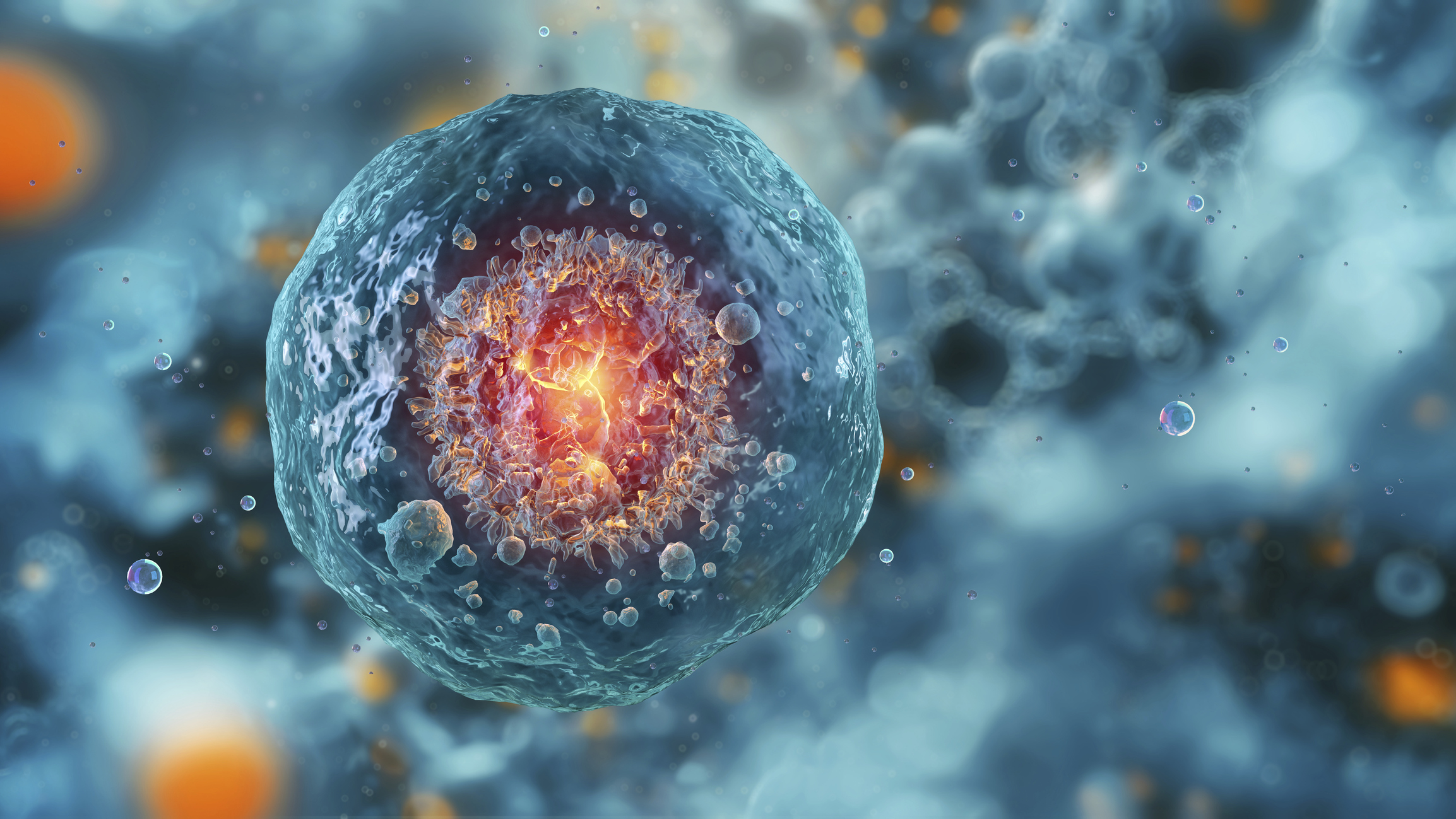
A family photo album can reflect how physical traits, such as blue eyes or lanky limbs, have been passed down — but a new analysis suggests that the way personality emerges across generations is far more complicated.
The meta-analysis, published Sept. 2 in the journal Nature, is the largest of its kind ever attempted. The work identified over 1,200 genes linked to five personality traits: extraversion, agreeableness, conscientiousness, openness to experience and neuroticism.
However, the scientists behind the study said its most important conclusion is that no single gene determines whether an individual will have a particular personality trait. Instead, thousands of genes exert tiny effects that are likely impossible to disentangle from environmental factors that shape our behavior.
Twins and DNA sequencers
Researchers have studied the link between genes and behavior for decades. Before the ability to analyze the genome in detail, many early studies looked to identical twins.
Knowing that identical twins share nearly 100% of their genetic material, scientists compared their traits with those of nonidentical twins and other siblings. Some studies compared twins raised in the same home with those raised in different homes in an attempt to separate the influence of upbringing from genetics. These efforts concluded that personality was roughly 50% heritable. ("Heritability" measures how much of the variance seen in a given trait can be explained by genes.)
Genetic studies of personality traits previously relied on comparisons between twins. Now, genetic sequencing technologies have advanced the field. (Image credit: Johner Images via Getty Images)
The advent of gene sequencing enabled geneticists to calculate the influence of genes with greater precision than twin studies did. The most powerful tool in this arsenal is the genome-wide association study (GWAS), which can link traits to gene variants in detail. These statistical analyses require sample sizes in the millions, though.
The new study was a large collaborative project called the Revived Genomics of Personality Consortium, which brought together 46 cohorts of participants who'd had their genomes sequenced. Each person had been asked something related to one of the Big Five personality traits. The team had data from roughly 600,000 people about the first four traits — extraversion, agreeableness, conscientiousness, openness to experiences — and data from over 1.1 million people about their levels of neuroticism.
Generally, as geneticists have leveraged larger and more powerful GWAS, many traits have shown increased heritability because these bigger studies can identify more subtle genetic links, said Brent Roberts, a psychologist at the University of Illinois at Urbana-Champaign who was not involved in the new study. But "with personality, it was really clear from the get-go that it was different," Roberts said.
Earlier GWAS had suggested that the heritability of personality from common gene variants was roughly 5% to 15%, far lower than the estimate suggested by twin studies. The new work has confirmed those low numbers, said study co-author Michel Nivard, a genetic epidemiologist at the University of Bristol in the U.K.
It may be that GWAS do not capture extremely rare variants that might link personality traits in certain families, which means the heritability may be slightly underestimated, Nivard suggested. Additionally, twins are more likely to rate their personalities as similar for reasons unrelated to genetics, so the twin studies may have overestimated the heritability of personality, Nivard noted.
Cutting out confounders
It can be difficult to control for confounding factors in GWAS. For example, if scientists were investigating the influence of genes on academic achievement, genetics only tells part of the story; a person's socioeconomic status and access to books and tutoring also influence their education.
But in this case, Nivard and his colleagues did not find confounders in measuring personality. The researchers found that the genetic effects on personality are the same within families as they are among unrelated individuals.
"We've basically been able to rule out that there's any straightforward, simple social processes by which your parents directly influence your personality," Nivard said.
A key takeaway from the study should be not to focus too much on the different contributions of genes versus environmental factors in a trait as complex as personality, Roberts said. Our moods and minds are a collaboration between the two factors, not a tug-of-war, he argued.
A study to properly measure this interplay would be vastly expensive and would require following participants for decades. "This is where the limitation of human genetics research is really profound," Roberts said. Part of the problem, he added, is that while our DNA sequence is fixed, how the body accesses and reads DNA varies significantly depending on the environment a person finds themselves in. In other words, which genes get turned on and when are variable factors.
Assessing this enmeshed relationship using only the DNA code we are born with is like trying to predict every move of a couple's dance by looking at how they hold each other before the music starts up.
Nivard and his team can now use this powerful data source to learn more about personalities within groups of people, he said. Nivard predicted that, even once geneticists have mapped all the rare gene variants that may influence personality, some genetic influence will still go unaccounted for; processes such as the interaction between the environment and gene expression are still not fully understood. He called these the "unknown unknowns." Identifying these factors is the next frontier for the genetics of personality.
Recurrent pregnancy loss affects an estimated one in a hundred couples trying to build a family, yet in a striking proportion of cases no cause is ever identified, leaving patients and clinicians alike without answers. A new study from researchers at the Institute of Medical Sciences, Banaras Hindu University in Varanasi, India, published in the journal Biochemical Genetics, offers a detailed picture of the genetic abnormalities lurking in lost pregnancies and points to a two-tier testing strategy that could substantially improve diagnostic yield. Combining rapid aneuploidy screening with high-resolution chromosomal microarray analysis and a layer of computational biology, the team has mapped not only which chromosomes go wrong in recurrent miscarriage but also which biological pathways those errors may disrupt.
The research team, led by Shivani Mishra and corresponding author Royana Singh, recruited 125 fetal specimens from pregnancies that ended in loss, ultimately analyzing 118 samples after applying predefined exclusion criteria designed to guard against contamination and degraded tissue. The specimens, which included products of conception and fetal tissues such as skin, muscle, and cartilage obtained during autopsy, underwent DNA extraction and quantitative fluorescent PCR, a technique that amplifies short tandem repeat markers scattered across chromosomes 13, 18, 21, X, and Y. By measuring the relative quantity of fluorescent signals at these markers, QF-PCR can detect the most common whole-chromosome imbalances, the trisomies and monosomies that account for the majority of chromosomally abnormal miscarriages, often within a day or two of sample receipt.
The results were sobering but clinically rich. Of the 118 samples analyzed, QF-PCR identified aneuploidy in 36 cases, a detection rate of 30.5 percent. Twenty of these were monosomies, in which an entire chromosome is missing from the fetal genome, and sixteen were trisomies, in which an extra copy is present. The remaining 82 samples appeared normal by this rapid screen. That nearly one in three lost pregnancies carried a whole-chromosome imbalance confirms the central role of aneuploidy in early pregnancy failure, and it also carries a practical message for genetic counseling: when an aneuploidy is identified as the cause of a loss, the recurrence risk for future pregnancies is generally low, information that can meaningfully reduce anxiety for couples facing the aftermath of repeated loss.
Beyond the raw detection numbers, the study teased out clinical associations that matter for patient management. Maternal age showed a statistically significant relationship with the presence of chromosomal abnormalities, echoing decades of reproductive biology linking the aging oocyte to meiotic segregation errors. More intriguing was the finding of a weak but statistically significant negative correlation between gestational age and aneuploidy risk, with a correlation coefficient of −0.238 and a p-value of 0.008855. In other words, the earlier the loss, the more likely it was to be chromosomally abnormal, a pattern consistent with the idea that grossly imbalanced embryos tend to fail early in development, while losses later in gestation are more often driven by other mechanisms, including immunological, endocrine, thrombotic, and anatomical factors.
But QF-PCR has a well-known blind spot. It interrogates a limited set of chromosomes and cannot see submicroscopic copy number variations, the deletions and duplications of genomic segments too small to alter chromosome number but large enough to remove or duplicate genes. To peer beneath that resolution floor, the team selected a subset of 30 samples, stratified by their QF-PCR outcomes, and subjected them to array comparative genomic hybridization. In aCGH, differentially labeled fetal and reference DNA are co-hybridized to a microarray carrying hundreds of thousands of immobilized genomic probes, and the fluorescence ratio at each probe reveals whether the corresponding region is deleted or duplicated in the fetal genome. This technique can detect imbalances ranging from roughly 100 kilobases upward, an order of magnitude finer than conventional karyotyping.
The aCGH analysis proved its worth. Within the 30-sample cohort, the researchers identified clinically relevant copy number variations spanning the spectrum of pathogenicity, including pathogenic and likely pathogenic variants as well as variants of uncertain significance, the latter category reflecting regions whose dosage sensitivity has not yet been firmly established. Each variant was interpreted according to the joint guidelines of the American College of Medical Genetics and the Clinical Genome Resource, the standard framework that weighs factors such as gene content, inheritance pattern, and overlap with established disease loci before assigning a clinical classification.
Among the genes caught in deleted or duplicated segments were several with compelling biological narratives. CFHR3, a member of the complement factor H-related gene family, participates in the regulation of the complement cascade, the arm of innate immunity whose dysregulation has been implicated in inflammatory damage at the maternal-fetal interface. TNFRSF4 encodes a tumor necrosis factor receptor superfamily member with roles in immune cell signaling and tolerance. UGT2B17 belongs to the uridine diphosphate glucuronosyltransferase family, a set of metabolic detoxification enzymes whose variation has previously been linked to recurrent miscarriage, possibly through altered clearance of environmental and endogenous compounds. CD24 and MSR1 are involved in immune signaling and macrophage scavenger function, respectively, while the pregnancy-specific glycoprotein gene family, or PSG genes, is expressed almost exclusively in the placenta and is thought to modulate maternal immune tolerance of the fetus.
To move from gene lists to mechanism, the authors turned the implicated genes loose on a battery of bioinformatics tools. Using FunRich and WebGestalt for functional annotation, KEGG for pathway enrichment, and STRING for protein-protein interaction network construction, the team asked what biological themes united the disrupted loci. The answer converged on a coherent set of processes: immune-inflammatory signaling, endocrine regulation, lipid metabolism, extracellular matrix remodeling, and placental development. The involvement of TNF receptor signaling, MAPK cascades, and interleukin-1-related pathways emerged from the network analyses, weaving the copy number variations into the same immunological tapestry that independent studies have implicated in unexplained recurrent loss, including work on maternal B cell signaling and natural killer cell-mediated immunopathology.
The authors are careful, appropriately so, to frame the pathway findings as exploratory. The cohort of 30 microarray samples is modest, and several of the variants of uncertain significance may prove benign as genomic databases mature. The statistical associations between individual CNVs and miscarriage phenotypes will require validation in larger, well-powered cohorts with matched controls before they can inform clinical decision-making. Still, the architectural message of the study is clear: sequential testing with QF-PCR followed by aCGH captures abnormalities that neither method detects alone, and it does so at a fraction of the cost and turnaround time of broader approaches such as whole-genome sequencing.
That message lands at a moment of ferment in reproductive genetics. European Society of Human Reproduction and Embryology guidelines define recurrent pregnancy loss as two or more consecutive losses, and professional societies have been wrestling with the optimal diagnostic workup of lost pregnancy tissue for years. Previous studies comparing karyotyping with molecular methods have shown that conventional cytogenetics suffers from culture failure and maternal cell contamination, problems that DNA-based methods largely sidestep. The Banaras Hindu University team’s approach, anchored in a real clinical diagnostic laboratory serving a population with limited access to advanced genomic testing, demonstrates that a tiered molecular protocol is feasible and productive outside of well-funded Western centers.
For couples enduring the grief of repeated loss, the study’s findings translate into a concrete recommendation worth discussing with a genetic counselor: comprehensive cytogenetic analysis of lost pregnancy tissue is not merely an academic exercise. Identifying an aneuploidy or a pathogenic copy number variant can close the diagnostic loop, refine recurrence risk estimates, and in some cases guide the management of subsequent pregnancies, including targeted prenatal or preimplantation genetic testing. Conversely, a normal molecular karyotype redirects the clinical search toward the non-genetic contributors, from antiphospholipid syndrome to uterine anomalies.
The work also adds to a growing map of the genomic landscape of pregnancy failure, in which the PSG gene cluster on chromosome 19, the complement factor H-related locus, and immune receptor genes recur across independent cohorts. As genome-wide datasets accumulate and functional studies test the dosage sensitivity of these loci, variants of uncertain significance will gradually be reclassified, and the exploratory pathway signals reported here may harden into causal mechanisms. Until then, the study stands as both a practical diagnostic template and a hypothesis-generating catalog of the fragile genetic architecture underlying one of medicine’s most quietly devastating problems.
Subject of Research: Genetic risk factors in recurrent pregnancy loss, identified through combined QF-PCR and aCGH chromosomal analysis with bioinformatics pathway enrichment analysis of fetal specimens
Article Title: Decoding Genetic Risk Factors in Recurrent Pregnancy Loss: An Integrative Chromosomal and Bioinformatics Approach
Article References: Mishra, S., Ashish, A., Rai, S., Yadav, A. K., & Singh, R. (2026). Decoding Genetic Risk Factors in Recurrent Pregnancy Loss: An Integrative Chromosomal and Bioinformatics Approach. Biochemical Genetics. https://doi.org/10.1007/s10528-026-11424-z
Using a new computational toolkit, Yale researchers recovered evolutionary clues from highly degraded DNA and identified two extinct lineages of giant tortoises. Some Galapagos giant tortoise specimens preserved in museums contained so little usable DNA that less than 1% of their genomes could be read. Yet Yale researchers were still able to recover enough genetic [...]
In one of the most comprehensive investigations of its kind, researchers have taken a major step toward untangling the biology underlying suicide risk, moving the field beyond long-standing debates about whether suicidal behavior is simply a consequence of psychiatric illness. By integrating large-scale genetic data with cutting-edge approaches that map how genes act in specific brain cells and circuits, an international team has identified shared genetic liability that cuts across psychiatric diagnoses while also pinpointing druggable biological pathways that could one day inform prevention strategies. The study, published in Translational Psychiatry, offers a sweeping synthesis of how inherited risk factors may translate into molecular vulnerabilities, and it does so with a level of resolution that previous candidate-gene approaches could never achieve.
Suicide remains one of the leading causes of death worldwide, claiming more than 700,000 lives each year according to global health estimates. Despite decades of research, clinical prediction remains stubbornly imprecise, and biological models of suicidal behavior have historically been hampered by small sample sizes, inconsistent findings, and a tendency to conflate suicide risk with the psychiatric disorders with which it so often co-occurs. Major depression, bipolar disorder, schizophrenia, and substance use disorders all carry elevated suicide risk, yet most people with these conditions never attempt suicide, while some individuals who die by suicide have no diagnosed psychiatric illness at all. This paradox has long suggested to researchers that suicidal behavior may possess partially distinct biological underpinnings, and the new study set out to test that hypothesis with unprecedented statistical power.
At the heart of the work lies genome-wide association study methodology, or GWAS, which scans hundreds of thousands to millions of genetic variants spread across the entire genome in very large groups of people, comparing individuals who have experienced suicidal thoughts or behaviors with those who have not. Each variant is tested for statistical association with the outcome, and the results can be combined into polygenic risk scores, numerical summaries of a person’s inherited liability based on the cumulative small effects of thousands of variants. Because no single common variant exerts a large effect, the field depends on aggregating data across massive biobanks and cohorts, a strategy that has only become feasible in the past decade as datasets containing hundreds of thousands of individuals became available.
The research team took particular care to address a central confound: psychiatric disorders themselves are highly heritable, and any genetic signal linked to suicide could simply reflect inherited liability to depression or schizophrenia rather than to suicide specifically. To disentangle this, the investigators applied sophisticated statistical techniques, including genetic correlation analysis and conditional modeling, which allow researchers to ask how much genetic overlap remains between suicidal behavior and psychiatric conditions after accounting for their shared heritability. Their findings revealed a complex architecture. Substantial genetic correlations confirmed that suicide risk shares a meaningful fraction of its inherited basis with major psychiatric disorders, supporting the idea of a common pool of genetic liability. Yet the analyses also uncovered signals that persisted beyond what could be explained by those disorders, pointing to biological mechanisms that may be more specific to the emergence of suicidal thoughts and actions.
A key conceptual innovation of the study is its framing of “shared genetic liability” as a bridge between diagnoses. Rather than treating suicide as an epiphenomenon of any single disorder, the researchers modeled it as a transdiagnostic phenomenon, one whose risk emerges partly from inherited vulnerabilities that cut across diagnostic categories. This perspective aligns with a growing movement in biological psychiatry that emphasizes symptom dimensions and mechanisms over traditional diagnostic boundaries. From this vantage point, the genetic variants that raise risk for depression, PTSD, and schizophrenia may do so partly because they converge on shared molecular pathways, and those same pathways may govern an individual’s threshold for suicidal crises regardless of which diagnosis they carry.
Identifying genetic variants, however, is only the first step. A GWAS signal typically points to a genomic region rather than a specific causal gene or mechanism, since many associated variants fall in non-coding stretches of DNA that regulate gene activity from a distance. To translate statistical associations into biological insight, the team deployed a battery of functional genomics approaches. Transcriptome-wide association studies, known as TWAS, integrate gene expression data to identify genes whose genetically predicted expression levels are linked to suicide risk. Tissue and cell-type enrichment analyses then ask whether the implicated genes are unusually active in particular organs or cell populations. The results converged strikingly on brain tissue, and more specifically on neuronal populations, providing support for the idea that inherited suicide risk is mediated largely through gene regulation within the central nervous system.
The study’s most clinically provocative findings concern druggable biology. Using databases that catalog the protein targets of approved and investigational medications, the researchers examined whether genes implicated in suicide risk were over-represented among known drug targets. The analysis identified several classes of candidate targets, including components of neurotransmitter signaling systems, ion channels, and intracellular signaling cascades that are already the focus of pharmacological development for other conditions. This “druggable genome” approach, which has gained traction in genetics research across many diseases, offers a pragmatic shortcut for translational science: rather than waiting decades for entirely new therapeutic classes to be invented, researchers can prioritize existing or in-development compounds whose molecular targets intersect with genetically implicated pathways.
Among the pathways receiving attention in the analysis were those involving glutamatergic and GABAergic neurotransmission, the primary excitatory and inhibitory systems of the brain, which have repeatedly surfaced in studies of mood disorders and stress response. The endocannabinoid system and inflammatory signaling pathways also appeared in the convergent analyses, consistent with a broader literature linking immune dysregulation and allostatic load to suicidal ideation. Importantly, the authors emphasize that these findings identify hypotheses rather than ready-made treatments. Genetic association with a drug target does not guarantee that modulating that target will reduce suicide risk, and any therapeutic application would require extensive preclinical validation and carefully designed clinical trials. Nevertheless, the work provides a principled, data-driven map for where to look, replacing decades of largely failed candidate-gene speculation with a genome-wide evidence base.
The methodological rigor underpinning the study deserves emphasis, because the genetics of suicidal behavior has a troubled history. Early candidate-gene studies, which typically examined a handful of variants in genes related to serotonin signaling, produced findings that largely failed to replicate and in some cases were later shown to reflect statistical artifacts, small samples, and publication bias. The field has since undergone a reckoning, and modern efforts adhere to far stricter standards: preregistered analyses, very large and ancestrally diverse cohorts, replication in independent samples, and conservative multiple-testing corrections that demand genome-wide significance thresholds on the order of five times ten to the negative eighth power. The new study reflects these lessons, applying sensitivity analyses to guard against confounding from psychiatric comorbidity and examining whether findings generalize across ancestral groups, an issue of considerable importance given that most genetic discovery to date has been conducted in populations of European ancestry.
The implications for clinical practice, while still distant, are worth considering. Polygenic risk scores for suicide-related outcomes currently explain only a small fraction of individual differences in risk, far too little to be used as stand-alone screening tools, and their predictive performance is degraded in populations not represented in the discovery data. Researchers in the field are careful to stress that genetic risk is not destiny: suicidal behavior arises from an intricate interplay of inherited vulnerability, mental illness, trauma, substance use, social isolation, access to lethal means, and the availability of support. What genetics offers is not a crystal ball but a window into mechanism, and mechanistic knowledge is the raw material from which better biomarkers, risk stratification tools, and ultimately targeted interventions are built. In the near term, the druggable pathway findings are most valuable as a guide for laboratory science, directing neurobiologists toward molecular systems whose manipulation might alter suicide-relevant phenotypes in animal models and cellular assays.
Looking forward, the study signals where the field must go next. Sample sizes for suicide-specific genetic analyses remain smaller than those for major psychiatric disorders, and expanding representation across ancestries is essential if the benefits of this research are to be shared equitably. Integrating genetic data with longitudinal measures of life stress, neural imaging, and digital phenotyping may eventually allow researchers to model how inherited liability interacts with environmental triggers across the lifespan. Functional experiments in induced pluripotent stem cell-derived neurons and brain organoids could test whether the implicated genes genuinely alter stress-response circuitry or resilience. And randomized trials in patients carrying high genetic liability, though ethically and logistically complex, represent the logical endpoint of the druggable-target approach.
What the study ultimately delivers is a reframe. Suicide risk, in this analysis, is not an amorphous clinical outcome but a measurable, partly heritable trait whose genetic architecture is now being resolved with the same tools that have transformed our understanding of heart disease, diabetes, and schizophrenia. By demonstrating that shared genetic liability links suicide risk across diagnostic boundaries and by mapping that liability onto specific, pharmacologically tractable biology, the researchers have converted a long-standing clinical puzzle into a concrete research agenda. The path from genetic correlation to effective prevention is long, and no genetic finding will soon replace the human work of crisis intervention, connection, and care. But for a problem that has defied biological explanation for generations, the identification of shared, druggable mechanisms represents a genuine and hard-won advance, one that may shape suicide research for years to come.
Subject of Research: Shared genetic liability and druggable biological pathways underlying suicide risk across psychiatric disorders
Article Title: From shared genetic liability to druggable biology in suicide risk
Article References: Yao, K., Zhuo, C., Chen, X., Li, C., Song, H., Zou, J., & Tian, H. (2026). From shared genetic liability to druggable biology in suicide risk. Translational Psychiatry. https://doi.org/10.1038/s41398-026-04428-1
Australian researchers have identified a promising new approach that could help stop one of the most aggressive forms of breast cancer from spreading. Triple-negative breast cancer becomes especially dangerous once tumor cells escape the breast and establish themselves elsewhere in the body. Australian researchers have now identified a molecular weakness that could eventually help determine [...]
Cluster headache causes extreme pain, but its origins remain unclear. New research links the disorder to genetics, inflammation, and tobacco smoke exposure. Unlike an ordinary headache, a cluster attack typically erupts without warning and causes excruciating pain around one eye. Episodes can last from 15 minutes to three hours and often return at similar times [...]
The story of a Medieval European plague is coming into focus through historic manuscripts, but not in the way you might think. Instead of providing mere historical accounts, the animal skins from which these Medieval volumes are constructed provide genetic evidence for reconstructing the 3,500-year story of the sheepox virus (SPPV), which decimated European livestock.
In a recent paper published in Science Advances, an international team of interdisciplinary researchers ranging from geneticists and virologists to historians and conservators explored the history of this pre-modern plague.
The new work combs through Medieval history to reveal the lineages of diseases plaguing modern herds, providing new insight into the viruses threatening modern animal agriculture.
Medieval Texts
Chosen for the work were some of the most important Medieval manuscripts that Europe has to offer, including the millennium-old York Gospels, one of the best surviving examples of an early Anglo-Saxon illuminated manuscript. Another work used in the study was one of the earliest known English dictionaries, the Corpus Glossary of Corpus Christi College, Cambridge. Beyond these notable examples, the team exploited a variety of English and continental European works.
Medieval volumes did not contain paper pages like modern books, but instead, vellum pages made of calfskin, as well as the skins of goats and sheep. Therefore, these works are preserved as hundreds-of-years-old biological specimens for modern geneticists.
SPPV tends to target younger animals, such as the calves from whom vellum is made, and is highly contagious, rapidly spreading through large portions of a herd. While these factors made the disease easier for modern researchers to study via vellum samples, it also made it devastating to wool, meat, and milk production.
Extracting DNA
“The recording of ancient DNA of pathogens has completely changed our understanding of infectious disease in the past, and here we show that parchment can also preserve animal pathogen DNA. It is possible that archives and libraries around the world also contain the genetic traces of disease outbreaks in animals in the past,” said lead author Louis L’Hôte, PhD candidate at the University of Cambridge, Dublin.
“These genomes help us understand how these pathogens evolved and how they impacted past societies – such as sheeppox virus, which we show has been affecting Eurasian sheep herds for at least three and a half millennia,” L’Hôte added.
With the rapid spread of SPPV across herds, many manuscripts contained SPPV DNA on multiple pages within the same manuscript. Additionally, finding SPPV DNA on pages made from both calf and goat skin marks it as a rare example of cross-species infection.
“Parchment making has been considered a vanishing craft, but we are beginning to understand just how much of the past it has retained. This project shows that parchments not only preserve our cultural history for hundreds of years, but also the landscape of disease in our livestock in the medieval period,” said supervising author Dr Kevin G. Daly, Associate Professor at UCD School of Agriculture and Food Science.
“This kind of exciting, surprising discovery is only possible when researchers from a range of disciplines come together to share their expertise and data,” Day said, calling this “a generosity and trust that collaborative science depends on.”
Exploring Medieval DNA
The Medieval manuscripts combine with even older surviving biological records, such as teeth, to show the virus reaching back 3,700 years. Between the manuscripts and archaeological finds, the team identified 41 samples containing related viruses.
Along with the goatpox virus and the lumpy skin disease virus, SPPV belongs to a category called capripoxviruses. The new research allowed the team to compare the ancient and modern sequences to determine that the division between the various viruses in the group occurred between 3,700 and 11,500 years ago, around the time animal domestication took hold in Europe as sheep came off the steppe.
“Ancient genomes can help us understand how these pathogens affecting us today evolved. We find that, unlike the smallpox virus, the sheeppox virus genome was very stable over the last few millennia,” L’Hôte concluded. “This might mean that genetic changes before this point were very important to how the sheeppox virus evolved to infect its host, which may help us fight the pathogen in the future.”
Ryan Whalen covers science and technology for The Debrief. He holds an MA in History and a Master of Library and Information Science with a certificate in Data Science. He can be contacted at ryan@thedebrief.org, and follow him on Twitter @mdntwvlf.
Over a century ago, German pathologist Georg Schmorl documented a strange phenomenon: cells from fetuses that had moved into the mother during pregnancy. He had discovered microchimerism — where the cells from one individual move into another and set up shop.
In the past few decades, scientists' understanding of microchimerism has grown, thanks to technological advances. And the implications are wild.
Cells transferred during pregnancy could mean you have cells from your grandmother living inside you. A twin can be "absorbed" within the womb, leaving a genetic trace of the lost sibling. And after a bone marrow transplant, a man ended up with the donor's DNA in his blood and semen. These are just a few examples of how microchimerism opens a new way to understand immunology, genetics and evolution.
Live Science spoke with Barnéoud about the book, why the idea of microchimerism resonates so deeply, and where the field could be headed next. "Hidden Guests" has been short-listed for the 2026 Royal Society Trivedi Science Book Prize.
Hannah Osborne: What first got you interested in microchimerism? How did your personal understanding change over the course of writing "Hidden Guests"?
Lise Barnéoud: The first time I heard about it, I was writing my previous book on vaccinations, and I had a chapter on our immunity, how our immune system works. I wanted to explain what we got wrong about how our immunity works. We are still learning — [we used to have this idea] that our immune system is kind of an army patrolling in our body and expelling any foreign elements. That's the image we have of our immune system. I wanted to tell another story, which is more accurate because we know now that it's not working like that.
I was looking for counterexamples, and, of course, one very famous counterexample is pregnancy. We tolerate the fetus that is half different than us. Another well-known counterexample is microbiota. Inside our body, we carry microbial cells as well as human cells — so cells coming from bacteria, viruses, fungi. We don't expel them; we need them. The third example I found was microchimerism.
The more I read about it, the more I was interested in it. It was challenging so many things. I've learned that the placenta is not that watertight barrier that we often think of, that the immune system is not as intolerant as we often think, and we can inherit cells from our kids like an inverted inheritance. All those elements popped up within my first reading, and I thought, "OK, I really need to dig more."
For your second question, I think the biggest change is that I realized those cells are not only kind of inactive tourists, just homeless drifters passively traveling in our body. They are really active. They can multiply, they can produce protein, they communicate with other cells, and they really influence our physiology, for better or for worse.
HO: We see how microchimerism is a relatively new field of research that has had lots of stops and starts. What was the biggest hindrance to the development of the field, and what changed?
LB: Maybe before speaking about the hindrances, just saying that the first time we discovered cells from other individuals within individuals was a century ago. The first one was in 1893, and it was a German pathologist who discovered cells from fetal origin in lungs from women who had died during pregnancy. Then it took a century to understand that this phenomenon happened to everyone and that it is of importance.
I think there are several hindrances. There's never one big one. The first one we can think of is the technology, because you need to study these microchimeric cells; you need tools that are able to study a tissue at the cellular level. This is quite new, so now we have these tools and we can make some progress. The second big hindrance is that this research field calls into question the traditional view of the immune system. If you see the immune system as a purely defensive and intolerant army, then you can't think of these foreign cells staying forever in your body, and that was a big hindrance, I think.
Maybe a third hindrance is that at the beginning of this field — I mean the new beginning in the '90s — they were mostly women working in this field. They told me that male colleagues often said, "Well, that's woman's stuff, you know; you're looking for some connection with your babies, but it's not really relevant," which is wrong, of course, because even men are affected by this phenomenon. They stayed nine months in the womb like us.
That might have, at least, challenged some of the first pioneers of the field. Now there are more and more men working in the field. In April, there was an international conference on microchimerism, and I think it was half women.
It's because it's such a new field and you have such open questions, there's room for many interpretations, many hypotheses.
HO: The book includes many anecdotes about people directly affected by microchimerism. Which story were you most affected by, and why?
LB: I guess it'd be the story of Lydia Fairchild because she nearly lost custody of her children because of microchimerism. She was 26 and living in the U.S., and she applied for a welfare benefit to help her raise her kids. In the U.S., you need to undergo a maternity test for that, and her DNA test ends up showing that she could not be the mother of her kids. Of course she tried to argue; she had photos; she had testimonies. But she was always told, "DNA is 100% foolproof; DNA doesn't lie," which implied that she was lying. But she was not, so she called several lawyers. But at the beginning, they were all saying, "No, DNA doesn't lie."
At that time, she was pregnant with her third child, and they decided to do a test immediately after birth. Again, the impossible happened. This third child that just emerged from her womb was not her son — genetically speaking, at least.
In fact, she was harboring cells from her vanished sister inside her. Most of her eggs carried her sister's genetic signature, which means that the mother of her kids never lived. Isn't it amazing? That's maybe one of the stories that blew my mind.
HO: The section on DNA and how microchimerism complicates things is fascinating. How much of an impact do you think hidden cells have on the reliability of DNA evidence?
LB: The truth is that I think we don't really know. If you have only some microchimeric cells, among thousands of your own cells, then it won't change the reliability of the DNA test. But in some cases, these microchimeric cells can make up a significant part of an organ and sometimes even an entire organ. This proportion might depend on when you received those microchimeric cells. For instance, if you received those cells like Lydia Fairchild at the very, very beginning of your development, they might end up making up the entire organ where they landed first. But probably, we don't know which percentage of us start our in utero life with an evanescent embryo.
An illustration of a human embryo (Image credit: RUSLANAS BARANAUSKAS/SCIENCE PHOTO LIBRARY via Getty Images)
I looked for this number because I didn't really know about evanescent twins, so I wanted to know how many pregnancies started with several embryos and suddenly one disappeared. And I couldn't find any good number because, of course, it's quite difficult to put any imaging there at the beginning of the condition. Some articles say that up to 30% of pregnancies will start with several embryos, which means a lot of us are affected by evanescent twins. But let's say it might be rare that we have so many microchimeric cells in one organ that it completely changed a DNA test.
But there's another example in my book that took place in Alaska in 2004. After a rape, the forensic investigators collected the semen from the scene, and they discovered that the DNA in this semen matched with a man who was already in the database. But this guy was behind bars. He was in jail, the day of the rape, so he couldn't be the rapist.
Finally, investigators discovered that this guy, the prisoner, received a bone marrow transplant a few years ago from his brother, and the cells from the bone marrow of his brother didn't stay quietly in the bone marrow and blood. They go outside the marrow, and some end up in the semen. And the true rapist was the brother, the one who donated the bone marrow, and not the receiver. Without the alibi of the prison, the brother who received the transplant would have been charged, and the true rapist would have been left free.
I guess there are some cases where microchimeric cells can really change the result of a DNA test. But for now, it's not really studied. I've been contacted by some forensic investigators to learn more about microchimerism, so maybe things will change.
HO: You point out that microchimerism is used for both pro- and anti-abortion arguments, and it's used by "manosphere" influencers to promote ideologies. Why do you think microchimerism research is so open to interpretation and used in this way?
LB: Next time you're at a dinner, just try to explain microchimerism. You're a science journalist, right? Well, usually when you tell the story you're working on, people are saying, "Oh, yeah, OK." And they just turn to another conversation. If you try mentioning microchimerism in your next dinner, then you'll see. Everyone will try to make some hypothesis, some connection; some will be moved that it's possible to keep some cells from an unborn child or a lost child. Some will be disgusted by the idea that they keep cells from their mother, like having their mother on their shoulder all the time. It's really the first time as a science journalist I experienced such resonance with a story I was working on among people around me, people who are really not into science.
As soon as the door opened, it revealed the fears and the desires of people. So yes, some conservatives use microchimerism to fight against abortion because they say the cells from those aborted will haunt the women for the rest of their life. [But] I've met scientists explaining that if you receive a shot of cells of fetal origin, it can also help you to regenerate your organs. It can be used also by some other people to rejuvenate, to try to stay young.
It's because it's such a new field and you have such open questions, there's room for many interpretations, many hypotheses. I mean, that's fine; that's how science works. You should always have hypotheses and try to answer your hypothesis with new experiments. But it's true that sometimes I didn't know how to handle all those ideas at the beginning. Then I realized it was like a window open on the intimate — their fears and their desires — and it reveals what they really think about their connection to the world they are in. From a sociological point of view, it would be really interesting to study that.
HO: In the book, biologist and evolutionary theorist Amy Boddy warns that microchimerism researchers love to hypothesize. When the implications of the field are so far-reaching, how do we get past this? Which hypothesis do you find most intriguing?
LB: Many scientists told me that until 2021 or something like that, there was this hypothesis that those cells could come from another generation than the mother. We knew that the mother would give cells to the fetus and the fetus could give cells to the mother, but then people ask, because the mother can still carry cells from her mother, maybe she could pass those cells to the fetus. Until the 2020s, scientists were thinking, "Well that's a crazy hypothesis; that can't happen." There could be only a few cells that won't go through the placenta and integrate with the new fetus — until one scientist proved that a newborn can carry cells from the grandma.
In this field, you always have crazy hypotheses, and I guess until we haven't proved that this hypothesis is wrong, you can still leave the hypothesis on the table. One of the craziest hypotheses, the first time I encountered it, I thought, "OK, they've gone really too far."
This hypothesis is that we could inherit cells from our sexual partner. I thought it was not possible because I thought the semen was full of spermatozoa, that they are only like half cells having only half chromosomes, so they cannot survive. But actually in semen, you have a lot more cells than spermatozoids — you have immune cells, for example.
There are many cells in the semen, and we can think that if you have some, there could be some path for those cells to escape and go into your blood. It's not completely crazy. For now, there's no formal proof of this new source of microchimerism, but I recently heard that some scientists are working on the topic on mice.
We should be careful about those interpretations. But I think scientists are not the ones who should care about this interpretation; otherwise, they would prevent themselves from doing their research. They should open all the doors with all kinds of hypotheses. If we do it in a scientific way, meaning if we try to answer a hypothesis with good experiments, then I think everything is fine. You will never prevent people around you from using this information to distort the facts and tell another story. That's how life goes.
HO: What do you think the most promising use for microchimerism research will be over the next 10 to 20 years? How do you see it unfolding?
LB: I guess it will be in the regeneration field. We have discovered that those cells can participate to regenerate tissue. Some scientists around the world are doing clinical studies to see whether we could use these cells as a therapy either after an infarctus [heart attack], but also a vascular accident [stroke] or when you have a wound on your skin. The hope is that by either attracting microchimeric cells that are within our bodies, or injected, these microchimeric cells maybe can help this kind of tissue to repair. I think that might be one of the promising uses of these cells in the future.
Maybe also in the transplantation field; maybe we can think about using the knowledge of how microchimeric cells can induce tolerance to avoid [immunosuppressive] drugs. When you receive the transplant, you need to take drugs [that suppress the immune system] for the rest of your life, and those drugs are like really strong drugs. If we can use microchimeric cells before the transplant to allow better tolerance, then we could either not use any drugs or maybe use less drugs. That would be really interesting, too.
This interview has been condensed and edited lightly for clarity.
Hidden Guests: Migrating Cells and How the New Science of Microchimerism Is Redefining Human Identity
What if some of your cells were not your own? What if they once belonged to someone else?
Part mind-bending medical mystery — part cutting-edge science — Hidden Guests uncovers the astonishing phenomenon of microchimerism: the presence of foreign cells inside our own bodies. The incredible story of how those cells got there — and what they do once they arrive — might change everything we know about the immune system, lineage, and identity.
For several decades, paleontologists have described Miracinonyx trumani -- an extinct species of cheetah-like felid that roamed the North American prairies and steppe terrains more than 13,000 years ago -- as North America’s answer to the African cheetah (Acinonyx jubatus).
Researchers have identified more than 1,200 locations in the human genome associated with personality traits, offering the most detailed look yet at the biological underpinnings of traits like sociability, anxiety and curiosity.
BatDNA may hold clues to a long and healthy life, according to an international team of researchers.
These small creatures grace the night skies with extremely long lifespans and low cancer rates—traits that may ultimately benefit humanity if scientists can figure out how bats achieve them. Now, in a recent paper published in Nature, scientists from the University of Vermont and Penn State University led an international team investigating bats’ immune systems.
The paper’s crucial discovery was that bat DNA reveals the creatures encode genomic changes to handle exposure to environmental pathogens.
The Miraculous Bat
“Pathogen adaptation, longevity, and cancer resistance—they are fundamentally linked,” explained co-lead author Elise Lauterbur, an assistant professor of evolutionary biology at the University of Vermont. “Many of the genes that have adapted to viruses in bats are genes that are also involved in longevity and cancer resistance.”
Bats make an ideal study subject as they are only second to rodents in diversity, allowing scientists to study a wide variety of features in a single order.
“If you look at two bats, the same size, one lives 3 years the other lives 30 years and they are very closely related, like humans and Neanderthals, you can essentially find a very small number of genetic changes that leads to a very big change in their lifespan,” said co-lead author Juan Manuel “Manny” Vazquez, an assistant professor at Penn State University. “And that is kind of the Holy Grail for evolutionary medicine.”
The work focused on Myotis, one of the largest bat genera, whose members are found across much of the globe outside Antarctica. Eight Myotis species came under the microscope, with the team experimenting on cultured cells and analyzing their genomes. Among the species sampled were little brown bats (Myotis lucifugus), known both for their long lifespans, even among bats, and their susceptibility to the deadly white-nose syndrome, a fungal disease.
Even the researchers’ sample-collection methods were novel. They developed a technique akin to ear-piercing to collect small tissue samples from bat wings, which were then used to establish cell cultures. The team says this both spared the animals’ lives and produced more scientifically useful samples.
Bat Evolution
In their detailed examination, the researchers identified evidence of past events in which Myotis bats encountered pathogens and how adaptations to those viruses were selected for during their evolution.
Their work identified specific genomic changes, including the duplication or deletion of genes, associated with adaptation to environmental pathogens. This illuminated important differences in bats’ responses to DNA and RNA viruses compared with humans and other primates, potentially contributing to bats’ long, healthy lifespans.
Comparatively, human evolution shows stronger selection involving proteins that interact with RNA viruses like influenza, while bats show stronger selection involving proteins that interact with DNA viruses such as herpesviruses. These differences may help explain the distinct ways bats and primates have adapted to viral threats.
Cancer Resistance
The team also identified genetic mechanisms that could help explain bats’ resistance to cancer and other age-related diseases. In some cases, duplicated genes can diversify their functions, allowing different copies to perform distinct roles. According to the researchers, this may provide immune advantages that help bats combat multiple viruses while also contributing to pathways associated with longevity.
Another important finding was that while most mammals contain one copy of the immune-related gene PRKN, some bat species contain two or even three copies, suggesting that additional copies may offer evolutionary advantages. The team says this could provide important insights for future cancer research, given bats’ unusual ability to repair or eliminate damaged cells.
The research doesn’t point to immediate treatments for humans, but it does provide starting points for reconsidering human illness in the context of bat immunology and evolution.
“Everything is connected,” Vazquez concluded. “Everything uses the same shared set of biology. … We don’t have to treat all of these different problems as silos. We can now start focusing on systems like bats to understand how we can solve multiple human age-related diseases.”
Ryan Whalen covers science and technology for The Debrief. He holds an MA in History and a Master of Library and Information Science with a certificate in Data Science. He can be contacted at ryan@thedebrief.org, and follow him on Twitter @mdntwvlf.
The genetic code is what life everywhere uses to convert the information contained in DNA into specific protein sequences. With minor variations, the same genetic code is used by every living thing on Earth, suggesting it was already present in the last common ancestor of all of them. It's not an easy thing to change, because so many things in every cell depend on it.
Nevertheless, some preliminary steps have been taken. Researchers have managed to add some new amino acids to a bacterial cell and were able to make proteins that were one amino acid less than usual. But it's a slog; for some of this work, people have had to re-engineer every single gene in a bacterial genome.
Now, researchers have found a way to operate two separate genetic codes simultaneously, avoiding the need to do any work to compensate for altering the code that every protein in a cell relies on. They didn't test it in an actual cell, and it might cause some problems there. But it's a creative solution that should accelerate some synthetic biology work.
New research involving tiny aquatic animals known as rotifers is providing scientists with a unique look at how information about maternal age may be passed from one generation to its offspring.
The findings offer insights into the ways a mother’s age can influence factors that range from health and lifespan to the behavior and even reproductive success of her offspring. This phenomenon, known as maternal age effects, has been documented throughout the animal kingdom—observed in everything from invertebrates to large land mammals like elephants, and even humans.
While advanced maternal age has often been associated with negative effects in offspring in past studies, questions remain about exactly how those effects are transmitted.
“Nearly all forms of life show some level of maternal age effect, and most are negative effects caused by advanced maternal age,” said Kristin Gribble, an associate scientist at the Marine Biological Laboratory’s Bay Paul Center, in a statement.
For their research, Gribble and her colleagues have focused on Brachionus manjavacas rotifers, a variety of microscopic aquatic creatures whose rapid reproduction abilities allow scientists a unique opportunity to study biological changes that occur across multiple generations.
Now, Gribble and her team’s findings point to something novel: that rather than being influenced primarily by accumulated DNA mutations, or even cellular damage, some maternal age effects appear to be epigenetic.
Going Beyond DNA Mutations
The new findings resulted from work undertaken in Gribble’s laboratory by researcher Alyssa Liguori, which looked at two different genotypes within a single species of rotifer.
The premise was simple: if mutations or cellular damage were what was behind maternal age effects, the consequences would likely be shown to worsen progressively across generations.
However, what the team found instead was that such effects could be reversed, and in some cases, within a single generation. The fact that this can occur so rapidly seems to imply that the underlying mechanism at play involves changes in the way genes are expressed, as opposed to alterations in the actual genetic code, as suggested in the past.
Based on this, one possibility the team is now exploring is that what are called histone modifications—essentially chemical changes that impact proteins that are encased with DNA, which can influence whether particular genes become activated, or suppressed.
Additionally, the researchers are now looking at whether mitochondrial DNA (mDNA), known to generally be inherited from an offspring’s mother, could play a key role in helping transmit information about maternal age from one generation to the next.
Further, genetic differences may influence how much an offspring is affected. One intriguing aspect of the recent research that Gribble emphasizes involves how one rotifer strain seemed to produce offspring from older mothers, which actually exhibited longer lifespans. This may suggest that some genetic variants could help to protect against, or possibly even reverse, factors that are generally negatively associated with advanced maternal age.
Effects That Echo Across Generations
Still, a few mysteries remain, such as what exactly causes maternal age effects to remain so widespread, especially if they often reduce an offspring’s evolutionary fitness.
Gribble has an idea: she suspects that natural selection may provide at least part of the answer, since reproduction in rotifers occurs relatively early in life. This could mean that evolutionary pressures that eliminate harmful effects that may arise from reproduction at advanced ages could be comparatively weak.
Ultimately, the team believes their research may help to illuminate a much broader question: how the experiences and biological state of one generation influence those that follow.
“It’s not just about what’s in your genome as an individual,” Gribble said, adding that this is mainly because “your health potentially depends on the health and environment of your mom and grandmother and great-grandmother.”
Fundamentally, if researchers can identify the mechanisms behind maternal age effects in rotifers, these unassuming aquatic creatures could help to significantly advance our understanding of the ways biological information is carried across generations, potentially offering new insights into human health and precision medicine.
Micah Hanks is the Editor-in-Chief and Co-Founder of The Debrief. A longtime reporter on science, defense, and technology with a focus on space and astronomy, he can be reached atmicah@thedebrief.org. Follow him on X @MicahHanks, and at micahhanks.com.
In 2006, geneticist Bryan Sykes, Professor of Human Genetics at the University of Oxford, published the results of a study that had taken him a decade to complete. His team had collected and analyzed the mitochondrial DNA and Y chromosome of 10,000 volunteers from England, Scotland, Wales, and Ireland, with the aim of reconstructing the […]
Scientists in Arkansas have developed a machine-learning approach that can identify potentially disease-causing strains of a poultry bacterium by analyzing not only which genes are present, but also how those genes are arranged across the genome. The method focuses on the “genetic neighborhoods” surrounding genes, offering a new way to distinguish harmful strains of Enterococcus cecorum from closely related strains that do not cause disease.
E. cecorum is commonly found in poultry environments, and many of its strains are harmless. However, pathogenic strains can cause arthritis, bone infections and lameness, creating serious animal-welfare problems and economic losses for poultry producers. Because pathogenic and nonpathogenic strains can share many of the same genes, identifying the genetic features associated with disease has been challenging.
The research team, led by Aranyak Goswami of the Arkansas Agricultural Experiment Station’s Center for Agricultural Data Analytics, examined the organization of genes within genomic islands. These are segments of DNA that bacteria often acquire from other microorganisms through horizontal gene transfer. Genomic islands can carry genes involved in antibiotic resistance, bacterial survival, movement of DNA and virulence, making them important regions for understanding how pathogens emerge.
Rather than treating the bacterial genome as a simple list of genetic components, the researchers analyzed it as a structured map. They investigated which genes occurred near one another, the order in which they appeared and the way groups of neighboring genes formed recurring units known as genomic-island cassettes. This architecture provided information that could be missed when genes are examined individually.
“The machine-learning model recognizes patterns in gene order much like it recognizes patterns in language,” Goswami said. In the same way that a language model can learn that certain words frequently appear together, the computational system learned that particular combinations and arrangements of bacterial genes were associated with pathogenic strains. The approach therefore captures relationships among genes rather than relying only on the presence or absence of individual DNA sequences.
For the proof-of-concept study, the researchers analyzed the genomes of 145 E. cecorum strains isolated from poultry. The dataset included 95 nonpathogenic strains and 50 pathogenic strains capable of causing illness. The analysis found that disease-associated strains were more likely to contain genomic islands enriched in genes linked to antimicrobial resistance and the transfer of genetic material between bacteria.
Those findings do not mean that every bacterium carrying such genes will necessarily cause disease. Instead, the researchers describe the system as an exploratory classification and research tool. It identifies patterns that may help scientists investigate why some bacterial lineages become harmful, while also providing clues about how resistance and virulence traits move through bacterial populations.
Current monitoring methods for E. cecorum often depend on culturing the bacterium or screening for specific genes. Such techniques remain valuable, but they may overlook broader genomic relationships. The new pipeline could complement these approaches by evaluating the wider context in which genes occur. Additional testing with larger and more geographically diverse datasets will be necessary before the method can be used for routine flock surveillance or diagnostic decision-making.
The researchers believe the strategy could eventually be adapted to other bacterial species affecting animals, humans, wildlife and plants. Goswami’s team is beginning to examine Enterococcus faecalis, a close relative of E. cecorum and a frequent cause of hospital-acquired infections in humans. They also plan to apply the pipeline to Escherichia coli and other bacteria to study how nonpathogenic lineages acquire the genomic configurations associated with disease. The study, published in Frontiers in Microbiology, demonstrates how machine learning and comparative genomics can reveal hidden signals in bacterial DNA and may help researchers track the evolution of emerging pathogens.
Subject of Research: Animals
Article Title: Genomic-island cassette architecture provides interpretable signal for exploratory classification of poultry-associated Enterococcus cecorum lineages
Researchers at the University of Copenhagen found that a signaling mechanism in the cells’ ‘antennae’ may help explain why some fetuses develop disease in both the heart and other organs. About two of every 100 babies worldwide are born with congenital heart disease, but the biological processes responsible for these conditions are not fully understood. [...]

 Login

 Ancient DNA suggests Rome’s fall reshaped Europe through gradual migration and intermarriage, not a sudden barbarian takeover.