 Login
A New Drug Lifts the Fog of Sleepiness for Those With Narcolepsy
9 September 2026 at 18:21
The first-of-its-kind drug offers a reprieve for people who live with the debilitating sleep disorder.

In a development that could reshape how scientists study the mechanics of human movement, a team of biomedical engineers and computer scientists has unveiled an open-source software tool capable of tracking the tiny, intricate bones of the wrist as they glide and rotate during motion. The new tool, called Hierarchical 3D Registration, or 3DH, is a module within the SlicerAutoscoperM extension of the widely used 3DSlicer computing environment. Described in a study published in BioMedical Engineering OnLine, the software was validated against previously established methods using both sequential three-dimensional computed tomography, or 3DCT, scans of living participants performing thumb tasks and dynamic four-dimensional CT, or 4DCT, images of cadaveric wrists moving through flexion and extension. The results demonstrate that the open-source approach can match the accuracy of independent, previously published methodologies while offering researchers everywhere a free and reproducible path to quantifying skeletal kinematics.
The wrist has long been one of the most challenging joints for biomechanists to study. It contains eight small carpal bones, including the scaphoid and lunate, along with the radius of the forearm and the metacarpal bones of the hand, all articulating in a tightly packed arrangement that allows the remarkable range of motion humans rely on every day. Understanding how these bones move relative to one another, a field of study known as arthrokinematics, is essential for unraveling the causes of wrist instability, osteoarthritis, ligament injuries and the long-term outcomes of surgical repairs. Yet measuring those movements in living, moving joints has historically required either invasive techniques, labor-intensive manual analysis or proprietary software that few laboratories can access or verify.
Quantitative tracking of skeletal structures in vivo, with both efficiency and high temporal resolution, matters to a wide range of questions in biomechanical research, and the authors of the new study argue that the field has been held back by a shortage of accessible, semi-automated registration tools. Image registration, the computational process of aligning one three-dimensional image volume to another, lies at the heart of motion tracking from CT data. When a bone moves between two scans, its position and orientation change, and a registration algorithm must find the transformation that best overlays the bone’s image from one time point onto the next. Done well, this yields precise measurements of rotation and translation. Done poorly or inconsistently, it introduces errors that can swamp the small movements of carpal bones, which may shift by only a few millimeters and a few degrees during normal function.
The new 3DH module addresses this problem with a hierarchical strategy for registering bones across sequences of image volumes. The researchers presented and evaluated the software using two distinct classes of data drawn from previous studies. The first consisted of sequential 3DCT datasets of human participants performing various thumb tasks, in which the radius, the first metacarpal and the trapezium were the target bones. The second consisted of dynamic 4DCT datasets capturing simulated flexion-extension motion in cadaveric wrists, providing time-resolved, three-dimensional image volumes of the scaphoid and lunate in motion. By testing the tool on both static sequential scans and true dynamic imaging, the team was able to demonstrate that 3DH is not limited to a single imaging paradigm but can serve as a unified tracking solution across modalities.
To determine whether the new open-source tool could be trusted, the investigators compared the arthrokinematics computed with 3DH against values that had been previously calculated using independent approaches for the same 3DCT and 4DCT datasets. The statistical framework for this comparison was the Bland-Altman analysis, a standard method for assessing agreement between two measurement techniques. Rather than simply correlating the two sets of results, Bland-Altman analysis quantifies the mean bias, the systematic difference between methods, and the 95 percent limits of agreement, the range within which most individual differences between the methods are expected to fall. This approach is particularly well suited to method-comparison studies because it reveals whether a new technique introduces systematic error and how widely individual measurements might deviate from those of the established gold standard.
The performance figures reported in the study are striking. For the 3DCT data, in which the radius, first metacarpal and trapezium were tracked, the mean bias for the helical angle, a single-axis description of three-dimensional rotation, was just minus 0.06 degrees, with 95 percent limits of agreement ranging between minus 1.87 degrees and 1.74 degrees. Translation measurements fared nearly as well: the mean bias was 0.12 millimeters, with limits of agreement spanning from minus 2.23 millimeters to 2.00 millimeters. In practical terms, the open-source tool reproduced the rotational and translational measurements of the earlier methods to within roughly two degrees and two millimeters at the extremes, differences small enough to preserve the biological signal in most wrist kinematics experiments.
The dynamic 4DCT results told a similar story for the carpal bones. Tracking the scaphoid and lunate through simulated flexion-extension, 3DH produced mean biases of the Euler angle rotations, which decompose three-dimensional rotation into three orthogonal components, of less than 0.77 degrees about any direction. The widest 95 percent limits of agreement ranged between minus 7.11 degrees and 8.65 degrees. For translations, mean biases were less than 0.5 millimeters in any direction, with the widest limits of agreement ranging between minus 2.36 millimeters and 1.43 millimeters. The wider limits of agreement for rotations in the dynamic data likely reflect the added challenges of time-resolved imaging, including motion blur and the smaller, more complexly shaped carpal bones, but the near-zero mean biases indicate that the tool does not systematically drift away from established values.
Crucially, all target bones were successfully tracked in both the 3DCT and 4DCT datasets, meaning the hierarchical registration algorithm converged on correct solutions in every case tested. This robustness matters because registration algorithms can occasionally fail, locking onto incorrect alignments or diverging entirely, and such failures typically demand time-consuming manual intervention. A semi-automated tool that reliably succeeds across diverse bones, motion tasks and imaging protocols removes a significant bottleneck. Manual bone tracking in CT volume sequences can take hours per bone per trial, and the labor multiplies rapidly in studies involving multiple participants, multiple bones and dozens of motion cycles. By automating this process within a validated framework, 3DH promises to accelerate the pace at which wrist biomechanics data can be collected and analyzed.
The choice to build the module inside 3DSlicer, and specifically within the SlicerAutoscoperM extension, is a deliberate strategic decision with implications well beyond the wrist. 3DSlicer is a free, open-source platform for medical image computing and visualization with a large international user base spanning research, clinical and educational communities. By embedding 3DH in this ecosystem, the developers have made the tool immediately available to any laboratory already working with 3DSlicer, complete with the platform’s established infrastructure for loading DICOM data, segmenting anatomical structures and visualizing results. The authors note that the ability to track bones from both 3DCT and 4DCT data sources, with agreement to prior methodologies, demonstrates that the 3DH approach is a capable tool that could promote collaboration and consistency of results through open-source software.
Consistency is a quietly urgent issue in biomechanics. When different laboratories use different proprietary tools with undisclosed algorithms, it becomes difficult to distinguish true biological differences between study populations from mere methodological discrepancies. Open-source registration changes that calculus: the code can be inspected, verified, extended and reused, and results from different institutions become directly comparable because they are produced by identical algorithms. This transparency also lowers the barrier to entry for research groups in settings that cannot afford commercial licenses, potentially broadening the global community capable of conducting rigorous skeletal kinematics research.
The study itself emerged from a collaboration spanning several institutions. The team included Joseph J. Crisco, Amy M. Morton and John D. Holtgrewe of the Department of Orthopedics at The Warren Alpert Medical School of Brown University and Rhode Island Hospital; Cesar Lopez, Andrew Thoreson and Kristin D. Zhao of the Assistive and Restorative Technology Laboratory and the Department of Physical Medicine and Rehabilitation at the Mayo Clinic in Rochester, Minnesota; and Jean-Christophe Fillion-Robin and Beatriz Paniagua of Kitware Inc. in Clifton Park, New York. The involvement of Kitware, a company known for its leadership in open-source scientific computing, underscores the blend of academic rigor and software engineering expertise required to produce a research tool robust enough for widespread distribution. Funding was provided by the National Institute of Arthritis and Musculoskeletal and Skin Diseases of the National Institutes of Health under award numbers AR059185, AR078924 and AR071338.
The implications of this work extend naturally to the future of musculoskeletal imaging. As newer CT technologies, including photon-counting detector systems, deliver ever-finer spatial resolution and as dynamic 4DCT becomes more widely available, the volume of image data available for kinematic analysis will grow dramatically. Tools like 3DH, which are fast, semi-automated and validated across modalities, will be essential for converting that imaging bounty into quantitative biomechanical knowledge. For patients with wrist disorders, the downstream promise is equally tangible: better measurements of how carpal bones actually move in living joints can inform more accurate diagnoses, improved surgical planning, more realistic computational models of joint loading and ultimately better-designed treatments for conditions ranging from scapholunate instability to thumb-base arthritis. With the tool now openly available in the 3DSlicer environment, researchers around the world can begin putting it to work.
Article Title: Kinematic tracking of the small bones of the wrist in sequential 3DCT and dynamic 4DCT volume images using open-source Hierarchical 3D Registration, a module within SlicerAutoscoperM
Article References: Crisco, J. J., Morton, A. M., Lopez, C., Thoreson, A., Zhao, K. D., Holtgrewe, J. D., Fillion-Robin, J.-C., & Paniagua, B. (2026). Kinematic tracking of the small bones of the wrist in sequential 3DCT and dynamic 4DCT volume images using open-source Hierarchical 3D Registration, a module within SlicerAutoscoperM. BioMedical Engineering OnLine. https://doi.org/10.1186/s12938-026-01601-x
Image Credits: AI Generated
DOI: 10.1186/s12938-026-01601-x
Keywords: Computed tomography, 3DCT, 4DCT, Bone tracking, 3DH, 3DSlicer, Image registration, Wrist kinematics, Arthrokinematics, Open-source software, SlicerAutoscoperM, Bland-Altman analysis
The Denisovans, the enigmatic archaic human relatives first identified from a finger bone and a handful of teeth in a Siberian cave, have long been known almost exclusively through their DNA. Now, a landmark study published in Nature has pulled back the curtain on how these ancient hominins actually lived — what they ate, how they hunted, what tools they made, and the environments they braved — through an extraordinary assemblage of fossils and artefacts from Bianfu Cave in Yunnan Province, southwestern China. The findings paint the most detailed portrait to date of Denisovan biology, behaviour and ecology in eastern Asia, and suggest that these vanished people left a deep genetic and cultural imprint on later populations across Southeast Asia and Oceania.
The research team, led by Qingfeng Ruan, Hua Li and Song Xing and their colleagues, conducted a comprehensive analysis of hominin fossils and the associated archaeological remains recovered from Bianfu Cave. The hominin fossils themselves date to roughly 167,000 to 134,000 years ago, placing them firmly in the Middle Pleistocene. But the cultural sequence preserved in the cave’s sediments is far longer, stretching from approximately 190,000 years ago to about 70,000 years ago. That span — some 120,000 years of continuous or near-continuous occupation — makes Bianfu Cave one of the longest cultural records that can most parsimoniously be attributed to Denisovans anywhere in the world.
The Denisovan identity of the fossils was established through two independent and technically demanding lines of evidence. The first involved analysis of the enamel–dentine junction, the internal boundary between the enamel cap and the underlying dentine of the teeth. This three-dimensional morphological interface acts something like a fingerprint of evolutionary relationships, revealing diagnostic traits of the tooth’s internal architecture that are invisible from the outside but characteristic of particular hominin lineages. The second line of evidence came from palaeoproteomics, the recovery and identification of ancient proteins preserved within fossilized tissues. Because proteins are far more abundant and durable than DNA, they can survive in warm, humid environments like those of southern China, where genetic material degrades rapidly. Sequencing these ancient proteins allowed the researchers to place the Bianfu hominins on the Denisovan branch of the human family tree with confidence.
The fossil collection itself is remarkable. It includes four teeth that substantially expand the known range of dental variation within the Denisovan lineage — a critical contribution, since Denisovans were previously represented by a very small number of dental specimens, making it difficult to gauge how variable their teeth could be. The new fossils also include two cranial fragments and a radius, a forearm bone, providing rare glimpses of Denisovan skull and postcranial anatomy that were previously almost entirely unknown. Together, these remains transform Denisovans from a genome and a scatter of isolated teeth into a population with recognizable skeletal biology.
Beyond the fossils, the archaeological record from Bianfu Cave offers something equally precious: a window into Denisovan daily life. The researchers reconstructed the ancient environment using faunal remains and pollen preserved in the cave deposits. These records indicate that across Marine Isotope Stages 6 through 4 — a period encompassing major glacial–interglacial climatic swings between roughly 190,000 and 57,000 years ago — the landscape around the cave was dominated by coniferous forest or a mosaic of forest and steppe. This was a cool, variable environment that Denisovans exploited successfully across tens of thousands of years, demonstrating a resilience to climatic change that adds to growing evidence of their adaptability, from the high altitudes of the Tibetan Plateau to the subtropical latitudes of mainland Southeast Asia.
The subsistence evidence is particularly striking. Analysis of the animal bones from the cave shows that its inhabitants practised specialized hunting focused on medium- to large-bodied prey. This was not opportunistic scavenging but a sustained, targeted hunting strategy — one that would have required knowledge of animal behaviour, coordinated effort and effective weaponry. Specialized big-game hunting of this kind has often been associated with modern humans and late Neanderthals in western Eurasia, so finding it in a Denisovan context more than 150,000 years ago dramatically narrows the perceived behavioural gap between these archaic lineages and our own species.
Yet the technological strategy documented at Bianfu Cave is, at first glance, surprisingly simple. The stone tool assemblage is characterized by expedient core reduction and tool production — that is, the inhabitants struck flakes from cores as needed, used them, and discarded them, rather than investing in carefully shaped, curated tools that were carried, maintained and resharpened over time. Alongside this, the Denisovans of Bianfu Cave made pervasive use of unmodified bones, employing skeletal elements of their prey directly as tools without altering them beforehand.
Taken together, the researchers interpret this pattern as an adaptive system that prioritized the exploitation of what they call object affordance — the inherent functional possibilities already present in a raw material or object — over intensive tool manufacture. A suitably shaped bone fragment could serve as a hammer, a digging implement or a scraping tool with little or no modification; a sharp flake struck from a convenient core could cut and butcher without further refinement. In the rich, resource-diverse environments of subtropical southwestern China, where raw materials and animal resources were plentiful, such a flexible, low-investment strategy may have been not a sign of simplicity but a highly efficient way of life. It suggests that Denisovans measured success not by the sophistication of their toolkit but by how skilfully they read and exploited the opportunities their landscape provided.
This behavioural portrait gains added significance when set against the wider geography of Denisovan presence. Over the past decade and a half, Denisovans have been documented across a staggering range: from the original genomic discoveries in the Altai Mountains of Siberia, first reported in 2010, to the mandible from the Tibetan Plateau described in 2019, Denisovan DNA recovered from sediments at Baishiya Karst Cave in 2020, a molar from the Annamite Chain of northern Laos announced in 2022, a male Denisovan mandible from Pleistocene Taiwan reported in 2025, and proteomic identification of the 146,000-year-old Harbin cranium, also in 2025. Bianfu Cave now anchors this story firmly in southwestern China, a region long suspected to have been a Denisovan heartland and a critical corridor for hominin movements between mainland Asia and Southeast Asia.
The study’s authors argue that the region’s Denisovans point to a substantial genetic and cultural legacy in later human populations. Modern populations in Southeast Asia and Oceania carry the highest known proportions of Denisovan ancestry anywhere in the world — a legacy of interbreeding that occurred as modern humans moved through and beyond island Southeast Asia. The new evidence from Bianfu Cave suggests that the ways of life practised by Denisovans in this region — their hunting strategies, their flexible technological approaches, their hard-won knowledge of tropical and subtropical environments — may have been part of the inheritance, alongside their genes, that helped later human populations thrive as they spread across the diverse and challenging landscapes of Asia and the Pacific.
For palaeoanthropologists, the significance of the Bianfu Cave discoveries is difficult to overstate. Denisovans have been the least anatomically documented of all known hominin groups — a lineage defined first by DNA and only slowly fleshed out with fragmentary fossils. The new teeth, cranial fragments and radius from Yunnan, combined with an unmatched 120,000-year archaeological sequence, mean that researchers can finally study Denisovans not just as a genome or a set of molecular signatures but as a living, adapting population: hunters of large game, makers and users of expedient tools, and inhabitants of conifer forests that swung between glacial cold and interglacial warmth. As analysis of the Bianfu material continues, it promises to reshape our understanding of how archaic humans lived in eastern Asia and how deeply their story is woven into our own.
Article Title: Denisovans from southwestern China and their subsistence strategies
Article References: Ruan, Q., Li, H., Xing, S., Zhao, K., Liu, J., Zanolli, C., Doyon, L., Delpiano, D., Peresani, M., d’Errico, F., Demeter, F., Hublin, J.-J., Xiao, P., Wang, Y., Jia, Z., Yang, Q., Fu, Q., Sun, Q., Zhao, Y., … Chen, F. (2026). Denisovans from southwestern China and their subsistence strategies. Nature. https://doi.org/10.1038/s41586-026-10997-4
Image Credits: AI Generated
DOI: 10.1038/s41586-026-10997-4
Keywords: Denisovans, Bianfu Cave, Yunnan, palaeoproteomics, enamel–dentine junction, Middle Pleistocene, subsistence strategies, expedient tool use, object affordance, specialized hunting, Marine Isotope Stages, Southeast Asia ancestry

A very low-carbohydrate ketogenic diet may improve some measures of metabolic health more than Mediterranean or low-fat eating when people lose the same amount of weight. In a small clinical trial, the keto diet produced the largest reduction in liver fat and the highest proportion of participants who no longer met the definition of prediabetes. […]
The post Keto Diet Shows Extra Benefits for Prediabetes and Fatty Liver appeared first on Knowridge Science Report.
A compound produced in the body after eating pomegranates improved heart function by up to 80% in experimental models of a type of heart failure with limited treatment options. For people with heart failure with preserved ejection fraction, the heart can still pump blood but struggles to relax and refill properly between beats. In experimental [...]For seafood lovers, there are few things more appealing than a simple slab of raw tuna — a fish whose fatty, savory, and tender muscle tissue has been a delicacy for thousands of years.
Unfortunately for those foodies, fresh raw fish comes with the distinct possibility of parasitic infection. Off the coasts of southern Brazil, that risk is rising, as hauls of the common skipjack tuna come with the near certainty of parasitic contamination.
That’s not an exaggeration. According to a new report published in the journal Ocean and Costal Research and flagged by ScienceAlert, a whopping 96 percent of a recent load of tuna was infested with gut-busting worms.
Out of 53 skipjack, marine researchers counted over 1,600 worms infesting nearly every organ of every fish hauled in from the ocean. Only the kidneys and hearts of the tuna remained worm-free, the researchers noted. Everything else, including the muscle tissue we humans eat, was festering with flatworms and roundworms.
Though most preparation methods will kill the parasites, the images are pretty off-putting. As the researchers explain, the sheer volume of parasitic infection is alarming.
“These results highlight the importance of sanitary inspection and emphasize the importance of immediate evisceration to avoid the migration of larvae from the viscera to the musculature,” the authors write.
Still, parasitologists say the worms aren’t all bad. Though increased presence of worms also comes with more risk of infection, University of Washington parasitologist Chelsea Wood told ScienceAlert in a prior story that they can actually be a sign that the fish is well-sourced.
“Everyone assumes that worms in your salmon is a sign that things have gone awry,” Wood said. “But the anisakid [roundworm] life cycle integrates many components of the food web. I see their presence as a signal that the fish on your plate came from a healthy ecosystem.”
More on food: Scientists Turn Plastic Waste Into Edible Cookies Because That’s the Future We Deserve
The post Scientists Find That 96 Percent of Delicious Tuna Is Infected With Parasitic Worms appeared first on Futurism.

After health officials in Pennsylvania found that two young children died from measles, the Centers for Disease Control and Prevention is refusing to count their deaths as being caused by the deadly virus.
“This week’s measles outbreaks update will not include the two measles-associated deaths reported by the Pennsylvania Department of Health on Aug. 25,” the CDC posted on its website, in a high unusual announcement.
It’s a precedent shattering action. The New York Times called it an “extraordinary move for the CDC,” breaking with the agency’s long-standing procedure of accepting data shared by states.
The challenge by the CDC comes amid a political showdown between health secretary Robert F. Kennedy Jr, who oversees the CDC, and Pennsylvania’s Democratic governor Josh Shapiro.
At a news conference last week, Shapiro announced the deaths of the two children from measles and condemned Kennedy for spreading vaccine misinformation, saying that “there’s real life consequences for spreading misinformation.”
RFK Jr responded on social media by accusing Shapiro of taking “giddy delight” in their deaths and even suggested he may even “fabricated” them. The CDC’s own announcement that it would not be counting the deaths as measles related came just days after the public spat.
According to the latest reporting, one of the deceased was a newborn with a ruptured spleen, and the other was a child of unknown age. Both were unvaccinated and tested positive for measles, the Pennsylvania Health Department said, per the NYT. They came from Lancaster County, the center of a measles outbreak where 242 people have been infected with the disease so far, the most in the state.
Conflicting information over the children’s deaths gave rise to the heated controversy.
Though the state’s health department said both deaths were due to measles, Lancaster County coroner Stephen Diamantoni, a Republican, said that his office’s pathologist determined that the newborn — which was born infected with virus which it inherited from its mother — died of a ruptured spleen, not measles.
But some top medical experts have pushed back against Diamontoni. Measles can enlarge a baby’s spleen and make them prone to rupturing during birth, wrote Paul Offit, a renowned vaccine and pediatric infectious disease expert at Children’s Hospital of Philadelphia, in a widely reported essay on Substack. Once the spleen ruptures, it’s difficult to determine if it was enlarged after the fact.
Offit called on Diamontoni to provide more details surrounding the newborn’s death — and that, if it wasn’t measles, to offer an alternative explanation for the baby’s ruptured spleen. Since ruptured spleens in newborns is exceedingly rare and can be caused only by a handful of disorders or infections, he said, if one of those conditions wasn’t found in the child, then the “only obvious cause left would be measles.”
None of this explains why the CDC also refused to count the other child’s death, few details on whom have been released ot the public.
The Department of Health and Human Services, which oversees the CDC, defended its decision in a statement.
“The two measles-associated deaths announced by Pennsylvania have not been confirmed based on the information currently available to CDC,” it said, per the NYT. “The information currently posted reflects the best available information and our commitment to transparency and telling the American people the truth — a foundational principle of the Trump administration.”
More on viruses: Authorities Alarmed When Person With Measles Spends Entire Day Frolicking Around Universal Studios
The post The CDC Is Now Denying That Specific Children Died From Measles, in a Scandal Potentially Beyond Anything in Its History appeared first on Futurism.
If you’re one of the many people chowing down on melatonin gummies to help get to sleep, you may want to check out this study.
A new piece of research published by the American Heart Association made an alarming discovery about long-term use of melatonin supplements, an over-the-counter sleep aid used by tens of thousands. Basically, those who use melatonin over a year or more had a 90 percent higher risk of developing congestive heart failure over a five-year period.
Before you go flush your gummies down the toilet, there are a few caveats. For starters, the findings weren’t based on a clinical trial, but on data from a large database of anonymous health records. Researchers drew from data on chronic insomniacs, specifically, splitting them into two groups: those who have dosed the sleep-regulating hormone over a period of 12 months or more, and those who suffer without.
Still, the results are clearly worrisome. Overall, the study found that 4.6 percent of the melatonin group went on to develop heart failure, compared to 2.7 percent for the control group.
That wasn’t the only data point suggesting it may be time to wean off the ‘tone. A secondary analysis performed by the researchers found that melatonin users were nearly 3.5 times more likely to be hospitalized for heart failure than those in the other group, with hospitalization rates of 19.0 and 6.6 percent, respectively.
“Melatonin supplements are widely thought of as a safe and ‘natural’ option to support better sleep, so it was striking to see such consistent and significant increases in serious health outcomes, even after balancing for many other risk factors,” Ekenedilichukwu Nnadi, the study’s lead author and chief resident in internal medicine at SUNY Brooklyn, said in a press release.
While more in-depth research is needed to confirm the study’s results, the early findings indicate that melatonin use comes with some serious risks in the long-term.
“Melatonin supplements may not be as harmless as commonly assumed,” as Nnadi explained. “If our study is confirmed, this could affect how doctors counsel patients about sleep aids.”
More on health studies: The Hectic Modern World Is Shredding Your Little Cave Man Brain, Scientists Confirm
The post Melatonin Use Linked to Massively Increased Risk of Heart Failure appeared first on Futurism.
A new paper published in the Journal of the American Medical Association this month concluded, after surveying hundreds of studies on AI in medicine, that not only is AI already an indispensable tool that supports doctors, but that it’s in many ways surpassing them, and will soon be able to care patients on its own better than a human physician could.
“In cognitive medical functions, AI-alone medical care is likely to be better than physician-only or physician-AI hybrid care,” the authors, the widely renowned bioethicist Ezekiel Emanuel and venture capitalist Vinod Khosla, flatly declare.
But many physicians aren’t having it, Wired reports. One of the paper’s chief critics is the CEO of the American Medical Association himself, John Whyte. According to Whyte, some of the studies it surveyed are simulations, instead of blinded experiments, the gold standard of medical studies. And some contradict the author’s conclusions, such as a Nature article from February that found that most patients had a hard time talking to AI chatbots to get the expertise they needed, he argued.
“The AMA does see the potential in these tools, but they have to be utilized in the context of a care plan that’s governed by a physician,” Whyte told Wired.
The controversial study cites the rapid progress of LLMs since being publicly introduced in November 2022 with the release of ChatGPT. In spite of the tech’s relatively young age, the paper claims it already surpasses doctors at five fundamental tasks, which include: eliciting relevant medical information from patients, diagnosing the patient and conducting diagnostic testing, and managing chronic diseases.
This gap between physicians and AI will widen because AI is “rapidly improving,” it predicts, while human doctors have their own capabilities diminished, or are “deskilled,” by becoming dependent on the very same AI models.
“We’re making a prediction for four years from now,” Emanuel told Wired. “You don’t think that [AI is] going to be way more advanced than a doctor?”
Taking this to its logical endpoint, the paper makes another bold claim: human physicians are better off not getting in AI’s way, finding that human-AI hybrids “actually degrade performance compared with AI alone.”
“Thus, as AI improves, having humans in the loop will likely worsen patient care,” it adds.
There’s a kernel of truth to this. AI-deskilling is already happening, with senior doctors warning of how med students are already heavily relying on AI tools. To be fair to the med students, though, so are actual doctors: internal data from OpenEvidence, an AI chatbot specifically designed for clinicians, suggests that around two thirds of physicians in the US are using the tool.
New ground in AI in medical applications, meanwhile, is being broken seemingly every day. Last week, a team of neurosurgeons in the UK announced that they’d used an AI tool that guided them as they performed surgery to remove a tumor from a patient’s brain.
Robert Wachter, head of medicine at the University of California, San Francisco (and a friend of Emanuel), cautions that AI and humans together are better for now, but that in the future, there “will be times when humans will muck up the performance,” he told Wired.
Still, he emphasized that human doctors will always be irreplaceable. He called the idea of AI automating doctor’s jobs an example of the “doorman fallacy”: the fear that doormen would be made obsolete with the introduction of automatic doors. Doormen stuck around, accepting packages while also providing an air of hospitality that a robotic door can’t. The idea that highly trained doctors who spend years of their life honing their craft will become a bougie novelty like the doorman, somehow, is meant to be comforting.
In any case, it’s common to hear highfalutin claims to come from advocates of AI technology, and nearly as common to see holes poked in them. Advocates of AI in education declared victory when a much-hyped study concluded that ChatGPT can have a “large positive impact on improving learning performance,” for instance, but the study was later retracted by the publisher Springer Nature after it found major discrepancies in its analysis.
More on AI: Alarming Study Suggests That Using AI Is Turning Humans More Robotic
The post American Medical Association Fires Back Against Paper Finding AI Is Now Outperforming Doctors appeared first on Futurism.
It's that time of year again, when we learn which lucky scientists are among the winners of the Ig Nobel Prizes. This year, the prizes honor research on designing the perfect splash-free urinal; using mosquito proboscises to "necroprint" tiny nozzles; studying composition rates of buried cotton underwear; and the aerodynamics of a healthy nose-blow, among other highlights.
Established in 1991, the Ig Nobels are a good-natured parody of the Nobel Prizes; they honor “achievements that first make people laugh and then make them think.” The unapologetically campy awards ceremony features miniature operas, scientific demos, and "24/7 lectures," whereby experts must explain their work twice: once in 24 seconds and the second in just seven words.
Acceptance speeches are limited to 60 seconds. And as the motto implies, the research being honored might seem ridiculous at first glance, but that doesn’t mean it’s devoid of scientific merit. In the weeks following the ceremony, the winners will also give free public talks, which will be posted on the Improbable Research website.
© YouTube/Improbable Research
Ever seen the guy online who eats raw liver? What about the podcast by the guy who only eats meat and a little spinach? Unlike tigers or sharks, people aren’t carnivores. But you’d never know that if you went on social media.
A lot of influencers on social media and podcasts promote diets with as much protein — and as few carbohydrates — as possible. Some diet influencers talk about carbohydrate-rich foods, such as potatoes and bread, as though they’re poison, not a source of nutrients.
Even the U.S. government has gotten on the low-carb bandwagon.
For more than 100 years, government agencies have helped people decide which foods to eat. Past guidelines often looked like a pyramid. The biggest section at the bottom — the foundation — comprised those foods that people should eat the most.
These images and guidelines changed over time as scientists learned more about how our bodies respond to food. But until 2026, the guidelines always promoted eating lots of bread, rice, cereals, fruits and veggies. These foods all have one important thing in common: carbohydrates. These foods are high in sugars, starch and fiber.
The latest food guidelines have now turned that pyramid upside down, literally.
In January, the U.S. government released its new version. The food pyramid now points downward, and carbohydrates make up just its tip. The image, and accompanying guidelines, suggest that people shouldn’t eat many carbs.
Scientists and doctors have worried that too many sugary treats can promote weight gain and poor health. And carbohydrates tend to be a big component of cookies, candy and soft drinks. Based on that, some people trying to promote health will suggest we eat as few carbohydrates as possible. There are even people who try to eat mostly meat and fats — and go on social media trying to get others to do the same.
But carbohydrates aren’t evil. Often, they’re exactly the burst of energy our bodies need.
Carbs come in many different types. Some provide quick energy for our brains and muscles. Others feed our gut bacteria or help us poop. Too many carbohydrates from sugars can be harmful in the long run. But cutting them from our diet isn’t the answer. Carbs can be an important — and delicious — part of healthful eating.
All living cells contain carbs. The word “carbohydrate” points to what these molecules are made of: a mix of carbon, hydrogen and oxygen atoms. (Carbo refers to carbon. Hydrate points to their hydrogen and oxygen atoms.) Which carbohydrate they are will depend on how many atoms come from each element and how they are arranged.
Every carbohydrate is built from sugar units called saccharides (SAK-uh-rydes). The smallest building block — a monosaccharide (MON-oh-sak-uh-ryde) — denotes that it has just one saccharide.
Glucose is a monosaccharide. Its chemical formula — C6H12O6 — denotes that it has 6 carbon atoms, 12 hydrogens and 6 oxygen atoms. This molecule is important to health. It provides energy for our cells, including our muscles and brain. The term blood sugar refers to the amount of this glucose circulating in our blood.
This is a chemical drawing of amylose, a type of starch that is a long chain of glucose molecules linked together. The brackets indicate a single glucose unit. The “n” at the bottom right corner is to show that there could be many more glucose molecules.
Other monosaccharides your body might encounter include fructose (FROOK-tose), galactose (gah-LACK-tose) and xylose (ZY-lose).
Sucrose, sometimes called table sugar, sweetens a lot of candy and baked goods. It’s a disaccharide (die-SAK-uh-ryde), meaning made of two sugar units — in this case, a glucose hooked onto a fructose molecule.
Then there are the polysaccharides. These “are usually long chains of multiple glucose molecules joined together,” explains Michael Gleeson. He’s a sport nutritionist at Loughborough University in England.
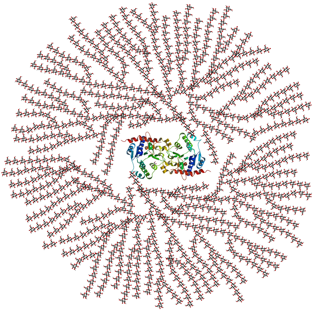
This is a chemical drawing of glycogen, a carbohydrate molecule that our bodies use to store glucose. The long chains here are made up of glucose molecules waiting to be used.
Although there are many types of polysaccharides, our bodies encounter three most often. One is glycogen. Animals use this molecule to store sugars as energy. “We find that in our muscles and our liver,” Gleeson says. The second, or starch, is “a storage form of energy in plants,” he explains. The third type is fiber, which our bodies can’t turn into energy.
When scientists talk about carbohydrates in food, they call monosaccharides and disaccharides “simple” sugars. These are common in processed foods. Polysaccharides such as starch are “complex” carbohydrates and more commonly turn up in less-processed foods, explains Kimber Stanhope. She’s a nutrition researcher at the University of California, Davis. “Rice and wheat and oats and corn — those foods are mostly starch,” she notes.
| Type of carbohydrate | Examples | Where to find them |
|---|---|---|
| Monosaccharides | Glucose, fructose, galactose, xylose | Glucose tablets or nutrition supplements |
| Disaccharides | Sucrose, lactose | Table sugar, sugar in milk |
| Oligosaccharides | Starches such as amylose and amylopectin | Starchy foods like bread or rice |
| Polysaccharides | Pectin, cellulose, hydrocolloids, guar gum | Whole fruits and vegetables, beans, jams and jellies, corn starch |
Foods we classify as “carb-rich” often contain a mix of different carbohydrate molecules. For example, a cookie will have plenty of sucrose. But it also has flour, which contains starch. Whole-wheat bread will have starch and some sugar, as well as fiber. Vegetables, such as carrots, are made of cells that contain starch, sugars and fiber.
Our bodies can use glucose molecules right away. Other carbohydrate molecules must first be broken down.
Complex carbohydrates “are relatively large molecules,” Gleeson explains. The gut can’t directly absorb them. They must be digested — their polysaccharide chains broken apart — to release their glucose units.
That digestion starts in the mouth, Gleeson explains. Saliva contains enzymes — molecules that speed up chemical reactions. “Your spit contains some enzymes, which break down the long-chain carbohydrates into disaccharide units,” he says. Those much simpler sugars head to the stomach. But, he adds, little carbohydrate digestion goes on there. Why? “The enzymes that are coming [in with] the saliva,” he explains, “are inactivated by [stomach] acid.”
After the stomach, “you’ve mixed everything up into a nice sort of soup-like consistency,” called chyme (KYME), Gleeson explains. “That’s sort of squirted, almost, into the small intestine. And there we secrete enzymes into the small intestine from the pancreas.”
Those enzymes finish the job, splitting disaccharides into monosaccharides such as glucose. Our blood then absorbs the glucose and moves it around the body.
When that blood reaches the liver, an enzyme grabs glucose from it. Called glycogen synthase (GLY-koh-jen SIN-thays), this enzyme links up the glucose molecules to form glycogen. Glycogen then gets stored in the liver and muscles. Fat cells also store glucose in the form of triglycerides (Try-GLIH-sur-ides). The body will draw on these sources when it needs more energy.
This liver-enzyme reaction has turned carbs from food into a stored fuel. The liver turns off this enzyme when it determines that it has made enough glycogen.
The body aims to keep blood glucose levels fairly constant. So when cells draw down this sugar, the liver begins breaking down some stored glycogen, releasing its glucose into the blood.
That’s especially important during exercise, as your muscles burn through glucose to power activity.
It’s a different story for fructose. This monosaccharide pairs up with glucose to make sucrose. But it’s also found on its own in fruits, vegetables and honey. Manufacturers have also sweetened many sugary foods and drinks with this sugar in the form of high-fructose corn syrup.
In the liver, an enzyme called fructokinase (Frook-toh-KY-nase) sets off the first step in turning fructose into glycogen. But unlike the enzyme that processes glucose, fructokinase “doesn’t seem to get turned off — ever,” explains Stanhope at UC Davis.
So if you’re drinking a sugary soft drink, “86 percent of all the fructose you’re consuming in that … drink goes into the liver,” Stanhope says. “The liver does the best it can with all this fructose, but now it’s overloaded.” The liver will now turn any excess into fat, she explains.
Too much fat can hamper how the liver reacts to hormones such as insulin. As a result, the organ stores even more sugars as fat. “You just keep going around in circles and making the situation worse,” Stanhope explains.
That’s why eating too much fructose can be a problem. The issue isn’t fructose from fruits and vegetables, though. Consider a sweet fruit, such as grapes, Stanhope says. “They basically average around 16 percent of their weight as sugar.” But fruits and veggies also contain fiber and other complex carbohydrates, as well as vitamins and other nutrients.
Most of the sugar in our diet doesn’t come from fruit but from sweetened baked goods and drinks. Oreos, for instance, are 38 percent sugar, of which fructose is half.
Many sweets have even higher levels of fructose than that. Some highly processed foods — not only soda but also canned soups and some sauces — can contain a type of corn syrup that is 55 percent fructose (five percent more fructose than in table sugar), Stanhope says.
What really worries her are some corn syrups where the share of fructose is higher still. One 2014 study tested sodas and juices containing high-fructose corn syrup. Many of these drinks contained 60 percent fructose — some even close to 70 percent. That’s a lot for the liver to handle.
While our bodies can break down starches and sugars for energy, other carbohydrates are tougher. Consider fiber.
Fibers are polysaccharides found in plants. Our bodies cannot fully break these down. Fibers include the cellulose that makes plant cell walls stiff. They include the pectins (PEC-tins) that firm up fruits. You’ll find them in whole fruits, vegetables, beans and whole-grain products (such as whole-wheat bread).
Our bodies can’t convert fiber into energy. But our bodies still find it useful, notes Leslie Cho. A cardiologist, or heart specialist, she works at the Cleveland Clinic in Ohio.
Fiber comes in two types. Soluble fiber, as its name suggests, dissolves in water. This type shows up in foods such as apples, bananas, avocados, citrus fruits, carrots, beans and oats. By acting like a sponge, Cho says, “it helps you lower your cholesterol and glucose.”
Cholesterol (Ko-LES-ter-ol) is a fatty material that drifts in our blood. Over time, it can collect in blood vessels and increase someone’s risk of heart attack or stroke. By soaking up cholesterol, soluble fiber helps eliminate it.
That sponge-like fiber can also trap glucose so that it takes longer for those sugar molecules to enter the blood stream. If you eat a lot of sugar and no fiber, Cho explains, “you absorb glucose very quickly and you tend to get hungry again.” Soluble fiber keeps you feeling fuller longer. And it’s especially helpful for people with high blood sugar, such as those with type 2 diabetes.
The other fiber — the insoluble type — “doesn’t dissolve in water, and it helps you with your stomach and your intestine,” Cho says. Vegetables, whole-wheat bread, potatoes, beans, nuts and seeds all contain this type.
This fiber even helps you poop; it’s one of the ways your body takes out the trash. How? Insoluble fiber adds bulk and pulls water into your stool. That keeps you pooping just often enough for comfort.
Soluble and insoluble fibers also serve as an important food for the bacteria in your microbiome. Those are the bacteria that live in your gut, Gleeson explains. “They have a big influence on your immune system and on your health.”
Bacteria can break some fiber down into molecules that affect our immune and other systems. These bacteria “also produce chemicals that can communicate with the brain,” Gleeson says. In this way, “They influence your mood and your sleep.”
Most people eat too little fiber, Gleeson says. Diets with lots of simple sugars don’t have enough fiber to keep our microbes happy. We’re talking about meals that contain lots of white bread or rice, cookies and cakes — and too few fruits, vegetables, beans or whole grains.
Submit your question here, and we might answer it an upcoming issue of Science News Explores
Like proteins and fats, carbohydrates are an important macronutrient. But do people need them?
The body needs certain fatty acids and amino acids to build cells and operate effectively. That’s why we need to eat fats; they provide some essential fatty acids that our bodies can’t make, notes Gleeson. And, he adds, “Protein is absolutely essential, because there are [also] amino acids in those proteins” that we can’t make on our own.
But technically speaking, he says, you can live without eating carbohydrates.
Glucose is an essential fuel for the brain, Gleeson explains. Fortunately, the body can make it from fats, amino acids and water. Getting glucose this way wouldn’t be a very tasty life, though. And people who live on extremely low-carb diets can miss out on fiber, vitamins and other nutrients.
But for some carbs, reducing how much of them you eat isn’t a bad idea. We don’t need nearly as much sugar — especially soft drinks, candy or cake — as we eat, observes Stanhope at UC Davis.
“There is simply no doubt,” she says: “Evidence shows sugar is detrimental to our health when consumed at high levels.” The problem: “We just don’t know for sure how high ‘high’ is.”
Because of worries about too much sugar, some people try diets with as few carbohydrates in them as they can manage — eating mostly meat and a few veggies. But such low-carb diets won’t necessarily result in a healthy body, Gleeson points out.
Living the low-carb life tends to be high in fats and protein. Long term, all that fat and protein can put people at risk for heart disease, type 2 diabetes and problems with their kidneys (the organs that produce urine). People on low-carb diets who don’t eat enough fiber also don’t poop enough, suffering constipation.
And if you’re an athlete, your body definitely needs carbs. Athletes perform their best with carbs, Gleeson has shown in his research. Complex and simple carbohydrates are the easy energy our bodies use to work hard.
“As soon as you start doing hard running, sprinting as fast as you can, performing any all-out efforts, then you’re going to be using carbohydrate,” he says.
“There is no food group that’s bad,” Cho adds. “You can’t eliminate one whole thing and think you’re doing a good job.” Although high doses of simple sugars can cause problems, other carbohydrates don’t. “We often [confuse] ‘good’ carbohydrates with ‘bad’ carbohydrates,” she notes. And unfortunately, that can give important nutrients a bad rap.
The trick is eating enough carbs — and a diverse mix of them — to give us energy and a great-tasting diet. Just don’t overdo it.
“Moderation is important, even though these things taste good,” Cho says. And much as cookies, soda and candy are delicious, they don’t contain fiber or other nutrients. “Instead of having five chocolate bars, maybe you can just have one.”
Editor’s note: The enzyme used to transform glucose to glycogen was originally misreported as phosphofructokinase. This was corrected to glycogen synthase on September 9. In addition, fat stores glucose not as glycogen, but as triglycerides.
In a study of 2,266 workers in Finland, evening-type people, or night owls, were up to three times more likely to be treated for depression or anxiety than morning types, with poor work-life balance identified as a key contributing factor.
The post Study: Night Owls Face Higher Mental Health Risks Tied to Work-Life Balance appeared first on Sci.News: Breaking Science News.
The Federal Bureau of Investigation looks almost unrecognizable under director Kash Patel’s leadership.
The agency’s head has been embroiled in a seemingly endless list of controversies, from allegations of excessive drinking to buying up location data on everyday American citizens to furthering of harebrained conspiracies.
Patel lacked virtually any degree of relevant working experience for the job before the Senate confirmed him — and now, the agency is opening the doors for even more highly questionable hirings.
As inside sources told CBS News, the FBI has quietly lowered standards of its background checks for potential federal agents and other personnel. The bureau is now officially looking the other way when it comes to prostitution, for instance — as long as recruits didn’t hire sex workers over three times.
Even more eyebrow-raising is that the FBI is even allowing the consideration of candidates who engaged in bestiality or animal cruelty, according to CBS‘ sources. However, any such incidents would’ve had to have taken place before the candidate turned 18.
The broadcaster’s sources also said there was no distinction between candidates who were forced into sexual activity with animals or if they did so willingly. An FBI spokesperson claimed that eligibility requirements were limited to a “narrow set of circumstances, such as in cases where a person was sexually abused with animals as a child.”
Experts, however, were appalled at the news, arguing that the FBI is undermining its responsibility in vetting applicants by changing background check standards.
“Bestiality is clearly a potential indication of a more serious emotional issue, and even in a circumstance where it is not, the pool of candidates should be extensive enough that the criteria should certainly not be eliminated,” former FBI agent James Davidson told CBS.
Before the changes, which were reportedly implemented earlier this summer, the FBI’s webpage was clear about automatically disqualifying special agents who engaged in “prostitution or bestiality.”
Both current and former FBI employees told the CBS that the bureau could now be hiring “people with questionable backgrounds,” allowing them to undermine the integrity of the service. Others warned that the news was evidence of a major “recruiting problem.”
Considering the fact that the agency is being helmed by a personality as controversial as Patel, these changes shouldn’t come as too much of a shock.
While he hasn’t personally acknowledged the latest changes, Patel has frequently called for new hiring standards at the bureau, arguing that “we are losing too many qualified candidates to the rigors of the requirements from yesteryear” in a July interview with former FBI agent John Nantz.
Patel’s motivation for lowering standards could be to bring in his closest associates. As ProPublica reported in November, his personal assistant didn’t clear their polygraph test and required a waiver, claims the FBI has since denied.
More on Patel: The Head of the FBI Just Admitted Something Moderately Horrifying
The post FBI Now Allowing History of Bestiality Among New Recruits appeared first on Futurism.
Diseases that cause nerve cells in the brain to die over time are known as neurodegenerative diseases. These cells, known as neurons, are vulnerable to DNA damage because they are highly active cells that require large amounts of energy. Neural DNA damage is a normal part of aging as changes to the DNA sequence, called mutations, build up over time.
Previous scientists have shown that neurons from people with the neurodegenerative diseases Alzheimer’s disease (AD), frontotemporal dementia (FTD), and amyotrophic lateral sclerosis (ALS) accumulate unusually high numbers of changes in the DNA sequence, called somatic mutations. However, researchers don’t know what causes the high number of mutations or whether the same process causes them in all 3 diseases.
To answer these questions, researchers at Boston Children’s Hospital and Harvard Medical School analyzed brain tissue from people who died with and without AD, FTD, or ALS. They compared the processes that cause DNA mutations in the 3 diseases by first accessing the DNA inside individual neurons from healthy and diseased postmortem brain tissue.
To do this, they broke apart the brain tissue and added a fluorescent marker that identified the center of each neuronal cell where DNA is housed, called its nucleus. They used a sorting machine to separate the nuclei from the rest of the cell material to access the neuron’s DNA. Using a DNA sequencing method called single-cell whole genome sequencing, the team then sequenced each neuron’s DNA to identify mutations.
They analyzed 159 neurons from people with AD, 61 neurons from people with FTD, 77 neurons from people with ALS, and 232 neurons from healthy individuals. This allowed the team to compare the number and types of DNA mutations between diseased and control (non-diseased) neurons. The researchers found that compared to the control neurons, which typically accumulate only 200 to 300 mutations over an 80-year lifespan, neurons from all 3 diseases contained an unusually high number of 2-base-pair deletions, often exceeding 1,000 mutations per cell.
When a biological process damages DNA, it leaves behind its own unique signature. By identifying these signatures, scientists can identify the biological process that likely caused the damage. Since these 2-base-pair deletions were so frequent in diseased neurons, the researchers wanted to know whether they were left behind by a biological process. The researchers used a machine learning algorithm to scan DNA sequences for recurring mutation patterns and identified a signature in neuronal DNA, which they named ID-4.
When the researchers compared the ID-4 signature with a database of previously characterized signatures, they found that it was associated with a DNA repair protein called TOP1. TOP1 temporarily cuts and rejoins DNA to relieve the tension that builds up during normal cellular activity. TOP1 typically detaches from the DNA after completing this process. However, if TOP1 fails to detach, it breaks one of the two DNA strands.
To test whether abnormal TOP1 activity was breaking the DNA, the researchers isolated DNA from control and diseased neurons. They visualized the DNA on a gel, where intact DNA forms bands while broken DNA spreads out into smears. The more fragmented the DNA is, the more intense the smear appears. Using a machine that quantified the intensity of smears, the researchers found that DNA from diseased neurons produced smears that were up to 3.2 times more intense than those from control neurons. They also found that neurons with more DNA fragmentation had more ID-4 signatures.
Based on these findings, the researchers concluded that abnormal TOP1 activity created frequent single-strand DNA breaks. They proposed that some of these breaks are repaired incorrectly, eventually leading to widespread DNA breaks in all 3 neurodegenerative diseases.
Overall, the researchers found that neurons from these 3 neurodegenerative diseases accumulated excessive DNA mutations despite differences in the disease symptoms and underlying causes. Their findings also provided evidence that abnormal TOP1 activity contributes to this mutation accumulation. Based on these findings, they stated that preventing TOP1 from becoming trapped on DNA, or improving the DNA repair process that removes trapped TOP1, could be a promising way to treat a wide range of neurodegenerative diseases.
The post Common DNA damage could cause neurodegenerative diseases appeared first on Sciworthy.
