The symptoms: The woman started occasionally hearing her name being called when she was alone, usually in quiet surroundings. Later, the sounds evolved into indistinct murmurs. These voices seemed to come from outside her head rather than from her own thoughts. They never spoke directly to her, commented on her actions or gave commands.
What happened next: In the next two years after she began hearing voices, the woman made several trips to emergency departments and had brief psychiatric admissions. Because hearing voices is commonly associated with psychosis — symptoms that occur when people experience a disconnect from reality — doctors diagnosed her with "unspecified psychosis" and attempted various treatments.
They first tried the antipsychotic risperidone, gradually increasing the dose, but the voices did not go away. Doctors then switched the patient to aripiprazole, but the hallucinations remained unchanged. Next, they tried haloperidol, and although the woman said the medication made her feel calmer and less distressed, the voices persisted.
As they conducted further psychiatric evaluations, the doctors noticed that the woman frequently leaned forward during conversations, cupped an ear and asked people to repeat themselves. About four to six months after her first contact with a psychiatric team, she was referred for hearing tests. These tests revealed hearing loss in both ears, ranging from moderate to severe in one ear and mild to profound in the other.
About two months later, the woman was fitted with hearing aids in both ears, which improved her hearing. The voices did not go away, however.
Brain scans showed no abnormalities, and blood tests came back normal. Neurological evaluations turned up no evidence of other explanations. Meanwhile, the woman continued working full time, managing her household and maintaining an active social life. She showed no signs of paranoia, delusions, disorganized thinking or the decline in daily functioning that doctors would typically expect to be associated with a psychotic disorder.
The diagnosis: Based on the woman's stable functioning, hearing impairment and lack of other clinical abnormalities, the doctors concluded that her auditory hallucinations stemmed from sensory deprivation caused by hearing loss.
Reduced sound input from the ears can make auditory regions of the brain unusually active, causing them to "fill in" missing sounds, the doctors explained in a report of the case. A related phenomenon is known as musical hallucinosis, in which people with hearing impairments hear songs, melodies or other forms of music that are not actually playing.
The treatment: Because neither antipsychotic medications nor hearing aids eliminated the patient's hallucinations, doctors shifted their focus toward helping the woman cope with them.
They taught her about the connection between hearing loss and hallucinations and advised her to pursue psychotherapy aimed at reducing the distress associated with the voices and improving her coping strategies for dealing with their persistence. At the time the report was published, in May 2026, she was waiting to begin this therapy.
What makes the case unique: In their report, the doctors noted that most published accounts of this phenomenon have described patients' hallucinations improving or resolving after their hearing impairment was treated. Persistent hallucinations after treatment are rare. In the woman's case, however, the voices persisted despite the use of hearing aids.
The doctors suggest that prolonged hearing loss may cause lasting changes in the brain that do not immediately reverse once hearing improves. (The case report does not note how long the woman's hearing had likely been impaired.)
The case also serves as a reminder that hearing voices does not always indicate psychosis, the doctors concluded. Because the woman's hearing loss was recognized only after years of symptoms and several unsuccessful medication trials, the report authors argued that doctors should consider running hearing assessments early in patients with isolated auditory hallucinations, especially when their insight and daily functioning remain intact.
Scientists say they have built a "synthetic cell" that can eat, grow and divide in a way that's remarkably similar to living cells.
The research, released to the preprint database bioRxiv July 2, has not been peer-reviewed yet. It introduces SpudCell, a new type of artificial cell, and marks a striking step toward creating living cells from scratch. But for study co-author Kate Adamala, a synthetic biologist at the University of Minnesota, that's far from the most interesting part of the work.
"I do not believe [SpudCell] is alive," Adamala told Live Science. Instead, she describes the system as a framework that could generate "all the chemicals we need for our civilization with biology." The thought is that SpudCell could serve as a tiny factory, pumping out medicines, fertilizers, plastics or any number of other compounds.
The work's announcement has sparked some controversy, with some scientists seeing it as a ploy to gain media attention for the simultaneous launch of the author's nonprofit Biotic, which aims to raise money to further develop the SpudCell platform. Adamala does not take umbrage to that criticism, as she is keen to bring more attention and funding to her field. If a synthetic cell can be perfected, she thinks, it could help humanity generate chemicals without relying as heavily on petroleum products.
"I feel this incredible stressful urgency that if we don't get to work on it now, then we're going to run out of time," she said, referring to the climate crisis. "We need to highlight that bioengineering can offer a solution. That's why I'm doing it."
A synthetic cell and a big promise
According to the preprint, Adamala and her team produced a lifelike system that closely resembles a living cell. To do so, they combined 36 purified enzymes and a fatty membrane with a pared-back genome about 50 times smaller than that of an average bacterial cell. By mixing these human-made elements together, the scientists generated a cell that could feed, grow, and divide — so, in essence, they created a full cell cycle in a petri dish.
"We built a cell-like system that is fully chemically defined, so there are no unknown building blocks in it," Adamala said. "It's capable of doing things that people up until now used to think only natural living cells can do." They call the system "SpudCell" because it looks similar to a potato, The New York Times reported. The name is also a nod to the Sputnik satellite, per CNN.
The concept of recreating the cell cycle in a dish is not entirely new. The J. Craig Venter Institute's 2016 "minimal cell" paper flirted with the concept by stripping as many genes from a bacterium as possible to leave only a minimal cell that could still replicate. However, the new study is the first time scientists have achieved feeding, growing and division using a "bottom-up" approach.
The work marks "a great advancement," said Mauro Rinaldi, a lecturer in biotechnology and biochemistry at the University of Hull in the U.K. who was not involved in the work. "It moves the needle because one of the key things about cells is division."
But there are important caveats. For one, the cells cannot yet create their own energy as our cells do with mitochondria. They also rely on externally provided fats, sugars and enzymes. The cells cannot make their own ribosomes, the machinery that turns genetic instructions into working parts of the cell. That means it relies on proteins being delivered from the outside.
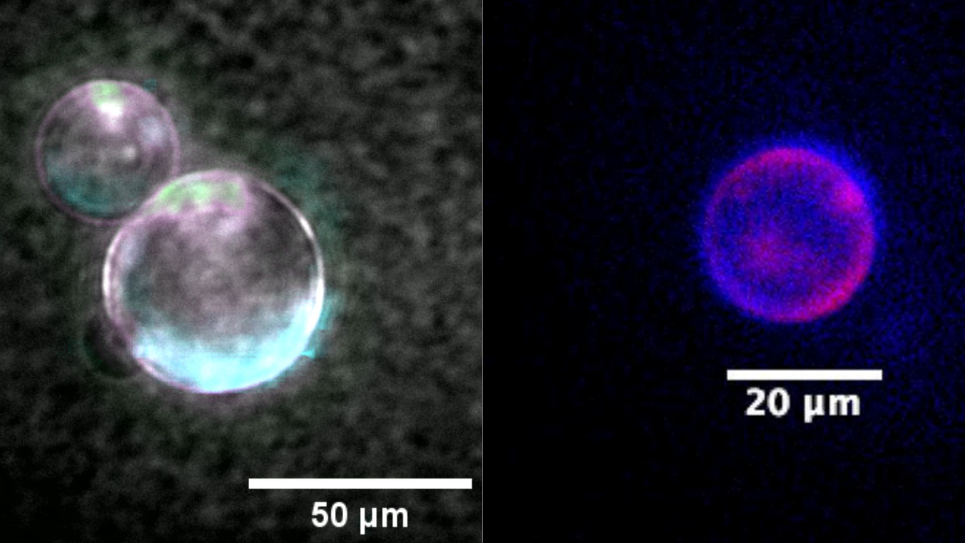
Left: A super-resolution image of SpudCell's liposomes with an encapsulated genome and active protein expression. Right: A SpudCell encapsulates a whole genome. The DNA of the genome and the synthetic cell membrane are stained with fluorescent dyes. (Image credit: Orion Venero, Adamala Lab)
Another difference is that SpudCell's genome is spread out over bits of DNA called plasmids, rather than being neatly packaged in chromosomes. It does not possess the skeleton that cells typically use to neatly split DNA during cell division, so consequently, the division of SpudCell's DNA to its daughter cells can be somewhat haphazard.
"The description of the results leaves me with substantial technical questions regarding the nature and the robustness of the findings," said Cees Dekker, a biophysicist at the Delft University of Technology in the Netherlands who was not involved in the work. "[Its] approach uses some ingenious engineering tricks as shortcuts to achieve complex functions such as growth, but a major challenge remains to create a truly autonomous cell that executes all these functions without external help," he said.
Dekker is among the scientists who would have preferred that the research made the news after being published in a peer-reviewed scientific journal. "If these findings are solid, that great media attention is definitely warranted; if peer review would, however, reveal weaknesses or issues, all the current media reporting is premature," he argued.
From cell to manufacturing platform
Adamala sees SpudCell as a blank slate for engineering. "We believe that if we make a cell from scratch, that's fully engineerable," she said.
Cells have long been used to produce chemicals for human use. Millions of people use synthetic insulin made in bacteria and yeast cells, for example. But cells resist making chemicals that could be harmful to them, and through evolution, they've developed systems to prevent them from doing so. A SpudCell-like system could circumvent those natural hurdles, Adamala and her colleagues think.
That kind of platform could also be useful for making newer generations of medicines, such as those based on mRNA or peptides. These drugs use molecular building blocks, like amino acids or nucleotides, that have been chemically tweaked to make the molecules more stable or harder for the body to break down. SpudCell could be engineered to produce such modified components directly, rather than scientists having to synthesize them in traditional chemistry labs. This could potentially shorten development timelines and lower costs, the team thinks.
Adamala and her colleagues also envision the cells being used as easily shippable laboratories. They could be dried out, shipped, stored without refrigeration, and then activated on-site to make chemicals, vaccines or proteins when and where they are needed.
But there's still a long road ahead. For now, SpudCell is only a proof of principle, and many hurdles must be addressed before it could become an industrial platform.
"[SpudCell] is not an engineering platform that can give you any useful product, but it's step one," Adamala said. "It's probably at least a couple of decades from now when we can actually scale it up to the point where we can replace all the petrochemicals with biology, but I do believe it's doable."
Adamala hopes the nonprofit Biotic will help funnel donations from philanthropists directly to research. "Biotic is a funding agency that is going to globally fund this work," she said.
While the technology is promising, "it needs to go through peer review," Rinaldi said. "I expect a lot of the hype and some of the terms that they're using to go away after a couple of years have passed."
Scientists uncovered a link between mild heart trouble and microscopic signs of brain damage that may raise the risk of memory problems down the line.
The new study, published Monday (July 6) in The Journal of Neuroscience, did not prove that the heart issues directly caused memory problems. But it "adds to the overall picture that preserving healthy brain-heart communication is key for healthy aging," said Dr. Jan Scheitz, a consultant stroke neurologist and head of the Brain-Heart Lab at the Charité University Hospital in Germany, who was not involved in the study.
"A healthy lifestyle that protects the cardiovascular system will not only benefit the heart but also the brain," he told Live Science in an email.
The work could one day help doctors identify patients at risk of memory problems early on through routine heart-function tests, said study co-author Dr. Xia Zhang, a doctoral researcher at the Max Planck Institute for Human Cognitive and Brain Sciences in Germany.
"The broader implication is that the brain may show subtle tissue-level changes related to cardiac dysfunction before we see obvious brain shrinkage or clinical dementia," Zhang told Live Science in an email.
So far, though, scientists have only studied changes in the brain in people already diagnosed with heart conditions. They haven't necessarily assessed the changes that take place earlier on.
In the new study, researchers followed 73 patients who had sought care for heart-related symptoms at the Heart Center Leipzig in Germany, some of whom had confirmed coronary artery disease and/or heart failure. They also assessed 95 people without any heart-related symptoms. The researchers measured heart function at the start of the study. Then, about 3.5 years later, they performed cognitive testing and an MRI scan of the brain to take a snapshot of its structure.
Among all 168 study participants, those whose hearts pumped blood less efficiently at the start of the study showed greater signs of tissue damage in their gray matter years later. This was true regardless of whether or not they had diagnosed heart failure.
The cognitive tests were given only to the heart-disease patients, and they assessed attention, executive function, learning and memory. Only memory showed a link to weaker heart pumping. The participants with weaker heart pumping showed more microscopic damage in memory-related brain regions, and those brain changes correlated with worse memory scores.
The degree of brain damage also correlated with the levels of stress hormones released by the heart, but this link was only seen in patients who already had heart failure.
"What surprised us most" was that subtle reductions in the heart's ability to pump were tied to later brain changes, even in patients who did not meet clinical criteria for heart failure, Zhang said.
One limitation of the study is that the researchers didn't take MRI scans at the start. That means "we do not know whether some changes were already present at the beginning of the study period," Scheitz noted. This is something that Zhang and her colleagues plan to test in future studies.
"The next step for the field is replication in larger cohorts with multiple time points, so that cardiac function, brain microstructure, and cognition can be followed more precisely over time," she said.
The brain damage flagged in the 73 patients happened in parts of the brain that are important for memory and are vulnerable in Alzheimer's disease. "These regions helped identify a possible brain pathway through which poorer cardiac function may contribute to later memory problems," Zhang said.
However, until this idea is tested directly in future studies, it is too soon to say if the heart function-related brain changes seen on the MRI scans point to a higher risk of Alzheimer's, Zhang cautioned. The team did not measure proteins related to Alzheimer's, so they cannot conclude that the patients with measurable brain changes are developing Alzheimer's disease.
The study also did not directly test the effect of exercise on heart and brain health, but the findings may help explain why regular exercise is often linked to better brain health and cognitive aging, Zhang said.
"Regular exercise supports cardiovascular function, vascular health, and cerebral blood-flow regulation," she said, "all of which may help protect brain tissue over time."
This article is for informational purposes only and is not meant to offer medical advice.
What do you know about the body's hardest-working muscle? Find out with our heart quiz!
The symptoms: The patient went to the emergency department after experiencing nausea and vomiting for about a month, along with decreased appetite. She told doctors that she'd also experienced a burning pain in her upper abdomen and right side of her torso, which wrapped around to her back. She'd tried treating these symptoms with two common over-the-counter medications for acid reflux, but the treatments didn't help.
In the year prior, the patient had started taking semaglutide, a type of drug known as a GLP-1 receptor agonist — the same class of drug as Ozempic. These injected drugs are used to treat type 2 diabetes and assist in chronic weight management; in this case, the woman had a history of type 2 diabetes and obesity. Since starting the medication, she'd lost about 40 pounds (18 kilograms), but notably, that weight loss had accelerated over the previous month, she noted.
What happened next: Doctors took a CT scan of the patient's abdomen and pelvis. This revealed that the bile ducts, which move bile from the liver to the gallbladder and small intestine, were slightly dilated. The woman also had "heavy stool burden" but no signs of obstructions in her bowels, and her stomach was slightly stretched out due to carrying "semisolid material."
To take a closer look, the medical team then used magnetic resonance cholangiopancreatography, a noninvasive scan that examines the bile ducts, gallbladder, pancreas and pancreatic ducts. The scan confirmed that the bile ducts were dilated and revealed a mass in the stomach that appeared to be trapped air. The doctors then examined the upper digestive tract, from the esophagus to the top of the small intestine, with an endoscope.
The diagnosis: The endoscopy revealed a large mass in the woman's stomach, known as a gastric bezoar. These masses are tightly packed blobs of partially digested or undigested materials.
The treatment: Semaglutide and other GLP-1 agonists typically cause delayed gastric emptying, which means they slow the rate at which the stomach empties its contents into the intestines. The woman was taken off that medication upon hospital admission.
The next step with a bezoar is to try and dissolve the mass while it's still in the stomach — and evidence suggests a good way to do this is to give the patient soda.
"Existing evidence, largely from case series and anecdotal experiences, supports the administration of 3 liters [0.8 gallons] of cola, either orally or through a nasogastric tube, within a 12-hour window," doctors noted in a report of the case. "It is not well understood whether acidity, carbonation, or another mechanism accounts for dissolution of the bezoar."
Due to the patient's history of diabetes, she was given diet cola. She also didn't enjoy carbonated beverages, so the typical prescription of 3 liters of soda was cut down to 1.5 liters (0.4 gallons). On the second day after starting this treatment, the patient noted a "tugging" sensation in her abdomen followed by relief of her nausea and abdominal discomfort. An endoscopic exam revealed that the bezoar was no longer stuck in her stomach.
The woman then transitioned to a typical diet in the hospital, and by the time she was discharged, she had no nausea, vomiting or abdominal pain. She did not restart semaglutide but was prescribed acid reflux medication to take daily. She reported that her appetite increased after hospital discharge and her weight rebounded a bit over the following months. Her abdominal symptoms had not returned within a few months of discharge.
What makes the case unique: Gastric bezoars are fairly rare, the report noted. They are found in less than 0.5% of endoscopies of the upper digestive tract. Common symptoms of the condition, such as pain, nausea, vomiting and abdominal discomfort, also appear in many more-common disorders, so they don't necessarily point to the presence of a bezoar in all cases.
The most common type of gastric bezoar is formed of plant fibers and called a "phytobezoar." Eating lots of certain foods, such as persimmons, pineapples, raisins or celery, can raise the risk of phytobezoars because the plants contain a high concentration of indigestible components, such as cellulose, lignins and certain tannins.
However, various medical conditions, procedures and drugs can also raise the risk of bezoars. These include anatomical changes related to bariatric or other gastric surgeries, autonomic nerve damage, and medications that cause delayed gastric emptying, such as GLP-1s. In the woman's case, semaglutide was likely to blame for her bezoar, and discontinuing the treatment helped resolve the problem.
The cola she consumed also helped. Although that course of treatment sounds strange on paper, it is the preferred first-line treatment over using a scope to physically break up the bezoar in the patient's stomach.
"Bezoars formed from food material may be initially managed with oral administration of cola in patients in a clinically stable condition," the report said. "This intervention is generally cost-effective and is associated with a lower risk of complications than invasive procedures."
Want to speed up your recovery after an injury or a workout? Some influencers are fans of shots of BPC-157 and TB-500 for that. Want scar-free, youthful skin and thick hair? Reddit posts rave about GHK-Cu and KPV. Want to build beautiful, bulky biceps? Some fitness influencers swear shots of ipamorelin will do wonders. This is the world of peptides — an assortment of chemicals that promise to boost your body and health — and people are self-injecting them in a growing wellness trend.
Promoted by bodybuilders and influencers, supporters of the "Make America Healthy Again" movement and Silicon Valley early adopters, peptides promise results that leave people better than well, fitter than fit. Yet despite getting glowing reviews on social media, most peptides have limited clinical evidence to support health benefits, whether it's helping to heal a rotator cuff injury, improving libido or building muscles. There's not much information out there on the drugs' effectiveness and even less on their safety.
In 2023 the Food and Drug Administration banned several peptides, including BPC-157, GHK-Cu, KPV and ipamorelin, from being produced in the U.S. by compounding pharmacies — facilities that legally make non-FDA-approved medications for individual use — because of "significant safety risks." Many peptides are imported from outside of the country, and many made here are marketed for research use only. People who desperately want the promise contained in the tiny vials seek them out on online on legally dubious gray markets.
But accessing the shots might soon get easier. On the podcast The Joe Rogan Experience on February 27, Robert F. Kennedy, Jr., secretary of the Department of Health and Human Services, proposed legalizing the compounding of 14 peptides — and the FDA seems poised to grant his wish. Earlier this week the agency released plans to hold a meeting with independent advisers in July to review whether some U.S. pharmacies should be allowed to manufacture certain peptides, including BPC-157, TB-500 and KPV.
If the lift on peptides happens, a flood of people could start using the drugs, regardless of any safety concerns.
A Peptide by Many Names
A peptide is a chain of two or more amino acids — building blocks of proteins. The body's cells can produce numerous peptides, which perform various tasks or serve as signaling molecules.
Insulin is a peptide. So is human growth hormone — and semaglutide, the active ingredient in glucagonlike peptide 1 (GLP-1) receptor agonist drugs such as Ozempic and Wegovy. (GLP-1s are FDA-approved for managing type 2 diabetes and for weight loss). Buzzy new injections such as BPC-157 and GHK-Cu — touted for tissue repair — are derivations or synthetic versions of naturally occurring peptides. The public and online health retailers are starting to use "peptides" as a catchall term for any chemical taken for well-being, energy, exercise recovery, and more, says Luke Turnock, a criminologist who studies how people use enhancement drugs at the University of Lincoln in England.
And while people are using them as treatments, peptides aren't often described as drugs. "'Drug' has a certain stigma or negative connotation attached to it," Turnock says. The popularity of peptides has its roots in the bodybuilding and powerlifting communities, he explains, where "drug" has historically been tied to steroids — which are generally banned for professional athletes.
The term "peptides," meanwhile, stresses the natural origin of the molecules, says Flynn McGuire, who studies sports medicine at the University of Utah. "Because it's 'natural,' it is better or different, even though they're just drugs," McGuire says.
A Surge in Popularity
In the past few years, orthopedic surgeon Omar Rahman has seen a spike in interest in peptides. "I'm seeing more patients asking about peptides, often driven by the longevity and wellness space," says Rahman, who practices at Pacific Coast Sports Medicine in Los Angeles.
Because peptides encompass many products — some available by prescription, others on the gray market — the exact number of people trying them is hard to pin down. The subreddit r/peptides currently has more than 70,000 weekly visitors, and the related subreddit r/biohackers, which frequently hosts conversations on peptides, has more than 600,000. A scroll through TikTok offers pages of people sharing their "stacks" — combinations of injections people take to meet their personal goals, from muscle-building and exercise recovery to increased brainpower or a better tan.
"The real cultural tipping point in my mind seems to have been around 2022, when the GLP-1s really blew up," says Turnock, who is researching the peptide boom. He says injecting drugs became "normalized" when GLP-1 drugs approved to treat diabetes, such as Ozempic, were also shown to be effective for weight loss — and eventually became an approved treatment for obesity.
Some are turning to peptides because they feel doctors aren't giving them the help they need, says Dan Cushman, a sports medicine doctor at the University of Utah. Injuries to certain tissues, such as tendons, "are very slow to heal" with conventional treatments, he says. Mainstream treatment can also be expensive and painful. To someone desperate for relief, peptides might seem worth a try.
Peptide use is also linked to a growing interest in health autonomy, Turnock says. This is the idea "that doctors, if they're not prescribing what you ask for or they're not offering you these solutions, are acting as a barrier to your good health."
Stacking Up
McGuire, Cushman and colleagues published a review last year on one peptide they encountered frequently: BPC-157. The peptide is promoted as a way to stimulate multiple cellular pathways involved in blood vessel formation, cell growth, muscle repair and inflammation. Their review, however, found that most of the evidence of such health effects was from rodent studies, and only three small pilot studies had looked at BPC-157 use in humans.
In a regimen called the "Wolverine" stack, many people combine BPC-157 with injections of TB-500, another peptide that is supposed to promote healing but has even less research behind it. The "Wolverine" stack is named after the rapidly healing X-Men character. Add injections of GHK-Cu and KPV, and the stack is called "glow" or "KLOW." People claim that GHK-Cu increases wound healing, decreases scarring and helps regenerate collagen — and that KPV, derived from a hormone naturally found in the body, reduces inflammation. GHK-Cu is found in blood plasma and is an FDA-approved ingredient in topical antiaging cosmetics — but it is currently banned as an injectable because of safety concerns, such as the risk of immune reactions caused by impurities.
For muscle-building, the peptides ipamorelin and CJC-1295 are advertised as stimulating growth hormone release. Both have little clinical evidence behind them, however. Some people stack ipamorelin with GLP-1 drugs to try to lose fat while building muscle, though the effects of taking these injections together have not been studied.
Use at Your Own Risk
Clinical trial data for most of these peptides are thin. Not only are the effects anecdotal, but so are the doses. FDA-approved drugs, Cushman says, have been tested and reviewed not just for their efficacy but also for their safety. There is very little existing information on most of these peptides, he says, let alone on what happens if they are combined.
In many cases, the provenance of these peptides is even grayer than the gray market would suggest. "Patients are accessing peptides through online vendors, wellness clinics and compounding pharmacies," Rahman says. "That variability in sourcing is one of the biggest concerns, particularly when it comes to purity, dosing consistency and overall quality control."
The FDA does not allow production of BPC-157, GHK-Cu, KPV and ipamorelin in the U.S., for example, by compounding pharmacies because of safety concerns, so people are ordering them from overseas — usually from China. Some are buying peptides labeled as "for research only." Not all are stuck in the legal gray areas; other peptides are available from compounding pharmacies.
In Kennedy's February remarks about his aim to allow compounding of more than a dozen peptides in the U.S., he suggested that peptides made within the country would be safer than ones acquired abroad.
"Americans deserve to know the quality of the products they are buying and deserve drugs that have been proven to be safe and effective," an HHS spokesperson told Scientific American in response to questions about Kennedy's comment and the timing of the lift. "The FDA's goal is to ensure that patients can obtain FDA-approved products, and when those aren't available or can't work because of a patient's unique situation, are made by licensed U.S. pharmacies."
But even if Kennedy's plan to make some peptides easier to compound in the U.S. is enacted, it does not necessarily mean the drugs are safe or effective. The FDA does not approve or review drugs from compounding pharmacies — it just monitors the active ingredients the facilities use.
If peptides become more readily available in the U.S. market, Cushman predicts, more "people are going to just start trying [them]" — whether there are data to back them up or not. The public may experience any benefits — and perils — of peptides before scientists quantify them in the clinic.
The hantavirus outbreak that struck a cruise ship in April, killing three people and sparking fears of further spread, is over, the World Health Organization (WHO) has announced.
"Today, the final contact of a person exposed to hantavirus on the cruise ship MV Hondius completed their quarantine period, tested negative and returned home. No further cases have been reported since the 25th of May," Tedros Adhanom Ghebreyesus, the WHO's director-general, said in his opening remarks at a news conference Thursday (July 2). "We are therefore very pleased to say that WHO considers the outbreak of hantavirus over."
The total number of cases linked to the outbreak was 13. All those affected were either passengers or crew on the ship.
The outbreak began aboard the Dutch-flagged cruise ship MV Hondius after it departed southern Argentina on April 1. It involved the Andes virus, the only known hantavirus that can spread between people.
Hantaviruses are rodent-borne viruses that are found in the Americas, Europe and Asia and spread to humans relatively rarely, usually via contact with the urine, droppings or saliva of infected rodents. The Andes virus, however, has sparked short chains of human-to-human transmission in the past.
Before public health authorities were informed of the cases on MV Hondius, several dozen people disembarked the vessel on the island of St. Helena. To find them, authorities launched an international contact-tracing operation, tracking down more than 650 contacts in total who were then followed by health authorities in 33 countries and territories, according to the WHO.
Additionally, health authorities coordinated the monitoring, care and transport of the remaining passengers and crew aboard MV Hondius.
Of these contacts, those considered at the highest risk of infection were then quarantined and monitored for up to 42 days, because sometimes, hantavirus symptoms don't show up until weeks after exposure to the virus. Depending on their jurisdiction and degree of exposure, some contacts quarantined at home while others stayed in specialized facilities.
The initial unchecked travel of some contacts, alongside the virus's long incubation period, prompted concern from the public that the cluster of cases could explode into an enormous outbreak, or even a global pandemic. Many infectious-disease experts emphasized that the Andes virus does not spread easily between people and that the containment effort was going well, so the risk of a large outbreak was low. Still, some experts raised concerns that the international travel of contacts could trigger pockets of the deadly disease, and some argued that the Andes virus does have "pandemic potential."
Now, with the final contact having completed their quarantine period and no further cases reported, the WHO has declared the outbreak over. All of the contacts who were repatriated to the U.S. completed their quarantine period by June 21.
"WHO will continue working with governments and partners to advance our understanding of this outbreak and of hantavirus more generally," Tedros said. "We are also coordinating a study involving 21 countries to understand how the disease develops, which will support the development of diagnostics, therapeutics and vaccines for future outbreaks."
Besides rigorous contact tracing, the WHO credited the coordinated actions of national public health bodies as being vital to preventing the further spread of the disease. These measures included the response of the Spanish government, which created a safe zone on the island of Tenerife for the ship's remaining passengers to disembark before they were repatriated under quarantine.
As the hantavirus outbreak concludes, there's an ongoing Ebola outbreak in the Democratic Republic of the Congo and an outbreak of the Marburg virus in Uganda. These and future outbreaks will require similarly robust international cooperation, the WHO emphasized.
"The outbreaks of hantavirus, Ebola and Marburg all show why there is no alternative to international cooperation in the face of international threats," Tedros said. "No country alone can fight."
This article is for informational purposes only and is not meant to offer medical advice.
An 11-year-old boy in Ontario, Canada, has died after being bitten by a rabid bat that was resting on his face as he slept. The boy had no obvious bite or scratch marks and didn't show symptoms for the first 19 days after the incident, his doctors report.
While on vacation with his parents at a cottage in northern Ontario in 2024, the boy was woken up in the night by a bat on his nose and mouth, according to a report published in the Canadian Medical Association Journal on June 29. His father caught the bat in a cooking pot and released it outside, and because the child had no visible injuries and the bat did not seem to be particularly aggressive, his parents decided not to seek a medical assessment, the report noted.
A few weeks later, the child began to experience tingling, numbness and swelling on the right side of his face. When he was examined at his local hospital his vital signs appeared to be normal, aside from an elevated heart rate and white blood cell count.
The next day, the boy's symptoms worsened: he lost feeling in the right side of his face and his speech was beginning to slur. While waiting in hospital, he developed a fever, confusion, hallucinations and difficulty swallowing, followed by excessive production of saliva.
After four days in intensive care at McMaster Children's Hospital in Hamilton, Ontario, the child tested positive for rabies, and after five days his brain stem reflexes were absent, indicating a total loss of brain stem function. Seventeen days after admission, following a consultation between his family and medical experts, he was taken off life support and died.
Rabies is a viral disease that primarily affects the central nervous system, according to the CDC. It is mainly spread through bites and scratches from infected animals, with bats accounting for the majority of cases in North America, according to the boy's doctors.
"Bats pose a particular risk because bites or scratches may be small and are easily overlooked, and patients may not recollect or recognize a bat exposure," the medical team wrote in their report.
According to a report in the Journal of Virology in 2013, silver-haired bats (Lasionycteris noctivagans), are responsible for the majority of rabies cases in Canada. Skunks, foxes and raccoons also often carry the disease.
The rabies virus must travel to the brain before symptoms can occur, but once a patient develops symptoms the infection is almost 100% fatal, with most deaths occurring within one to two weeks of symptom development. However, prompt treatment after exposure — including wound cleaning, antibody administration and a series of vaccines — is nearly always effective at preventing the disease, according to the CDC.
Rabies in humans is very rare in North America, with fewer than 10 deaths reported in the U.S. every year, according to the CDC. In Canada, only 28 human fatalities have been reported since 1924, according to the Canadian Veterinary Medical Association. Even so, the boy's doctors have emphasized the importance of prompt medical attention following exposure to bats and other animals that could carry the disease.
"Early recognition of exposure and timely PEP [postexposure prophylaxis] remain the only effective means of rabies prevention," they wrote. "Any direct human contact with a bat, even in the absence of a visible bite or scratch, is an indication for PEP."
They added that rabid bats do not always display the classic symptoms commonly associated with rabies, such as aggressive behavior and frothing at the mouth. "Any direct human contact with [a] bat is considered high risk," they added.
This article is for informational purposes only and is not meant to offer medical advice.
The symptoms: The man visited an eye clinic after noticing floaters — small, dark shapes drifting across his field of vision — along with brief flashes of light in his right eye for six days. He was otherwise healthy and had no history of eye disease, physical trauma to the eye, or drug use. He had mild nearsightedness, for which he wore glasses.
What happened next: Doctors performed a thorough eye exam while the patient wore glasses, finding that his corrected vision measured 6/6 in both eyes, the metric equivalent of 20/20. His eye pressure was also normal.
However, a closer examination of the patient's retinas, the light-sensitive layer lining the back of the eye, revealed significant damage in both eyes, according to a report of the case.
In the right eye, doctors found multiple retinal tears and bruising, as well as a retinal dialysis, a rare type of tear in which the retina begins to separate from its outer edge where it connects to other tissues of the eye. Without treatment, retinal dialysis can progress to full retinal detachment and cause permanent vision loss. The left eye also had bruising and six small, horseshoe-shaped retinal tears: three at the top and three on the side of the retinal tissue.
Since these injuries are typically caused by sudden trauma to the eye — from a punch or sports injury, for instance — doctors revisited the patient's history and asked whether anything unusual had happened involving his eyes.
The patient, described in the case report as a "hesitant historian," later disclosed that he had been using a percussive massage gun directly on and around his eyes for the past three months. He had bought the device himself and used it weekly for several minutes at a time without medical guidance, in an effort to relieve a "feeling of tiredness" in his eyes.
The diagnosis: Doctors concluded that the patient's retinal injuries were most likely caused by this improper use of the massage gun, according to the case report. These devices deliver rapid, high-frequency pulses of pressure that are intended to relieve muscle tension. However, they can cause severe damage to delicate tissues like those found in the eyes.
The eye is filled with a gel-like fluid that helps it maintain its shape. The case report authors suggested that, each time the device put the eye under pressure, it may have briefly distorted the eye's shape. Over time, this repeated stress may have placed strain on the retina, eventually leading to the small tears.
The man's doctors suspected that his use of the massage gun had warped the shape of his eyes, placing pressure on the retinas that led to tearing. (Image credit: Mark Garlick/science Photo Library via Getty images)
This pattern of injury was also different from what's seen in typical retinal dialysis cases, they noted. The condition most often occurs in the lower, outer part of the retina, along its bottom edge. In this patient, however, the injury was found in the retina's upper region, which his doctors said may reflect repeated forward pressure from the device rather than a conventional lateral impact, such as blow to the head.
The treatment: Around 8% to 15% of retinal dialysis cases can progress to retinal detachment, which can lead to vision loss. In this case, however, the patient's vision was preserved.
The retinal tears in both eyes were treated with laser therapy; targeted laser burns create scar tissue that helps seal the retina in place and thus prevent fluid from seeping underneath. The right eye's retinal dialysis was treated with "barrier laser therapy," which similarly creates a protective scar around the affected area to reduce the risk of progression.
At a follow-up six months later, the patient's condition remained stable, with no further damage or cataract formation (clouding of the eye's lens). Doctors reported that the positive outcome was "likely due to the patient's prompt presentation soon after noticing symptoms and the immediate initiation of treatment."
What makes the case unique: Eye injuries linked to massage guns are rarely reported. The small number of cases that appear in the medical literature suggest they can sometimes cause serious or permanent damage.
In one such case, a 69-year-old man developed severe eye complications, including lens dislocation and glaucoma (a sudden rise in eye pressure) after using a massage gun near his eye for several weeks. In another case, a 38-year-old woman developed a dense traumatic cataract — a type of cataract that forms due to physical injury — and severe vision loss after using a similar device over her temple and eye region.
The Edinburgh man's case is the first to report retinal dialysis and multiple retinal tears linked to massage gun use. Such injuries are usually associated with high-energy trauma, such as road traffic accidents, sports injuries or a direct blow to the eye, or they occur in people with underlying risk factors, including severe nearsightedness or age-related eye changes, the case report authors noted.
In this case, the young man had none of these known risk factors. The extent of the damage in the absence of these risk factors raises concerns about the effects of improper use of massage guns near the eyes, his doctors concluded.
Massage guns are widely sold for at-home use, but there are currently no standardized safety guidelines for using them on or near sensitive areas, such as the eyes. The authors of the case report said the man's case highlights the need for clearer manufacturer warnings and greater awareness of the potential risks of improper massage gun use.
'A silent pandemic': How Japan is curbing antibiotic resistance, $5 at a time
Antimicrobial resistance is a "silent pandemic," posing huge threats to public health while raising little attention. To curb resistance, doctors must use antibiotics sparingly and responsibly. This report is the first in a series comparing antibiotic use in Japan and the United States, with a focus on outpatient pediatrics. It was supported by a reporting fellowship from the Association of Health Care Journalists and The Commonwealth Fund.
About a decade ago, the Japanese government spotted a worrying pattern: Pediatricians were doling out a ton of antibiotics, well beyond what should be needed to treat the bacterial infections coming through their doors.
Antibiotics treat bacterial infections, not those caused by viruses, fungi or parasites. Yet doctors were often sending young patients home with antibiotics for illnesses unlikely to be bacterial. Treating nonbacterial infections with antibiotics can fuel antimicrobial resistance (AMR) and the rise of superbugs by unnecessarily exposing bacteria to the drugs, pressuring those bacteria to evolve strategies to survive. Resistant bacteria can then spread their adaptations to others, thereby compounding the problem.
In the long run, resistance could make common infections impossible to treat with existing drugs, raising the risk of serious illness and death across the population.
When pediatricians in Japan did treat bacterial infections, they were overusing the antibiotics that are likely to fuel resistance — "broad-spectrum" drugs that target many bacteria at once. When compared against 35 other high-income countries in 2015, Japan ranked dead last in the appropriateness of antibiotic choices for kids under 5.
One way to slow the development of AMR is to get antibiotic prescriptions under control. So Japan focused on one of its biggest sources of problematic antibiotic use: pediatric outpatient clinics.
"The clinics are a particular problem," said Dr. Yusuke Okubo, chief of clinical epidemiology and health services research at the National Center for Child Health and Development, a research center and hospital in Tokyo. Looking at Japan's overall antibiotic use, "90% of prescriptions are [from] outpatient clinics, not hospitals," Okubo told me. (Outpatient practices account for a similar proportion of antibiotic prescriptions in the U.S.)
A large proportion of problematic prescriptions were being written for children under 3 years old, especially those with upper respiratory tract infections, which nine times out of 10 are caused by viruses, Okubo said. The stomach bug gastroenteritis was another biggie for overprescription, despite most often being caused by a virus.
Government officials hatched an idea: What if each time a pediatrician chose not to prescribe an antibiotic in these cases, they earned a small financial reward — a tip for making a better choice? Each tip would be small, amounting to about $5 per claim at today's exchange rate, but could translate to thousands of extra dollars of annual income for individual clinics. That's no small matter for Japanese pediatricians, who Okubo estimates earn roughly $90,000 to $100,000 a year.
This incentive program, rolled out in 2018, has proved a success, so much so that it's since been expanded to cover more patients, more clinics and a wider variety of diseases. But what made the program work? I went to Japan to find out what systemic and cultural factors contributed to the program's success. Ultimately, I wanted to see whether other countries, like the U.S., could learn from this nationwide experiment.
Japanese pediatricians historically overused antibiotics, but an insurance policy is helping to change that. (Image credit: Susumu Yoshioka via Getty Images)
The quietly rising threat of resistance
With bacteria, as with animals, it's survival of the fittest. When exposed to antibiotics, a percentage of bacteria die, while others survive. That surviving population has traits that help them withstand the drug's effects, which are encoded in "resistance genes." Bacteria can transfer those genes to the next generation by multiplying, as well as physically pass those genes to nearby bacteria. Plus, they can pick up new resistance genes through random DNA mutations.
All antibiotics come with the risk of pressuring bacteria to evolve resistance — it's an inherent feature of the drugs. However, broad-spectrum drugs carry the greatest risk, because they place pressure on a wider variety of bacteria than narrow-spectrum drugs do. The 2015 cross-country comparison found that Japanese doctors prescribed antibiotics with the lowest risk of resistance only 35% of the time, meaning most antibiotic prescriptions were for broader-spectrum drugs.
It's sometimes necessary to use broad-spectrum drugs — such as when an infection is resistant to narrow-spectrum options — but using broad-spectrum antibiotics when they're not needed hastens the development of AMR. So does using antibiotics for nonbacterial infections. In both scenarios, you're introducing evolutionary pressure that could have otherwise been avoided.
Data showed that pediatricians in Japan often prescribed antibiotics for common childhood infections that were likely viral. Overprescription was a particularly big problem for children under 3. (Image credit: recep-bg via Getty Images)
Japan's youngest residents are also at risk, though, as resistant bacteria can sometimes pass from mothers to newborns at birth and can cause serious complications, like sepsis. Resistance has also been detected among respiratory bugs that frequently infect kids, such as Mycoplasma pneumoniae, said Dr. Takemi Murai, deputy head of the Infectious Diseases Division at Nagano Children's Hospital in Azumino. "There have been outbreaks of Mycoplasma that are resistant to antibiotics," he said.
Yet not long ago, the unrestrained use of antibiotics was a mainstay of Japanese medicine. (I'll dig into the myriad reasons why in later installments of this series.)
National insurance data sampled from 2005 showed that 60% of patients in Japan with nonbacterial upper respiratory tract infections were prescribed antibiotics, mostly broad-spectrum ones like third-generation cephalosporins, macrolides and quinolones. Most of those prescriptions came from clinics.
Something had to shift.
Tips for appropriate treatment
In 2016, Japan got serious about reducing its antibiotic misuse, releasing its first National Action Plan on Antimicrobial Resistance. It aligned with a global plan from the World Health Organization, which aimed to raise awareness of AMR and optimize the use of antimicrobials, including antibiotics, among its member states.
Two of Japan's big goals were to slash overall antibiotic use by 33% and broad-spectrum antibiotic use by 50% by 2020. The country came very close to hitting those ambitious targets by the deadline, and doing so was no small feat. The tipping program was just one of a slew of initiatives introduced to improve AMR awareness and antibiotic use.
The Japanese government created posters to raise awareness of AMR. This example features the popular anime character Amuro Ray, whose first name is similar to "AMR," so his inclusion in the campaign plays off of a pun. (Image credit: Courtesy of Dr. Yusuke Okubo)
Prior to the incentive's introduction, Japan's Ministry of Health, Labour and Welfare (MHLW) — whose role is somewhat analogous to the U.S. Department of Health and Human Services — rolled out educational campaigns for doctors and patients and wrote a manual for antibiotic use, with the first edition directed primarily at outpatient doctors. It emphasized that most acute respiratory tract infections and acute diarrheal diseases don't require antibiotics.
Among kids, children under 5 saw the highest antibiotic prescription rates, often receiving the drugs for respiratory infections. Data showed this overprescription problem was the worst in children under 3 being assessed for upper respiratory infections or acute gastroenteritis. The government's solution? Pay doctors extra to withhold antibiotics when faced with cases that don't warrant them.
"If the clinicians provide more appropriate medical services, we add additional payment," said Dr. Takuma Kato, a counselor at the Permanent Mission of Japan to the United Nations who previously worked on the incentive program for MHLW. In this case, they pay "a little bit more" when doctors don't give patients antibiotics for illnesses that are likely viral, he said.
A "little bit more" is accurate. Each tip is 800 yen, equivalent to about $7.20 when the program launched in April 2018 and about $5 at today's exchange rate.
Here's how it works: A caregiver brings in their sick infant or toddler for an initial visit, and the pediatrician determines the child likely has an acute upper respiratory tract infection or gastroenteritis. These illnesses are typically caused by viruses, so the doctor decides not to provide an antibiotic. The doctor explains this rationale to the caregiver and provides guidance for home care. If the appointment checks those boxes, the clinic can claim an extra 800 yen when they seek reimbursement.
Because mild viral infections typically resolve on their own in a few days, a strategy called "watchful waiting" can help clarify if a bacterium is actually at fault. So if a doctor does prescribe an antibiotic at the first visit, they'll often encourage the caregiver to bring the child back if their condition remains the same or worsens within a few days. The incentive and this "waiting" strategy go hand in hand.
There are a few technicalities. For example, the children being assessed must have no underlying conditions that might complicate their case, such as a weakened immune system. If children test positive on a formal diagnostic test for influenza or COVID-19, the incentive cannot be claimed. To qualify for the incentive, clinics must specialize in pediatrics and use a "comprehensive" payment system, meaning patients pay a standardized amount for the whole appointment rather than the itemized "fee for service" that is ubiquitous in the U.S.
Despite this fine print, the perk is pretty appealing for the clinics that claim it. "I think pediatric doctor associations are really happy," Okubo said.
Doctors say "small incentives add up"
The Japanese government generally incentivizes doctors to adjust their behaviors, rather than penalizing them for poor practices, Okubo said.
"It's a constructive message from the government: 'You changed your behavior, so we'll pay something,'" Okubo said. "This constructive approach motivates physicians, especially pediatricians, to apply their common sense to their actual practice."
This system is readily accepted by Japanese doctors, who have historically held a lot of political power, Kato noted. Just like in the U.S., their professional groups, such as the Japan Medical Association, lobby the government and typically push against policy proposals that they view as potential threats to their bottom line.
By contrast, an incentivizing approach is "very, very welcome, especially by the doctors' associations," said Dr. Norio Ohmagari, director of disease control and prevention at the National Center for Global Health and Medicine, part of the Japan Institute for Health Security (JIHS) in Tokyo. Ohmagari also leads the AMR Clinical Reference Center, which collaborates with the WHO on AMR countermeasures.
Dr. Yusuke Shibata has been treating patients at the Shibata Pediatric Clinic in Tokyo since the 1990s. He appreciates the incentive in that it both boosts his profits and aligns with his stance that the careless use of antibiotics should be avoided. (Image credit: Nicoletta Lanese)
Doctors I asked confirmed that they like the incentive, namely because it boosts their profits.
"I apply for the pediatric antibiotic appropriate use support premium each time" it's applicable, said Dr. Yusuke Shibata, who runs the Shibata Pediatric Clinic in Asakusa, a historic district in Tokyo's Taito ward. "I appreciate the premium, as pediatric clinics already have low profits" compared with clinics that care for adults, Shibata told me in an email after I visited his clinic.
For first visits with kids under 6, clinics are paid a base rate of 6,040 to 7,210 yen, or about $38 to $45 at current exchange rates. An extra 800 yen (about $5) increases that fee by more than 10% — "a huge amount," Okubo emphasized.
Shibata estimates that his clinic sees about 30 to 40 patients with an acute respiratory infection or diarrhea each week, depending on the season. He can potentially claim the incentive for the first visit with each of these patients, assuming they don't have any conditions that would disqualify the claim.
On the high end, Shibata estimates that he might claim the incentive 180 times in a single busy month, which would total 144,000 yen, or about $900.
Dr. Atsushi Miyahara of the Karugamo Clinic in Tokyo frequently claims the antibiotic incentive. He's long been careful about antibiotic use, and the incentive rewards him for those efforts. (Image credit: Nicoletta Lanese)
Dr. Atsushi Miyahara, who runs the Karugamo Clinic in the Setagaya ward in Tokyo, said he was already conservative about using antibiotics so the incentive rewards him for sticking with his status quo.
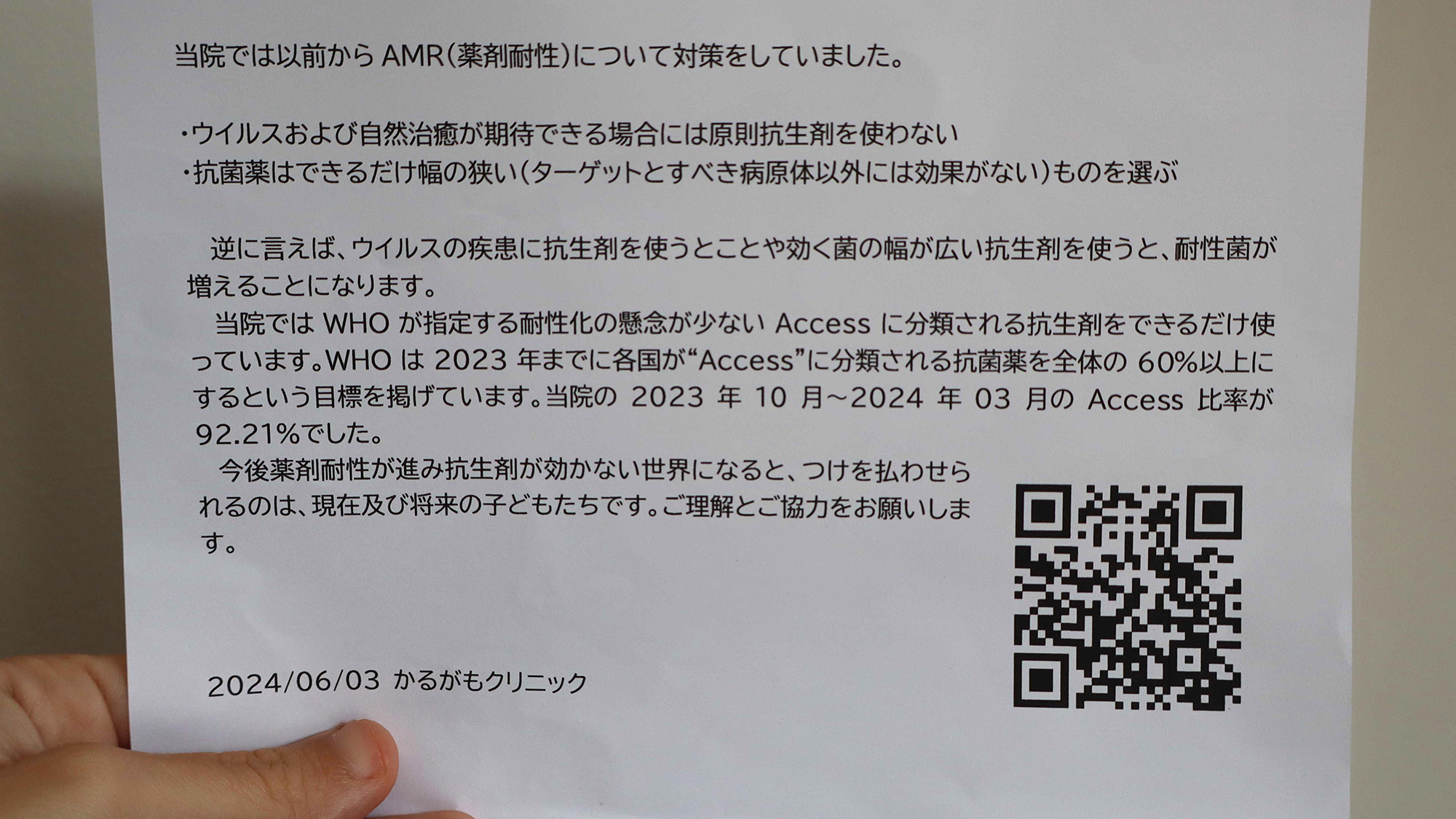
Fifteen years ago, when Miyahara opened his clinic, he noticed that other physicians prescribed a lot of antibiotics, and he questioned the practice due to the potential to fuel resistance. He provides his patients with informational flyers that explain the risks of resistance and how avoiding unnecessary prescriptions can reduce that risk. When antibiotics are needed, he predominantly uses narrow-spectrum drugs that pose a relatively low risk of resistance.
Miyahara said the local government and medical associations announced the antibiotic incentive when it was launched, and he felt its introduction has been very positive. It's increased his revenue and encouraged him to continue his stewardship practices. He estimates that for every 50 of his first visits with patients, he claims the incentive 10 to 15 times, so it applies to at least 20% of those visits.
An informational flyer Dr. Atsushi Miyahara provides to his clients. It states that the clinic takes measures against AMR, explaining that antibiotics are not used for viral illnesses, and for bacterial infections, the narrowest-spectrum options are prioritized. (Image credit: Nicoletta Lanese)
The incentive's impact
Because some pediatric clinics qualified and others didn't — due to fee-for-service clinics being excluded — Okubo and his colleagues could directly measure whether the policy worked.
To assess the incentive's effects in its first year, the researchers looked at insurance claims from over 10,000 medical facilities from just before and after the policy's introduction. About 3,000 of the facilities qualified and claimed the incentive 316,770 times, totaling 253 million yen ($2.29 million at the time). These eligible clinics saw a 17.8% reduction in their total antibiotic use over a year without any negative effects for patients, such as higher hospitalization rates.
Science Spotlight
(Image credit: Marilyn Perkins / Future)
Science Spotlight takes a deeper look at emerging science and gives you, our readers, the perspective you need on these advances. Our stories highlight trends in different fields, how new research is changing old ideas, and how the picture of the world we live in is being transformed thanks to science
To see if those effects lasted, the team drew years of data from over 165,000 young children who went to either eligible or ineligible clinics. Within the first month of the policy's implementation, the former group of children saw a nearly 45% reduction in total antibiotic prescriptions, compared with the other kids. Cumulatively over the next four years, their overall antibiotic use and broad-spectrum-antibiotic use was 20% and 24% lower, respectively.
The decrease in antibiotic prescriptions did not come with an uptick in hospitalizations or healthcare costs, although there was a slight increase in the total number of doctor's visits. But that's what you'd expect as doctors track an infection over time, Okubo explained, meaning they likely employed the watchful-waiting strategy and had parents bring their kids back in if they didn't improve quickly.
Okubo's team has continued to track pediatricians' antibiotic use, and he noted that they're seeing "spillover effects" among age groups not covered by the incentive. In the under-20 age group, outpatient antibiotic prescriptions fell by 50% between 2011 and 2022. He thinks the incentive is a key driver of this trend, directly reducing prescriptions for the youngest kids while also triggering ripple effects in older groups. (This research will soon be published in a peer-reviewed journal.)
That said, there's room to improve doctors' selection of antibiotics when they are used, as the ratio of broad- to narrow-spectrum drugs is still too high. "Total antibiotic use was reduced, but its quality should be improved further," Okubo said.
Okubo conducts research at the National Center for Child Health and Development in Tokyo. As a research center and Japan's largest children's hospital, it aims to deepen the understanding of children's health and development while providing advanced medical care. (Image credit: Nicoletta Lanese)
The incentive's evolution
While the 800-yen tip has proved significant to clinicians, the incentive represents a very small slice of overall government healthcare spending, which totaled 468 billion yen ($3.1 billion) in 2022.
"This program is not large compared to the whole budget," Dr. Kosuke Sasaki, who works in the MHLW's health insurance bureau, told me. The program's budget has no upper limit, so if the number of claims from clinics increased, there isn't a cap on how many could be paid out. "The number of doctors using this program has increased while the number of antibiotic prescriptions has decreased since the start," Sasaki's colleague Dr. Tomonori Aoki added, noting that the government isn't concerned about how to pay that rising bill.
The program's measurable impact and low price tag may explain its growth over the years.
Every two years, Japan's Ministry of Finance hands the MHLW its slice of the government budget, and MHLW then revises the pricing for drugs, medical devices and healthcare services. The antibiotic incentive falls under this revision process and has been expanded several times.
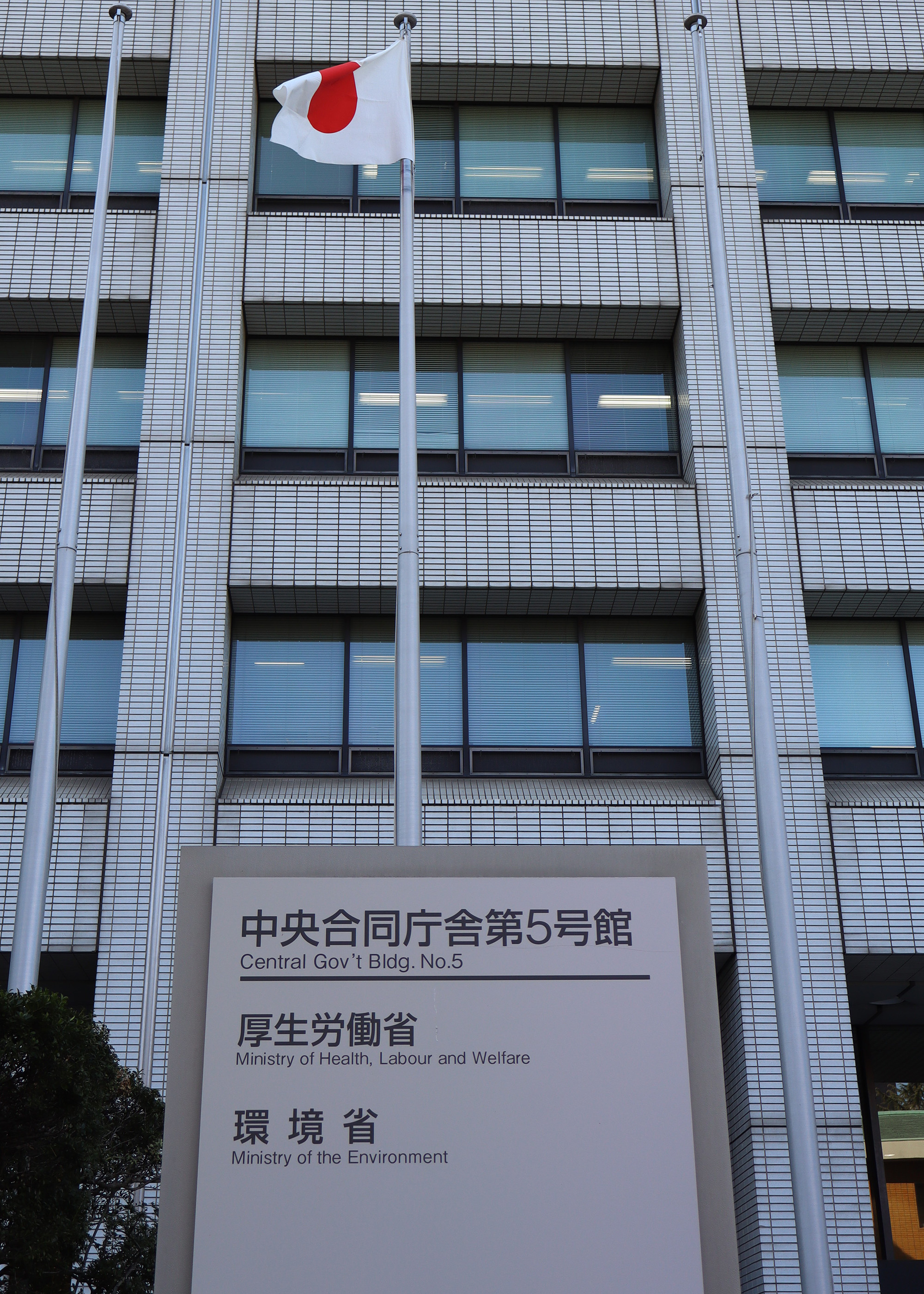
The Ministry of Health, Labour and Welfare is housed in an unremarkable building in Central Tokyo. It sets the prices for pharmaceuticals, medical devices and healthcare services nationwide. (Image credit: Nicoletta Lanese)
During its first revision in 2020, the incentive was extended to children under 6. In 2022, ear, nose and throat specialists (ENTs) newly qualified for the incentive; like pediatricians, they treat many acute infections in kids and tend to overuse antibiotics, insurance data suggested. That same year, doctors could start claiming the fee for ear infections and sinus infections.
"I see a tendency for pediatric clinics to avoid prescribing antibiotics, but I do see some ENT clinics prescribing antibiotics carelessly," Shibata, the clinic owner in Asakusa, Tokyo, told me. So ENTs seem to be a logical next target.
In 2024, a separate, facility-level incentive was introduced as a complement to the 800-yen incentive. It encourages clinics to submit data to a government database that tracks antibiotic use. If first-line, narrow-spectrum antibiotics make up a certain percentage of the clinic's overall prescriptions, that clinic earns extra money, Okubo explained.
Ultimately, the 800-yen incentive helped put AMR on the radar of doctors who didn't take it as seriously as experts like Kato, who told me "AMR is kind of my life's work." Kato and researchers like Okubo see the program as a success, although they pointed to room for improvement in antibiotic selection. Ministry officials like Sasaki and Aoki said the program is easy to implement and makes a difference. Clinic doctors like Shibata and Miyahara appreciate the incentive and use it consistently. All in all, at just $5 a claim, the incentive has been remarkably effective.
In speaking with experts in Japan and the U.S., I've learned that U.S. doctors have historically faced the same pressures and showed similar lapses in antibiotic use that Japanese doctors have. However, the U.S. does not have an incentive program like Japan's. Should it launch one?
In the next installment of this series, I'll explore a central feature of this problem: the motivations behind pediatricians' antibiotic misuse. What are they, and do those motivations differ between doctors in the U.S. and Japan? Answers to those questions will help determine whether a similar incentive might have the same impact in both places.
In a groundbreaking study poised to reshape the landscape of inflammatory bowel disease (IBD) treatment, researchers at the University of Louisville have elucidated the molecular mechanisms by which a natural metabolite derived from dietary sources orchestrates a protective immune response in the gut. IBD, encompassing debilitating conditions such as Crohn’s disease and ulcerative colitis, affects millions globally and is characterized by persistent inflammation and compromised intestinal barrier integrity. The intestinal barrier plays a critical role in maintaining homeostasis by allowing nutrient absorption while preventing the translocation of harmful microbial agents. In patients with IBD, this barrier is disrupted, leading to exacerbated inflammation and tissue injury.
Central to this new discovery is urolithin A (UroA), a microbial metabolite produced in the human gut following the ingestion of ellagitannin-rich foods such as pomegranates, walnuts, and berries. The research team led by Dr. Venkatakrishna Rao Jala unveiled how UroA selectively activates the aryl hydrocarbon receptor (AHR), a ligand-activated transcription factor expressed in various cell types, including those that constitute the intestinal epithelium. Unlike conventional views of AHR as a mediator of toxicological responses to environmental contaminants, this study highlights a nuanced role for AHR signaling in immune modulation and barrier function maintenance.
In their experimental design, investigators employed a combination of cell culture models, intestinal organoids, and human tissue samples from IBD patients to delineate the cellular and molecular pathways engaged by UroA. This comprehensive approach allowed them to pinpoint intestinal epithelial cells as the primary sites of AHR activation by UroA. Activated epithelial AHR, in turn, stimulates the assembly of the NLRP6 inflammasome, a multiprotein complex traditionally associated with inflammatory cytokine maturation and pyroptotic cell death. Contrary to the canonical inflammatory paradigm, NLRP6 activation here initiates a controlled release of protective mediators that promote epithelial repair, mucus production, and antimicrobial peptide secretion, collectively enhancing barrier integrity.
This dualistic nature of inflammasome signaling reveals an elegant example of cellular context-determined outcomes of innate immune pathways. While systemic inflammasome activation is linked to pathological inflammation, its epithelial-specific engagement appears to serve as a homeostatic mechanism preserving mucosal immunity and preventing microbial dysbiosis. This insight furthers our understanding of the immune microenvironment in the gut and challenges existing dogmas that broadly classify inflammatory signaling as detrimental in chronic intestinal diseases.
The exploration of UroA’s mechanism also sheds light on the intricacies of diet-microbiota-host interactions that underpin gut health. Dietary metabolites have emerged as critical modulators of signaling networks that maintain tissue equilibrium. This study positions urolithin A as a pivotal molecular bridge linking dietary intake to cellular defense strategies, mediated through receptor-mediated inflammasome activation. By elucidating this axis, the findings offer a compelling rationale for dietary or microbiota-targeted interventions designed to harness endogenous metabolites for therapeutic benefit.
Importantly, the protective pathway abrogates damage caused by intestinal injury without invoking broad immunosuppression, a common drawback of current IBD therapies that can predispose patients to infections and malignancies. Targeted activation of epithelial AHR by UroA selectively augments mucosal barrier function while modulating local immunity, offering a precision medicine approach with potentially fewer side effects.
From a translational perspective, these insights could catalyze the development of novel therapeutics that mimic or potentiate UroA’s activity, aiming to restore gut barrier homeostasis in individuals suffering from IBD and other gastrointestinal disorders. Unlike systemic immunomodulators, such agents could enhance intrinsic repair mechanisms, reinforcing the mucosal shield against microbial translocation and inflammation relapse.
Furthermore, this research underscores the importance of endothelial and epithelial cellular compartments in orchestrating immune responses that are finely tuned to environmental and nutritional cues. The aryl hydrocarbon receptor emerges as a nodal point integrating extrinsic and intrinsic signals, modulating inflammasome dynamics uniquely in epithelial cells as opposed to immune cells such as macrophages or dendritic cells, where AHR activation may produce opposing outcomes.
The team’s validation using human intestinal biopsies strengthens the clinical relevance of their findings, demonstrating that UroA-induced AHR-NLRP6 activation is not merely an experimental artifact but a genuine biological phenomenon with implications in human disease states. The investigation also highlights the potential inter-individual variability in microbiota capacity to generate UroA, which may influence susceptibility or response to treatment in IBD patients.
Concluding remarks from the lead investigators emphasize the broader implications of delineating this pathway. By distinguishing harmful from beneficial inflammatory cascades, the study advocates for selective immunomodulation strategies that leverage inherent protective mechanisms instead of indiscriminate suppression. This paradigm shift could revolutionize therapeutic approaches, fostering better clinical outcomes and improved quality of life for those affected by chronic intestinal inflammation.
Collectively, these findings mark a significant advance in gut immunology and open new avenues for precision dietary-based therapeutics. The interaction between diet-derived microbial metabolites, host epithelial signaling, and inflammasome activation represents a complex yet promising frontier in combating chronic inflammatory diseases. As our understanding deepens, integrating nutritional sciences with immunopharmacology may yield innovative treatments that redefine gut health maintenance.
Subject of Research: Human tissue samples
Article Title: Urolithin A activates aryl hydrocarbon receptor-NLRP6-mediated pathways in intestinal epithelial cells to modulate mucosal immunity and strengthen gut barrier integrity
News Publication Date: 23-Jun-2026
Web References: http://dx.doi.org/10.1038/s41467-026-73760-3
References: Published in Nature Communications
Image Credits: University of Louisville
Keywords: Human gut microbiota, Metabolism, Metabolomics, Metabolites, Diseases and disorders, Digestive disorders, Inflammatory bowel diseases, Gastrointestinal tract, Gastrointestinal disorders, Inflammasomes
In the ongoing battle against obesity and its associated cardiovascular risks, new scientific insights are shedding light on the complex interplay between weight loss strategies and vascular health. Recent research from the S-LiTE trial, presented in a landmark study by Sandsdal et al., unfolds groundbreaking findings on how exercise and the GLP-1 receptor agonist liraglutide profoundly influence vascular function and systemic inflammation during weight loss maintenance. This investigation not only advances our understanding of therapeutic approaches but also charts a promising path for improving cardiovascular outcomes in individuals struggling with obesity.
Obesity, recognized as a critical driver of cardiovascular disease, involves pathological alterations in the vascular system and chronic low-grade inflammation. These pathological changes elevate the risk of atherosclerosis, hypertension, and ultimately, heart failure. Consequently, effective weight loss maintenance after initial reductions is paramount for sustaining vascular benefits and mitigating long-term health risks. Despite existing weight loss regimens, relapse rates are high, and how these interventions impact vascular integrity during the maintenance phase remains incompletely understood. The S-LiTE trial rigorously targeted this knowledge gap, merging pharmacological and lifestyle components for a nuanced examination.
Exercise has been universally endorsed as a cornerstone intervention in weight management and cardiovascular disease prevention. Its benefits extend beyond simple calorie expenditure, encompassing modulation of endothelial function, arterial stiffness, and inflammatory profiles. Liraglutide, a glucagon-like peptide-1 (GLP-1) receptor agonist, has garnered attention for its potent weight-lowering effects through appetite regulation and metabolic improvements. However, the independent and combined vascular effects of exercise and liraglutide during the maintenance phase after weight loss had yet to be elucidated prior to this study.
The S-LiTE trial meticulously randomized participants who had achieved significant weight reduction to either an exercise program, liraglutide treatment, a combination of both, or placebo, allowing for a comprehensive comparison across groups. Over the course of the trial, vascular health parameters such as carotid artery compliance, endothelial function assessed via flow-mediated dilation, and systemic inflammatory markers including C-reactive protein (CRP) and interleukins were closely monitored. The multidimensional assessment unveiled striking differences attributable to intervention modalities.
Exercise alone was associated with notable improvements in arterial compliance and a favorable reduction in markers of vascular inflammation. The mechanical stimuli generated during physical activity promote shear stress on vessel walls, which in turn enhance nitric oxide bioavailability and endothelial repair mechanisms. These physiological adaptations are critical for maintaining vascular elasticity and preventing atherogenic processes. This study underscores the indispensable role of exercise not just in weight regulation but as a direct modulator of vascular integrity.
Liraglutide, beyond its appetite-suppressant and glycemic control effects, demonstrated intrinsic vascular benefits. The study highlighted liraglutide’s capacity to enhance endothelial function independently of exercise, potentially through GLP-1 receptor-mediated pathways that reduce oxidative stress and inhibit pro-inflammatory signaling cascades. This offers compelling evidence supporting the use of GLP-1 receptor agonists as adjunct therapies in cardiovascular risk reduction within the obese population.
Intriguingly, the combination of exercise and liraglutide yielded additive benefits, with participants exhibiting the most pronounced improvements in vascular compliance and the greatest suppression of inflammatory cytokines. This synergistic effect suggests that the pharmacological and mechanical influences converge on complementary pathways, amplifying the overall vascular protective effect. Such findings advocate for integrative treatment frameworks that leverage both lifestyle and pharmacotherapy in obesity management.
Delving deeper, the study’s prespecified secondary analysis elucidated how these interventions influenced various immune cell populations that are pivotal contributors to vascular inflammation. It was evident that the combined approach modulated monocyte and macrophage activation states, steering them towards anti-inflammatory phenotypes. This immune modulation is instrumental in curbing chronic inflammation and subsequent endothelial dysfunction, which are foundational in the pathogenesis of vascular disease.
Beyond the vascular sphere, the inflammatory milieu impacts insulin sensitivity and adipose tissue dynamics, central to metabolic health. The study posits that improvements in vascular health might interplay with metabolic regulation, facilitating a virtuous cycle of weight loss maintenance and cardiovascular risk attenuation. This holistic perspective on obesity interventions highlights the interconnected nature of metabolism, immunity, and vascular function.
Crucial to real-world applicability, the study design mirrored long-term maintenance conditions, providing practical insights for clinicians. The sustainability of vascular improvements with continued exercise and pharmacological support affirms the need for ongoing adherence and monitoring. Moreover, the tolerability profile and patient-centered outcomes reported provide a realistic framework for incorporating such regimens in routine care.
This research not only emphasizes the potent vascular benefits tied to weight loss maintenance but also brings to light the nuanced biological effects of different modalities. The implications extend beyond cardiovascular disease prevention, touching on broader aspects of healthy aging and quality of life, as vascular health is integral to multiple organ systems. Hence, the S-LiTE trial’s findings hold substantial promise for shaping future clinical guidelines.
In the broader context of obesity treatment paradigms, these insights challenge the notion that weight loss alone is sufficient. Instead, they advocate for multidimensional interventions addressing vascular function and inflammation, which are crucial mediators of obesity-related morbidity. Such transformations in therapeutic strategies could pivot the field towards more personalized and mechanistically informed approaches.
While the study is robust, the authors acknowledge limitations including the homogeneity of the cohort and potential confounders inherent to lifestyle interventions. Future investigations are warranted to explore molecular signaling pathways in greater detail, diverse populations, and longer-term cardiovascular outcomes. Additionally, integrating imaging modalities and biomarker profiling could refine our understanding of treatment impacts at the microvascular level.
On a scientific and societal level, the S-LiTE trial represents a milestone in conceptualizing weight loss maintenance not simply as weight stabilization but as an active process with profound systemic effects. Public health strategies might evolve to promote combined regimens, potentially alleviating the healthcare burden posed by obesity-related vascular diseases. This research, therefore, stands at the confluence of basic science, clinical innovation, and health policy.
The findings from Sandsdal and colleagues’ work resonate with an emerging narrative in metabolic medicine: holistic, multimodal approaches yield superior benefits than isolated interventions. With obesity prevalence continuing to climb globally, translating these findings into accessible, scalable solutions remains both a challenge and an imperative. The S-LiTE trial ignites hope for more effective, durable solutions to one of the most pressing health crises of our time.
In summary, the S-LiTE trial reveals that exercise and liraglutide individually and synergistically improve vascular health and reduce inflammation during weight loss maintenance. This study offers compelling evidence for the inclusion of GLP-1 receptor agonists alongside structured exercise programs to sustain cardiovascular health in individuals maintaining weight loss. These innovative insights redefine obesity treatment and open new avenues for reducing the global burden of cardiovascular disease through integrated, targeted therapeutic strategies.
Subject of Research: Effects of exercise and GLP-1 receptor agonist liraglutide on vascular health and inflammation during weight loss maintenance.
Article Title: Effects of exercise and liraglutide on vascular health and inflammation during weight loss maintenance: a prespecified secondary analysis of the S-LiTE trial.
Article References:
Sandsdal, R.M., Holt, J., Alkhefagie, H.G.A. et al. Effects of exercise and liraglutide on vascular health and inflammation during weight loss maintenance: a prespecified secondary analysis of the S-LiTE trial. Nat Metab (2026). https://doi.org/10.1038/s42255-026-01554-4
In a groundbreaking study poised to reshape our understanding of Parkinson’s disease (PD), researchers have unveiled a complex molecular mechanism linking metabolic stress to the disruption of circadian rhythms, with far-reaching implications for neurodegenerative disorders. This deep dive into cellular bioenergetics identifies a glucose-responsive oscillation involving the AMP-activated protein kinase (AMPK), ten-eleven translocation methylcytosine dioxygenase 2 (TET2), and 5-hydroxymethylcytosine (5hmC) axis as a pivotal mediator of metabolic stress responses in the brain. The discovery connects metabolic alterations directly to circadian transcriptional regulation, thereby illuminating previously obscure pathways influencing the onset and progression of Parkinson’s disease.
At the heart of this research lies the energy-sensing enzyme AMPK, a master regulator of cellular metabolism that responds dynamically to changes in cellular energy levels, especially glucose availability. AMPK’s role extends beyond energy balance; it modulates numerous signaling pathways critical for maintaining cellular homeostasis. When glucose levels fluctuate, AMPK activity adjusts accordingly, triggering a cascade of epigenetic modifications mediated by TET2, an enzyme known for its involvement in DNA demethylation and gene expression regulation through oxidative modification of 5-methylcytosine. This AMPK-TET2 interaction sets the stage for rhythmic changes in 5hmC, a key epigenetic mark that fine-tunes circadian gene expression.
The circadian clock is a complex transcriptional network orchestrating daily physiological and behavioral rhythms. Its disruption has been increasingly implicated in neurodegenerative conditions, yet the molecular underpinnings linking metabolism, circadian dysregulation, and neuronal vulnerability have remained elusive. The researchers’ demonstration that metabolic stress-induced AMPK activation regulates TET2-dependent 5hmC oscillations provides a compelling mechanistic bridge. Through this mechanism, energy fluctuations dynamically shape the transcription of circadian genes, potentially disturbing neuronal homeostasis and contributing to PD pathogenesis.
This study utilized a combination of cutting-edge genomic, epigenomic, and proteomic techniques to unravel the dynamics of the AMPK-TET2-5hmC circuit. High-resolution temporal profiling revealed that the oscillations of 5hmC marks at key circadian gene loci are not static but fluctuate in response to metabolic cues. These fluctuations integrate signals of cellular energy status and translate them into rhythmic transcriptional outputs. Importantly, the investigators found that these oscillatory patterns are disrupted in experimental models of Parkinson’s disease, suggesting that the fidelity of metabolic circadian signaling is compromised in neurodegeneration.
A significant aspect of the findings is the direct link between glucose availability and neuronal epigenetic regulation. Metabolic stress, often characterized by reduced glucose uptake or utilization, is a hallmark of aging and neurodegenerative disease states. The AMPK-TET2-5hmC axis serves as a metabolic sensor and effector that translates glucose scarcity into epigenetic reprogramming, thus influencing gene networks that sustain neuronal survival and function. This axis might represent a critical checkpoint where metabolic perturbations exert long-lasting effects on the neuronal epigenome and transcriptome, thereby driving PD progression.
One of the most intriguing revelations of this research is the bidirectional nature of the relationship between metabolic stress and circadian transcription. Not only does metabolic status influence circadian gene expression via epigenetic modulation, but disruptions in circadian regulation feedback to exacerbate metabolic dysfunction. This feedback loop could create a vicious cycle that accelerates neurodegenerative processes, underscoring the importance of maintaining metabolic and circadian integrity for brain health.
The implications of this study extend beyond basic science, offering exciting therapeutic potential. Targeting the AMPK-TET2-5hmC axis may open new avenues for interventions aimed at restoring metabolic and circadian homeostasis in Parkinson’s disease patients. Pharmacological modulators of AMPK activity or epigenetic editors capable of fine-tuning 5hmC dynamics could serve as innovative strategies to halt or slow neurodegeneration. Moreover, optimizing glucose metabolism through lifestyle or pharmacological means might indirectly preserve epigenetic and circadian function, offering a multi-faceted approach to disease management.
The research also highlights the importance of integrating metabolic and epigenetic perspectives to fully capture the complexity of neurodegenerative diseases. Traditional approaches focusing solely on genetic or protein dysfunction have fallen short in explaining the multifactorial etiology and progression of Parkinson’s disease. By illuminating the interplay between energy metabolism and epigenetic regulation, this study sets a new paradigm that could drive future research towards more holistic models of brain pathology.
In addition to elucidating molecular mechanisms, the study provides valuable insights into the temporal dimension of Parkinson’s disease pathology. The rhythmic oscillations of 5hmC marks influenced by glucose and AMPK activity emphasize that timing and metabolic context are crucial factors in disease progression. This may explain the variability in symptom manifestation and disease course among patients and suggests that chronotherapeutic approaches—aligning treatment with the circadian system—could enhance clinical outcomes.
Technologically, the application of multi-omics profiling coupled with advanced computational modeling allowed the researchers to dissect complex regulatory networks with unprecedented precision. Such integrative methodologies are essential for capturing dynamic biological processes that span different molecular layers and timescales. This approach underscores the future direction of biomedical research, where systems biology and temporal dynamics take center stage in unraveling disease mechanisms.
Furthermore, this study draws attention to the overlooked role of 5-hydroxymethylcytosine in the adult brain. Traditionally regarded as mere intermediates in DNA demethylation, 5hmC marks are now recognized as stable epigenetic modifications with distinct regulatory functions. Their oscillatory behavior in response to metabolic cues reveals them as critical mediators linking extracellular environmental factors to nuclear transcriptional machinery, thereby influencing neuronal fate and function.
The association of metabolic stress with circadian dysregulation through the AMPK-TET2-5hmC circuitry also provides a plausible explanation for the observed links between lifestyle factors—such as diet, sleep patterns, and physical activity—and Parkinson’s disease risk. Disruptions in any of these factors can alter glucose metabolism and circadian rhythms, potentially perturbing the delicate balance maintained by this molecular axis and predisposing individuals to neurodegeneration.
Importantly, the study’s findings could redefine biomarker discovery in Parkinson’s disease. Epigenetic oscillations of 5hmC, detectable in neuronal tissues and potentially biofluids, might serve as dynamic indicators of metabolic-circadian health and disease state. This could facilitate earlier diagnosis, tracking of disease progression, and evaluation of therapeutic interventions—ushering in a new era of precision medicine tailored to the metabolic and epigenetic status of patients.
Altogether, the elucidation of the glucose-responsive AMPK-TET2-5hmC oscillation offers a transformative lens through which to understand the intersection of metabolism, epigenetics, and circadian biology in Parkinson’s disease. By identifying a molecular conduit that links metabolic stress to circadian transcriptional misregulation, this research paves the way for innovative diagnostic and therapeutic strategies. As metabolic and circadian dysfunction climb into prominence as central pathogenic drivers, targeted modulation of this axis may hold the key to altering the trajectory of neurodegenerative diseases for millions worldwide.
This pioneering work heralds a new frontier in neuroscience, where cellular energetics, epigenetic remodeling, and circadian rhythms converge to shape neuronal destiny. Understanding and harnessing these complex oscillatory mechanisms offers unprecedented hope for combating the devastating impacts of Parkinson’s disease—a promise that echoes far beyond this single disorder and resonates with the broader quest to unravel the biological rhythms at the core of human health and disease.
Subject of Research: The study investigates the molecular mechanism linking metabolic stress, circadian transcriptional regulation, and Parkinson’s disease, focusing on the glucose-responsive AMPK-TET2-5-hydroxymethylcytosine (5hmC) oscillation.
Article Title: Glucose-responsive AMPK-TET2-5hmc oscillation links metabolic stress to circadian transcription and Parkinson’s disease relevance.
Article References: Chu, L., Wang, S., Wang, L. et al. Glucose-responsive AMPK-TET2-5hmc oscillation links metabolic stress to circadian transcription and Parkinson’s disease relevance. npj Parkinsons Dis. (2026). https://doi.org/10.1038/s41531-026-01447-z
The foods people eat every day may have a bigger impact on digestive health than many realize. A large international study has found that eating high amounts of ultraprocessed grain products may increase the risk of developing inflammatory bowel disease, a serious condition that affects millions of people worldwide. The findings were published in the […]
Plastic has transformed modern life. It is cheap, lightweight, and found in almost every home, office, and hospital. From food packaging and cosmetics to medical tubing and cleaning products, plastics are deeply woven into everyday living. Yet scientists are increasingly concerned that some of the chemicals used in plastics may come with hidden health costs. […]
The symptoms: The man visited a hospital because two months earlier, some foods and beverages began to taste extremely unpleasant to him. Over time, more foods had become difficult for him to stomach. Sour and salty flavors were the first to trigger disgust. Oily foods were next, followed by fresh vegetables, cold water and juice, warm water and, finally, sweets.
The man ate less due to the taste aversions, and he unintentionally lost a few pounds. He told doctors at the hospital that he felt lethargic, they wrote in a report of the case.
What happened next: The patient, who was a physician himself, reported that eight years ago, he had been diagnosed with bipolar disorder. For the prior three years, he had been managing the condition with lithium, a mood stabilizer commonly used to treat bipolar disorder.
He was taking a prescribed dosage of 800 milligrams per day and historically hadn't had any unusual side effects. (This is slightly higher than the typical dose recommended for chronic management of bipolar disorder, but that said, doctors may tweak patients' doses based on their individual needs.)
The diagnosis: Doctors performed bloodwork and found that the level of lithium in the patient's blood was 1.28 milliequivalents per liter (mEq/L). Safe levels of lithium are between 0.6 and 1.2 mEq/L; any higher than that can cause a condition called lithium toxicity, which can be fatal if untreated.
When the doctors checked the man's medical records, they found that prior to the onset of his symptoms, the levels of lithium in his blood were between 0.4 and 0.9 mEq/L. In the report, the authors did not investigate the cause of the toxicity, nor did they hypothesize as to why lithium was accumulating in the patient's blood.
The treatment: The doctors told the man to stop taking lithium, and they instead prescribed another mood-stabilizing drug, called valproate. As the lithium levels in his blood dropped, the patient's energy returned and the foods and beverages that previously disgusted him recovered their normal flavors. Notably, this happened in the reverse order that their flavors had changed.
At a follow-up visit one year later, the man was still taking valproate and his symptoms of fatigue and taste changes had not returned.
What makes the case unique: Although they're uncommon, side effects of lithium use can include confusion, increased thirst, frequent urination, irregular or slow heartbeat, tiredness and weight gain. Rarer side effects include dizziness, headaches, vision problems and heat loss in the arms and legs. Lithium toxicity, also known as lithium intoxication, is known to increase the risk of these adverse symptoms.
Nevertheless, "it has not been generally known that lithium can lose or change taste in some patients," the case report authors wrote. Based on their findings, they suggested that in the future, taste loss or changes in taste perception should be flagged as a potential indicator of mild lithium intoxication.
This article is for informational purposes only and is not meant to offer medical advice.

 Login