Normal view
Shared Auditory Maps Predict Human and Mouse Decisions
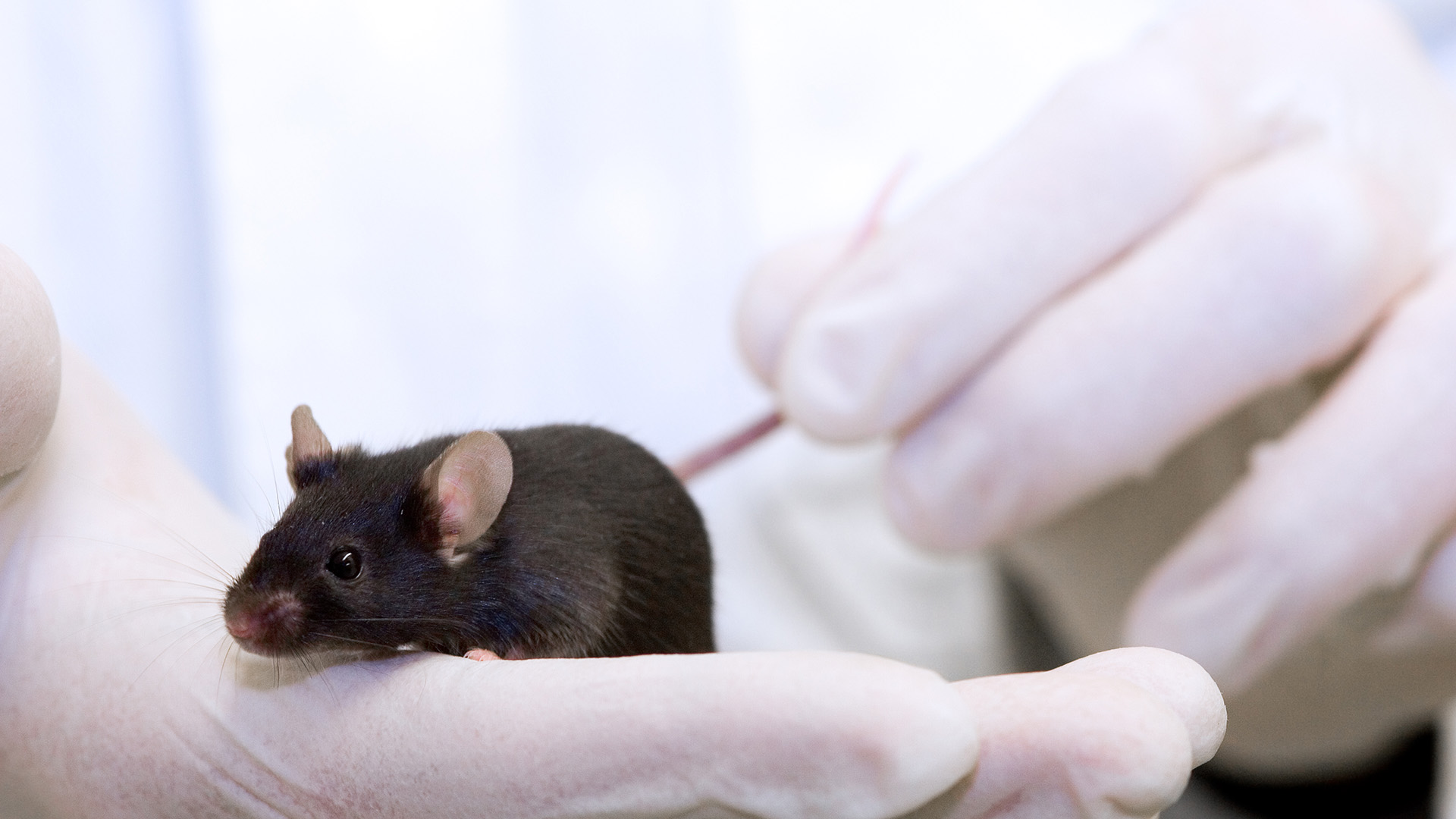
In a groundbreaking study poised to redefine our understanding of auditory perception across species, researchers have unveiled strikingly similar auditory perceptual maps in both humans and mice. This discovery not only bridges a crucial gap between animal models and human sensory processing but also offers predictive insight into how perceptual decisions are formed during auditory discrimination learning. The implications ripple across neuroscience, psychology, and artificial intelligence, promising new avenues for studying sensory perception and developing advanced hearing-related technologies.
Auditory perception—the way organisms interpret and make sense of sound stimuli—is a complex process that involves integrating multifaceted sound attributes such as frequency, intensity, and temporal patterns. While mouse models have long been foundational in auditory research due to their genetic malleability and experimental accessibility, skepticism has persisted about the extent to which their perceptual frameworks parallel those of humans. This novel research offers compelling evidence that these frameworks are not only comparable but also exhibit shared underlying structures.
The study employed a sophisticated experimental design in which both human participants and mice were exposed to a wide array of tonal stimuli varying systematically across multiple auditory dimensions. Using advanced computational modeling and neural decoding techniques, the researchers were able to construct perceptual maps—conceptual frameworks representing how different sounds are internally organized within the brain’s auditory space. These maps enabled a quantifiable comparison of perceptual similarity and distance between sounds for both species, revealing a remarkable congruence in their auditory representations.
What makes these findings particularly compelling is the demonstration that these perceptual maps are predictive of behavioral outcomes during discrimination tasks. In other words, the way an individual’s brain groups different sounds predicts how well that individual can distinguish between them in learning scenarios. This link between neural representation and perceptual decision-making underlines a fundamental cognitive mechanism shared across mammalian species, offering crucial insights into the principles guiding sensory learning processes.
Beyond the fundamental science, the implications for translational research are profound. Understanding shared auditory perceptual structures facilitates the development of more accurate animal models for studying hearing impairments and neurological disorders affecting auditory processing. It also guides the design of auditory prosthetics, such as cochlear implants, by informing the algorithms that mimic natural sound perception. By aligning animal and human perceptual frameworks, therapies can be tailored with greater precision and efficacy.
A key technical achievement of the study was the use of multidimensional scaling techniques combined with machine learning algorithms to decode auditory neural responses with unprecedented granularity. The researchers collected high-resolution neural data using electrophysiological recordings in mice and non-invasive neuroimaging in humans. This dual approach allowed cross-species alignment of perceptual space, overcoming previous methodological challenges that hindered direct comparisons between human and rodent sensory processing.
Moreover, the study explored dynamic aspects of perceptual learning by measuring how auditory maps evolve through repeated training sessions. The maps exhibited plasticity reflective of learning-induced changes, emphasizing the adaptive nature of auditory perception. This plasticity was mirrored across both species, suggesting an evolutionarily conserved mechanism for tuning sensory systems based on environmental demands and experience.
One of the study’s most innovative aspects lies in its predictive modeling of discrimination performance. By mapping perceptual distances within the auditory space, the researchers could forecast individual differences in learning speed and accuracy, an advancement that holds enormous promise for personalized auditory training protocols. Such protocols could optimize rehabilitation strategies for individuals with auditory processing deficits by targeting specific perceptual aspects most relevant to their unique neural patterns.
The shared structure of auditory perceptual maps also raises intriguing questions about the evolutionary trajectory of sensory systems. The findings suggest that despite divergent evolutionary paths, fundamental auditory processing architectures remain conserved, supporting efficient sound discrimination necessary for survival and communication. This conservation across species underpins the generalizability of animal research findings to human sensory neuroscience.
Furthermore, these insights pave the way for advancing artificial sensory systems, especially in the realm of auditory artificial intelligence. By modeling computational auditory perception after natural neural maps, engineers can design more sophisticated speech recognition systems and assistive devices that better emulate human-like sound processing. The study’s integrative framework thus acts as a blueprint for creating tonal perceptual algorithms with enhanced contextual understanding.
This research also contributes importantly to theoretical debates surrounding sensory representation. By empirically demonstrating a shared, structured perceptual space, the study challenges views that sensory maps are strictly idiosyncratic or species-specific. Instead, it supports a paradigm that sensory systems leverage common organizational principles, rooted perhaps in fundamental neural coding constraints and environmental pressures shaping auditory perception.
The study’s methodology and results were meticulously detailed in a recent publication, where the authors delineated the computational techniques, behavioral paradigms, and statistical analyses that underscored their conclusions. This transparency fosters reproducibility and invites further interdisciplinary collaboration between computational neuroscientists, experimentalists, and cognitive psychologists aiming to unravel the complexities of sensory perception.
Importantly, these findings hold societal relevance beyond academic circles. Hearing loss and auditory processing disorders affect millions worldwide, often hindering communication and quality of life. Technologies and therapies inspired by a unified perceptual mapping approach could revolutionize auditory rehabilitation, offering hope for more effective interventions grounded in a thorough understanding of perceptual neuroscience.
In conclusion, this pioneering work illuminates the common auditory perceptual code shared by humans and mice, demonstrating that the brain’s representation of sound is not merely a byproduct of species-specific adaptations but a fundamental, conserved neural language. This discovery not only furthers scientific knowledge but also catalyzes innovation in medicine and technology, underscoring the power of cross-species research in unraveling the mysteries of the sensory brain.
As this field develops, future investigations are likely to explore how these perceptual maps interact with other cognitive systems such as memory and attention, and how factors such as age, experience, and pathology modulate the structure and function of auditory perception. With burgeoning advances in neurotechnologies and computational methods, the confluence of cross-species neuroscience and perceptual science promises to unlock new dimensions of understanding sensory experience.
Ultimately, the revelation that humans and mice share auditory perceptual maps that predict learning efficacies serves as a testament to nature’s optimization strategies, propelling a unified framework for deciphering sensory perception. This convergence of biology, computation, and psychology heralds a new chapter in auditory neuroscience with far-reaching implications for science and society alike.
Subject of Research: Auditory perceptual maps and discrimination learning in humans and mice
Article Title: Auditory perceptual maps in humans and mice share common structures and predict perceptual decisions in discrimination learning
Article References:
Seiler, J.PH., Cazzetta, G., Ghobadi, A. et al. Auditory perceptual maps in humans and mice share common structures and predict perceptual decisions in discrimination learning. Commun Psychol 4, 98 (2026). https://doi.org/10.1038/s44271-026-00485-w
Image Credits: AI Generated
- Live Science
- Estrogen levels in both the male and female brain may shape memory's resilience in face of stress
Estrogen levels in both the male and female brain may shape memory's resilience in face of stress
High estrogen in the brain's memory center may worsen one's resilience against traumatic events, swaying the tendency to develop memory problems or post-traumatic stress in the aftermath, a recent study in mice suggests.
The research, published in April in the journal Neuron, explored the effects of estrogen in the mouse brain. It zoomed in on the hippocampus, a key part of the brain involved in learning and memory. Both male and female mammals produce significant amounts of estrogen in the hippocampus, despite it often being framed as a "female" hormone.
"We're so biased to think of female high estrogen, male low estrogen," said study co-author Elizabeth Heller, an associate professor of pharmacology at the University of Pennsylvania Perelman School of Medicine. But "in this local brain region, where you have local production of estrogen, actually sometimes the males are higher than the females depending on the female's cycling," Heller told Live Science. Estrogen levels in the female hippocampus rise and fall in line with the body-wide hormone cycle, while its levels in the male hippocampus remain fairly steady.
The study suggests that these local estrogen concentrations may influence one's vulnerability to memory problems following major acute stress. Although the research was conducted in mice, the authors think it likely has relevance to humans.
"I think this is highly translatable," study senior author Dr. Tallie Z. Baram, a professor, developmental neuroscientist and child neurologist at the University of California, Irvine, told Live Science.
Estrogen isn't always a memory booster
Traumatic experiences can cause memory disturbances, including difficulty remembering specific personal experiences and having fearful reactions to formerly safe, familiar situations. When these issues persist and are accompanied by intrusive memories of the traumatic event, they are classified as post-traumatic stress disorder (PTSD).
About 10% to 12% of women experience PTSD in their lifetime, compared with 5% to 6% of men. Some of that difference may stem from variance in men's and women's lived experiences; for instance, women have higher rates of sexual assault at young ages than men do. Biological differences between women and men are another potential factor, but their contribution to the phenomenon is poorly understood.
The new study highlights hippocampal estrogen as one difference that might matter. "The research has uncovered important new avenues for research on PTSD," Victoria Luine, a professor emerita of psychology at Hunter College in New York City who wasn't involved in the work, told Live Science in an email.
In the study, researchers simulated acute traumatic events by exposing lab mice to multiple stressors at the same time, including bright lights, loud music and the odors of other stressed-out mice. They ran the mice through various memory tests before and after the stressful experience and compared these rodents with a group that did not experience such stressors.
Compared with unstressed mice, the stressed-out male mice performed worse on the various memory tests, and those deficits persisted for weeks. "Even a month later, they had a memory deficit — so it's a really perseverative effect," Heller said.
A similar pattern was seen in female mice that were stressed out during proestrus, the phase of their hormone cycle when estrogen peaks and the body prepares for ovulation. Both sets of mice learned to associate certain cues with the stressful experience and avoid them, with females being more sensitive to those cues than males were.
But interestingly, female mice that were stressed during estrus, when estrogen plummets and ovulation occurs, showed resilience. Their behavior and memory remained comparable to those of unstressed mice. "The female mice that had low levels of estrogen laughed it off — they were completely protected," Baram said.
Studies suggest hippocampal estrogen levels are similar in male and proestrus female mice, while estrus females have lower levels. The researchers confirmed this using a technique called mass spectrometry, finding that estrus mice had half the amount of hippocampal estrogen that the males and proestrus females did.
In this context, that lack of estrogen in the hippocampus appeared to guard against the negative effects of stress. This finding was surprising, Baram noted, because estrogen is generally thought to promote memory function in both sexes and declines in estrogen, as seen during menopause, are tied to memory problems. That said, menopause takes place over a much longer timeline than the female mouse hormone cycle, which takes only four or five days.
A connection to DNA
Why do estrogen levels matter for memory? "Estrogen receptors directly control gene expression," Heller said. By binding to its receptors, estrogen turns the activity of certain genes up or down.
Heller's lab studies mechanisms that control gene activity in the context of psychiatric disorders. One of those mechanisms is chromatin remodeling, meaning changes in how DNA is packaged in a cell that can shift which genes can be activated at a given time. A portion of the chromatin can be "open," exposing genes to machinery that turns them on, or "closed," which typically shuts genes down.
It turns out that the high hippocampal estrogen in male mice and proestrus female mice opens up their chromatin in a way that might leave them vulnerable to memory issues ushered by severe stress. Female mice in estrus, by contrast, have a totally distinct chromatin profile that appears to be protective.
What is it about women at that stage in life that makes them more vulnerable to memory loss with aging?
Tallie Z. Baram, professor, developmental neuroscientist and child neurologist at the University of California, Irvine
"We can see that the function of many of those [open] genes relates to synapse biology," Heller said. Synapses are the points at which different neurons meet and exchange electrical signals, and they're central to the physical structure of memories in the brain.
It may be that, in most circumstances, it's useful to have high levels of hippocampal estrogen because they "open" the chromatin, enabling the hippocampus to forge new memories quickly in response to new experiences, Baram noted. But when these experiences consist of severe acute stress, "that same plasticity, that same ability of the brain to learn, turns problematic," she said. If the results carry over to humans, women may be particularly vulnerable to these memory impacts in certain phases of their menstrual cycles or points in their lifespans when estrogen is high.
In males and females, different flavors of estrogen receptor were responsible for the stress-induced memory issues. The reasons for this difference will be a matter of future study, Baram said. Additionally, future research could attempt to pinpoint exactly where the different estrogen receptors are located throughout the hippocampus, Heller said.
The study provides a "strong demonstration that estrogens drive sex-dependent, stress-induced changes in chromatin networks which can dramatically alter neural functions like memory," Luine said. What's more, "these results present cogent evidence that sex is a powerful biological variable."
Historically, female lab animals were excluded from studies because it was thought that their hormone cycles were too complex and would muck up the findings. The field of neuroscience exemplified this trend. In recent years, the U.S. National Institutes of Health (NIH) has required that scientists take sex differences into account when designing NIH-funded human and animal studies, but progress has been slow on both fronts — and current federal leadership has signaled a lack of support for the initiative.
It's important to include both sexes in research to truly understand how the brain functions and responds to external factors, like stress, Luine said. "An important aim of this and other studies is to protect humans against PTSD," she added, and this study strongly suggests preventive treatments for PTSD might need to be tailored by sex.
Beyond PTSD, Baram thinks the research could have implications for women's risk of aging-related memory problems and dementia.
The decline of estrogen in menopause is thought to raise this risk, but prior to menopause comes perimenopause — a period with massive spikes in estrogen. The study's findings hint that if stress shows up during perimenopause, the combination of stress and high estrogen levels may contribute to memory problems. Thus, perimenopause may represent another time when women are particularly vulnerable to memory disturbances, Baram suggested.
"We need to start thinking a little bit differently," she said. "What is it about women at that stage in life that makes them more vulnerable to memory loss with aging?"
This article is for informational purposes only and is not meant to offer medical advice.
This article was first published May 4, 2026.
See how much you know about the most complex organ in the human body with our brain quiz!
Scientists Let People Play Video Games Using Only Their Thoughts
- SciTechDaily
- Women’s Brains May Be More Vulnerable to Dementia Risk Factors Than Scientists Realized
Women’s Brains May Be More Vulnerable to Dementia Risk Factors Than Scientists Realized
Scientists Say We’ve Been Wrong About the Aging Brain
Consciousness is Not Exclusive to Earth’s Biology, Philosophers Argue
University of California, Riverside’s Professor Eric Schwitzgebel and University of Lisbon postdoctoral researcher Jeremy Pober argue that consciousness is substrate flexible, meaning it can arise not just in the biological tissue we find on Earth, but potentially in radically different physical materials found elsewhere in the cosmos.
The post Consciousness is Not Exclusive to Earth’s Biology, Philosophers Argue appeared first on Sci.News: Breaking Science News.
- Science Mag
- Innovative Vibrating Pill Shows Promise in Predicting Relapse Risk Among Anorexia Nervosa Patients
Innovative Vibrating Pill Shows Promise in Predicting Relapse Risk Among Anorexia Nervosa Patients
A groundbreaking study from UCLA Health introduces a pioneering approach to understanding anorexia nervosa, a complex psychiatric disorder notorious for its high relapse rates and elevated mortality. Researchers employed an ingestible, vibrating capsule to probe the nuanced ways in which the nervous system of individuals with anorexia nervosa processes bodily sensations originating from the gastrointestinal tract. This innovative technology not only illuminates the enigmatic interplay between gut sensations and brain function but also offers promising predictive capabilities regarding relapse risk, potentially transforming clinical management of the disorder.
Anorexia nervosa is marked by a persistent restriction of energy intake, an intense fear of weight gain, and a distorted body image, resulting in dangerously low body weight. Despite state-of-the-art treatments aimed at restoring healthy body weight, relapse rates remain alarmingly high—up to 50% within a year post-treatment. Suicidal behavior is also a significant concern, often cited as the leading cause of death among afflicted individuals. This desperate clinical landscape underscores the urgent need for objective biomarkers and more profound mechanistic insights to improve therapeutic outcomes.
The inadequacy of current understanding centers around a crucial chasm: while physicians focus on weight normalization, many patients continue to suffer from entrenched symptoms that drive relapse, suggesting that the disorder’s etiology extends beyond mere weight regulation. The nervous system’s interpretation and integration of visceral signals, especially from the gut, could hold the key to these persistent challenges. Yet, before this study, precise tools to quantify and manipulate these signals in vivo remained elusive.
The UCLA research team harnessed a clinical trial design involving an ingestible vibrating capsule capable of delivering controlled, gentle mechanical stimuli to the stomach. This non-invasive method allowed for real-time interrogation of gastrointestinal interoception—the brain’s perception and processing of internal bodily sensations. By remotely activating the capsule, the researchers could produce varying intensities of stomach vibrations, simulating subtle gut sensations that are typically difficult to access or measure reliably.
In the study, 62 female patients who had been hospitalized for anorexia nervosa and whose body weight had been restored to clinical norms, were compared against 57 healthy controls. Participants swallowed the vibrating capsule and were instructed to indicate, via button press, whenever they perceived a vibration. Concurrent physiological monitoring tracked brain activity through neuroimaging techniques, cardiac rhythms, and stomach motility, providing a multidimensional understanding of the gut-brain axis in real time. Self-reported metrics on hunger and bodily awareness further enriched the dataset.
Employing sophisticated computational modeling, the researchers analyzed participants’ ability to detect and interpret the gastric signals. These models were designed to estimate participants’ expectation of gut sensations, their reliance on incoming sensory signals, and their adaptability in updating these expectations when signals fluctuated. This approach transcended simple sensory detection, probing fundamental cognitive processes involved in interoceptive learning and prediction error correction.
The results revealed a striking divergence between anorexia nervosa patients and healthy controls. Those with the disorder exhibited diminished accuracy in sensing subtle stomach vibrations, often unaware that a stimulus was occurring even when the capsule was active. They demonstrated a cognitive bias towards expecting an absence of gut sensations and were significantly slower to revise these expectations in the presence of confirmed visceral signals. This implies an altered central processing mechanism rather than a peripheral sensory deficit, reflecting a disconnection or miscommunication within the gut-brain axis.
Crucially, these interoceptive abnormalities persisted despite the restoration of healthy body weight, challenging the conventional clinical assumption that weight normalization is tantamount to recovery. The findings suggest that long-lasting neurobiological alterations in interoceptive processing may underlie the stubborn persistence of anorexic behaviors and cognitions, thus offering a plausible explanation for the high relapse propensity witnessed in clinical practice.
Linking these mechanistic insights to clinical outcomes, the study reported a robust association between impaired gastric signal detection and relapse risk during a six-month post-discharge follow-up period. Patients whose nervous systems exhibited the greatest bias towards ignoring bodily signals were significantly more likely to experience a relapse. This highlights the potential clinical utility of the vibrating capsule as a predictive tool, enabling healthcare providers to stratify relapse risk and tailor interventions accordingly.
From a translational perspective, the use of this ingestible technology marks a seminal advance in biomarker discovery for anorexia nervosa. It opens new avenues for personalized medicine where objective physiological metrics, rather than solely subjective symptom reports, guide therapeutic decisions. Moreover, it positions gastrointestinal interoception as a critical target for future treatment strategies, including neuromodulation, pharmacotherapy, and cognitive retraining designed to recalibrate gut-brain communication pathways.
Nevertheless, the authors caution that these findings currently pertain to a relatively homogeneous sample, primarily young female patients, and must be validated across larger and more diverse populations to confirm generalizability. Future research should also explore how different treatment modalities influence interoceptive processing and whether modulation of this axis can directly mitigate relapse risks.
In conclusion, this UCLA-led study redefines our understanding of anorexia nervosa by exposing the subtle but profound alterations in gastrointestinal interoception that endure beyond weight restoration and presage relapse. It suggests that true recovery necessitates healing not only the body but the fundamental neural processes that govern the perception and integration of internal bodily states. This insight sets the stage for an era where interoceptive biomarkers and bioelectronic medicine intersect to revolutionize the management of this devastating disorder.
Subject of Research: Gastrointestinal interoception and relapse prediction in anorexia nervosa
Article Title: Altered Gastrointestinal Interoception in Anorexia Nervosa Predicts Relapse
News Publication Date: 17-Jun-2026
Web References: http://dx.doi.org/10.1001/jamapsychiatry.2026.1301
References: Published in JAMA Psychiatry, 2026
Keywords: Anorexia nervosa, gastrointestinal interoception, relapse prediction, gut-brain axis, ingestible vibrating capsule, psychiatric biomarkers, neurobiology of eating disorders
This Copper Drug Clears Alzheimer’s Brain Toxins and Boosts Memory
This Surprising Hair Type Could Hold the Key to Chronic Itch Relief
Experimental Copper-Based Drug Clears Alzheimer’s Plaques and Boosts Memory in Mice
Copper diacetyl bis(4-methyl-3-thiosemicarbazone), or Cu(ATSM), restored a key waste-removal system in the brain, reducing toxic amyloid-beta buildup and improving spatial memory in lab models of Alzheimer’s disease, according to a team of researchers at Monash University.
The post Experimental Copper-Based Drug Clears Alzheimer’s Plaques and Boosts Memory in Mice appeared first on Sci.News: Breaking Science News.
Scientists Mapped Every Neuron in a Fruit Fly and the Brain Wasn’t Running the Show
Scientists Found a Hidden Brain Signal That Predicts Social Behavior
Neuroscientists are searching for the 'cellular substrate of loneliness'
The Brain May Not Need Full Sleep To Recover, New Research Finds
Scientists Reveal the Hidden Way Caffeine Sabotages Sleep
- The Debrief
- A ‘Memory Switchboard’ in the Hippocampus May Help Explain How the Brain Learns Without Overwriting Old Memories
A ‘Memory Switchboard’ in the Hippocampus May Help Explain How the Brain Learns Without Overwriting Old Memories
Scientists have often wondered how the brain can take in new experiences without erasing stored memories. However, a new study in Nature now identifies a circuit-level mechanism that may help answer this question—and it may also offer clues for improving artificial intelligence.
Researchers at NYU Langone Health recorded the activity of hundreds of neurons simultaneously across several brain regions in mice. By tracking how signals move from the hippocampus to the neocortex, where long-term memories are kept, they found that a subset of hippocampal neurons appears to participate in routing information between regions in distinct patterns. These cells participate in both incoming and outgoing signaling, but do so using distinct patterns of activity.
Separate Channels Through the Same Cells
The study focused on a series of interconnected brain regions: CA3, which processes rapidly changing information; CA1, which serves as a central hub; and the retrosplenial cortex, which is important for navigation and spatial memory. About one in four CA1 neurons receives most of the signals from CA3. When these same neurons send signals to the retrosplenial cortex, they fire in a different pattern, creating a separate outgoing channel using the same cells.
Rather than using new neurons, the brain separates these channels by changing how the same neurons fire together. The researchers suggest this arrangement may help reduce interference between new learning and more stable cortical memories.
“Our findings help explain how memory can be both moldable and enduring,” said co-lead author Joaquín Gonzalez, PhD, a postdoctoral fellow in the Department of Psychiatry at NYU Grossman School of Medicine. “By changing how the same cells fire together instead of turning on new cells, the brain can keep information organized and protect older memories.”
What Happens During Sleep
The CA1 neurons involved in learning during the day also stay active at night. The researchers found that these cells remain busy during sleep events called sharp-wave ripples, which are bursts of activity the hippocampus uses to replay and strengthen memories from the day.
Since overlapping neurons are recruited during both waking activity and sleep replay, the same circuit elements appear to participate in both learning and consolidation. This theoretically allows the brain to use a single system for both learning and storing memories, without the two processes interfering with each other.
“Our study shows how learning and memory consolidation can coexist in the same network,” said co-lead author Mihály Vöröslakos, MD, PhD. “Our discovery was made possible because for the first time, we were able to record hundreds of individual neurons across all the key regions simultaneously in animals that were moving around naturally.”
Implications for Disease and AI
The system identified in this research may help explain how memory disorders begin. In Alzheimer’s disease, for example, the hippocampal circuits examined here are some of the earliest to deteriorate. These networks are essential for sorting and organizing new experiences.
“Our discovery of a ‘memory switchboard’ deep in the hippocampus may provide clues as to how memory circuits fail in Alzheimer’s disease,” said co-senior author Zhe S. Chen, PhD, a professor in the Departments of Psychiatry and Neuroscience at NYU Grossman School of Medicine.
Artificial intelligence systems have a hard time holding onto what they’ve already learned when faced with new tasks, a problem researchers call catastrophic forgetting. The human brain appears able to update memories without catastrophic forgetting by altering the use of existing neural circuits. By studying how our brains pull this off, scientists hope to design AI that can keep learning new things without forgetting the old.
“By showing how the mammalian brain can safeguard memories during learning, our research may offer a biological blueprint for designing next-generation AI technology that can update itself continuously without overwriting what it has already acquired,” said co-senior author György Buzsáki, MD, PhD, the Biggs Professor of Neuroscience at NYU Grossman School of Medicine.
The researchers plan to investigate whether similar switchboard-like channels exist in other memory circuits. The authors caution that their findings in mice under controlled conditions do not yet allow conclusions about the human brain.
Austin Burgess is a writer and researcher with a background in sales, marketing, and data analytics. He holds an MBA, a Bachelor of Science in Business Administration, and a data analytics certification. His work focuses on breaking scientific developments, with an emphasis on emerging biology, cognitive neuroscience, and archaeological discoveries.
- The Debrief
- High-Dose Psilocybin Case Report Suggests Hidden Function May Persist in Advanced Alzheimer’s
High-Dose Psilocybin Case Report Suggests Hidden Function May Persist in Advanced Alzheimer’s
For five years, an elderly woman with advanced Alzheimer’s disease had spoken mostly in single words, needed help walking, and showed little spontaneous interaction with the people around her. Then, roughly 19 hours after receiving a high dose of psilocybin-containing mushrooms, she emerged from a prolonged, deep sleep-like state and began talking about her life.
The striking episode, described in a new case report published in Frontiers in Neuroscience, does not show that psilocybin reverses Alzheimer’s disease. The researchers are careful to stress that this was a single patient, observed in a real-world private clinical setting without brain imaging, formal sleep monitoring, or standardized cognitive testing.
Still, the study’s authors say the case raises the interesting possibility that in late-stage dementia, some functional abilities may indeed remain dormant rather than be completely lost.
“Advanced Alzheimer’s disease is generally regarded as a stage of irreversible functional decline,” researchers write. “This case documents transient multidomain functional improvement in advanced Alzheimer’s disease following psilocybin administration.”
The case involved a Japanese-American woman in her 80s with a 10-year history of Alzheimer’s disease. The patient was described as being in an advanced stage of dementia, with the previous five years being noted for severe decline.
Her speech had become mostly monosyllabic, and she had chronic urinary incontinence, executive dysfunction, dysphagia, dependent mobility, flat affect, prolonged somnolence, and a severe reduction in spontaneous communication.
Her diagnosis had been made clinically about a decade earlier, based on a steadily progressive neurodegenerative course, memory impairment, language reduction, executive dysfunction, and functional decline.
The intervention was a single oral dose of 5 grams of psilocybin-containing mushrooms from the “Enigma” strain. Researchers note that no established psilocybin dosing framework exists for advanced dementia, and the dose was high by almost any comparison.
Recreational users commonly describe 1 to 3 grams of dried Psilocybe cubensis mushrooms as a typical range, while doses around 3.5 grams or more are generally considered strong. A 5-gram dose is often treated as an especially intense experience. However, direct comparisons are imprecise because psilocybin content can vary widely between mushroom species, strains, batches, and even different parts of the same mushroom.
The study also notes that the dose was “relatively high compared with dosing approaches commonly used in modern clinical trials,” which typically use carefully measured amounts of purified psilocybin rather than whole mushrooms.
One month later, after some improvements persisted, the patient underwent a second supervised session using 3 grams.
The first session produced a dramatic acute response. The patient experienced autonomic activation, profuse sweating, suspected hyperthermia, and what the researchers describe as a prolonged deep sleep-like state. Exact temperature measurements were not available.
The study’s timeline shows that the first 12 hours were dominated by autonomic and sleep-like effects. At about 19 hours, the patient spontaneously awakened and produced roughly four hours of autobiographical speech.
By the first day, she showed increased alertness and recognition of family. By the second day, she was independently walking. By days two and three, she was dressing herself and showing spontaneous initiative. Around the same time, her diapers remained dry, including at night.
The improvement in continence was particularly notable to researchers because urinary control depends on more than a single simple reflex. It requires awareness of bodily signals, executive inhibition, and coordination among brain networks affected by dementia. The report states that the patient remained continent one month after the initial session, which was one reason a second session was performed.
During that second session, the patient reportedly remained more verbally expressive and described positive imagery involving surfing with her son on a peaceful island.
Researchers also observed improved facial expressivity, emotional reciprocity, spontaneous humor, and greater agility while walking. At one point during follow-up, the patient spontaneously said, “It is pleasant to come here.”
The study’s broader importance is in what it may suggest about the brain in advanced neurodegenerative disease. Alzheimer’s is usually understood as a progressive condition in which neurons and networks progressively deteriorate, stripping away memory, communication, mobility, and autonomy. By the advanced stage, treatment is largely supportive, and meaningful functional recovery is generally considered unlikely.
Psilocybin, however, is known to transiently alter large-scale brain dynamics via activation of the serotonin 5-HT2A receptor. Researchers point to prior human neuroimaging studies showing changes in default mode network integrity, network segregation, and functional connectivity after psilocybin administration. They also cited preclinical research suggesting psychedelics may promote structural and functional plasticity, including dendritic growth and synaptic remodeling.
That does not mean psilocybin repaired the patient’s Alzheimer’s disease. Instead, researchers suggest a more measured interpretation. The drug may have temporarily altered brain network activity, allowing remaining functional systems to reintegrate or become accessible again, at least for a limited period.
The report does not claim that plaques, tangles, or neurodegeneration were reversed. It also does not show that other patients would experience similar results. Because this was a single case, there was no control group, no placebo comparison, no formal biomarker confirmation, and no standardized cognitive testing to measure the degree of change.
Researchers acknowledge that mixed or alternative neurodegenerative contributions, including vascular components, cannot be fully excluded. Although they found no evidence of acute delirium, intoxication, or another acute neurological diagnosis during the observation period, the case remains observational. Causality cannot be established, and fluctuations in neurodegenerative disease cannot be completely ruled out.
The safety picture is also incomplete. While no severe persistent adverse effects, prolonged agitation, clinically significant cardiovascular instability, persistent psychotic symptoms, delayed neurological deterioration, or delayed medical complications were observed during follow-up, the acute phase included suspected hyperthermia and profuse sweating. In frail elderly patients, especially those with advanced dementia, such responses could carry serious risks.
For that reason, the findings should not be read as a green light for unsupervised psychedelic use in dementia patients. Psilocybin is still a powerful psychoactive compound, and older adults with complex neurological disease may be especially vulnerable to complications.
Ultimately, the value of the report doesn’t lie in providing a treatment recommendation but in documenting an unusual clinical observation that researchers say warrants controlled investigation.
The case also lands amid a flood of scientific interest in psychedelics as tools for studying brain plasticity, perception, memory, and large-scale network dysfunction.
As The Debrief has previously reported, recent brain-imaging work has found that psilocybin can temporarily dissolve familiar brain-network patterns tied to the sense of self, while other studies suggest psychedelics may redirect visual processing toward memory-related regions. Additional research has analyzed how a single dose of psilocybin may produce lasting brain changes, how the compound can reshape neural pathways associated with depression, and how psychedelic effects may be influenced by factors such as exercise, food restriction, and immune signaling.
However, most clinical work with psilocybin has centered on psychiatric conditions such as depression, anxiety, PTSD, and substance use disorders.
More recently, researchers have begun asking whether similar neuromodulatory effects might have relevance for people with mild cognitive impairment or early Alzheimer’s disease.
Published clinical data involving advanced dementia remain extremely limited, which is why the new report is best understood not as evidence of a treatment, but as a provocative case study that may help guide future controlled research.
This recent case does not prove efficacy, but it questions assumptions about what may remain possible in a brain already deeply affected by Alzheimer’s disease.
If future studies find similar effects under supervised conditions, the implications could be significant, not only for dementia care but also regarding understanding how much latent function can persist beneath the outward appearance of severe decline.
“Residual functional capacity may persist in advanced Alzheimer’s disease and may become transiently accessible following psilocybin induced modulation of large-scale brain networks,” researchers conclude. “Systematic investigation is warranted.”
Tim McMillan is a retired law enforcement executive, investigative reporter and co-founder of The Debrief. His writing typically focuses on defense, national security, the Intelligence Community and topics related to psychology. You can follow Tim on Twitter: @LtTimMcMillan. Tim can be reached by email: tim@thedebrief.org or through encrypted email: LtTimMcMillan@protonmail.com

Turning the Psychedelic Experience into a Math Problem
Extended DMT trips could help scientists probe a new theory of reality that puts consciousness first
The post Turning the Psychedelic Experience into a Math Problem appeared first on Nautilus.

The Healing Power of Dreaming Under Anesthesia
This new five-step protocol could make surgery a lot less painful
The post The Healing Power of Dreaming Under Anesthesia appeared first on Nautilus.