❌

Normal view

Catchment Lithology Shapes Antarctic Peninsula Carbon Balance

16 June 2026 at 22:34

In a groundbreaking study set to reshape our understanding of polar carbon dynamics, researchers have uncovered that the lithology of catchment areas along the Antarctic Peninsula plays a pivotal role in regulating the net carbon balance of these fragile ecosystems. This revelation not only challenges previous assumptions regarding carbon sequestration in polar regions but also highlights the complex interplay between geology and climate change in one of the planet’s most sensitive areas.

For decades, scientists have grappled with accurately quantifying carbon fluxes in polar environments due to the extreme conditions and limited accessibility. The Antarctic Peninsula, stretching northwards from the main Antarctic continent, represents a unique geophysical interface where ice, rock, and ocean converge. Prior research predominantly focused on biological and climatic factors controlling carbon balance but seldom accounted for the geochemical contributions borne from the underlying lithology. This new research sets a transformative precedent by rigorously dissecting how rock types influence carbon absorption and release processes, thereby painting a more nuanced picture of the peninsula’s carbon budget.

The study relied heavily on state-of-the-art field measurements coupled with advanced geochemical modeling to parse out the contributions of catchment lithology—essentially the rock makeup and mineral composition of drainage basins—to net carbon balance. Geological substrates act not only as passive backdrops but as active participants in biogeochemical cycles. Chemical weathering of silicate and carbonate minerals can sequester atmospheric carbon dioxide, while other rock forms may facilitate carbon release via mineral dissolution or hydrological transport mechanisms. The research team meticulously sampled various catchment zones along the peninsula, integrating data from frozen soils, sediment runoff, and meltwater streams to decode these lithological influences.

One of the most striking findings from the investigation was the spatial variability of carbon fluxes directly correlated with geological heterogeneity. Catchments dominated by silicate-rich bedrock exhibited higher carbon sequestration capabilities through rock weathering processes compared to those underlain by carbonate lithologies, which tend to release carbon dioxide during dissolution. This variability suggests that predictions of polar carbon dynamics need to incorporate detailed geological maps and lithological classifications for greater precision. Such insights could significantly improve climate models that currently struggle with polar region parameterization.

Moreover, the researchers illuminated the role of mineral weathering as a long-term sink for atmospheric CO2, contrasting the widely documented biological sinks such as phytoplankton blooms and moss growth. In particular, the slow yet persistent process of silicate mineral weathering draws down carbon dioxide, converting it into bicarbonate ions that are flushed into the ocean. This naturally occurring geological carbon pump could be particularly impactful in the Antarctic Peninsula where rapid glacial retreat exposes fresh silicate rock surfaces, enhancing weathering rates. These observations offer compelling evidence that geological processes may buffer climate change effects, at least regionally, by locking away carbon in mineral forms.

Intriguingly, temporal data spanning multiple melt seasons revealed that net carbon balance is highly sensitive to seasonal variations in temperature and hydrology, which themselves are modulated by lithological factors. For example, catchments composed of permeable volcanic rocks exhibited more pronounced meltwater infiltration, accelerating mineral weathering and carbon sequestration during warm months. Conversely, less permeable sedimentary formations tended to trap water and organic carbon within soils, affecting the timing and magnitude of CO2 fluxes to the atmosphere. This complex feedback between lithology, hydrology, and climate underscores the necessity of interdisciplinary approaches when assessing polar carbon systems.

The implications of these findings reverberate far beyond academic curiosity. As the Antarctic Peninsula experiences some of the fastest warming rates on Earth, understanding the controls on carbon balance is critical for forecasting future climate trajectories. If lithological variability leads to differential carbon sequestration zones, then localized geological disruptions caused by melting glaciers or human activity could substantially alter regional carbon budgets. This knowledge is paramount for policymakers designing effective climate mitigation strategies, particularly in sensitive high-latitude environments where carbon feedbacks may accelerate global warming.

In addition to geochemical and climatological impacts, the study also highlights the potential for emerging technologies such as remote sensing and machine learning to map lithological controls at unprecedented resolution. By integrating satellite imagery with field data, researchers can create predictive models that swiftly identify carbon hotspots and areas vulnerable to carbon loss. This approach holds promise not only for Antarctic research but for global efforts to monitor and manage terrestrial carbon stocks in the face of escalating environmental change.

Furthermore, the interdisciplinary team behind this research underscores the importance of collaboration across geology, ecology, and atmospheric science disciplines. Only through such synergies can the subtle yet powerful effects of geological substrates on biogeochemical cycles be fully appreciated. This holistic perspective marks a paradigm shift in polar science, encouraging future studies to transcend traditional boundaries between earth sciences and climate research.

The work also invites a reevaluation of how catchment-scale processes are incorporated into Earth system models. Historically, these models have neglected fine-scale lithological heterogeneities, instead favoring generalized assumptions about soil and sediment carbon storage. The evidence provided by this study advocates for embedding lithological parameters into model architectures, enabling more accurate simulations of polar carbon dynamics under variable climatic scenarios. Such advancements are vital for anticipating tipping points in Antarctic ecosystems where abrupt changes could trigger widespread carbon release.

As global attention intensifies on polar regions as bellwethers of climate change, the intricate connection between rocks beneath our feet and atmospheric carbon cycles comes sharply into focus. The Antarctic Peninsula serves as a natural laboratory, revealing that the geology beneath evolving ice landscapes is not merely inert background but an active, dynamic component influencing the Earth’s carbon footprint. These insights champion a new era where geosciences play a central role in unraveling planetary carbon mysteries and inform global climate stewardship.

To realize the full potential of these findings, continuous monitoring and expanded field campaigns will be indispensable. The study sets a benchmark for future Antarctic research by demonstrating how integrative methodologies can decode complex environmental phenomena. With accelerating climate pressures threatening polar ecosystems, unraveling the lithological control on carbon balance becomes not just an academic endeavor but an urgent imperative for humanity’s environmental resilience.

In conclusion, this landmark research elegantly reveals how the mineral composition and geological characteristics of Antarctic catchments exert profound control over carbon exchanges between land, atmosphere, and ocean. By amalgamating detailed geochemical analyses with ecological and climatic data, the study enriches our comprehension of polar carbon cycling and offers tangible pathways to improve global climate models. As the Antarctic Peninsula continues to reshape itself under warming skies, understanding the lithological underpinnings of its carbon dynamics will be essential for predicting its role in the Earth’s future climate narrative.


Subject of Research: Geology and carbon cycling in the Antarctic Peninsula catchments

Article Title: Catchment lithology controls net carbon balance along the Antarctic Peninsula

Article References:
He, S., Zhang, X., Hu, H. et al. Catchment lithology controls net carbon balance along the Antarctic Peninsula. Nat Commun (2026). https://doi.org/10.1038/s41467-026-74667-9

Image Credits: AI Generated

NASA’s Webb Catches Exoplanet Getting Roasted

16 June 2026 at 22:15
 
4 Min Read

NASA’s Webb Catches Exoplanet Getting Roasted

Illustration of a planet and a star, labeled artist’s concept at the bottom left. The planet fills more than a quarter of the image to the upper right, with the bright star to its lower left. The planet is white hot on the star side, fading to yellow that mixes with swirls of bright red across its middle, and eventually fading to black on the side most distant from the star. Distant stars dot the background of space, which is black near the edges of the frame.
This artist’s concept shows exoplanet HD 80606 b being “roasted” as its orbit approaches periastron, the point at which it is closest to its host star, which is similar to our Sun.
Credits: Artwork: NASA, ESA, CSA, Joseph Olmsted (STScI)

4 min read

NASA’s Webb Catches Exoplanet Getting Roasted

One well-done gas giant, coming right up! That’s the latest from researchers analyzing NASA’s James Webb Space Telescope’s observations of HD 80606 b, an exoplanet four times the mass of Jupiter with an extremely elliptical orbit that sweeps close by its Sun-like star. The research team is presenting their study and preliminary findings Tuesday at the 248th meeting of the American Astronomical Society in Pasadena, California.

“Hot Jupiters are already considered some of the most extreme exoplanets we know of, but even among that population, HD 80606 b is one of the most extreme,” said Tiffany Kataria, the study’s principal investigator at NASA’s Jet Propulsion Laboratory in Southern California. “We typically think of hot Jupiters as hot gas giants sitting right next to their stars, but this planet’s highly eccentric orbit creates a completely different beast.”

As the planet plunges close to its star, Webb shows its temperature skyrockets by 1,100 degrees Fahrenheit. Previous studies have shown that radical temperature swings can cause an exoplanet’s chemistry and clouds to change in real time. According to the research team, the dynamic conditions of HD 80606 b make the planet an ideal target to observe these changes with Webb’s powerful instruments.

Image: Artist’s concept exoplanet HD 80606 b

Illustration of a planet and a star, labeled artistu2019s concept at the bottom left. The planet fills more than a quarter of the image to the upper right, with the bright star to its lower left. The planet is white hot on the star side, fading to yellow that mixes with swirls of bright red across its middle, and eventually fading to black on the side most distant from the star. Distant stars dot the background of space, which is black near the edges of the frame.
This artist’s concept shows exoplanet HD 80606 b being “roasted” as its orbit approaches periastron, the point at which it is closest to its host star, which is similar to our Sun.
Artwork: NASA, ESA, CSA, Joseph Olmsted (STScI)

“Observing a planet like HD 80606 b is actually very efficient because its unusual orbit, with the corresponding swings in temperature and chemical composition, allow us to gather data under varying conditions in just hours and apply those findings to other hot Jupiters or more conventional exoplanets,” said Laura C. Mayorga, co-investigator on the study and an exoplanet astronomer at the Johns Hopkins Applied Physics Laboratory in Laurel, Maryland.

Measurements of temperature and chemical composition were done with spectroscopy, a technique scientists use to break light into its component colors to reveal information about the composition, temperature, motion, and physical properties of objects in space. The team used Webb’s MIRI (Mid-Infrared Instrument) for an extended observation of HD 80606 b before, during, and after its periastron, or closest pass by its star. During periastron, the planet also passed behind the star from Webb’s perspective in what’s known as a secondary eclipse. The observation was years in the planning, as scheduling the time to catch the planet at this point was complex given its extremely elliptical 111-day orbit, and Webb’s own restrictions on where it can look during specific times of the year, based on Earth’s position in orbit around the Sun.

Researchers say they have only begun to peel back the layers of an incredibly rich dataset, but they can clearly see a dramatic shift in the exoplanet’s temperature. “Webb has shown that the planet’s increase in temperature was even more extreme than we anticipated based on Spitzer data,” said Kataria.

In fact, the planet had already been dubbed the “roasted exoplanet” and even got its own poster in NASA’s popular series. NASA’s now-retired Spitzer Space Telescope laid the groundwork of infrared observations of HD 80606 b, showing that more detailed spectroscopic data from Webb would be especially compelling.

“Spitzer did amazing work on this exoplanet, and now Webb is building on that legacy by enabling us to drill down to distinguish specific chemical signatures like methane and carbon dioxide, which is just amazing progress,” said Ryan Challener, co-author and research associate at the Cornell Center for Astrophysics and Planetary Science. “There’s so much to learn from this one dataset here — we really are just getting started deciphering what Webb has to tell us.”

The James Webb Space Telescope is the world’s premier space science observatory. Webb is solving mysteries in our solar system, looking beyond to distant worlds around other stars, and probing the mysterious structures and origins of our universe and our place in it. Webb is an international program led by NASA with its partners, ESA (European Space Agency) and CSA (Canadian Space Agency).

For more information on Webb, visit:

science.nasa.gov/webb

Downloads & Related Information

The following sections contain links to download this article’s images and videos in all available resolutions followed by related information links, media contacts, and if available, research paper and Spanish translation links.

Related Images & Videos

Illustration of a planet and a star, labeled artistu2019s concept at the bottom left. The planet fills more than a quarter of the image to the upper right, with the bright star to its lower left. The planet is white hot on the star side, fading to yellow that mixes with swirls of bright red across its middle, and eventually fading to black on the side most distant from the star. Distant stars dot the background of space, which is black near the edges of the frame.

HD 80606 b (Artist’s Concept)

This artist’s concept shows exoplanet HD 80606 b being “roasted” as its orbit approaches periastron, the point at which it is closest to its host star, which is similar to our Sun.

Related Links

Este Artículo en Español – “Telescopio Webb de la NASA Detecta un Exoplaneto Asandose”

Read:  Webb’s Impact on Exoplanet Research

Watch:  How to Study Exoplanets: Webb and Challenges

Explore:  Eyes on Exoplanets

Print: The Roasted Exoplanet Poster

More Webb: News | Images | Science | Home Page

Share

Details

Last Updated
Jun 16, 2026
Contact
Media

Laura Betz
NASA’s Goddard Space Flight Center
Greenbelt, Maryland
laura.e.betz@nasa.gov

Leah Ramsay
Space Telescope Science Institute
Baltimore, Maryland

Hannah Braun
Space Telescope Science Institute
Baltimore, Maryland

The post NASA’s Webb Catches Exoplanet Getting Roasted appeared first on NASA Science.

Medical School Researcher Secures $2.2 Million NIH Grant to Investigate Causes and Treatments of Retinal Diseases

16 June 2026 at 21:32

In a groundbreaking advancement for ocular science, a multidisciplinary research team from the University of Minnesota Medical School has embarked on a four-year investigative journey funded by a substantial $2.2 million grant from the National Eye Institute. This highly anticipated project aims to elucidate the intricate molecular foundations of retinal diseases, particularly focusing on age-related macular degeneration (AMD) and the uncommon but debilitating Doyne honeycomb retinal dystrophy, also known as Malattia Leventinese (DHRD/ML). By leveraging cutting-edge approaches in molecular biology and ophthalmologic research, the team aspires to pioneer transformative therapies that address the multifaceted challenges posed by these degenerative eye conditions.

Central to this research initiative is the exploration of extracellular matrix proteins, a class of biomolecules that form the structural and functional scaffolding outside cells. Of particular interest within this complex microenvironment is fibulin-3, an extracellular matrix glycoprotein implicated in maintaining the integrity and homeostasis of retinal tissues. Mutations and alterations in the fibroblast-derived fibulin-3 have been identified as key contributors to the pathogenesis of various blinding disorders, positioning this protein at the forefront of retinal disease research. Understanding fibulin-3’s biological role and its dysregulation promises to unlock novel therapeutic avenues for preventing vision loss.

Professor John Hulleman, PhD, an esteemed ophthalmology and visual neurosciences researcher at the University of Minnesota Medical School, emphasizes the significance of this endeavor. He remarks that the project not only intensifies the focus on fibulin-3’s molecular dynamics but also strives to develop sophisticated tools to dissect its functional interactions within the retinal extracellular matrix. This approach underscores a shift towards targeting the extracellular matrix environment itself, rather than solely focusing on intra-cellular pathways, thus broadening the horizon for innovative treatment modalities.

Age-related macular degeneration, a leading cause of irreversible vision loss among the elderly, is characterized by progressive degeneration of the macula, the central region of the retina responsible for sharp, central vision. Current therapies, while somewhat effective in managing symptoms, often fail to halt or reverse the underlying pathology. The research spearheaded by the University of Minnesota seeks to transcend these limitations by identifying molecular targets within the extracellular matrix that mediate AMD progression, which could lead to breakthrough treatments with enhanced efficacy.

Alongside AMD, the project addresses Doyne honeycomb retinal dystrophy, a rare congenital disorder exhibiting clinical and pathological overlap with AMD. DHRD/ML is marked by the deposition of abnormal extracellular materials within the retina, leading to impaired photoreceptor function and progressive vision deterioration. Dissecting the mechanistic pathways governing extracellular matrix remodeling in DHRD/ML through the lens of fibulin-3 aberrations provides a unique opportunity to develop targeted interventions that can ameliorate or even prevent the disease’s progression.

The research team will undertake a comprehensive pharmacological screening of existing FDA-approved drugs with the objective of repurposing them to mitigate AMD symptoms. This strategy leverages the pharmacokinetic profiles and safety data of known medications, potentially accelerating the timeline for clinical application. Simultaneously, the investigation will delve into the biological mechanisms regulating the synthesis, assembly, and degradation of retinal extracellular matrix proteins, aiming to delineate the homeostatic balances integral to healthy retinal function.

A significant aspect of the study involves characterizing the cellular pathways that control fibulin-3 expression and turnover, which are pivotal in maintaining extracellular matrix stability. Disruptions in these processes can lead to pathological accumulation or deficiency of critical matrix components, contributing to disease pathology. By elucidating these control mechanisms, the team aims to identify molecular checkpoints amenable to therapeutic modulation.

To facilitate these objectives, the researchers employ an array of sophisticated methodologies, including advanced imaging techniques, proteomic analyses, and in vitro cellular models that recapitulate retinal architecture and extracellular matrix interactions. These technologies enable high-resolution tracking of protein dynamics and intercellular communication in both normal and diseased states, generating critical insights into retinal degeneration.

Moreover, the initiative recognizes the intricate interplay between genetic predispositions and environmental factors in retinal disease etiology. Investigations into how fibulin-3 mutations influence extracellular matrix composition under various physiological conditions will enhance understanding of disease variability and patient-specific responses. This knowledge is essential for the future development of personalized medicine approaches tailored to individual molecular profiles.

The translational potential of this project is fortified by its dual approach, combining fundamental scientific inquiry with a pragmatic search for viable therapeutics. The anticipated outcomes include the generation of novel diagnostic biomarkers and the formulation of innovative treatment protocols that could significantly improve patients’ quality of life. This work exemplifies the integration of biomedical research with clinical imperatives, embodying a future where vision preservation is attainable.

Currently active under grant number 2R01EY027785-07A1, this endeavor not only epitomizes the University of Minnesota Medical School’s commitment to scientific excellence but also signifies a beacon of hope for millions affected by retinal degenerative diseases worldwide. Through rigorous investigation and cutting-edge technology, the research team is poised to reshape the landscape of vision science and ocular therapeutics.

As the study progresses, continuous updates and detailed findings are expected to stimulate further scientific inquiry and collaborative efforts across the biomedical community. The potential to bridge gaps between molecular understanding and clinical innovation positions this project as a landmark in the quest to combat retinal blindness.

Subject of Research: Molecular mechanisms and therapeutic development targeting extracellular matrix protein fibulin-3 in retinal diseases including age-related macular degeneration and Doyne honeycomb retinal dystrophy.

Article Title: University of Minnesota Researchers Secure $2.2M NIH Grant to Target Fibulin-3 in Retinal Disorders

News Publication Date: June 16, 2026

Web References:
https://med.umn.edu/bio/john-hulleman-phd
https://med.umn.edu/

Keywords:
Retinal diseases, Age-related macular degeneration, Doyne honeycomb retinal dystrophy, Malattia Leventinese, Fibulin-3, Extracellular matrix, Ophthalmology, Visual neuroscience, Drug repurposing, Molecular biology, Retinopathy, Therapeutic development

UCLA Scientists Uncover Potential Method to Repair Damaged Kidneys

16 June 2026 at 21:29

A groundbreaking discovery at the University of California, Los Angeles (UCLA) has unveiled a promising new therapeutic avenue for kidney regeneration, leveraging a drug initially designed to repair heart tissue following myocardial infarction. This drug, known as AD-NP1, targets a specific protein—ENPP1—that plays a critical role in impeding the body’s natural healing processes in injured tissues. The revelation that AD-NP1 can also accelerate kidney repair heralds a significant advancement in regenerative medicine, especially for patients suffering from chronic kidney disease (CKD), a condition that affects millions globally.

The heart of this research revolves around a protein named ectonucleotide pyrophosphatase/phosphodiesterase 1 (ENPP1). When tissues such as the heart or kidney sustain injury, ENPP1 production escalates, initiating a cascade of metabolic disturbances that hinder energy synthesis within cells. This disruption compromises cellular function and ultimately stalls effective tissue regeneration. In the context of the kidney, which is vital for maintaining systemic homeostasis, such impediments can lead to progressive scarring and functional decline, culminating in kidney failure.

Through years of meticulous experiments, the UCLA team delineated the detrimental impact of ENPP1 on the kidney’s reparative capacity. They observed that injured renal tissue upregulates ENPP1 expression, which in turn resets the metabolic landscape of cells in the damaged microenvironment. This altered metabolic signaling disrupts mitochondrial function, thereby curtailing ATP production essential for cell proliferation and repair. By employing genetic knockout models in mice, where ENPP1 expression was inhibited, the researchers documented a striking enhancement in renal recovery post-injury. These mice demonstrated reduced fibrotic scarring and improved renal biomarkers, signaling restored kidney function.

Central to translating these findings into potential clinical interventions is AD-NP1, a sophisticated monoclonal antibody engineered to specifically neutralize ENPP1. Unlike broad-spectrum immunosuppressants or anti-inflammatory agents, AD-NP1 offers targeted therapeutic precision by binding solely to human ENPP1, circumventing off-target effects. Initially conceptualized to mitigate cardiac fibrosis and promote myocardial regeneration following heart attacks, AD-NP1’s mechanism exerts a similar rejuvenating influence on kidney tissues. The antibody effectively halts the ENPP1-induced metabolic sabotage, facilitating an environment conducive to cellular proliferation and tissue repair.

In experimental paradigms, the administration of AD-NP1 in mice subjected to nephrotoxic diets and chemically induced renal injuries yielded compelling outcomes. Within merely seven days of treatment, subjects exhibited marked improvements in kidney function tests and histological assessments showed diminished collagen deposition and scarring. These findings underscore the drug’s potential to transform the management of acute and chronic kidney injuries by reinstating the organ’s intrinsic regenerative programs, which are often stifled in disease states.

Moreover, the team’s translational approach integrated analyses of human kidney biopsies acquired from individuals with chronic kidney disease. These samples consistently manifested elevated ENPP1 levels relative to healthy controls, substantiating the protein’s pathological role in human renal disorders. This correlation not only reinforces the relevance of the preclinical models used but also paves the way for clinical evaluation of AD-NP1’s efficacy in human subjects suffering from renal ailments.

The broader implications of this discovery extend into the metabolic regulation of tissue repair. ENPP1’s role appears multifaceted, orchestrating cellular energy dynamics and intercellular communication networks that dictate regenerative outcomes. Inhibiting this protein intervenes in maladaptive signaling cascades, thereby restoring bioenergetic balance and enabling clonal expansion of healthy cells adjacent to injury sites. This paradigm shift in understanding organ regeneration emphasizes metabolic modulation as a cornerstone of effective healing strategies.

Prior to this research, the therapeutic targeting of ENPP1 was primarily confined to cardiac medicine. The successful repurposing of AD-NP1 for kidney injury illustrates the concept of cross-organ regenerative mechanisms, where molecular pathways governing tissue healing are conserved albeit contextually modulated. This discovery propels the field into a new frontier, advocating for the exploration of shared regenerative targets that can be manipulated across diverse organ systems.

AD-NP1’s journey into clinical trials commenced recently with FDA approval of a Phase 1 safety study in human patients recovering from heart attacks. This milestone reflects the rigorous preclinical validation of the drug’s pharmacodynamics and safety profile. Building on these milestones, ongoing efforts at UCLA aim to extend these trials to include patients with progressive kidney disease, setting the stage for comprehensive evaluation and potential future clinical application.

Underlying this innovative research are substantial investments from the National Institutes of Health, California Institute of Regenerative Medicine, and the U.S. Department of Defense. This underscores the strategic importance and translational potential of regenerative medicine therapies in addressing chronic diseases that impose heavy societal burdens. The interdisciplinary collaboration among cardiovascular scientists, nephrologists, and molecular biologists at UCLA exemplifies the synergy needed to unlock complex biological processes for therapeutic gain.

In summation, the identification of ENPP1 as a metabolic gatekeeper that impedes tissue repair, combined with the development of AD-NP1 to neutralize its effects, represents a significant leap forward in regenerative medicine. The drug’s ability to foster organ regeneration by restoring metabolic and cellular homeostasis offers hope for millions afflicted by kidney disease and opens novel avenues for organ repair strategies. As clinical trials progress, the scientific community eagerly anticipates further validation of these findings, potentially heralding a new era where molecularly targeted therapies can substantially reverse organ damage and restore quality of life.

Subject of Research: Kidney regeneration, metabolic regulation of tissue repair, ENPP1 protein inhibition, monoclonal antibody therapy

Article Title: UCLA Researchers Discover Novel Role for ENPP1 Inhibition in Enhancing Kidney Repair via AD-NP1

News Publication Date: Not specified (based on provided content)

Web References: Cell Stem Cell Journal Article

Keywords: ENPP1, kidney injury, chronic kidney disease, tissue regeneration, monoclonal antibody, AD-NP1, metabolic signaling, renal repair, fibrosis, UCLA, regenerative medicine, Phase 1 clinical trial

Scientists Develop New Technique to Protect Privacy of Electrocardiogram Data

16 June 2026 at 21:27

In a groundbreaking development addressing the burgeoning concern of privacy in modern medical technology, researchers at the University of Kansas have unveiled a transformative artificial intelligence (AI) model designed specifically to safeguard sensitive biometric data embedded within electrocardiograms (ECGs). This innovation is poised to reshape how medical institutions share and analyze vital cardiovascular data without compromising patient confidentiality. Electrocardiograms have long been renowned for their role in capturing the heart’s electrical activity to diagnose and monitor cardiac health; however, as AI integration deepens, these signals inadvertently reveal more than just clinical information — exposing personal attributes such as sex, age, race, and even uniquely identifiable biometric markers.

The research team at Kansas, led by doctoral candidate Fairuz Shadmani Shishir in collaboration with the KU Medical Center, has pioneered the privacy-preserving variational autoencoder (PP-VAE). This novel AI architecture is specially engineered to retain the clinical utility of ECG data while systematically obfuscating sensitive biometric features that might otherwise be inferred through advanced AI analysis. The dual objective of PP-VAE is both technically sophisticated and ethically imperative, ensuring that predictive diagnostic accuracy remains uncompromised while minimizing the risk of privacy breaches inherent in sharing raw biomedical signals.

Electrocardiograms, traditionally viewed as a straightforward diagnostic tool, have grown in complexity with modern AI techniques that can extrapolate a multiplicity of patient traits beyond the cardiovascular parameters they were designed to measure. Shishir notes that these advanced systems can uncover “soft biometrics” such as demographic information, which raises pressing data governance and patient privacy challenges. By developing PP-VAE, the researchers aim to reimagine ECG data processing whereby sensitive attributes are masked without degrading the signal quality necessary for important medical prognoses, such as identifying patients at risk of left ventricular ejection fraction (LVEF) abnormalities, a critical indicator linked to heart failure and mortality risk.

The technical underpinning of PP-VAE involves training independent convolutional neural networks to disentangle and suppress identifiable biometric markers while preserving clinically relevant features within the ECG signal. This balancing act required careful engineering to ensure that the encoded ECG data remained diagnostically rich, capable of supporting predictions related to conditions such as left ventricular hypertrophy and forecasting five-year mortality risk. The research showcased that their method consistently outperformed or competed with other state-of-the-art AI models, marking a significant stride in machine learning’s ability to harmonize privacy with clinical efficacy.

Importance of such privacy-preserving strategies becomes particularly clear against the backdrop of the healthcare industry’s increasing reliance on data sharing for collaborative research, AI model development, and multi-institutional patient care coordination. The KU team articulates that unrestricted data sharing without privacy controls introduces tangible risk vectors for patient re-identification and misuse. PP-VAE proposes a scalable approach toward enabling secure exchange of ECG data, thus fostering innovation and improving medical outcomes while staunchly protecting individual privacy rights.

This research also touches on the profound issue of bias within AI-driven medical diagnostics, which has historically contributed to disparities in healthcare delivery among marginalized populations. The team consciously incorporated balanced datasets reflective of gender and racial diversity in their model training to mitigate bias and improve generalized performance across varied demographic groups. While initial validations were primarily conducted using data from KU Medical Center and publicly available datasets, future work is projected to expand this cross-regional training, enhancing the robustness and impartiality of the model when applied globally.

Another compelling facet of this breakthrough is the planned public release of the PP-VAE model, reflecting the researchers’ commitment to transparency, collaboration, and democratization of AI tools in medicine. By allowing institutions worldwide to access and further refine the model with local datasets, the innovation promises to catalyze a new era of secure, privacy-conscious healthcare analytics. Adoption of PP-VAE could alleviate prevalent trust issues among patients concerned about the confidentiality of their biometric health information—a critical barrier to broader acceptance of AI-driven healthcare technologies.

Furthermore, the researchers discuss the broader implications for data-driven diagnosis, illustrating how balancing the protection of sensitive attributes with clinical utility could become a standard principle in biomedical AI applications beyond cardiology. This approach offers a blueprint for addressing ethical and privacy challenges that increasingly pervade the intersection of health data science and artificial intelligence.

The University of Kansas team’s work is also distinguished by its multidisciplinary nature, featuring collaboration between electrical engineering, computer science, and cardiovascular medicine experts. Such an integrated approach enabled the synthesis of advanced machine learning expertise with intimate clinical insight, culminating in a pragmatic solution tailored precisely to real-world healthcare needs.

This pioneering study was recently published in Scientific Reports, showcasing detailed methodology, experimental evaluations, and comparative performance analyses that underscore the efficacy and potential transformative impact of PP-VAE. Supported by the American Heart Association, this research is an exemplar of innovative technological stewardship addressing one of the most pressing challenges in digital health.

As AI continues to proliferate across medical diagnostics, models like PP-VAE will undeniably play a critical role in shaping ethical frameworks and technological architectures for future patient data management. By protecting sensitive biometric identifiers embedded in ECG data, the model not only preserves patient autonomy but also enhances the potential for AI-driven insights to be safely harnessed for better clinical outcomes worldwide.

The journey ahead involves validating and deploying this technology in diverse healthcare environments globally to ensure scalability, efficacy, and fairness remain intact as data heterogeneity increases. Yet, with the foundational work from the University of Kansas, a robust and privacy-conscious approach to ECG analysis and beyond appears well within reach, heralding a new era of secure AI-driven cardiovascular care.


Subject of Research: Privacy-preserving AI models for electrocardiogram data analysis
Article Title: Safeguarding Patient Privacy in Electrocardiogram Analysis with AI-driven Models
Web References: Scientific Reports Article
Image Credits: Fairuz Shadmani Shishir / University of Kansas

Keywords

Privacy-preserving AI, Electrocardiogram (ECG), Variational Autoencoder, Convolutional Neural Networks, Biomedical Data Security, Cardiovascular Diagnostics, Left Ventricular Ejection Fraction, Patient Privacy, Machine Learning Bias, Medical Data Sharing, Artificial Intelligence in Healthcare, Clinical Decision Support

UC Irvine Scientist Awarded Nearly $4 Million NIH Grant to Develop Therapeutic Vaccine Targeting Genital Herpes

16 June 2026 at 21:26

University of California, Irvine Innovates Therapeutic Vaccine Strategy to Combat Recurrent Genital Herpes

In a groundbreaking development that could transform the management of genital herpes, UC Irvine immunologist Lbachir BenMohamed has secured nearly $4 million in funding from the National Institutes of Health to advance a novel therapeutic vaccine approach. This ambitious five-year project aims to tackle recurrent genital herpes caused by herpes simplex virus type 2 (HSV-2), a persistent viral infection that affects over 60 million Americans and more than half a billion individuals globally. Despite extensive research efforts historically, no approved therapeutic vaccine exists to date, leaving millions vulnerable to recurrent outbreaks and transmission.

BenMohamed’s research focuses on a next-generation vaccine strategy termed “Prime/Pull/Keep,” designed to establish a robust, localized immune defense precisely at the mucosal sites where HSV-2 resides and reactivates. Conventional vaccines predominantly elicit circulating antibodies, which are insufficient to control HSV-2’s latent reservoirs in the nerve ganglia and mucosal tissues. This innovative approach instead targets tissue-resident memory T cells (TRM), a specialized subset of immune cells critically positioned at the barrier sites to provide immediate defense against viral reactivation.

The Prime/Pull/Keep methodology encompasses a three-step immunization process: initial priming of protective T cells via vaccination, mobilization of these cells to the infected tissue through chemokine signals (pull), and sustained retention and functional maintenance at the site of infection (keep). Together, these steps ensure a sustained population of antiviral T cells, capable of rapid detection and elimination of reactivated virus, potentially curbing recurrent disease manifestations and viral shedding.

Previous investigations by BenMohamed’s team have demonstrated promising results with mRNA-based therapeutic vaccines in preclinical models. These candidates were effective in reducing clinical signs of recurrent genital herpes and enhancing the recruitment of protective T cells within genital mucosal tissues. Additionally, exploring immune-signaling molecules such as chemokines has validated the feasibility of attracting and retaining TRM populations, thereby improving local immune surveillance and long-term antiviral immunity.

The current NIH-funded project intends to rigorously evaluate the safety profile and immunogenic potential of various Prime/Pull/Keep vaccine candidates in preclinical animal models, examining how augmenting the quantity, quality, and durability of TRM cells correlates with protection from HSV-2 reactivation. This represents a critical translational step toward bridging laboratory research and clinical trials, responding to NIH’s strategic priority to develop novel therapeutic interventions for genital herpes.

BenMohamed articulates the significance of this research succinctly: “People living with recurrent genital herpes have long awaited effective therapeutic vaccines. Our innovative immune-based approach not only amplifies protective T cells but directs and maintains them where the virus hides. This could revolutionize prevention strategies for millions affected worldwide.” The team’s vision is to develop a vaccine capable of reducing recurrent outbreaks and viral transmission, substantially improving quality of life for patients.

HSV-2 represents a formidable pathogen due to its ability to establish lifelong latent infection in sensory neurons, periodically reactivating to cause painful genital lesions and facilitating onward transmission. Existing antiviral therapies focus on suppressing viral replication but do not eliminate reservoirs or prevent recurrence. The targeted boosting of tissue-resident immune memory represents a paradigm shift in immunotherapy, promising durable tissue-specific immunity where it is needed most.

The Prime/Pull/Keep vaccination strategy could also circumvent issues faced by traditional systemic vaccines, which often fail to generate sufficient mucosal immunity or induce long-lasting memory T cell populations within genital tissues. By tailoring antigen delivery and immune modulation, this approach aims to enhance the local microenvironment and foster a self-renewing population of sentinel T cells poised to respond immediately to viral reactivation.

As the project advances through preclinical stages, rigorous assessment of immune correlates of protection will inform optimization of vaccine formulations, dosing schedules, and adjuvant combinations. Understanding the mechanisms underpinning TRM cell maintenance and functionality remains an active area of investigation critical to maximizing therapeutic efficacy and durability.

The successful identification of an optimal Prime/Pull/Keep candidate would pave the way for human clinical trials, marking a historic milestone in the fight against genital herpes. Such a vaccine holds the promise not only to alleviate recurrent symptoms but also to significantly curtail viral shedding, thereby decreasing transmission rates and public health burden.

Beyond genital herpes, the conceptual framework underlying Prime/Pull/Keep may inspire novel immunotherapeutic strategies for other persistent viral infections and chronic diseases where localized immune memory is pivotal. Collaborative efforts among immunologists, virologists, and clinical scientists will be essential to translate these findings into effective interventions.

With UC Irvine’s robust infrastructure, cutting-edge facilities, and interdisciplinary expertise, BenMohamed’s laboratory is well-positioned to spearhead this transformative endeavor. The $3.93 million NIH R01 grant underscores the scientific community’s confidence in this pioneering approach and its potential to redefine therapeutic vaccine development for chronic viral infections.

In conclusion, the UC Irvine-led Prime/Pull/Keep vaccine project represents a beacon of hope for millions plagued by recurrent genital herpes, offering a sophisticated and strategic means to harness the immune system’s power at the precise sites of viral latency and reactivation. As research progresses, the scientific world eagerly anticipates the realization of a durable, tissue-targeted therapeutic vaccine capable of changing the landscape of HSV-2 management and prevention.

Subject of Research: Development of a novel therapeutic vaccine to prevent recurrent genital herpes by targeting tissue-resident memory T cells

Article Title: UC Irvine Advances Prime/Pull/Keep Therapeutic Vaccine Strategy to Combat Recurrent Genital Herpes

News Publication Date: June 16, 2026

Web References:
– University of California, Irvine: www.uci.edu
– UC Irvine News: news.uci.edu
– NIH Media Resources: https://news.uci.edu/media-resources/

Keywords: herpes simplex virus type 2, HSV-2, genital herpes, therapeutic vaccine, tissue-resident memory T cells, Prime/Pull/Keep, immunotherapy, mRNA vaccine, viral latency, NIH funding, recurrent infections, mucosal immunity

New Research Reveals Rays Use Chemical Signals to Warn Others of Danger

16 June 2026 at 21:22

In a groundbreaking discovery that broadens our understanding of marine life communication, researchers at Oregon State University have identified a novel chemical signaling mechanism among bat rays (Myliobatis californica) that warns conspecifics of looming threats. This chemical disturbance cue, previously documented only in bony fish, now emerges as a key anti-predator strategy in cartilaginous fishes—specifically rays—revealing a layer of behavioral complexity that challenges long-held assumptions about elasmobranch social interactions.

Dr. Joshua Bowman, leading the study from OSU’s Big Fish Lab, devised an elegant experimental setup employing three isolated tanks interconnected through water flow but segregated visually and acoustically to eliminate confounding sensory influences. Each tank housed an individual bat ray, with one designated as the “signaler” and the other two as “receivers.” By simulating predatory pressure through a gentle chase of the signaler ray, the experimenters induced a natural defensive response while ensuring no physical harm.

Strikingly, within mere seconds of the signaler tank’s water being directed towards the receiver tanks, the bat rays in these isolated environments exhibited marked behavioral shifts characterized by increased swimming speed and evasive maneuvers. These responses mimic a flight reaction, confirming the transfer of a chemical alarm cue through the aquatic medium—a sensory pathway previously undocumented in cartilaginous species.

This discovery bears profound implications for understanding elasmobranch ecology and survival strategies. Bat rays, along with sharks and skates, belong to a class of cartilaginous fishes characterized by their distinct evolutionary lineage and physiology. These findings suggest sophisticated intraspecies chemical communication channels functioning as early-warning systems against predation risks, potentially reshaping our perception of their behavioral ecology.

Moreover, the research carries significance beyond bat rays. Whites sharks, apex predators renowned for their fearsome reputation, are known to exhibit avoidance behaviors when orcas, their potential predators, are nearby. Yet, these sharks often do not have direct visual contact with orcas. This study’s insights into chemical alarm signaling hint at unseen communication mechanisms that might underlie such evasive behavior, opening new avenues for exploration into shark sensory biology and their interactions within the marine predator-prey network.

The experiment’s methodological rigor enhances the credibility of these findings. By isolating variables—eliminating acoustic and visual cues—the team conclusively linked behavioral changes to chemical signals. High-resolution overhead video recordings captured nuanced shifts in locomotion dynamics, quantitatively analyzing the rays’ rapid swimming velocity increases immediately following exposure to the alarm chemicals. These data underscore a robust flight response triggered solely by chemical disturbance cues.

While the precise chemical compound or combination responsible for the alarm signaling remains unidentified, the study establishes a foundation for future biochemical and molecular investigations. Identifying the molecular structure of the alarm cue could unlock transformative understandings of elasmobranch chemical ecology, potentially informing conservation strategies and offering biomarkers for monitoring stress responses in marine populations.

Importantly, the research foregrounds broader ecological and ethical considerations. The evolutionary origin of these chemical disturbance cues underscores their critical role in survival and natural behavior modulation. Human interactions—whether through scientific research, fisheries, or recreational activities—may inadvertently trigger stress dissemination across populations via such chemical signals, amplifying disturbance impacts beyond the initially affected individual.

The OSU Big Fish Lab, recognized as a leading shark research facility on the U.S. West Coast, continues to spearhead innovative studies uncovering the intricate lives of marine cartilaginous fishes. This collaboration, involving co-authors Jamie Cornelius and Mauricio Cantor, integrates expertise spanning marine ecology, physiology, and animal behavior, thereby enhancing multidisciplinary perspectives crucial to advancing marine science frontiers.

By employing bat rays as a proxy for studying elusive white shark behavior, the researchers circumvent logistical challenges inherent to large predator studies. Bat rays’ accessibility and manageable size provide an ethical and practical model to dissect communication modalities applicable across related species, ensuring experimental feasibility while preserving ecological validity.

This study, recently published in the Journal of Experimental Zoology Part A: Ecological and Integrative Physiology, represents a significant milestone in marine biology. It not only documents, for the first time, chemically mediated disturbance cues in cartilaginous fishes but also stimulates critical reflection on the sensory worlds of sharks and rays, organisms whose behavioral sophistication may be far richer than previously appreciated.

Collectively, these findings herald a paradigm shift in our understanding of underwater communication networks. They remind us that aquatic ecosystems host complex, often hidden, language systems integral to species survival. As we deepen our inquiry into these chemical dialogues, we unlock new insights essential for protecting marine biodiversity in a rapidly changing ocean environment.

Subject of Research: Animals
Article Title: Behavioral Evidence for a Chemical Disturbance Cue in Bat Rays (Myliobatis californica)
News Publication Date: 10-Jun-2026
Web References: http://dx.doi.org/10.1002/jez.70102
References: Journal of Experimental Zoology Part A: Ecological and Integrative Physiology
Keywords: chemical disturbance cue, bat rays, cartilaginous fish, elasmobranchs, shark behavior, marine communication, anti-predator strategy, predator-prey interactions, aquatic chemical signaling, flight response, marine ecology, behavioral study

RNA Barcoding Uncovers Hidden Virus-Host Connections

16 June 2026 at 21:21

In a groundbreaking advance for microbiome science and viral ecology, researchers at Rice University have unveiled a revolutionary RNA barcoding system that reveals intricate interactions between bacteriophages—viruses that infect bacteria—and their bacterial hosts within complex microbial communities. This novel technique, recently described in the prestigious journal Nature Communications, offers an unprecedented lens through which scientists can decode the vast and hidden landscape of phage-host dynamics, potentially transforming microbiome engineering and therapeutic interventions.

Bacteriophages, the most abundant biological entities on our planet, orchestrate microbial ecosystems by infecting bacteria, altering their metabolic functions, and facilitating horizontal gene transfer—a natural mechanism for distributing genetic material across bacterial populations. Despite their ubiquity and ecological importance, unraveling which specific phages interact with which bacterial hosts in natural and engineered environments has remained an elusive challenge, primarily due to limitations in traditional culture-based methods and the complexity of microbial consortia.

The team, led by Lauren Stadler, Associate Professor of Civil and Environmental Engineering at Rice University, has pioneered a synthetic biology platform leveraging RNA-addressable modification, originally designed to monitor gene transfer via bacterial conjugation. Central to this approach is an engineered ribozyme that inserts unique molecular barcodes directly into the 16S ribosomal RNA of bacterial recipients following successful DNA transfer from a phage. This molecular tagging allows precise identification of host organisms through RNA sequencing, bypassing the need for labor-intensive bacterial culturing and enabling high-throughput analysis directly within environmental samples.

Applied to the well-characterized bacteriophage P1—known for its DNA transfer capabilities among enteric bacteria—the system revealed not only anticipated host range patterns but also uncovered a surprising new group of hosts in environmental wastewater samples: members of the order Aeromonadales, including Aeromonas hydrophila. This finding is particularly striking given that Aeromonas hydrophila had not previously been identified as a P1 host, underscoring the technology’s capability to unmask previously hidden phage-bacterial relationships within complex ecosystems.

The ability to detect these cross-order interactions is more than a mere academic curiosity; it offers critical insights into gene flow mechanisms that may fuel the spread of antibiotic resistance and influence microbial community stability. Traditional methodologies often confuse surface attachment of phages with genuine infection and DNA transfer. However, the RNA barcoding approach distinctly traces successful transduction events, allowing researchers to discriminate true host interactions from transient viral contacts.

Moreover, the Rice research team demonstrated that subtle genetic variations in phage tail fibers—protein structures critical for host recognition and binding—can drastically redefine a phage’s host range. By engineering phage particles with different tail fiber compositions and deploying their barcoding system within wastewater microbial consortia, they mapped how each fiber variant targeted a unique subset of bacteria. These insights provide an invaluable blueprint for designer phages with tailored host specificity, potentially catalyzing advancements in phage therapy and targeted microbiome modulation.

The implications of this technology extend far beyond wastewater treatment plants. As microbiome research continues to gain momentum in medicine, agriculture, and environmental science, the ability to efficiently monitor and understand viral gene exchange pathways promises to accelerate the development of phage-based alternatives to antibiotics, precision microbiota editing, and enhanced bioremediation strategies. Because the system is compatible with standard amplicon sequencing techniques, it lends itself to scalable deployment for large environmental surveys and clinical studies alike.

This innovative research was carried out through an interdisciplinary collaboration spanning Rice University’s civil and environmental engineering, biosciences, bioengineering, and chemical and biomolecular engineering departments, as well as the Systems, Synthetic and Physical Biology Graduate Program. The team included notable contributors such as James Chappell, associate professor of biosciences, and Jonathan Silberg, Stewart Memorial Professor of BioSciences, alongside doctoral graduate Zachary LaTurner—now a postdoctoral researcher at UC Berkeley’s Innovative Genomics Institute—and Rice graduate students Matthew Dysart, Samuel Schwartz, and Elizabeth Zeng.

In a broader context, this study spotlights the transformative potential of synthetic biology tools to decode biological complexity, enabling researchers to untangle the dense web of microbial interactions that govern health and disease in humans, animals, and natural environments. By providing a scalable, sensitive, and accurate method to chart phage-host relationships, the RNA barcoding system represents a major leap forward—a new frontier in the quest to harness viruses as precision instruments in biotechnology and medicine.

As bacteriophages gain recognition as powerful alternatives to traditional antimicrobial therapies amidst rising antibiotic resistance, tools like this RNA-based system will be crucial for understanding and safely deploying phage interventions. The meticulously engineered approach that allows direct observation of viral gene transfer events opens avenues for designing phages that deliver beneficial genes or selectively eliminate harmful microbial species with surgical precision, revolutionizing future therapeutic and environmental applications.

Rice University’s pioneering work injects fresh momentum into the fast-evolving field of microbiome science, providing a versatile, high-throughput technique to explore viral influences on bacterial populations in situ. This innovation promises to inspire a wave of research initiatives aimed at mapping the viral “dark matter” within microbiomes—complex microbial ecosystems influencing global biogeochemical cycles, human health, and industrial processes.

As scientists embrace this RNA barcoding platform, the microbial world’s hidden dialogues—encoded in viral gene transfers—may soon be decoded with clarity and scale that were previously unimaginable. Unlocking these intricate viral-bacterial conversations not only enriches our fundamental understanding of microbial ecology but also propels the design of next-generation biotechnologies, setting the stage for a paradigm shift in how we interact with the microbiome and its viral inhabitants.


Subject of Research: Bacteriophage-host interactions and microbial gene transfer dynamics using RNA barcoding technology

Article Title: Cross-order detection of bacteriophage transduction in microbial communities using RNA barcoding

News Publication Date: 23-Mar-2026

Web References: https://www.nature.com/articles/s41467-026-70995-y

References: DOI 10.1038/s41467-026-70995-y

Image Credits: Rice University

Keywords

Bacteriophages, RNA barcoding, microbial communities, bacteriophage host range, synthetic biology, horizontal gene transfer, phage therapy, wastewater microbiology, microbial ecology, viral-host interactions, microbiome engineering, DNA transduction

Semmelweis University Launches NATO Battlefield Surgery Training and Surgical Simulation Courses at New Innovation Center

16 June 2026 at 19:32

Semmelweis University’s recently inaugurated Anatomy & Innovation Center for Education and Research (AICER) has swiftly emerged as a revolutionary hub for surgical and trauma education. Officially launched in April, this state-of-the-art facility integrates cutting-edge simulation technologies and advanced anatomical resources, setting a new benchmark for clinical training. Among the first programs hosted were NATO battlefield surgery and trauma courses, underscoring the center’s commitment to equipping healthcare professionals with the critical skills needed for life-threatening injuries in conflict zones.

The NATO-affiliated courses, developed collaboratively by Semmelweis University, the Hungarian Defence Forces, and NATO partners, provided an immersive experience where participants managed complex hemorrhagic trauma scenarios under intensely realistic conditions. Expert instructors, including military medical personnel from the United States, guided these sessions, which emphasized hands-on application in a fully equipped environment replicating battlefield medical exigencies. This pioneering approach bridges the gap between theoretical knowledge and clinical application, ensuring a seamless transition of skills into real-world medical emergencies.

Behind the scenes, the Anatomy & Innovation Center’s infrastructure was rigorously tested through these initial courses. The center’s sophisticated operating suites, integrated support systems, and robust technological platforms were evaluated under operational loads that simulate real clinical pressures. Feedback from participants highlighted the facility’s efficacy in delivering high-fidelity training, noting the critical value of performing procedures in an environment mirroring actual patient care settings. This pilot phase demonstrates the center’s readiness to serve as a cornerstone for future medical education endeavors.

Beyond military applications, AICER extends its capabilities to a broader array of medical disciplines. Early programs included intensive training in oral surgery, orthopedic interventions, and ultrasound-guided joint injections. The incorporation of ultrasound techniques, particularly for rheumatologists and rehabilitation specialists, allows practitioners to refine minimally invasive procedures on cadaveric models. This is crucial for advancing the safe treatment of joint disorders, providing tactile and visual feedback that conventional classroom or textbook learning cannot replicate.

Looking ahead, the center’s calendar boasts specialized courses in upper-limb surgery, advanced airway management, and cadaver-based knee surgery protocols designed for experts in traumatology, orthopedics, anesthesiology, and critical care medicine. Such training opportunities reinforce AICER’s multidisciplinary approach, fostering cross-specialty collaboration and knowledge exchange. This integrated educational strategy is poised to elevate clinical proficiency and ultimately improve patient outcomes across various surgical fields.

Integral to AICER’s mission is the utilization of donated human cadavers, ethically sourced for teaching and research. These specimens underpin a realistic platform that enables physicians to experiment with novel surgical techniques, honing their competencies before clinical implementation. This approach substantially mitigates risks associated with adopting new interventions, directly enhancing patient safety by allowing repeated practice in lifelike anatomical contexts.

What distinguishes the center is its fusion of traditional anatomy with pioneering digital innovations. Through advanced imaging modalities, detailed digital reconstructions of cadavers are generated, creating three-dimensional virtual models—or “digital cadaver twins.” These models serve as invaluable adjuncts for preoperative planning, educational simulations, and translational medical research. The convergence of physical and digital tools embodies the future of surgical education, offering customizable, interactive experiences that cater to diverse learning needs.

AICER also serves as an incubator for translating medical technology innovations from concept to clinical practice. By providing an environment where new devices, diagnostic tools, and procedural techniques can be safely tested and refined, the center accelerates the pace of medical advancement. This translational pipeline ensures that emerging healthcare solutions meet stringent safety and efficacy standards before widespread adoption, reinforcing the university’s role as a leader in evidence-based medical education.

Currently in its pilot phase, the center is actively refining operational workflows, educational methodologies, and facility capabilities based on ongoing user feedback. This iterative process ensures that both technical and pedagogical elements align with evolving clinical demands. Semmelweis University’s vision extends beyond national boundaries, aiming to position AICER as a premier educational resource within Central and Eastern Europe, fostering regional collaboration and elevating medical training standards.

Expanding its specialty portfolio in the near future, AICER plans to introduce courses in thoracic surgery, vascular surgery, gynecologic surgery, spinal surgery, abdominal surgery, and endoscopy. These additions reflect a comprehensive approach to surgical education, addressing an array of clinical challenges. Furthermore, the center aims to support a continuum of medical education, from undergraduates and residents to seasoned specialists, enhancing lifelong learning and professional development.

The integration of sophisticated simulation tools, including a beating-heart model, further enhances the tactile and visual realism of training scenarios. These dynamic models replicate physiological behaviors, enabling practitioners to appreciate the complexities of living tissue interactions during surgical procedures. Such innovations foster deeper understanding and improve manual dexterity, essential qualities for mastering intricate operations.

Ultimately, AICER exemplifies a transformative platform for surgical innovation and education, marrying anatomical fidelity with technological prowess. Its comprehensive, multidisciplinary training programs offer a blueprint for future medical education centers worldwide, emphasizing patient safety, procedural mastery, and rapid clinical translation. As the center advances, it is expected to contribute significantly to the augmentation of healthcare quality, ensuring that tomorrow’s surgeons and clinicians are profoundly prepared to meet the challenges of modern medicine.


Subject of Research: Advanced surgical education and trauma training through simulation and cadaver-based methodologies at Semmelweis University’s Anatomy & Innovation Center for Education and Research (AICER).

Article Title: Semmelweis University’s Anatomy & Innovation Center Pioneers Next-Generation Battlefield and Surgical Training

News Publication Date: June 2026

Web References:

Image Credits: Photos by Bálint Barta, Boglárka Zellei – Semmelweis University, Budapest, Hungary

Keywords: Anatomy & Innovation Center, Surgical education, Battlefield surgery, Trauma training, NATO medical courses, Simulation-based learning, Cadaver training, Digital cadaver twin, Medical innovation, Patient safety, Multidisciplinary medical education, Semmelweis University

“Geriatric” Butterfly Species Lives Almost Three Times Longer Than Its Relatives, Study Finds

16 June 2026 at 19:30

In the dense, vibrant rainforests stretching across South and Central America, a remarkable evolutionary marvel is unfolding in the form of a tropical butterfly genus that defies traditional biological limits on lifespan. According to a groundbreaking study led by researchers at the University of Bristol, as published recently in Nature Communications, Heliconius butterflies have evolved a sophisticated anti-aging mechanism that dramatically extends their lifespans compared to their close relatives. This discovery not only challenges existing paradigms surrounding insect longevity but also opens a new frontier in understanding the molecular and physiological bases of aging in natural populations.

Heliconius butterflies, widely distributed throughout neotropical ecosystems, have long intrigued biologists due to their exceptional longevity. Unlike most butterfly species, which complete their adult phase within a few weeks, Heliconius butterflies exhibit lifespans that soar to nearly a full year, with median lifespans averaging around three times those of closely related species. Specifically, Heliconius hewitsoni can live up to 348 days in the wild, a staggering contrast to the mere 14 days observed in its relative Dione juno. This magnified difference—in terms of maximum lifespan—exceeds a 25-fold disparity, presenting a natural laboratory to investigate the factors that contribute to longevity.

Delving into the evolutionary dynamics behind this phenomenon, the research team employed an integrative methodology that combined field observations, controlled insectary experiments, and mark-release-recapture studies across multiple Heliconiini tribe species. This multifaceted approach allowed the scientists to quantify not only lifespan but also the patterns of physiological aging, including metrics such as physical performance decline via grip-strength assessments. Intriguingly, in some Heliconius species like Heliconius hecale, the expected decline in physical capabilities that accompanies aging in most organisms was virtually absent, underscoring an evolved resistance to senescence.

The research highlights a unique ecological adaptation: adult pollen feeding. Heliconius butterflies are one of the few lepidopteran groups capable of digesting pollen throughout their adult lives, contrasting with the nectar-reliant feeding habits of most butterflies. This dietary specialization appears intricately linked to their extended healthspan and longevity. Pollen is a rich source of proteins and amino acids, which are hypothesized to support enhanced somatic maintenance and cellular repair mechanisms, potentially underpinning slower rates of aging and sustained physiological function.

To rigorously test the relationship between diet and aging, the study contrasted Heliconius hecale with its non-pollen-feeding relative Dryas iulia under controlled conditions that varied access to pollen. Remarkably, while Heliconius hecale individuals sustained their muscular function and body mass longer and evaded typical age-related deterioration, even those deprived of pollen retained a longevity advantage relative to Dryas. This finding implies that while pollen feeding contributes nutritionally, intrinsic genetic or epigenetic adaptations also play a critical role in extending lifespan, suggesting a complex interplay between evolved traits and ecological factors.

From an evolutionary biology perspective, the divergence in longevity and aging rates within the Heliconiini tribe exemplifies natural selection’s power in shaping life-history traits in response to ecological niches and resource utilization. The accelerated lifespan and delayed senescence in Heliconius suggest that adaptive shifts—such as adult pollen feeding—can drive profound modifications in physiology and perhaps modulate molecular pathways linked to aging, including oxidative stress resistance, protein homeostasis, and metabolic regulation.

The implications of this research extend beyond the entomological sphere, offering a promising model for the biology of aging and lifespan extension. Insects have already revealed fundamental insights into genetic regulation and cellular processes, and the Heliconius genus introduces a new dimension by combining extraordinary longevity with ecological and evolutionary relevance. Decoding the molecular mechanisms enabling Heliconius butterflies to circumvent typical aging processes could illuminate targets for biomedical research into human healthspan and age-related diseases.

Jessica Foley, the study’s lead author, emphasized the significance of these findings within the broader context of longevity research. Highlighting the vast interspecies disparity in lifespan—from ephemeral mayflies surviving mere days to ant and termite castes living decades—she notes that Heliconius butterflies offer a naturally evolved system to decode the underlying biology that governs such diversity. Their relatively recent evolutionary divergence from short-lived relatives strengthens the value of this natural comparative experiment.

Furthermore, the study underscores the utility of integrating ecological, physiological, and evolutionary data to unravel complex biological phenomena. By blending fieldwork with laboratory precision, the researchers were able to identify robust patterns of aging and performance that are rarely accessible in wild insect populations. This holistic approach models a pathway forward for future studies aimed at dissecting the multifactorial influences on longevity and healthspan.

In conclusion, the revelation that Heliconius butterflies not only live exceptionally long lives but also maintain functional integrity well into old age represents a profound breakthrough in understanding natural aging mechanisms. Their case underscores how dietary innovations and evolutionary pressures can collectively sculpt lifespan, challenging the limits traditionally imposed by physiology. As research progresses, the Heliconius genus is poised to emerge as a vital model system, shedding light on the deep evolutionary roots of ageing and longevity and potentially inspiring novel therapeutic approaches for age-associated conditions in humans.


Subject of Research: Animals

Article Title: Evolution of increased longevity and slowed ageing in a genus of tropical butterfly

News Publication Date: 16-Jun-2026

Web References: 10.1038/s41467-026-73635-7

Image Credits: Louise Bestea

Keywords: Insects, Lepidoptera, Rainforests, Tropical forests, Evolutionary biology

NASA Webb, Hubble Reveal History of Relic of Milky Way’s Formation

16 June 2026 at 18:15
 
7 Min Read

NASA Webb, Hubble Reveal History of Relic of Milky Way’s Formation

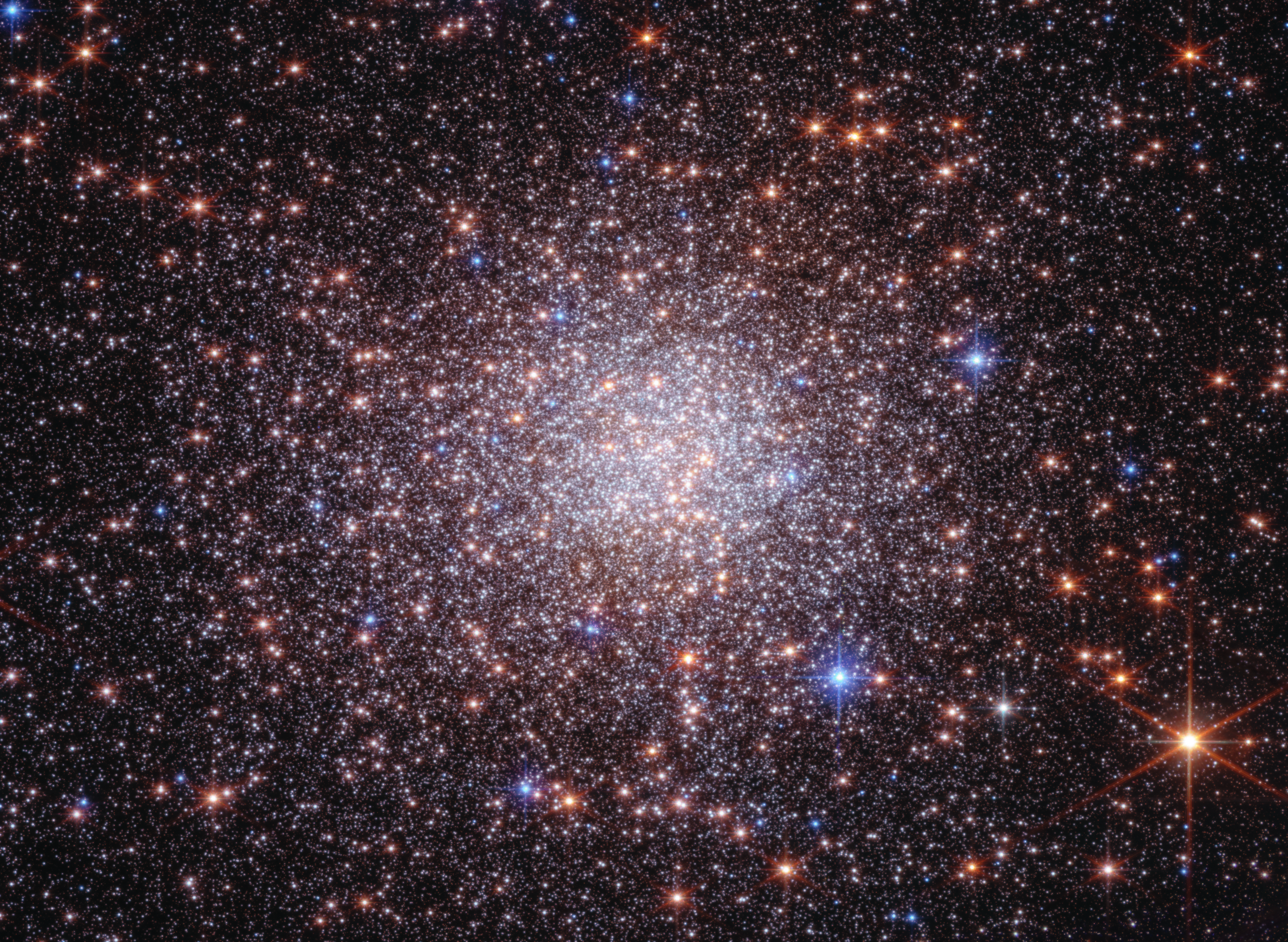
A dramatically crowded starfield that looks like a just-shaken snow globe. The black background of space, which is clearer at the edges, is covered by thousands of tiny white, orange, and blue points of light, which are stars. The stars are most concentrated in the center, forming a roughly circular orb, and sparser at the edges of the image. Several larger orange stars, particularly those largest near the edges of the frame, have prominent diffraction spikes.
New observations from Webb combined with multiple observations from Hubble prove that Terzan 5 is a self-contained, self-enriching stellar system that contains up to four distinct star populations. It orbits within our Milky Way galaxy’s central bulge.
Credits:
Image: NASA, ESA, CSA, STScI, Giorgia Zullo (University of Bologna), Francesco Ferraro (University of Bologna); Image Processing: Alyssa Pagan (STScI)

Researchers using two of humanity’s most powerful observatories — NASA’s James Webb and Hubble Space Telescopes — have definitively shown that Terzan 5 is not a globular star cluster as it was once classified, offering new insight into how galaxies like our own form and evolve over time. A globular star cluster typically has only one ancient star population. New data not only confirms the existence of two distinct populations of stars in Terzan 5, but also provides evidence for two more recent rounds of star formation. Although located within the crowded bulge of our Milky Way, our galaxy’s central, spherical region of older stars, Terzan 5 was massive enough to maintain its separate identity while lighter weight systems spread out and mixed to form the bulge billions of years ago. It’s like a lump in an otherwise well-mixed cake batter.

“Webb’s new near-infrared observations, cross-referenced with Hubble’s archival observations, have given us a much clearer picture of the history of Terzan 5,” said Giorgia Zullo, who led the research and is a PhD student at the University of Bologna in Italy.

These results were presented at a press conference Tuesday at the 248th meeting of the American Astronomical Society in Pasadena, and were published in Astronomy & Astrophysics.

Image: Bulge Fossil Fragment Terzan 5 (Webb and Hubble Image)

A dramatically crowded starfield that looks like a just-shaken snow globe. The black background of space, which is clearer at the edges, is covered by thousands of tiny white, orange, and blue points of light, which are stars. The stars are most concentrated in the center, forming a roughly circular orb, and sparser at the edges of the image. Several larger orange stars, particularly those largest near the edges of the frame, have prominent diffraction spikes.
New observations from Webb combined with multiple observations from Hubble prove that Terzan 5 is a self-contained, self-enriching stellar system that contains up to four distinct star populations. It orbits within our Milky Way galaxy’s central bulge.
Image: NASA, ESA, CSA, STScI, Giorgia Zullo (University of Bologna), Francesco Ferraro (University of Bologna); Image Processing: Alyssa Pagan (STScI)

Four generations of stars

Discovered in 1968 by astronomer Azop Terzan, Terzan 5 resembles a globular cluster in many ways. However, in 2009 this system was discovered to harbor two distinct populations of stars. In 2016 Hubble provided the first estimate of their ages, showing that one formed roughly 12 billion years ago — as the Milky Way itself was assembling — and the other about 5 billion years ago, just before Earth started forming. This pointed to a more complex history than a typical globular cluster.

Studying Terzan 5 is complicated by its location in a region of our galaxy crowded with stars and heavily obscured by dust. This is where Webb stepped in. Its infrared view allowed the research team to peer through the dust and catalog many more stars, and fainter stars, than previous work. By measuring star colors and brightnesses, astronomers can classify them into populations of different ages and chemistries.

Webb was able to measure these key properties for every star within the field of view in the sky — both stars within Terzan 5 and unrelated foreground stars. To isolate the stars of Terzan 5, the team relied on the power and longevity of Hubble. The 12-year separation allowed the team to measure very small movements of individual stars, known as proper motions, to determine which stars belong to Terzan 5 and which are part of the Milky Way bulge.

By combining data from both Webb and Hubble, the researchers found strong evidence for two more stellar populations, one that formed 3.8 billion years ago and another only 2.5 billion years ago. They also were able to determine the ages of the previously known stellar populations with unprecedented precision, finding that they formed 12.5 billion and 4.7 billion years ago.

With the previously known two generations of stars, astronomers could not rule out the possibility that Terzan 5 interacted with another object, like a globular cluster or a giant molecular cloud, becoming enriched with new gas and dust that set off a second round of star formation. With four stellar generations, those explanations are ruled out.

Measurements of the stellar composition of Terzan 5 populations made at the W. M. Keck Observatory and European Southern Observatory’s Very Large Telescope also point toward very distinct populations. “Along with the ages of these populations, the cluster preserves a fossil record of progressive enrichment of heavy elements by supernovae,” said co-author R. Michael Rich, a research astronomer at the University of California, Los Angeles.

Terzan 5 formed multiple generations of stars because it was able to retain the necessary raw materials. There is evidence of powerful supernova explosions in Terzan 5 that forged heavier elements that were swept up by subsequent generations of stars. In lighter weight systems, the force of the explosions themselves could have ejected the resulting elements as well as sweeping out leftover gas and dust. The progenitor of Terzan 5 had enough mass to retain those stars’ ejections, allowing new generations of stars to form over billions of years.

‘Bulge fossil fragment’

The results show that Terzan 5 is most likely the remnant of a much more massive stellar system that initially formed 12.5 billion years ago. Terzan 5 is extraordinary because it survived — and never merged or fully “mixed in” with the Milky Way’s bulge. “For some reason, this peculiar clump of stars formed separately from the bulge and was not destroyed as the bulge itself formed,” said Francesco R. Ferraro, a professor at the University of Bologna and principal investigator of the Webb observations. “Terzan 5 is what we now call a bulge fossil fragment because it resembles the primordial clumps that contributed to the formation of the bulge.”

To date, there’s one other known cosmic object like Terzan 5. Liller 1 was the second to be reclassified from a globular star cluster to a bulge fossil fragment. It also contains multiple generations of stars. There may be more objects like it. Between 40 to 50 additional globular clusters that orbit within the bulge will be examined by Ferraro’s team to determine if their stellar populations are all the same, like globular clusters, or have several generations, like bulge fossil fragments. 

Video: Zoom to See Terzan 5 Near Our Milky Way Galaxy’s Bulge

Zoom in to Terzan 5, a star cluster that lies within the crowded central region of our Milky Way galaxy known as the bulge. The scene starts with a ground-based image of our Milky Way bulge and zooms in on and circles Terzan 5, ending with the composite image of the star system from the James Webb and Hubble Space Telescopes.
Video: NASA, ESA, CSA, Alyssa Pagan (STScI); Acknowledgment: ESO, Pan-STARRS, DSS2, Akira Fujii

Potential parallels for galaxy formation near, far

Ultimately, this research may improve what we know about how the central bulges of galaxies form over hundreds of millions of years. “Based on observations and in-depth simulations, we think that galaxies in the early universe had huge disks of gas that fragmented into clumps and formed stars. These clumps migrated to the center of the galaxies, and many merged to form their bulges,” said Barbara Lanzoni, a co-author and associate professor at the University of Bologna. For example, Webb has turned up several examples of “clumpy” galaxies that were actively forming when the universe was only a few hundred million years old, like the clumps in the Firefly Sparkle galaxy. “Terzan 5 may provide direct evidence that can help explain how bulges formed in galaxies throughout the universe,” Lanzoni said.

The James Webb Space Telescope is the world’s premier space science observatory. Webb is solving mysteries in our solar system, looking beyond to distant worlds around other stars, and probing the mysterious structures and origins of our universe and our place in it. Webb is an international program led by NASA with its partners, ESA (European Space Agency) and CSA (Canadian Space Agency).

The Hubble Space Telescope has been operating for over three decades and continues to make ground-breaking discoveries that shape our fundamental understanding of the universe. Hubble is a project of international cooperation between NASA and ESA (European Space Agency). NASA’s Goddard Space Flight Center in Greenbelt, Maryland, manages the telescope and mission operations. Lockheed Martin Space, based in Denver, also supports mission operations at Goddard. The Space Telescope Science Institute in Baltimore, which is operated by the Association of Universities for Research in Astronomy, conducts Hubble science operations for NASA.

To learn more about Webb, visit:

https://science.nasa.gov/webb

To learn more about Hubble, visit:

https://science.nasa.gov/hubble

Downloads & Related Information

The following sections contain links to download this article’s images and videos in all available resolutions followed by related information links, media contacts, and if available, research paper and Spanish translation links.

Related Images & Videos

A dramatically crowded starfield that looks like a just-shaken snow globe. The black background of space, which is clearer at the edges, is covered by thousands of tiny white, orange, and blue points of light, which are stars. The stars are most concentrated in the center, forming a roughly circular orb, and sparser at the edges of the image. Several larger orange stars, particularly those largest near the edges of the frame, have prominent diffraction spikes.

Bulge Fossil Fragment Terzan 5 (Webb and Hubble Image)

New observations from Webb combined with multiple observations from Hubble prove that Terzan 5 is a self-contained, self-enriching stellar system that contains up to four distinct star populations. It orbits within our Milky Way galaxy’s central bulge.

Image titled u201cJames Webb and Hubble Space Telescopes; Terzan 5,u201d with compass arrows and color key. A dramatically crowded starfield that looks like a just-shaken snow globe. The black background of space is covered by thousands of tiny white, orange, and blue points of light, which are stars. The stars are most concentrated in the center, forming a roughly circular orb. At the bottom left are compass arrows indicating the orientation of the image on the sky. The east arrow points toward 12 ou2019clock. The north arrow points toward 3 ou2019clock. At the bottom right is a scale bar labeled 2 light-years. The length of the scale bar is about one seventh of the total image. Below the image is a color key showing which Hubble ACS/WFC and Webb NIRCam filters were used to create the image, and which visible-light color is assigned to each filter. Hubble ACS filters, from left to right: F606W is blue and F814W is teal. Webb NIRCam filters: F115W is orange, F200W is red.

Terzan 5 (Webb and Hubble Compass Image)

This image of bulge fossil fragment Terzan 5 was captured by the James Webb and Hubble space telescopes. Webb’s data are from its NIRCam (Near-Infrared Camera) and Hubble’s from its Advanced Camera for Surveys (ACS). The image shows a scale bar, compass arrows, and co…

Telescope image shows the Milky Way. The Milky Wayu2019s central region runs from top left to bottom right and has dark lanes of dust overlapping bright regions of stars. At the center is a label, Milky Way Center. The entire image is mostly black with pinpoints of light throughout. Thin blue lines connect several stars of two constellations, including the shape of a teapot representing Sagittarius at left and a long, arced line representing Scorpius at right. On the left, a large illustration of a centaur represents Sagittarius, with the teapot over his right arm. On the right, a large illustration of a scorpion represents Scorpius, with the line running throughout the center of its body.

Zoom to See Terzan 5 Near Our Milky Way Galaxy’s Bulge

Zoom in to Terzan 5, a star cluster that lies within the crowded central region of our Milky Way galaxy known as the bulge. The scene starts with a ground-based image of our Milky Way bulge and zooms in on and circles Terzan 5, ending with the composite image of the star system f…

Related Links

Read more: Hubble’s star clusters

Explore more: ViewSpace | Forms of light: the Cluster Omega Centauri

Watch: Globular Clusters, Stellar Pockets

Watch: Sorting the Stars in Omega Centauri

Webb: News | Images | Science | Home Page

Hubble: News | Images | Science | Home Page


Share

Details

Last Updated
Jun 16, 2026
Contact
Media

Laura Betz
NASA’s Goddard Space Flight Center
Greenbelt, Maryland
laura.e.betz@nasa.gov

Christine Pulliam
Space Telescope Science Institute
Baltimore, Maryland

Claire Blome
Space Telescope Science Institute
Baltimore, Maryland

Levothyroxine Trends in Congenital Hypothyroidism: 10-Year Study

16 June 2026 at 18:19

In recent years, the global medical community has observed an alarming rise in the incidence of congenital hypothyroidism (CH), a condition characterized by insufficient thyroid hormone production in newborns. This endocrine disorder, if undiagnosed or untreated, can lead to profound developmental delays and intellectual disabilities. Against this backdrop, a landmark study published in Pediatric Research sheds new light on the long-term administration and cessation patterns of levothyroxine—a synthetic thyroid hormone replacement—in patients assumed to have CH. This nationwide cohort investigation, spanning over a decade, provides unprecedented insights into treatment paradigms and patient management on a population scale.

The study meticulously tracked levothyroxine usage across a broad demographic, encompassing thousands of newborns clinically suspected of congenital hypothyroidism. Using comprehensive health databases, researchers were able to delineate initiation rates, dosage adjustments, duration of therapy, and the critical junctures at which discontinuation occurs. They highlight how early diagnosis, often through neonatal screening programs, has revolutionized patient outcomes but also raised new questions regarding optimal treatment length and criteria for safely discontinuing therapy.

One of the foundational aspects explored in this investigation is the temporal trend of levothyroxine initiation. The data convincingly demonstrate a marked increase in the frequency of diagnoses and subsequent treatment initiation over the 10-year observation period. This escalation aligns with heightened screening sensitivity and revised diagnostic thresholds, suggesting that what was once considered atypical or borderline hypothyroidism is now more frequently targeted for intervention. Such shifts bring about complex clinical decisions around the balance between necessary hormone supplementation and potential overtreatment.

In delineating the course of levothyroxine administration, the study underscores that a significant subset of patients undergoes treatment discontinuation, reflecting evolving clinical judgments based on follow-up evaluations. These discontinuations often result from confirmatory testing that challenges the initial diagnosis or reveals transient hypothyroidism, a condition that resolves spontaneously without lifelong hormone dependency. Understanding when and how to discontinue therapy safely remains a cornerstone in preventing unnecessary treatment burden and its accompanying risks.

Moreover, the investigation probes into the demographic and clinical determinants influencing levothyroxine discontinuation. Variables such as age at diagnosis, initial thyroid function test results, and the presence of thyroid dysgenesis or dyshormonogenesis significantly sway therapeutic decisions. The study’s granular stratification reveals that patients with milder biochemical abnormalities or transient hypothyroidism are more likely to cease treatment within the first few years of life, emphasizing the importance of individualized care pathways.

Intriguingly, the analysis also touches upon the challenges in differentiating permanent congenital hypothyroidism from transient cases during the neonatal period. These diagnostic ambiguities are amplified by biochemical fluctuations, maternal thyroid conditions, and environmental factors such as exposure to iodine or goitrogens. The study’s extensive dataset allows for a nuanced understanding of these confounders, paving the way for refined diagnostic algorithms and personalized monitoring protocols.

The ramifications of sustained levothyroxine therapy extend beyond hormone replacement; they encompass metabolic, cardiovascular, and neurodevelopmental domains, especially if treatment is misaligned with the patient’s true thyroid status. Over-replacement can lead to iatrogenic thyrotoxicosis, while under-treatment perpetuates hypothyroid sequelae. This cohort study underscores the imperative for vigilant clinical and biochemical monitoring, leveraging advances in assay technologies and genetic profiling to tailor interventions precisely.

From a public health perspective, the rising levothyroxine use in presumed CH presents implications for healthcare resource allocation, long-term follow-up infrastructure, and parental counseling. National screening programs must adapt to integrate not only early detection but also robust tracking systems that ascertain treatment necessity, effectiveness, and safety over time. This study’s nationwide scope lends critical data to guide policy refinements and equity considerations in thyroid disease management.

Furthermore, the researchers advocate for a paradigm shift towards dynamic therapeutic frameworks, where treatment initiation is complemented by scheduled re-evaluations to determine the persistence of hypothyroidism. By promoting trial discontinuations under strict supervision, clinicians can limit unnecessary exposure to levothyroxine, thereby mitigating potential adverse effects and preserving health system sustainability.

Equally important, the study highlights gaps in current clinical guidelines, which often lack consensus on precise criteria for levothyroxine cessation. These ambiguities contribute to variability in practice and patient outcomes. The authors call for concerted efforts among endocrinologists, neonatologists, and healthcare policymakers to develop evidence-based protocols informed by longitudinal data such as theirs.

In terms of methodology, the utilization of expansive health claims and prescription data validates the reliability and real-world applicability of the findings. The analytical approach, employing survival analysis and multivariate regression modeling, unravels complex patterns of drug utilization and discontinuation, while adjusting for confounding factors. This rigorous epidemiological framework sets a benchmark for future pharmacoepidemiological research in pediatric endocrinology.

It is also noteworthy that the study addresses psychosocial dimensions indirectly, recognizing that parents and caregivers play pivotal roles in adherence and decision-making regarding levothyroxine therapy. Long-term treatment regimens can strain families emotionally and financially, underscoring the need for supportive care models and transparent communication strategies.

The implications of the rising incidence of CH—and correspondingly, levothyroxine use—also evoke considerations about environmental and genetic influences driving this trend. Although not the primary focus, the comprehensive data collected may inspire ancillary investigations into prenatal exposures, epigenetic modifications, and population-based genetic predispositions contributing to this public health challenge.

Ultimately, this landmark investigation by Kim, Woo, Lee, and colleagues portrays a nuanced portrait of levothyroxine use in presumed congenital hypothyroidism, emphasizing the dual imperatives of timely treatment and judicious discontinuation. Their ten-year nationwide cohort analysis unearths critical trends that will undoubtedly inform clinical practice, guideline development, and health policy designed to optimize outcomes for affected infants and children worldwide.

As pediatric endocrinology advances, the insights gleaned from this study serve as a timely reminder of the complexity underlying ostensibly straightforward diagnoses. The nuanced interplay between biochemical parameters, diagnostic certainty, patient heterogeneity, and long-term treatment strategies requires clinicians to balance evidence-based medicine with individualized care. This study not only contributes pivotal epidemiological evidence but also catalyzes a re-examination of management philosophies in congenital hypothyroidism.

In conclusion, the increasing incidence of congenital hypothyroidism and the corresponding escalation in levothyroxine use illuminate a critical area for continued research and healthcare innovation. The ten-year nationwide cohort study effectively captures the evolving landscape of diagnosis, treatment, and discontinuation, reinforcing the need for integrated multidisciplinary approaches. By fostering personalized medicine approaches grounded in large-scale data analysis, healthcare systems can aspire toward improved thyroid health in newborns and beyond.


Subject of Research: Use and discontinuation patterns of levothyroxine treatment in patients with presumed congenital hypothyroidism over ten years in a nationwide cohort.

Article Title: Levothyroxine use and discontinuation in presumed congenital hypothyroidism: a 10-year nationwide cohort study.

Article References:
Kim, S.Y., Woo, H.A., Lee, E.H. et al. Levothyroxine use and discontinuation in presumed congenital hypothyroidism: a 10-year nationwide cohort study. Pediatr Res (2026). https://doi.org/10.1038/s41390-026-05153-7

Image Credits: AI Generated

DOI: 16 June 2026

NASA Webb, Hubble Reveal History of Relic of Milky Way’s Formation

16 June 2026 at 18:15
 
7 Min Read

NASA Webb, Hubble Reveal History of Relic of Milky Way’s Formation

A dramatically crowded starfield that looks like a just-shaken snow globe. The black background of space, which is clearer at the edges, is covered by thousands of tiny white, orange, and blue points of light, which are stars. The stars are most concentrated in the center, forming a roughly circular orb, and sparser at the edges of the image. Several larger orange stars, particularly those largest near the edges of the frame, have prominent diffraction spikes.
New observations from Webb combined with multiple observations from Hubble prove that Terzan 5 is a self-contained, self-enriching stellar system that contains up to four distinct star populations. It orbits within our Milky Way galaxy’s central bulge.
Credits: Image: NASA, ESA, CSA, STScI, Giorgia Zullo (University of Bologna), Francesco Ferraro (University of Bologna); Image Processing: Alyssa Pagan (STScI)

Researchers using two of humanity’s most powerful observatories — NASA’s James Webb and Hubble Space Telescopes — have definitively shown that Terzan 5 is not a globular star cluster as it was once classified, offering new insight into how galaxies like our own form and evolve over time. A globular star cluster typically has only one ancient star population. New data not only confirms the existence of two distinct populations of stars in Terzan 5, but also provides evidence for two more recent rounds of star formation. Although located within the crowded bulge of our Milky Way, our galaxy’s central, spherical region of older stars, Terzan 5 was massive enough to maintain its separate identity while lighter weight systems spread out and mixed to form the bulge billions of years ago. It’s like a lump in an otherwise well-mixed cake batter.

“Webb’s new near-infrared observations, cross-referenced with Hubble’s archival observations, have given us a much clearer picture of the history of Terzan 5,” said Giorgia Zullo, who led the research and is a PhD student at the University of Bologna in Italy.

These results were presented at a press conference Tuesday at the 248th meeting of the American Astronomical Society in Pasadena, and were published in Astronomy & Astrophysics.

Image: Bulge Fossil Fragment Terzan 5 (Webb and Hubble Image)

A dramatically crowded starfield that looks like a just-shaken snow globe. The black background of space, which is clearer at the edges, is covered by thousands of tiny white, orange, and blue points of light, which are stars. The stars are most concentrated in the center, forming a roughly circular orb, and sparser at the edges of the image. Several larger orange stars, particularly those largest near the edges of the frame, have prominent diffraction spikes.
New observations from Webb combined with multiple observations from Hubble prove that Terzan 5 is a self-contained, self-enriching stellar system that contains up to four distinct star populations. It orbits within our Milky Way galaxy’s central bulge.
Image: NASA, ESA, CSA, STScI, Giorgia Zullo (University of Bologna), Francesco Ferraro (University of Bologna); Image Processing: Alyssa Pagan (STScI)

Four generations of stars

Discovered in 1968 by astronomer Azop Terzan, Terzan 5 resembles a globular cluster in many ways. However, in 2009 this system was discovered to harbor two distinct populations of stars. In 2016 Hubble provided the first estimate of their ages, showing that one formed roughly 12 billion years ago — as the Milky Way itself was assembling — and the other about 5 billion years ago, just before Earth started forming. This pointed to a more complex history than a typical globular cluster.

Studying Terzan 5 is complicated by its location in a region of our galaxy crowded with stars and heavily obscured by dust. This is where Webb stepped in. Its infrared view allowed the research team to peer through the dust and catalog many more stars, and fainter stars, than previous work. By measuring star colors and brightnesses, astronomers can classify them into populations of different ages and chemistries.

Webb was able to measure these key properties for every star within the field of view in the sky — both stars within Terzan 5 and unrelated foreground stars. To isolate the stars of Terzan 5, the team relied on the power and longevity of Hubble. The 12-year separation allowed the team to measure very small movements of individual stars, known as proper motions, to determine which stars belong to Terzan 5 and which are part of the Milky Way bulge.

By combining data from both Webb and Hubble, the researchers found strong evidence for two more stellar populations, one that formed 3.8 billion years ago and another only 2.5 billion years ago. They also were able to determine the ages of the previously known stellar populations with unprecedented precision, finding that they formed 12.5 billion and 4.7 billion years ago.

With the previously known two generations of stars, astronomers could not rule out the possibility that Terzan 5 interacted with another object, like a globular cluster or a giant molecular cloud, becoming enriched with new gas and dust that set off a second round of star formation. With four stellar generations, those explanations are ruled out.

Measurements of the stellar composition of Terzan 5 populations made at the W. M. Keck Observatory and European Southern Observatory’s Very Large Telescope also point toward very distinct populations. “Along with the ages of these populations, the cluster preserves a fossil record of progressive enrichment of heavy elements by supernovae,” said co-author R. Michael Rich, a research astronomer at the University of California, Los Angeles.

Terzan 5 formed multiple generations of stars because it was able to retain the necessary raw materials. There is evidence of powerful supernova explosions in Terzan 5 that forged heavier elements that were swept up by subsequent generations of stars. In lighter weight systems, the force of the explosions themselves could have ejected the resulting elements as well as sweeping out leftover gas and dust. The progenitor of Terzan 5 had enough mass to retain those stars’ ejections, allowing new generations of stars to form over billions of years.

‘Bulge fossil fragment’

The results show that Terzan 5 is most likely the remnant of a much more massive stellar system that initially formed 12.5 billion years ago. Terzan 5 is extraordinary because it survived — and never merged or fully “mixed in” with the Milky Way’s bulge. “For some reason, this peculiar clump of stars formed separately from the bulge and was not destroyed as the bulge itself formed,” said Francesco R. Ferraro, a professor at the University of Bologna and principal investigator of the Webb observations. “Terzan 5 is what we now call a bulge fossil fragment because it resembles the primordial clumps that contributed to the formation of the bulge.”

To date, there’s one other known cosmic object like Terzan 5. Liller 1 was the second to be reclassified from a globular star cluster to a bulge fossil fragment. It also contains multiple generations of stars. There may be more objects like it. Between 40 to 50 additional globular clusters that orbit within the bulge will be examined by Ferraro’s team to determine if their stellar populations are all the same, like globular clusters, or have several generations, like bulge fossil fragments. 

Video: Zoom to See Terzan 5 Near Our Milky Way Galaxy’s Bulge

Zoom in to Terzan 5, a star cluster that lies within the crowded central region of our Milky Way galaxy known as the bulge. The scene starts with a ground-based image of our Milky Way bulge and zooms in on and circles Terzan 5, ending with the composite image of the star system from the James Webb and Hubble Space Telescopes.
Video: NASA, ESA, CSA, Alyssa Pagan (STScI); Acknowledgment: ESO, Pan-STARRS, DSS2, Akira Fujii

Potential parallels for galaxy formation near, far

Ultimately, this research may improve what we know about how the central bulges of galaxies form over hundreds of millions of years. “Based on observations and in-depth simulations, we think that galaxies in the early universe had huge disks of gas that fragmented into clumps and formed stars. These clumps migrated to the center of the galaxies, and many merged to form their bulges,” said Barbara Lanzoni, a co-author and associate professor at the University of Bologna. For example, Webb has turned up several examples of “clumpy” galaxies that were actively forming when the universe was only a few hundred million years old, like the clumps in the Firefly Sparkle galaxy. “Terzan 5 may provide direct evidence that can help explain how bulges formed in galaxies throughout the universe,” Lanzoni said.

The James Webb Space Telescope is the world’s premier space science observatory. Webb is solving mysteries in our solar system, looking beyond to distant worlds around other stars, and probing the mysterious structures and origins of our universe and our place in it. Webb is an international program led by NASA with its partners, ESA (European Space Agency) and CSA (Canadian Space Agency).

The Hubble Space Telescope has been operating for over three decades and continues to make ground-breaking discoveries that shape our fundamental understanding of the universe. Hubble is a project of international cooperation between NASA and ESA (European Space Agency). NASA’s Goddard Space Flight Center in Greenbelt, Maryland, manages the telescope and mission operations. Lockheed Martin Space, based in Denver, also supports mission operations at Goddard. The Space Telescope Science Institute in Baltimore, which is operated by the Association of Universities for Research in Astronomy, conducts Hubble science operations for NASA.

To learn more about Webb, visit:

https://science.nasa.gov/webb

To learn more about Hubble, visit:

https://science.nasa.gov/hubble

Downloads & Related Information

The following sections contain links to download this article’s images and videos in all available resolutions followed by related information links, media contacts, and if available, research paper and Spanish translation links.

Related Images & Videos

A dramatically crowded starfield that looks like a just-shaken snow globe. The black background of space, which is clearer at the edges, is covered by thousands of tiny white, orange, and blue points of light, which are stars. The stars are most concentrated in the center, forming a roughly circular orb, and sparser at the edges of the image. Several larger orange stars, particularly those largest near the edges of the frame, have prominent diffraction spikes.

Bulge Fossil Fragment Terzan 5 (Webb and Hubble Image)

New observations from Webb combined with multiple observations from Hubble prove that Terzan 5 is a self-contained, self-enriching stellar system that contains up to four distinct star populations. It orbits within our Milky Way galaxy’s central bulge.

Image titled u201cJames Webb and Hubble Space Telescopes; Terzan 5,u201d with compass arrows and color key. A dramatically crowded starfield that looks like a just-shaken snow globe. The black background of space is covered by thousands of tiny white, orange, and blue points of light, which are stars. The stars are most concentrated in the center, forming a roughly circular orb. At the bottom left are compass arrows indicating the orientation of the image on the sky. The east arrow points toward 12 ou2019clock. The north arrow points toward 3 ou2019clock. At the bottom right is a scale bar labeled 2 light-years. The length of the scale bar is about one seventh of the total image. Below the image is a color key showing which Hubble ACS/WFC and Webb NIRCam filters were used to create the image, and which visible-light color is assigned to each filter. Hubble ACS filters, from left to right: F606W is blue and F814W is teal. Webb NIRCam filters: F115W is orange, F200W is red.

Terzan 5 (Webb and Hubble Compass Image)

This image of bulge fossil fragment Terzan 5 was captured by the James Webb and Hubble space telescopes. Webb’s data are from its NIRCam (Near-Infrared Camera) and Hubble’s from its Advanced Camera for Surveys (ACS). The image shows a scale bar, compass arrows, and co…

Telescope image shows the Milky Way. The Milky Wayu2019s central region runs from top left to bottom right and has dark lanes of dust overlapping bright regions of stars. At the center is a label, Milky Way Center. The entire image is mostly black with pinpoints of light throughout. Thin blue lines connect several stars of two constellations, including the shape of a teapot representing Sagittarius at left and a long, arced line representing Scorpius at right. On the left, a large illustration of a centaur represents Sagittarius, with the teapot over his right arm. On the right, a large illustration of a scorpion represents Scorpius, with the line running throughout the center of its body.

Zoom to See Terzan 5 Near Our Milky Way Galaxy’s Bulge

Zoom in to Terzan 5, a star cluster that lies within the crowded central region of our Milky Way galaxy known as the bulge. The scene starts with a ground-based image of our Milky Way bulge and zooms in on and circles Terzan 5, ending with the composite image of the star system f…

Related Links

Read more: Hubble’s star clusters

Explore more: ViewSpace | Forms of light: the Cluster Omega Centauri

Watch: Globular Clusters, Stellar Pockets

Watch: Sorting the Stars in Omega Centauri

Webb: News | Images | Science | Home Page

Hubble: News | Images | Science | Home Page

Share

Details

Last Updated
Jun 16, 2026
Contact
Media

Laura Betz
NASA’s Goddard Space Flight Center
Greenbelt, Maryland
laura.e.betz@nasa.gov

Christine Pulliam
Space Telescope Science Institute
Baltimore, Maryland

Claire Blome
Space Telescope Science Institute
Baltimore, Maryland

The post NASA Webb, Hubble Reveal History of Relic of Milky Way’s Formation appeared first on NASA Science.

Young Frogs Adopt Cautious Behavior in Response to Disease Outbreak

16 June 2026 at 18:04

For a diminutive amphibian measuring no larger than a single fingernail, the allocation of energy can mark the fine line between survival and demise. Recent groundbreaking research from the University of Florida delves into the intricate decisions juvenile frogs make regarding energy distribution, especially when confronted with lethal pathogens. By leveraging a blend of empirical data and computational modeling, the study uncovers nuanced strategies these tiny creatures adopt, balancing growth and immune defense — insights that not only illuminate amphibian biology but also shed light on broader ecological and evolutionary dynamics.

Central to this study is the common coqui frog (Eleutherodactylus coqui), an emblematic species native to Puerto Rico known for its direct development, bypassing the traditional tadpole stage. This biological peculiarity means these frogs hatch as fully formed miniature adults, profoundly influencing how energy constraints impact their early life stages. Given their extraordinarily small size, approximately equivalent to a human pinky nail, tracking individual frogs in natural environments is virtually impossible. Consequently, the research team integrated robust field and experimental observations into sophisticated computer models, simulating how these frogs allocate scarce resources across growth and immunity throughout a typical year.

The research focuses poignantly on the interplay between the coqui frogs and a devastating fungal pathogen, Batrachochytrium dendrobatidis (Bd), commonly known as chytrid fungus. This pathogen has wreaked havoc on amphibian populations globally, making it a critical subject for understanding disease ecology. Leveraging data encompassing seasonal fluctuations in food availability and infection risk, the models capture the dynamic energy budgeting decisions these amphibians undertake. Intriguingly, findings reveal a compelling survival strategy: juvenile frogs prioritize rapid growth during early life phases, deferring costly immune investments until infection intensity escalates to dangerous levels.

This strategic delay in immune response is hypothesized to optimize survival probabilities. Small, slow-growing individuals face heightened predation risks, making early growth paramount. By investing in immunity only when faced with a significant pathogenic threat, frogs effectively balance the trade-offs between immediate survival pressures and long-term health. The models demonstrate that this energy allocation strategy maximizes fitness, highlighting the sophistication of amphibian physiological responses to environmental and pathogenic stressors.

Temporal variation emerges as a decisive factor modulating these energy allocation decisions. Seasonal oscillations in temperature and precipitation orchestrate changes in food resources and pathogen prevalence. Frogs hatching during favorable summer conditions, when prey is abundant, can allocate energy more flexibly across growth and immune defense, enhancing their chances for survival and successful maturation. Conversely, cohorts born during cooler and drier periods encounter constrained energy budgets, compelling a more frugal distribution that compromises both growth rates and immune competence, with dire consequences for survival.

This research also illuminates the broader implications of climate change for amphibian populations. Extending or intensifying cool, dry periods could exacerbate energetic limitations faced by juvenile frogs, impeding rapid growth and compromising immune defenses. Such shifts threaten to undermine population resilience against pathogens like chytrid fungus, potentially precipitating more severe declines. Understanding these vulnerabilities is crucial for conservation efforts aiming to buffer amphibian species against accelerating environmental perturbations.

Methodologically, the study exemplifies the power of integrating mechanistic disease models with empirical data sets. By simulating individual-level energy trade-offs in relation to pathogen exposure within fluctuating environments, the researchers transcend traditional population-level disease modeling. This approach unveils heterogeneity in individual responses, a key dimension often masked in aggregate analyses, enabling a more precise understanding of disease dynamics and host resilience.

The study’s lead author, Zuania Colón-Piñeiro Ph.D., emphasizes the importance of dissecting individual heterogeneity in disease ecology: “Not all individuals respond identically to pathogens, and understanding this heterogeneity is vital for predicting population outcomes.” Her collaboration with senior author Ana V. Longo Ph.D. underscores the synergy between empirical natural history and theoretical modeling, with Longo highlighting the unique biology of coqui frogs that facilitates this research and provides broader insights into amphibian developmental strategies.

The implications of these findings extend beyond the realm of wild amphibians. The computational framework developed by the team offers a valuable tool for conservationists managing captive breeding programs. By modeling energy allocation strategies under varying environmental conditions and pathogen pressures, conservationists can optimize rearing protocols to boost survival rates post-release, enhancing the efficacy of reintroduction programs. This translational potential exemplifies the study’s impact, providing a bridge between fundamental ecological theory and practical wildlife management.

Importantly, the research team advocates for the continued integration of fieldwork and modeling. Models, while powerful, rely on accurate and extensive biological data to yield meaningful predictions. Field studies that track environmental variables, pathogen prevalence, and individual growth trajectories remain indispensable. This dual approach ensures that theoretical insights remain grounded in biological reality, ultimately guiding more effective interventions to safeguard vulnerable species.

The study, entitled “Playing it safe at early life stages: Balancing energy allocations to maximize fitness under seasonal pathogen dynamics,” stands as a testament to interdisciplinary collaboration, involving expertise across multiple departments at the University of Florida, the University of Puerto Rico, and partner institutions. By illuminating how minute amphibians navigate the energetic tightrope of survival amidst disease threats and fluctuating environments, it adds a significant chapter to our understanding of life history evolution and disease ecology.

As amphibian declines continue to raise alarms on global biodiversity fronts, insights from such detailed mechanistic studies are invaluable. They remind us that survival strategies are seldom straightforward and that even the tiniest creatures deploy sophisticated physiological tactics honed by evolution. Understanding these strategies underpins efforts to stem biodiversity loss, highlighting the intricate tapestry connecting organismal biology, environmental dynamics, and disease.


Subject of Research: Animals

Article Title: Playing it safe at early life stages: Balancing energy allocations to maximize fitness under seasonal pathogen dynamics

News Publication Date: 15-Jun-2026

Web References: http://dx.doi.org/10.1111/1365-2656.70282

Image Credits: Alberto López

Keywords: Frogs, Amphibians, Developmental biology, Evolutionary developmental biology, Immune system, Adaptive immune system, Immune regulation

How Scientific Advances Are Transforming Our Understanding of the Biology of Ageing

16 June 2026 at 18:02

In the pursuit of understanding the enigmatic phenomenon of ageing, modern biology finds itself at a crossroads more complex than ever before. Once described as “an unsolved problem in biology” by Nobel laureate Peter Medawar in the mid-20th century, ageing has since evolved into a multifaceted enigma that transcends simple explanations or singular mechanisms. Recent advances in the field have empowered scientists to probe the molecular and genetic underpinnings of ageing at unprecedented depth, studying thousands of genes within individual cells and dissecting the interplay of diverse biological pathways. Yet this surge in knowledge reveals not clarity but the intricate complexity inherent to the ageing process.

Dr. Piotr Chmielewski from Wroclaw Medical University offers a timely and critical review that reshapes our understanding of ageing biology. His article published in the journal Biogerontology challenges the long-held quest for a universal cause of ageing and posits instead that ageing is a product of dynamic, overlapping biological processes operating concurrently across various organizational levels. In this paradigm shift, the once singular biological quandary becomes a tapestry of interconnected cellular and molecular systems whose interactions collectively propel the ageing process.

The leaps in technology over recent decades have drastically expanded the horizons of ageing research. Signaling networks such as mTOR, AMPK, FOXO, and IGF-1 have been identified as crucial regulators of growth, metabolism, and longevity, illuminating intracellular pathways that influence lifespan. The development of epigenetic clocks—biological age estimators derived from patterns of DNA methylation—has enabled researchers to quantify biological age beyond chronological markers. Furthermore, advancements in single-cell sequencing technologies now allow unprecedented resolution in observing cellular changes during ageing. Despite these breakthroughs, Dr. Chmielewski emphasizes the paradox that greater knowledge reveals deeper uncertainties concerning the fundamental nature of ageing.

Historically, the pursuit of a singular ageing mechanism has dominated the field, with hypotheses including oxidative stress, accumulation of DNA damage, telomere attrition, mitochondrial decline, epigenetic alterations, and chronic low-level inflammation each proposed as primary drivers. Over time, however, it has become apparent that none of these factors alone account for the totality of ageing-related changes. Instead, ageing appears akin to a gradual loss of systemic organization, a complex biological network gradually unraveling through cumulative and interacting defects rather than a discrete failure attributable to any one cause.

This multidimensional view underpins the widely accepted concept of the hallmarks of ageing, encompassing biological features such as genomic instability, telomere shortening, epigenetic alterations, proteostasis loss, mitochondrial dysfunction, cellular senescence, stem cell exhaustion, and altered intercellular communication. These hallmarks do not function in isolation but in intricate crosstalk, amplifying each other’s effects and contributing to the systemic degeneration observed in ageing organisms.

A pivotal advance driving contemporary age biology research lies in the use of epigenetic clocks, tools that estimate biological age by profiling DNA methylation patterns at select genomic loci. While these clocks correlate strongly with chronological age and predict disease risk and mortality, their exact biological significance remains uncertain. Dr. Chmielewski articulates a critical question facing the field: do epigenetic clocks capture core ageing mechanisms, or are they merely biomarkers reflecting downstream effects of other processes? The distinction is monumental because it determines whether interventions targeting epigenetic marks will achieve true rejuvenation or merely modulate surrogate ageing indicators.

Experimental therapies such as senolytics have shown promise by selectively eliminating senescent cells, which accumulate with age and contribute to tissue dysfunction and inflammation. Yet the complexity of ageing biology calls for caution—what works in rodent models may not directly translate to humans. Fundamental disparities in lifespan, metabolic rate, cellular turnover, tissue architecture, immune function, and repair capacity separate rodents from humans, limiting the applicability of animal data to human therapeutics. This gap underscores the critical need for human-centric clinical research in ageing interventions.

A profound insight emerging in recent years is the recognition that youthfulness is not the mere absence of cellular damage but rather a robust capacity for adaptation and repair. Living organisms continually face metabolic errors, DNA mutations, and protein misfolding; however, their resilience lies in effective stress response systems, regenerative potential, and metabolic plasticity. Interventions such as caloric restriction, intermittent fasting, and physical activity illustrate this principle: they do not halt damage accumulation but enhance the body’s adaptive responses, maintaining functionality and delaying age-related decline.

Shifting research focus accordingly, Dr. Chmielewski suggests a new narrative framing ageing research. Instead of solely asking “What causes ageing?”, the field is increasingly exploring “How do some organisms maintain function, resilience, and adaptive capacity despite ongoing damage?” This reframed question moves the scientific community towards identifying factors that preserve healthspan and mitigate functional loss rather than searching for elusive “anti-ageing” magic bullets.

Such an outlook heralds a future where ageing interventions emphasize support for systemic maintenance, resilience, and repair mechanisms. This direction could translate into therapeutic strategies that bolster cellular quality control, metabolic regulation, and immune competence, collectively sustaining organismal health beyond current expectations. In refocusing ageing as an emergent property of complex systems rather than a singular failure, biogerontology embraces a more holistic understanding that aligns with the observed heterogeneity and dynamism of ageing biology.

Ultimately, Dr. Chmielewski’s review challenges simplistic narratives and advocates for embracing complexity as the path forward. The realization that ageing is a multi-layered phenomenon demanding integrative study promises to inspire innovation in research approaches and therapeutic design. By charting this nuanced course, the scientific community stands poised to unlock the mysteries of longevity, delay the onset of age-related diseases, and fundamentally improve human health across the lifespan.


Subject of Research: Not applicable

Article Title: Ageing was never a singular problem in biology: implications for mechanisms, measurements and interventions

News Publication Date: 20-Apr-2026

Web References: http://dx.doi.org/10.1007/s10522-026-10436-x

References: Chmielewski, Piotr Paweł. “Ageing was never a singular problem in biology: implications for mechanisms, measurements and interventions.” Biogerontology (2026).

Image Credits: Wroclaw Medical University

Keywords: Gerontology, Ageing populations, DNA, Life sciences, Molecular biology

BU’s Sushrut Waikar, MD, MPH, Recognized by American Society of Nephrology

16 June 2026 at 18:01

In a remarkable acknowledgment of his contributions to nephrology, Dr. Sushrut S. Waikar has received the American Society of Nephrology (ASN) Distinguished Mentor Award. This prestigious accolade honors individuals who have profoundly impacted the kidney health community through their dedication to mentorship, fostering the careers of emerging clinicians and researchers. Dr. Waikar’s influence extends far beyond routine clinical practice; he has been instrumental in inspiring trainees to dedicate themselves to nephrology, emphasizing innovation in both research and healthcare practice.

Dr. Waikar currently holds the position of Chief of Nephrology at Boston Medical Center and serves as the Norman G. Levinsky Professor of Medicine at Boston University’s Chobanian & Avedisian School of Medicine. As a clinician-scientist, Dr. Waikar has maintained continuous funding from the National Institutes of Health (NIH) for over two decades, highlighting his sustained commitment to advancing kidney disease research. His expansive research agenda incorporates epidemiologic methodologies, translational science, and interventional clinical trials designed to address urgent clinical challenges.

Central to Dr. Waikar’s research portfolio is the refinement of diagnostic tools for kidney diseases, an area critical for early detection and improved patient outcomes. His laboratory has developed novel biomarkers and testing algorithms that aim to enhance diagnostic accuracy, thereby allowing for more timely and precise therapeutic interventions. Such advancements are vital in a field where early-stage kidney dysfunction often goes undetected until it progresses to irreversible damage.

Another significant thrust of Dr. Waikar’s research involves kidney biopsy studies, which offer invaluable insights into the pathophysiological mechanisms underlying disease progression within the renal parenchyma. By analyzing tissue samples, his team seeks to elucidate the cellular and molecular pathways that drive kidney deterioration, providing a foundation for the future design of targeted therapies. This level of detailed study bridges the gap between clinical observation and basic science, enabling a more comprehensive understanding of renal pathology.

Dr. Waikar’s investigations also spotlights a particularly devastating public health concern known as Chronic Kidney Disease of uncertain etiology (CKDu). This disease has emerged as a lethal epidemic primarily afflicting young agricultural workers in Central America and South Asia. Unlike traditional forms of chronic kidney disease associated with diabetes or hypertension, CKDu’s etiology remains elusive and multifactorial. His research endeavors aim to clarify environmental, occupational, and genetic risk factors contributing to this silent crisis, with hopes of informing preventative strategies and policy interventions.

Within the realm of precision medicine, Dr. Waikar acts as principal investigator for the NIH Kidney Precision Medicine Project. This ambitious initiative is engineered to leverage cutting-edge genomic, proteomic, and proteomic technologies to tailor kidney disease treatment at an individual level. By integrating these high-throughput techniques with clinical data, this project aspires to revolutionize management paradigms for kidney disorders, ultimately improving personalized patient care outcomes.

In addition to his leadership in precision medicine, Dr. Waikar coordinates the CKD of Unknown Etiology Consortium, a collaborative network of investigators dedicated to elucidating CKDu. This consortium underscores the importance of multidisciplinary cooperation in tackling complex health challenges, combining epidemiology, environmental sciences, and nephrology to develop holistic solutions to kidney health crises.

Dr. Waikar’s intellectual journey began with an undergraduate education in English and neuroscience at Amherst College, fostering a broad intellectual foundation that supports his interdisciplinary approach. He then earned his medical degree at Yale University, further complementing his scientific expertise with clinical acumen. His postgraduate training in internal medicine and nephrology at highly regarded institutions such as the University of California, San Francisco, and the Mass General Brigham system was distinguished by leadership roles, including chief medical resident, reflecting his early commitment to medical education and mentorship.

His academic trajectory also includes earning a Master’s degree in Public Health from Harvard University, which underpins his rigorous approach to epidemiology and population-based studies. This public health background allows Dr. Waikar to navigate and address the systemic factors impacting kidney health globally, marrying clinical insight with health policy and preventive medicine.

The American Society of Nephrology, founded in 1966, stands as the largest professional society devoted to kidney disease research and education worldwide. To further acknowledge the critical contributions of midcareer professionals, ASN established the Midcareer Awards Program in 2019, celebrating individuals like Dr. Waikar who demonstrate exceptional clinical expertise, innovative research, and dedicated service to the nephrology field and the society’s mission.

Dr. Waikar’s recognition by the ASN Distinguished Mentor Award not only celebrates his individual achievements but also highlights the critical role that mentorship plays in shaping the future of nephrology. His ability to guide, inspire, and cultivate the development of emerging leaders ensures that advances in kidney research and patient care continue to accelerate, ultimately benefiting countless individuals suffering from kidney disease globally.

Through the integration of basic science, clinical application, and public health strategies, Dr. Waikar exemplifies the modern physician-scientist who transforms knowledge into meaningful biomedical progress. His contributions serve as a catalyst for ongoing discoveries and innovations that hold the promise of reducing kidney disease burden worldwide.

Subject of Research:
Kidney disease diagnostics and research, Chronic Kidney Disease of uncertain etiology (CKDu), kidney biopsy studies, precision medicine in nephrology.

Article Title:
Not provided.

News Publication Date:
Not provided.

Web References:
Not provided.

References:
Not provided.

Image Credits:
Not provided.

Keywords:
Nephrology, kidney disease, chronic kidney disease, CKDu, NIH Kidney Precision Medicine Project, kidney biopsy, biomarkers, epidemiology, translational research, mentorship, American Society of Nephrology, public health.

NASA’s Quantum Lab Aboard Space Station Gets Chilly Upgrade

16 June 2026 at 17:50
Astronaut Jessica Meir inspects optical fibers while installing hardware updates to NASA’s Cold Atom Lab, or CAL, aboard the International Space Station on May 8, 2026. About the size of a minifridge, CAL enables researchers to explore quantum physics.
NASA

Astronauts aboard the International Space Station have switched on NASA’s newly upgraded Cold Atom Lab, a one-of-a-kind facility designed to improve how scientists explore the fundamental workings of matter and develop new quantum technologies. By leveraging the unique environment of microgravity in space, the lab can accomplish cutting-edge science impossible to do anywhere else.

Quantum science is the study of matter at the smallest scales, like atoms, electrons, and single particles of light. While it’s easy to imagine atoms as billiard balls bouncing off one another, they also exhibit wave-like behavior, can exist simultaneously in two places at once, and may even pass through one another.

About the size of a minifridge and operated from Earth, the Cold Atom Lab chills atoms to temperatures below minus 459 degrees Fahrenheit (minus 237 degrees Celsius). At this extreme cold, just above absolute zero, atoms form a large quantum object called a Bose‑Einstein condensate, or BEC, a collection of matter waves that is a fifth state of matter beyond solids, liquids, gases, and plasma. This object follows the rules of quantum mechanics despite being much larger than subatomic particles, and the microgravity of low Earth orbit helps make the waves even larger.

“At the coldest temperatures, matter behaves drastically different from anything we have experienced,” said Jason Williams, project scientist for Cold Atom Lab at NASA’s Jet Propulsion Laboratory in Southern California, which built the facility. “The wavelike nature of matter dominates, and ultracold matter can behave in ways that are not only unexpected, but that also enable extremely precise measurements of time, gravity, and motion. The lab has lots of tools — especially with this latest upgrade — to let us probe the nature of the universe.”

The project supports five international teams studying fundamental physics. It also tests the space-readiness of quantum tools that could support future Earth science and space exploration missions.

How it works

The heart of the Cold Atom Lab is a complex set of instruments called its science module. An upgraded module launched on April 11 as part of a Commercial Resupply Services mission to the space station, enabling new kinds of experiments.

For each experiment, a strip of rubidium or potassium metal is heated to as high as 750 F (400 C) — hot enough to form a gas within the facility’s vacuum chamber. Lasers tuned to specific frequencies are then fired at the gas, draining the energy from these atoms, and cooling them by slowing them down. Once this gas has completed the laser-cooling stage, a magnetic trap captures and holds the gas in place. Through a series of complex techniques, the laboratory reduces an atom cloud’s energy further, bringing it close to a standstill and maximizing its time in microgravity.

While facilities for studying ultracold gases exist on Earth, the Cold Atom Lab can study quantum gases in microgravity for longer periods of time and at even lower temperatures. Conducting these experiments in low gravity allows scientists to study larger quantum waves that also interact for longer times with gravity. To harness these benefits, the Cold Atom Lab essentially shrinks an atom physics lab, typically the size of an entire room filled with lasers and tabletop mirrors, to fit within an experiment rack aboard the space station.

“As the first project to create Bose-Einstein condensates in orbit, we’re demonstrating that we can make quantum technology work reliably in space,” said Ethan Elliott, deputy project scientist for Cold Atom Lab at JPL. “In the previous century, there was a quantum revolution that led to lasers, cellphones, and MRIs for medical imaging. We’re performing quantum 2.0 — direct manipulation of large quantum states — and we hope for similar gains in quantum tech by advancing this science in orbit.”

The latest upgrade is the fourth since the Cold Atom Lab arrived at the space station in 2018. Key improvements include a newly designed magnetic trap that changes the shape of the quantum gas clouds, allowing scientists to test different properties related to their atoms. The upgrade also features redesigned metal strips that act as sources for those gas clouds.

“It’s the closest thing we have to controlling the boundary of the quantum world,” said Kamal Oudrhiri, project manager of Cold Atom Lab at JPL, referring to those low temperatures. “This new upgrade pushes that boundary even further.”

The upgrade, Oudrhiri added, “demonstrates NASA’s ability to maintain U.S. leadership in space-based quantum technologies while maturing future quantum instruments, such as matter-wave interferometers for fundamental physics missions, positioning, navigation, timing, and gravity sensing of Earth, the Moon, and beyond.”

More about Cold Atom Lab

Managed by Caltech in Pasadena, JPL designed, built, and operates the Cold Atom Lab, which is sponsored by the Biological and Physical Sciences division of NASA’s Science Mission Directorate at the agency’s headquarters in Washington. The division pioneers scientific discovery and enables exploration by using space environments to conduct investigations that are not possible on Earth. Studying biological and physical phenomena under extreme conditions allows researchers to advance the fundamental scientific knowledge required to go farther and stay longer in space, while also benefiting life on Earth. 

To learn more about Cold Atom Lab, visit:

https://nasa.gov/cold-atom-laboratory/

Media Contact

Andrew Good
Jet Propulsion Laboratory, Pasadena, Calif.
818-393-2433
andrew.c.good@jpl.nasa.gov

2026-039

The post NASA’s Quantum Lab Aboard Space Station Gets Chilly Upgrade appeared first on NASA Science.

NASA’s Quantum Lab Aboard Space Station Gets Chilly Upgrade

16 June 2026 at 17:50
Astronaut Jessica Meir inspects optical fibers while installing hardware updates to NASA’s Cold Atom Lab, or CAL, aboard the International Space Station on May 8, 2026. About the size of a minifridge, CAL enables researchers to explore quantum physics.
NASA

Astronauts aboard the International Space Station have switched on NASA’s newly upgraded Cold Atom Lab, a one-of-a-kind facility designed to improve how scientists explore the fundamental workings of matter and develop new quantum technologies. By leveraging the unique environment of microgravity in space, the lab can accomplish cutting-edge science impossible to do anywhere else.

Quantum science is the study of matter at the smallest scales, like atoms, electrons, and single particles of light. While it’s easy to imagine atoms as billiard balls bouncing off one another, they also exhibit wave-like behavior, can exist simultaneously in two places at once, and may even pass through one another.

About the size of a minifridge and operated from Earth, the Cold Atom Lab chills atoms to temperatures below minus 459 degrees Fahrenheit (minus 237 degrees Celsius). At this extreme cold, just above absolute zero, atoms form a large quantum object called a Bose‑Einstein condensate, or BEC, a collection of matter waves that is a fifth state of matter beyond solids, liquids, gases, and plasma. This object follows the rules of quantum mechanics despite being much larger than subatomic particles, and the microgravity of low Earth orbit helps make the waves even larger.

“At the coldest temperatures, matter behaves drastically different from anything we have experienced,” said Jason Williams, project scientist for Cold Atom Lab at NASA’s Jet Propulsion Laboratory in Southern California, which built the facility. “The wavelike nature of matter dominates, and ultracold matter can behave in ways that are not only unexpected, but that also enable extremely precise measurements of time, gravity, and motion. The lab has lots of tools — especially with this latest upgrade — to let us probe the nature of the universe.”

The project supports five international teams studying fundamental physics. It also tests the space-readiness of quantum tools that could support future Earth science and space exploration missions.

How it works

The heart of the Cold Atom Lab is a complex set of instruments called its science module. An upgraded module launched on April 11 as part of a Commercial Resupply Services mission to the space station, enabling new kinds of experiments.

For each experiment, a strip of rubidium or potassium metal is heated to as high as 750 F (400 C) — hot enough to form a gas within the facility’s vacuum chamber. Lasers tuned to specific frequencies are then fired at the gas, draining the energy from these atoms, and cooling them by slowing them down. Once this gas has completed the laser-cooling stage, a magnetic trap captures and holds the gas in place. Through a series of complex techniques, the laboratory reduces an atom cloud’s energy further, bringing it close to a standstill and maximizing its time in microgravity.

While facilities for studying ultracold gases exist on Earth, the Cold Atom Lab can study quantum gases in microgravity for longer periods of time and at even lower temperatures. Conducting these experiments in low gravity allows scientists to study larger quantum waves that also interact for longer times with gravity. To harness these benefits, the Cold Atom Lab essentially shrinks an atom physics lab, typically the size of an entire room filled with lasers and tabletop mirrors, to fit within an experiment rack aboard the space station.

“As the first project to create Bose-Einstein condensates in orbit, we’re demonstrating that we can make quantum technology work reliably in space,” said Ethan Elliott, deputy project scientist for Cold Atom Lab at JPL. “In the previous century, there was a quantum revolution that led to lasers, cellphones, and MRIs for medical imaging. We’re performing quantum 2.0 — direct manipulation of large quantum states — and we hope for similar gains in quantum tech by advancing this science in orbit.”

The latest upgrade is the fourth since the Cold Atom Lab arrived at the space station in 2018. Key improvements include a newly designed magnetic trap that changes the shape of the quantum gas clouds, allowing scientists to test different properties related to their atoms. The upgrade also features redesigned metal strips that act as sources for those gas clouds.

“It’s the closest thing we have to controlling the boundary of the quantum world,” said Kamal Oudrhiri, project manager of Cold Atom Lab at JPL, referring to those low temperatures. “This new upgrade pushes that boundary even further.”

The upgrade, Oudrhiri added, “demonstrates NASA’s ability to maintain U.S. leadership in space-based quantum technologies while maturing future quantum instruments, such as matter-wave interferometers for fundamental physics missions, positioning, navigation, timing, and gravity sensing of Earth, the Moon, and beyond.”

More about Cold Atom Lab

Managed by Caltech in Pasadena, JPL designed, built, and operates the Cold Atom Lab, which is sponsored by the Biological and Physical Sciences division of NASA’s Science Mission Directorate at the agency’s headquarters in Washington. The division pioneers scientific discovery and enables exploration by using space environments to conduct investigations that are not possible on Earth. Studying biological and physical phenomena under extreme conditions allows researchers to advance the fundamental scientific knowledge required to go farther and stay longer in space, while also benefiting life on Earth. 

To learn more about Cold Atom Lab, visit:

https://nasa.gov/cold-atom-laboratory/

Media Contact

Andrew Good
Jet Propulsion Laboratory, Pasadena, Calif.
818-393-2433
andrew.c.good@jpl.nasa.gov

2026-039

Dragon Undocks to Return Science Experiments to Earth

16 June 2026 at 17:40
The SpaceX Dragon cargo spacecraft is seen shortly after undocking from the International Space Station's forward port on the Harmony module.
The SpaceX Dragon cargo spacecraft is seen shortly after undocking from the International Space Station’s forward port on the Harmony module.
NASA+

At 12:25 p.m. EDT, the unpiloted SpaceX Dragon spacecraft undocked from the forward‑facing port of the International Space Station’s Harmony module after a command from SpaceX ground controllers. Flight controllers delayed the undocking slightly to power‑cycle a navigation sensor and restore full redundancy before departure.

Dragon will reenter Earth’s atmosphere on Wednesday, June 17, before splashing down off the coast of California at approximately 5:08 a.m. PDT. NASA will not stream the splashdown but will post updates on its space station blog.

Learn more about station activities by following the space station blog, @space_stationon X, as well as the ISS Facebook and ISS Instagram accounts.

Get the latest from NASA delivered every week. Subscribe here.

The post Dragon Undocks to Return Science Experiments to Earth appeared first on NASA Science.

SpaceX Dragon Prepares for Undocking and Return to Earth

16 June 2026 at 16:46
A SpaceX Dragon cargo spacecraft approaches the International Space Station
A SpaceX Dragon cargo spacecraft approaches the International Space Station for an automated docking to the Harmony module’s forward port on May 17, 2026. Dragon delivered nearly 6,500 pounds of food, supplies, and equipment for the Expedition 74 crew.
ESA/Sophie Adenot

NASA’s live coverage of the undocking and departure of 34th SpaceX commercial resupply services mission to the International Space Station for NASA is underway on NASA+, Amazon Prime, and the agency’s YouTube channel. Learn how to watch NASA content through a variety of online platforms, including social media.

The unpiloted SpaceX Dragon spacecraft will undock at approximately 12:05 p.m. EDT from the forward-facing port of the station’s Harmony module and fire its thrusters to move safely away from the orbital complex.

On Wednesday, June 17, Dragon will reenter Earth’s atmosphere before splashing down off the coast of California at approximately 5:08 a.m. PDT. NASA will not stream the splashdown but will post updates on its space station blog. 

Dragon will return to Earth with thousands of pounds of cargo in one of the most research-packed returns in commercial resupply mission history, carrying samples that could shape future space exploration and life on Earth. Research returning on this mission includes bioprinted organ and cartilage tissue, data on improving cryogenic fuel storage for future space missions, and DNA‑inspired materials to develop new cancer treatments. The returning hardware includes an ocular imaging device used to monitor crew members’ eye health, a sorbent bed that filters trace contaminants from cabin air, and a separator pump from the waste and hygiene compartment.  

Loaded with nearly 6,500 pounds of crew cargo and science experiments, Dragon arrived at the station on May 17 after launching two days earlier on a Falcon 9 rocket from Space Launch Complex 40 at Cape Canaveral Space Force Station in Florida. 

Learn more about station activities by following the space station blog, @space_stationon X, as well as the ISS Facebook and ISS Instagram accounts.

Get the latest from NASA delivered every week. Subscribe here.

The post SpaceX Dragon Prepares for Undocking and Return to Earth appeared first on NASA Science.

❌