UK medical technology start-up Siloton has been awarded an £8.4 million contract to accelerate the development of its portable eye imaging system. Based on optical coherence tomography (OCT), the company’s GiraffeOCT device generates detailed retinal images for detection and monitoring of a range of treatable eye diseases.
OCT is a non-invasive optical technique that creates high-resolution, depth-resolved images of biological tissue. While OCT is the clinical gold standard for diagnosing retinal diseases, existing systems are large and expensive and typically only available in hospitals and optometry clinics. By using photonic chip technology to shrink the bulky optical components onto a single coin-sized chip, Siloton is developing an easy-to-use, handheld binocular-style system.
The contract, from Innovate UK, will massively accelerate development of both the in-clinic GiraffeOCT system and the at-home version, which will have built-in automation and guidance so that users can reliably image themselves. “The contract will reduce the time to market for the medical device by one to two years,” says Alasdair Price, CEO of Siloton. “It will also allow us to grow the company, bringing in new skills around manufacturing and regulation, both of which are critical to the next stage of the business.”
Continuing clinical trials
In 2024, Siloton was the first commercial organization to demonstrate sub-surface imaging of a human retina, using its first-generation Akepa OCT photonic chip. The following year, the company trialled its device with patients for the first time, imaging their retinas to identify the presence and progression of age-related macular degeneration (AMD). Testing continued as the system developed, with a second cohort of patients imaged earlier this year.
In addition, the Project Ginan consortium in Australia has purchased a GiraffeOCT research system and independently evaluated its retinal imaging performance with a group of healthy volunteers.
Over the course of the two-year Innovate UK contract, Siloton will work with Sunderland Specialist Eye Hospital in the UK to perform clinical trials of the in-clinic device, giving patients access to the technology during its development. “These trials will be based around the prototype for the full medical device, providing additional post-processing and improved ergonomics compared to our research-use only product line,” Price explains.
The funding will also support the development and internal testing of Siloton’s second-generation CheetahOCT system, a research-use device that will measure different layers of the retina, expanding the number of conditions that it can detect. CheetahOCT incorporates the company’s second‑generation BlueJay chip – the same architecture used in the system that Siloton is building for astronauts – and will be available from early 2027.
“The image performance will be significantly improved, increasing the number of biomarkers that it can be used to measure. This means you will be able see more of the eye and in finer detail,” Price tells Physics World. “CheetahOCT will also be much more compact, as we continue to miniaturize other parts of the system that fall outside the photonic chip.”
Retinitis pigmentosa is an inherited degenerative condition that destroys photoreceptors in the retina and can eventually lead to irreversible blindness. In a clinical trial first, an international research team has demonstrated that optogenetic therapy is safe to use and shows potential for restoring visual function in patients with late stages of this disorder.
Optogenetics is a method that uses light to characterize and manipulate the activity of neurons in the brain – and which earlier this week won its inventors the 2026 Nobel Prize in Physiology or Medicine. The optogenetic therapy developed by Sahel, Roska and collaborators combines gene therapy that makes surviving ganglion cells (neurons in the retina) sensitive to light with special goggles that stimulate the altered cells.
“Although patients with retinitis pigmentosa lose their rod and cone cells, which normally detect light, many other cells within the retina can remain present for years after vision loss,” explains Sahel, director of the UPMC Vision Institute and first author on this study. “Optogenetics enables us to introduce a light-sensitive protein into these surviving cells, allowing them to respond to light and transmit visual information to the brain. The therapy bypasses the damaged photoreceptors and takes advantage of retinal circuits that remain intact.”
Sahel notes that retinitis pigmentosa can be caused by mutations in any one of hundreds of different genes, making optogenetic therapy a particularly suitable approach as it can potentially help regardless of the underlying mutation.
Safe to use
In the study, 10 participants with blindness due to advanced retinitis pigmentosa were injected in their worse-seeing eye with a gene that produces ChrimsonR – a light-sensitive protein with a peak response at around 590 nm (amber). The team divided the subjects into three dose-escalation groups, receiving 5.0 x 10¹⁰, 1.5 x 10¹¹ or 5.0 x 10¹¹ vector genomes per eye.
The primary aim was to evaluate the safety of the optogenetic therapy. The researchers observed 33 mild and moderate adverse ocular events in nine of the participants, such as temporary inflammation and increases in eye pressure, plus one severe event, which resolved within minutes after treatment. They concluded that, within the limits of the study, the treatment was safe.
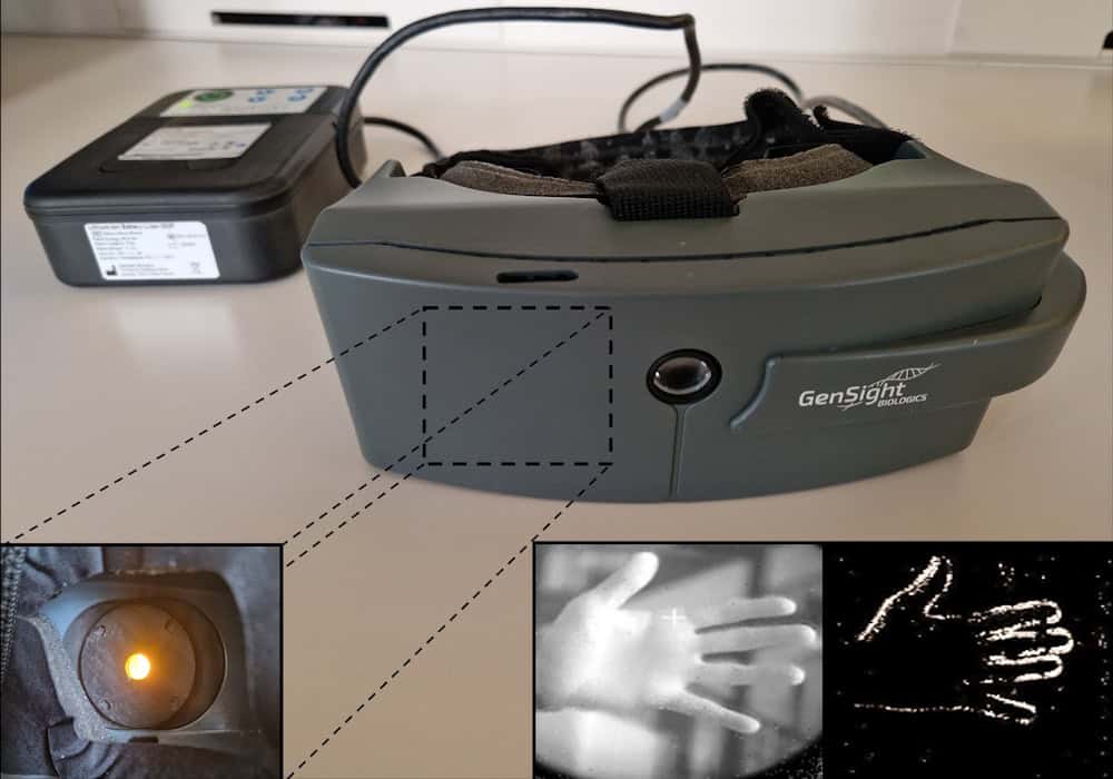
Visual stimulation system The specialized goggles (centre) send visual information to a processor (upper left) that converts the information into patterns of light. A projector within the glasses (lower left) sends these patterns back to the eye. Lower right: the system converts the image of a hand into a simplified light pattern that’s projected onto the retina. (Courtesy: Chloé Pagot, Streetlab, Paris)
Using the light-stimulating goggles, they then assessed the visual performance of treated eyes, before and after injection. The goggles contain a camera that detects changes in light intensities pixel-by-pixel and converts this visual data into pulses of amber light. This light is projected onto the eye to activate ChrimsonR in the modified retinal ganglion cells. Vision tests revealed that the treatment increased light sensitivity in seven of the 10 participants, by a factor of 2.0 to 62.3, with six patients showing a clinically meaningful improvement.
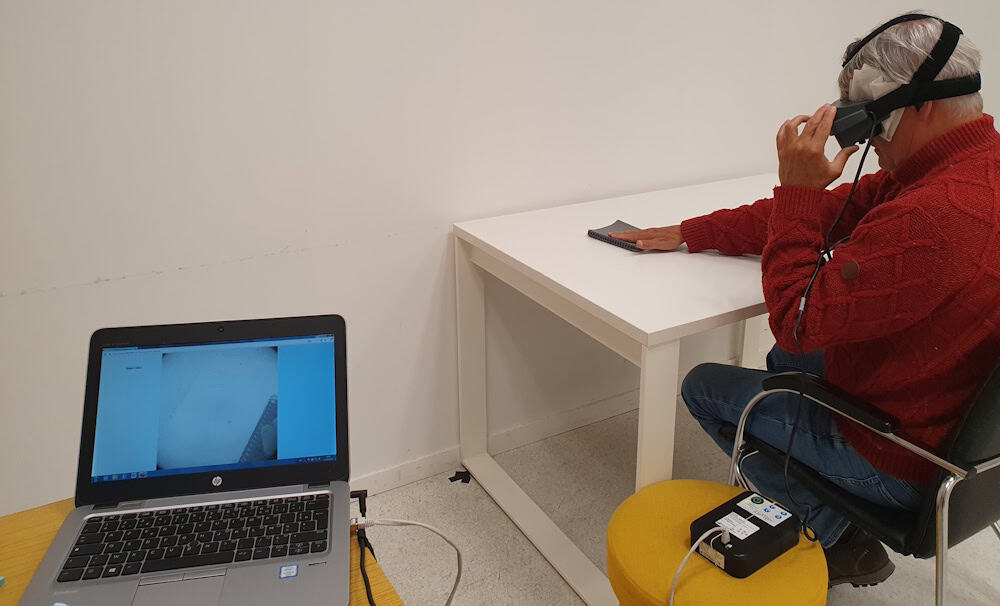
The patients also performed a series of tasks to detect, localize and touch a notebook and a staple-box. In the notebook task, five of eight participants had higher accuracy when wearing the goggles than when not wearing them, while four showed improved accuracy in the staple-box task. In a task determining the orientation of a single bar, accuracy was higher with the goggles in five of eight participants.
To provide an objective measurement of brain activity in response to light, the researchers obtained electroencephalographic (EEG) recordings in five patients performing two visual tests: detection of a tumbler placed in front of them (with the eyes open or closed); and passive viewing of periodic visual stimuli.
In the tumbler test, EEG decoding accuracy was higher with goggles than without when the eyes were open, but not when they were closed – implying a genuine improvement in visual processing rather than an artefact. The second test also showed increased decoding accuracy, which correlated with mean improvements in the notebook and staple-box tasks.
Visual behavioural test After receiving optogenetic therapy, participants used a visual-stimulation system to complete tasks. Here, a participant reaches for a notebook. (Courtesy: Chloé Pagot, Streetlab, Paris)
A bright future
The changes in visual performance were consistent across multiple tasks and testing sessions, with responses observed in all three dose groups. Participants who spent longer being trained to use the goggles performed better in the visual behavioural tests. The researchers point out, however, that functional changes were modest: improving object detection and discrimination capabilities but not restoring normal sight. The small sample size also limited their ability to characterize treatment response.
Several projects are currently underway to further enhance the technology. This includes digital holographic goggles with an eye tracker being developed by the team of Valentina Emiliani at the Institut de la Vision in Paris. The system will project 20 µm-wide images that stimulate individual cells in the retina.
“With the eye tracker, we can make sure we are projecting exactly onto the area of the retina that we want. We’ve already developed a prototype, and it is moving forward,” says Sahel.
Elsewhere, University of Pittsburgh scientists are working with Roska’s group to develop cell-specific gene therapy vectors. “There are different types of cells: some respond to the onset of light, some to the offset of light, some to movement and direction,” Sahel explains. “We are trying to selectively stimulate the ‘on’ and the ‘off’, and discriminate the signal we are sending.”
“We are optimistic about advancing this approach toward future clinical trials,” he tells Physics World.
The 2026 Nobel Prize in Physiology or Medicine has been awarded to Karl Deisseroth, Peter Hegemann and Georg Nagel for “their discoveries concerning light-gated ion channels and optogenetics”.
Optogenetics is a method that uses light to characterize and manipulate the activity of neurons in a living brain. Hegemann and Nagel discovered the light-sensitive protein channelrhodopsin, while Deisseroth transformed the protein into a light-controlled switch for nerve cells.
“Optogenetics provides opportunities for mapping the brain in a way that we could once only dream of,” says Per Svenningsson, chair of the Nobel Committee for Physiology or Medicine.
From the pond to the brain
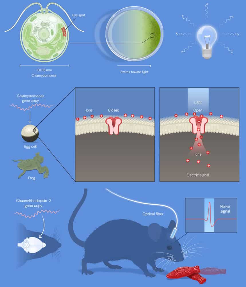
Speaking in the press conference following the announcement, neuroscientist Abdel El Manira from the Karolinska Institute described how the breakthrough began with the study of a single-celled green alga, Chlamydomonas, known for its ability to swim towards light using a tiny “eyespot” on its surface as a sensor. Hegemann was intrigued as to how this simple organism could transform light into action.
Using electrical measurements to solve this mystery, Hegemann discovered that the eyespot contains a light-sensitive protein that almost instantly converts light into an electrical signal, enabling the alga to move. Hegemann next teamed up with Nagel, who used the gene for Chlamydomona to produce the protein in frog egg cells. They discovered that the protein itself was a light-gated ion channel that opens when exposed to light, allowing positively charged ions to flow into the cell and rapidly generate an electrical signal.
“Nagel and Hegemann realised exactly what they had found. They had just discovered the switch neuroscientists had long dreamed of – a light-gated ion channel they named channelrhodopsin,” says El Manira.
Appreciating the potential of this discovery, Deisseroth – at the time studying ways to establish causal links between specific brain circuits and behaviour – decided to test whether channelrhodopsin could be used as a switch to control the activity of neurons with light. By introducing the DNA encoding the protein (supplied by Nagel) into mammalian neurons and illuminating them with light, he observed a remarkable response: the neurons reacted on command, instantaneously and with millisecond precision.
The real test was whether channelrhodopsin could control neurons in living animals. Deisseroth and colleagues demonstrated that this was possible. By introducing the gene for channelrhodopsin into specific nerve cells in the brains of living mice and illuminating these cells via a thin optic fibre, they activated selected group of neurons and elicited a distinct behaviour.
“For the first time, causal links between specific brain circuits and behaviour had been achieved. The technology, which soon became known as optogenetics, transformed neuroscience,” notes El Manira. “Optogenetics was rapidly and widely adopted all over the world.”
Future medical potential
The field of optogenetics has now enabled researchers to establish causal links between neural activity and behaviour, identifying, for instance, neural circuits associated with pain, social behaviours, thirst, food consumption, reward and attention. Beyond the healthy brain, the technique also sheds light on how specific brain circuits can be disrupted and how this impacts psychiatric and neurological conditions.
And while optogenetics is primarily a tool for basic research and understanding of disease, optogenetic therapy is also showing early promise as a medical treatment. In particular, a landmark study has demonstrated partial recovery of visual function in a blind patient with retinitis pigmentosa, by insertion of a channelrhodopsin-like protein in their retina.
“In this disorder, retinitis pigmentosa, patients lose their photoreceptors, the rods and cones in the retina,” explains Svenningsson. “But there are still healthy cells in the retina and researchers have used optogenetics to stimulate these healthy cells and thereby activate the optic nerve and generate vision perception in the brain. This has been done and there are several clinical trials ongoing with this approach.”
Karl Deisseroth was born in 1971 and received his PhD and MD from Stanford University in the US, where he performed the work for which he was awarded the prize. He is currently an investigator at the Howard Hughes Medical Institute and professor at Stanford University.
Peter Hegemann was born in 1954 and received his PhD at the Max-Planck-Institute for Biochemistry in Martinsried, Germany, where he made his prize-awarded discoveries. He is now a senior professor of neuroscience at Humboldt University of Berlin, Germany.
Georg Nagel was born in 1953 and received his PhD at the University of Frankfurt, Germany. His prize awarded discoveries were made at Max Planck Institute for Biophysics in Frankfurt. He is currently professor of molecular plant physiology at the University of Würzburg, Germany.
Thomas Perlmann, secretary-general of the Nobel Assembly, described his call to the laureates earlier this morning: “All three were very surprised and all three said the same thing, that it was absolutely wonderful to receive the prize with the other two, called them ‘my friends’, and that it was such an honour to receive them together”.
Read about the use of organic dyes in optogenetics, in a book chapter co-authored by Peter Hegemann, now free to read on IOP Science.
SmarAct proudly supports Physics World‘s Nobel Prize coverage, advancing breakthroughs in science and technology through high-precision positioning, metrology and automation. Discover how SmarAct shapes the future of innovation at smaract.com.
At this week’s ASTRO 2026 meeting in Boston, MA, researchers shared the results of clinical trials addressing important and longstanding questions in cancer care. In a dedicated news briefing highlighting noteworthy research studies, two speakers presented new evidence that could help inform how we deliver care to patients with brain metastases.
Brain metastases – cancerous cells that spread to the brain from another site in the body – are typically removed surgically or treated with radiotherapy. For most types of cancer, the preferred approach is stereotactic radiosurgery (SRS), in which high-dose radiation is precisely directed to individual lesions while limiting dose to surrounding healthy brain tissue.
Randomized trials have shown that SRS results in superior cognitive preservation and quality-of-life compared with whole-brain radiotherapy (WBRT), where the entire brain is irradiated to ensure treatment of both visible tumours and microscopic disease. There’s an exception, however. Historically, for brain metastases arising from small cell lung cancer (SCLC), a rare, fast-growing cancer, WBRT remains the standard-of-care. Results of a new trial challenge this approach.
Chad Rusthoven from the University of Colorado Anschutz School of Medicine shared the findings of the first phase III randomized trial to compare SRS versus WBRT for brain metastases from SCLC. The trial found that after adjusting for patient and disease characteristics, SRS was associated with a 40% lower risk of death than WBRT.
Rusthoven noted that recent advances have improved outcomes for patients receiving WBRT. “This includes addition of memantine [a drug that helps protect cognitive function], as well as hippocampal avoidance techniques,” he explained. “As a result, HA-WBRT plus memantine is now the preferred whole-brain treatment strategy for cognitive preservation.”
As such, the NRG-CC009 trial – which included 151 patients in the US and Canada – randomly assigned participants to receive either focused SRS to visible metastases or HA-WBRT plus memantine. All subjects had a median of two SCLC metastases, although some had more than 20.
For the primary endpoint – the time before a patient experienced cognitive decline – the study found no significant difference between the two treatment groups. However, patients treated with SRS had a median overall survival of 17.4 months, compared with 8.6 months for those in the HA-WBRT group. Rates of serious treatment-related side effects or death from neurologic causes were similar for the two cohorts.
Although no differences in neurocognitive failure were observed, the superior overall survival with SRS addresses a fundamental historical objection to SRS alone for small cell lung cancer brain metastases. That is, the concern that omission of WBRT would lead to worse overall survival in this population. Overall these findings support SRS as a treatment option for patients with brain metastases from small cell lung cancer,” Rusthoven concluded.
The power of fractionation
Speaking at the same briefing, Ayal Aizer from Mass General Brigham/Dana-Farber Cancer Institute explained how fractionated radiosurgery can reduce the risk of cancer recurrence after surgery for large brain metastases, without significantly increasing side effects. Fractionation – in which the radiation dose is divided into several treatments – gives healthy tissue time to recover between doses while still effectively treating the surgical cavity.
Large metastases, which can cause pressure on the brain leading to symptoms such as seizures, headaches or difficulties with speech or movement, can be removed via surgery. This is usually followed by radiotherapy to destroy any microscopic cancer cells left behind in the surgical cavity. Aizer explained that current guidelines recommend treating the cavity with SRS, following two pivotal trials demonstrating favourable outcomes for SRS compared with postoperative WBRT and observation.
For larger surgical cavities, however, the challenge, is how to deliver enough dose to eliminate any remaining cancer cells without exposing too much surrounding healthy brain to radiation. “In both of these trials where single-fraction SRS was utilized, the one-year surgical bed recurrence rates were high: 40% in one trial, 28% in the other,” he explained. “Meanwhile, alternative approaches such as fractionated SRS to the surgical bed had been emerging.”
Aizer described Alliance A071801, the first randomized phase III trial to directly compare single-fraction versus fractionated SRS for resected brain metastases. The multi-centre trial enrolled 242 patients with one to four brain metastases, one of which was larger than 2 cm and surgically removed. The metastases arose from different primary tumour types, the most common being lung cancer.
The team randomly assigned patients to receive either a single session of SRS, with dose based on target size, or fractionated SRS: 27 Gy in three fractions for smaller targets or 30 Gy in five fractions (9%) for larger ones.
One year after treatment, 87% of patients who received fractionated SRS were free from recurrence at the surgical site, compared with 81% of those who received single-session radiosurgery. Side effects such as radiation necrosis and cerebral oedema were low in both treatment groups, with no significant differences between the two.
The overall survival was also improved after fractionated SRS, with patients surviving a median of 29 months, compared with 20 months for those treated in a single session. The researchers note that this survival difference was unexpected and it is not yet clear why this occurred.
“In conclusion, we can say that fractionated SRS improved surgical bed control compared with single-fraction SRS after resection of a brain metastasis,” said Aizer. “The improvement in surgical control was achieved without an apparent increase in toxicity. For patients undergoing resection of a larger brain metastasis, postoperative fractionated SRS should be considered the standard-of-care.”
The annual meeting of the American Society for Radiation Oncology is the place to be to discover the latest radiotherapy technology developments and new product launches. And this year was no exception. The ASTRO 2026 meeting, held this week in Boston, MA, saw Siemens Healthineers unveil its newest radiotherapy system – Accela – which combines precision delivery with speeds fast enough to enable stereotactic treatments in a single breath-hold.
Accela achieves this by implementing a new delivery technique known as NeoArc, or dynamic modulated arc therapy (DMAT). DMAT is a type of arc therapy, in which the treatment beam rotates continuously around the patient during treatment, with the beam shape and intensity controlled to deliver the prescribed treatment plan.
But while conventional arc-based approaches such as volumetric-modulated arc therapy (VMAT) primarily optimize treatment plans for the dose distribution, DMAT evaluates dosimetric objectives and delivery efficiency simultaneously. This enables clinicians to tailor treatments for both speed and precision, to optimize the balance between them and, ultimately, to reduce the patient time on the treatment table.
Sasa Mutic, head of radiation oncology solutions at Siemens Healthineers, tells Physics World that the product launch at ASTRO was a great success. “The response to Accela from customers and the medical community has been overwhelming and overwhelmingly positive,” he says. “It was extremely gratifying to see that the ASTRO audience instantly grasped the significance of what we’ve accomplished and the impact it will have on clinicians and patients.”
Done in 60 seconds
Accela integrates high-speed gantry rotation of up to 2.5 rpm, dose rates of up to 40 Gy/min and a dual-layer multileaf collimator (MLC) with leaf speeds of up to 6.5 cm/s. This combination means that, as well as single breath-hold stereotactic treatments, Accela can deliver multi-site stereotactic body radiotherapy (SBRT) in as little as 60 s and go from imaging to beam-off in as low as 90 s – including on-couch imaging with a large HyperSight cone-beam CT panel, couch shifts and the treatment delivery.
An additional benefit of ultrafast radiotherapy is the potential to actually alter the body’s biological response.
“Because the treatment times are potentially as quick as a single breath hold, there is much less blood irradiated,” explains Dee Khuntia, chief medical officer at Varian, the Siemens Healthineers cancer-care business. “This allows for preservation of lymphocytes, which are necessary for helping the body heal, fight infection and fight cancer. It is also well documented that patients undergoing radiation or chemo-radiation who have lymphopenia [not enough lymphocytes in the blood] have lower survival rates.”
The first patient treatments with Accela are scheduled for December in Europe. By this time, the system will have the CE mark, with FDA clearance expected early next year. One of the first centres to use Accela will be Maastro in the Netherlands, which plans to initially treat small lung tumours, lung metastases, brain tumours and metastases in the vertebrae – sites that require a high radiation dose administered extremely precisely, while sparing healthy tissue and nearby vulnerable organs.
Updated on 6 October 2026 to amend the imaging to beam-off time and add a clarification from Siemens Healthineers: Accela and NeoArc are 510(k) pending. Not available for sale or quotation. No guarantee of commercialization or feature availability.
This episode of the Physics World Weekly podcast features the medical physicist Nick Zacharopoulos, who is CEO of US-based Aktina Medical.
In conversation with Physics World’s Tami Freeman, Zacharopoulos reflects on the twists and turns in his career developing new technologies for radiation oncology. His journey began when he was a child helping his father build radiotherapy components in the family’s garage and led to academic and commercial success.
Zacharopoulos talks about his role in the development of a pivotable product for Aktina and how his persistence – and a chance lunch with a colleague – led to his enrolment in a PhD programme at McGill University.
Delivering drugs directly to the skin provides a targeted means of treating dermatological conditions while minimizing side effects. The approach is limited, however, by the stratum corneum – the skin’s outer layer – which acts as a barrier that can restrict the effectiveness of most topically applied drugs. To overcome this, a team at Georgia Tech has developed biodegradable “STAR particles” that use microscopic needles to painlessly puncture the skin and increase its absorption of medication.
“Very few drugs can be absorbed effectively into the skin, which means that many drugs in dermatology are given by mouth or injection,” explains team leader Mark Prausnitz. “This exposes the whole body to the drug, often causing side effects and reducing drug efficacy. STAR particles painlessly make micropores in the skin that allow drug that is rubbed on the skin to be absorbed. This targets drug delivery exactly to the site where it is needed.”
Previously, the researchers fabricated the STAR particles from a ceramic material (titania), which is safe to use on the skin, but not biodegradable, raising possible environmental concerns. In their latest work, reported in Advanced Healthcare Materials, they designed polymer STAR particles that dissolve or biodegrade after use, reducing their potential environmental impact.
STARs effectively create micropores
Prausnitz and colleagues created STAR particles from three polymers: water-soluble poly(vinyl alcohol) (PVA), enzyme-degradable cellulose acetate (CA) and hydrolysable polylactic acid (PLA). All three materials have previously been used to create microneedle patches, which enhance drug delivery but can generally only be used on small areas of skin.
“For dermatological conditions like eczema and psoriasis, patients need to treat skin with variable and sometimes large areas,” says Prausnitz. “STAR particles provide the power of a microneedle patch to increase skin permeability with the flexibility to apply them over large and variable areas by simply rubbing a gel or cream containing STAR particles on the skin.”
The researchers fabricated the polymer STAR particles using femtosecond laser micromachining to create star-shaped structures with sharp, well-defined microneedle tips and a tapered profile. They tested the ability of the various particles to puncture pig skin samples.
While PVA STAR particles cannot be applied using water-based vehicles, when suspended in non-aqueous formulations such as isopropyl palmitate (a non-toxic ingredient widely used in dermatology), they successfully punctured the skin. The STAR particles remained intact after use and the formulations worked equally well after storage for one week.
Similarly, CA and PLA STAR particles in water formulations demonstrated consistent skin-puncturing ability, even after a week’s storage, with no visible damage to the particles after application. The researchers note that titania STAR particles in water (examined as a control) generated more pores than the polymer particles, due to their higher hardness.
Drug delivery demonstration
The team next investigated how the STAR particles could enhance the delivery of three drugs – tacrolimus, methotrexate and copper tripeptide-1 – into pig skin samples.
Tacrolimus is used to treat inflammatory skin conditions and is soluble in non-aqueous solvents. After topical drug delivery, rubbing the skin with PVA STAR particles in isopropyl palmitate for 10 s or 30 s increased drug levels within the skin 1.7- or 3.2-fold, respectively, compared with control samples.
For CA STAR particles, the researchers examined methotrexate, which is commonly used for psoriasis treatment but cannot be delivered topically due to its very low skin permeability. Treatment with CA STAR particles for 10 or 30 s increased intradermal delivery of methotrexate 5.4- or 25.2-fold, respectively.
Finally, they used PLA STAR particles to deliver copper tripeptide-1, a skincare ingredient employed for anti-aging, wound healing and skin regeneration applications. The particles enhanced intradermal drug delivery 12.1- or 37-fold, after 10 or 30 s application, respectively.
Safety considerations
As the STAR particles are designed to create micropores in skin, it’s possible that they could unintentionally end up in other parts of the body, where the microneedles could cause damage. While this risk is likely small (as the STAR particles require forceful application to be effective), the water-soluble PVA STAR particles eliminate any potential risk as they rapidly dissolve upon contact with wet tissues.
The CA and PLA STAR particles will likely retain their structure immediately after use, but will eventually become blunt and weak upon enzymatic degradation or hydrolysis. Likewise, any potential environmental consequences of non-degradable STAR particles are reduced or eliminated with the water-soluble and biodegradable materials.
The team concludes that the biodegradable STAR particles address environmental and safety concerns while enhancing drug delivery to the skin. The approach paves the way for improved patient outcomes and broader applications, enabling delivery of hydrophilic drugs and larger molecules that are usually blocked by the stratum corneum barrier. Prausnitz tells Physics World that the research is primarily targeted at treatment of dermatological diseases that can spread over large areas of skin – such as psoriasis, eczema, vitiligo and allergic rashes – as well as cosmetic applications.
“We have licensed the STAR particle technology to a company that is preparing for a clinical trial next year using STAR particles to deliver siRNA as a novel eczema treatment,” he says. “At Georgia Tech, we are focused on advanced materials, manufacturing and formulations for the next generation of STAR particles.”
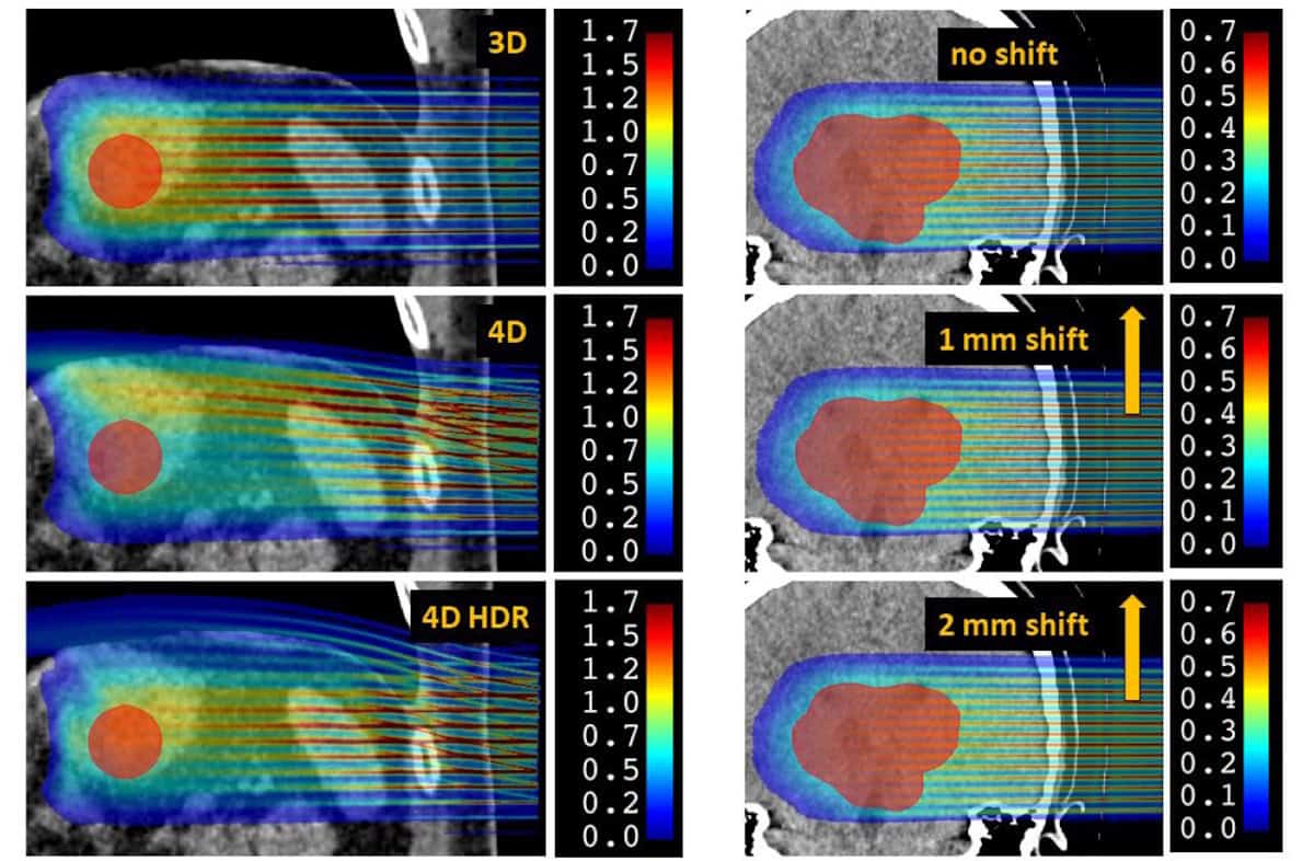
Dose calculations Left: dose distributions for thoracic pMBRT without motion (3D), with respiratory motion (4D) and with respiratory motion at high dose rate (4D HDR). Right: dose distributions for a brain treatment with zero shift, a 1 mm shift and a 2 mm shift. (Courtesy: Phys. Med. Biol. 10.1088/1361-6560/ae8ef8)
Proton minibeam radiotherapy (pMBRT) is an innovative new cancer treatment that uses an array of narrow proton beams to create alternating regions of high and low dose, before converging to a homogeneous dose distribution within the target volume. Numerous studies in small animals have shown that this spatially modulated dose distribution can destroy tumours while sparing healthy tissue and reducing side effects.
To implement pMBRT in the clinic, it’s important to first assess the impact of organ motion during treatment, which could lead to overlapping of the dose peaks and valleys, and potentially diminish the sparing effect. With this aim, a team at Institut Curie in France has developed a Monte Carlo (MC)-based framework to quantify the dose distribution for realistic clinical treatments, reporting their findings in Physics in Medicine & Biology.
To deliver pMBRT, a multi-slit collimator is used to create narrow beams, typically 0.3 to 1 mm wide, spaced 2 to 6 mm apart. As this collimator blocks much of the initial proton beam, longer treatments are needed to deliver the prescribed dose, increasing the likelihood of organ motion.
“Given the longer irradiation times, possible patient movement could deteriorate the heterogeneous spatial dose distribution of pMBRT,” explains senior author Ludovic De Marzi. “This is all the more problematic because it is this heterogeneous distribution that is believed to be responsible for the technique’s biological sparing effect.”
The dose reconstruction workflow
De Marzi and colleagues developed a 4D dose calculation workflow based on time-resolved MC simulations to determine how motion affects pMBRT dose distribution. The tool requires two main inputs: a 4D CT dataset capturing the patient’s anatomy and breathing motion over one respiratory cycle; and a proton pencil-beam scanning (PBS) treatment plan created on a reference CT.
To validate their workflow, the researchers irradiated a thoracic motion phantom with a conventional PBS plan and compared the measured dose with the simulations. For a 3%/3 mm gamma index (a standard quality check tool), the mean pass rate for nine phantom irradiations was 97.3%, confirming that the calculation performs reasonably well for 4D dose modelling in conventional PBS.
They then used the tool to assess the impact of motion in representative clinical cases, first examining a thoracic treatment with large breathing movements. They calculated 4D dose distributions for three scenarios: 3D pMBRT without motion; 4D pMBRT with 20 mm breathing motion; and 4D high-dose rate pMBRT, with the instantaneous dose rate increased tenfold to reduce beam-on time by a factor of ten.
The calculated dose distributions showed that breathing motion introduced significant variations in the patterns of peaks and valleys, impacting a crucial parameter: the peak-to-valley dose ratio (PVDR). At a depth of 5 mm, for example, the mean PVDR dropped from 11.9 for the 3D scenario to 6.7 in 4D. The high-dose rate scheme slightly mitigated this degradation, but not significantly.
“The PVDR is an index that takes into account both doses in the valleys (which are likely correlated with the biological sparing effect on healthy tissue) and the maximum delivered doses (which are likely responsible for the anti-tumour effect),” De Marzi explains. “It can also be used to help optimize the 3D dose distribution of the treatment plan.”
Breathing motion also degraded the dose delivered to the tumour, with the mean target dose and D95% (the minimum dose received by 95% of the target volume) reduced by 30% and 35%, respectively, for 4D pMBRT. Nearby organs-at-risk (OARs) were also impacted, with a decrease in dose to the stomach and a significant increase in mean dose to the spleen for the 4D cases.
The researchers next examined an intracranial treatment. While there is no intra-organ motion in this case, the patient’s head undergoes small rigid translations – generally limited to less than 1 mm using thermoplastic masks. As such, they modelled patient motion as a simple continuous 1 or 2 mm translation.
Motion did not significantly impact the mean dose or D95% for the target or OARs. A 1 mm shift was, however, enough to degrade the peak–valley patterns, reducing the mean PVDR by 10% from the static case (at 15 mm depth). For a 2 mm shift, this increased to a 24% reduction in mean PVDR.
A valuable tool
The researchers conclude that their 4D dose reconstruction workflow provides a useful tool for assessing interplay effects caused by organ motion during pMBRT. Without a specific motion management strategy, only intracranial treatment with movements below 1 mm was robust to interplay effects. A similar workflow could prove valuable for estimating the sparing effect of pMBRT while accounting for patient motion and adapting treatment plans accordingly.
Looking forward, it may be possible to minimize delivery times by combining pMBRT with the ultrahigh-dose rate delivery used for FLASH treatments.
“This idea is appealing because it could both solve the problem of reduced dose rate in pMBRT and add an additional biological effect (FLASH) to further spare healthy tissue,” says De Marzi. “However, its feasibility remains to be demonstrated, as does the value of developing a technique that combines such considerable complexities. A simple increase in dose rate – without going as far as FLASH – would already be of interest.”
In their next step towards clinical application, De Marzi and colleagues are working to validate calculation tools and measurement procedures tailored to this pMBRT technique. “Ones that are fast, accurate and usable by clinicians,” he says.
This episode of the Physics World Weekly podcast features Katharine Hunt, Head of Radiosurgery Physics at Sheffield Teaching Hospitals NHS Foundation Trust in the UK. She explains how Gamma Knife radiosurgery is being used to treat arteriovenous malformations (AVMs) and other benign brain conditions.
Sheffield is home to one of the world’s longest-running Gamma Knife centres, and Hunt shares the facility’s insights into treatment planning, dose precision, paediatric care, and long-term outcomes.
This podcast is sponsored by Elekta, which produces the Gamma Knife radiosurgery system.
Patient-specific quality assurance (PSQA) plays an essential role in the safe and effective delivery of radiotherapy to cancer patients. Enabling a “dry run” in which the treatment plan is delivered to a physical device before the patient enters the room, PSQA provides a final safety net before treatment. The aim: to ensure that the correct dose is delivered to the correct location for every patient and every single fraction.
That said, measurement-based PSQA is not sensitive enough to catch all errors – some still slip through. What’s more, pre-treatment measurements are not feasible in online adaptive radiotherapy, where treatment is re-planned and delivered within minutes. There’s simply no time to deliver the new plan to a phantom while the patient waits on the couch
And as clinics increasingly adopt adaptive workflows, incorporate artificial intelligence (AI) into the treatment chain and deliver more complex, more automated and faster radiotherapy than ever before – is it time to rethink radiotherapy QA? Could properly commissioned delivery techniques render routine measurement-based PSQA obsolete?
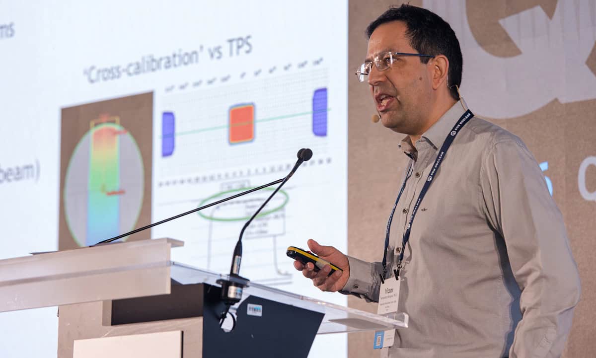
This was the question discussed at the QA & Dosimetry Symposium (QADS) hosted earlier this year by Sun Nuclear. In an animated debate, Victor Hernandez of Hospital Universitari Sant Joan de Reus in Spain and Dirk Verellen of the University of Antwerp and Iridium Netwerk in Belgium, considered the options.
Far fewer PSQA measurements
“We are debating today because we have a problem,” said Hernandez. “Measurement-based PSQA is not sensitive, not efficient and just not working.” The good news, however, is that a simple independent dose calculation – as provided by software-based PSQA – is an order of magnitude better at detecting failures than measurements.
Victor Hernandez “We need to commission and standardize all the steps of the radiotherapy process.” (Courtesy: Sun Nuclear)
Hernandez emphasized that to make this shift, it’s essential to commission and standardize the whole radiotherapy process, including not just the linac and treatment planning system (TPS) but the PSQA system itself, to optimize how it is used and understand its limitations. Perhaps the most important step is commissioning the treatment planning process – or “QAing” the plan characteristics. “Less complex plans tend to be more accurate and more robust to uncertainties,” Hernandez explained. “So you need to evaluate and minimize plan complexity during treatment planning.”
He proposed a simple process for QA of treatment planning, based on class solutions (pre-defined dosimetric objectives and geometric parameters) that incorporate complexity metrics in their definition. These class solutions are validated using measurements and used to standardize treatment planning. If a final plan falls outside of the class solution, it will require verification using both measurements and software. But most clinical plans will be within a class solution, where software-based verification will suffice.
Hernandez noted that if you’re performing less PSQA, then machine QA becomes more important and must be reinforced. This could, for example, involve measurements of a fixed set of complex plans every few weeks to make sure that the linac is stable. “But that’s no longer PSQA – it’s part of your machine QA programme,” he emphasized.
“You can perform treatment planning without controlling or evaluating the plan parameters, but then there’s no consistency in your plans and you need pre-treatment measurements of all plans because you’re not in control,” he explained. “Or you can control the plan parameters. Then you reduce variability, improve robustness and quality, and you don’t really need PSQA measurements anymore because that’s part of QA, which is of course, much more efficient.”
“That’s my vision for enhancing safety and advancing QA,” Hernandez concluded.
The unknown errors
Verellen was less convinced that measurement-based PSQA is obsolete. Contemplating Hernandez’s arguments, he agreed that phantom-based pre-treatment PSQA is “just recommissioning over and over again”, noting that “you won’t see any errors in your PSQA unless you have done your commissioning wrong.”
Dirk Verellen “For me at this moment, patient specific pre-treatment QA is my backup.” (Courtesy: Sun Nuclear)
He concurred that accurate commissioning should pick up any mechanical or dosimetric errors, dose tracking can account for anatomical variations, and surface- and image-guided radiotherapy can address positioning errors. “And we also have in vivo dosimetry,” he added. “So in principle, we are covered.”
But sometimes, the things that go wrong are the things nobody thought to look for. “You might have a very good plan, but are you really sure that everything is covered?” he said.
One obstacle is knowing which metrics to use to define a complex plan. Another problem lies in the onslaught of new technologies such as online adaptive that create and deliver a plan in minutes. Verellen pointed out that companies are selling complex adaptive systems as a plug-and-play option that can be commissioned and treating patients in just a few weeks. “Your department is not ready for that. You don’t have the procedures. People are not trained for that,” he emphasized.
And as software is updated, hardware is upgraded and AI is rapidly becoming inherent in ever more stages of the radiotherapy workflow, it becomes challenging to rely on existing procedures. “With this large-scale introduction of AI and automation, we really start to rethink our role as humans in monitoring that process,” said Verellen.
The big challenge is to anticipate each and every kind of error that could possibly occur. And as Verellen pointed out, radiotherapy is much more than just the treatment machine. “There are a lot of people [involved] and they may make mistakes,” he said. “The interactions of the different system components make things so complicated that it’s very difficult to really anticipate all the things that might go wrong.”
What’s needed is a way to evaluate, validate and monitor the entire treatment chain, and a way to detect and avoid catastrophic events. Currently, pre-treatment PSQA is still an important safety barrier in this respect, he emphasized, by detecting the small, unexpected errors that could add up to create a catastrophic event. Moreover, in vivo dosimetry (IVD) will become even more important as a final safeguard of quality that, in the case of online adaptive radiotherapy, will need to evolve into real-time IVD.
“We need rigorous commissioning, but we need to realise that this cannot predict every situation,” said Verellen. “That’s my main point: as my father used to say, ‘don’t throw away your old shoes before you have the new pair’ – this is why we still need pre-treatment patient-specific QA at this stage … until we’ve figured out how to deal with the new developments.”
Near agreement
Remarkably for a conference debate, Hernandez and Verellen agreed on many points – in particular, that the field requires a redefinition of radiotherapy QA. “AI and automation are like a tsunami coming over us and we are not prepared for it,” said Verellen. “We have to step back and rethink the QA of the process.”
Where they differed, however, was on whether it is safe to abandon current PSQA measurement practices right now. “We know where we want to go, but we’re not there yet,” said Verellen.
“I agree with Dirk about many things,” said Hernandez. “But pre-treatment verification to a phantom is not patient safety. That’s why we need to make sure that our plans are accurate and robust, then you can verify it in every single fraction.”
“That works if the flow goes according to plan,” Verellen countered. “But unpredictable things happen.” He likened the risk to that of skydiving, where sky divers spend much time meticulously folding the parachute to guarantee that it will open. “But I’ve never seen a sky diver jumping without a reserve parachute,” he said. “For me, pre-treatment PSQA is my backup, until we have a solution that’s more or less watertight. And at this moment, it is not.”
Both agreed that the way forward is to focus, not just on the machine or the plan, but on understanding and verifying the entire treatment process – particularly the treatment planning and delivery processes. Treatment verifications such as real-time beam monitoring systems and IVD are also crucial for patient safety. “We need to think about how to better monitor all these processes to really advance QA and improve safety for patients,” said Hernandez.
Top billing In one of the most talked-about sessions at QADS15, delegates crowded in to learn how treatment quality can be maintained as radiotherapy becomes more complex, more automated and faster than ever before. (Courtesy: Sun Nuclear)
The next frontier
While the QADS debate was framed as “measurement versus calculation”, Greg Robinson from Sun Nuclear reiterates that the real evolution is the shift from checking a device to monitoring the entire process, driven by the introduction of automation and AI.
“The debate was interesting because they were arguing for both extremes, but I think the field is going to land somewhere in the middle,” Robinson tells Physics World. “It’s not that physical measurements are going away, but the frequency by which clinics are doing these measurements is dropping.”
Verellen pointed out that to enable the uptake of software-only PSQA, “we need QA tools that monitor the process”. One option for completely virtual pre-treatment QA is the DoseCHECK secondary 3D dose calculation algorithm and PerFRACTION, both part of SunCHECK from Sun Nuclear.
“SunCHECK brings machine and patient QA together in a single platform,” explains Robinson. “[This] lets a clinic see relationships that isolated tools miss; for instance, when a change in machine performance begins to show up in patient results.” Meanwhile, for adaptive online radiotherapy, AdaptCHECK provides an independent secondary check of the adapted plan within the session time frame.
Looking further ahead, Robinson predicts that the next frontier will be “turning connected QA data into understanding”. In other words, ensuring that errors are not just detected, but can be understood and prevented. “It isn’t enough to tell a physicist that a result passed or failed – the value is in the why: what’s trending, what’s related, what’s likely to drift next,” he explains. “Sun Nuclear is developing new solutions to help physicists untangle the meaning behind all of this data, and make connections across machine and patient data much more seamless.”
Robinson concludes that measurement-based PSQA is not obsolete, but can no longer stand alone. “The future is an independent, connected, insight-driven QA layer that spans machine and patient, works where measurement can’t, and explains the results it produces,” he says. “Quality first, delivered by automation and intelligence rather than replaced by them. That’s exactly where Sun Nuclear is investing.”
A collaboration of researchers in North America has reported their findings from the first diagnostic X-ray imaging performed in space. X-ray images recorded during an orbital spaceflight exhibited similar overall quality to those taken preflight on Earth, demonstrating the feasibility of adding radiography as a diagnostic tool for crew health.
With humans venturing further into space and spending more time in this harsh environment, the risk of adverse medical events increases. Accurate and timely diagnosis and treatment is a must-have. Currently, the only reliable medical imaging modality available to astronauts in spaceflight is ultrasonography – which is operator dependent, requires substantial training and relies on limited acoustic windows.
X-ray imaging, on the other hand, benefits from low operator dependence, rapid acquisition and typically higher spatial resolution than ultrasound. Digital radiography could also offer superior diagnostic capabilities for many medical conditions of concern during spaceflight, including dental disease, musculoskeletal trauma, inhalational injury, collapsed lung and arthritis.
Traditional X-ray scanning systems are bulky, produce a lot of radiation and are sensitive to motion – making them less than ideal for sending into space. But a new generation of portable X-ray scanners shows potential to survive the spaceflight and be operated by crew members with minimal training.
“It’s been a dream for aerospace medicine to have more than one imaging modality for diagnosing illnesses and injuries in space,” says lead researcher Sheyna Gifford, an assistant professor of aerospace medicine at the Mayo Clinic in Rochester, MN, in a press statement.
Scanning in space
In a study led by co-author David Lerner, the researchers first demonstrated the feasibility of X-ray imaging in simulated microgravity in 2022. During a parabolic flight, they used a commercial off-the-shelf digital radiography system to successfully capture radiographs of a human subject and a phantom during lunar gravity (1/6g), Martian gravity (1/3g) and microgravity.
In this latest project, reported in in Radiology, Gifford and her team investigated the performance of a similar portable digital radiography system aboard the Dragon spacecraft, during the Fram2 spaceflight mission, a 3.5-day polar orbital flight launched on 31 March 2025 on a SpaceX Falcon 9 rocket. The imaging system comprised a commercial off-the-shelf, FDA-cleared, portable, digital X-ray generator (Impact Wireless; MinXray) along with an FDA-cleared flat-panel detector.
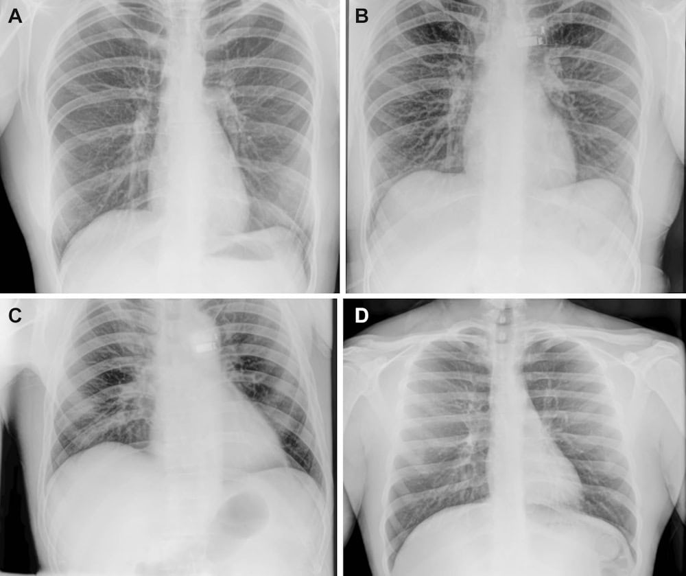
Chest radiographs Representative X-ray images acquired preflight by a crewmember (A), in-flight on day 3 after launch by a crewmember (B, C) and postflight by a non-crew operator using the same imaging protocol (D). (Courtesy: RSNA)
Prior to the mission, three crew members received 4 hr of training on the portable X-ray system and acquired preflight X-ray images of the hand, forearm, abdomen, pelvis, chest and a quality control phantom. They then recorded the same anatomic and phantom images whilst in space, and also imaged a smartwatch to investigate the potential for non-destructive testing.
During anatomic imaging, the devices were handheld by the crew members, while the phantom and smartwatch were secured to the detector for imaging. The crew reported that they found it easy to use the X-ray system and follow the imaging protocols.
Back on Earth
To assess the quality of the seven X-ray images acquired by the crew, three independent radiologists evaluated and compared the preflight and in-flight anatomic radiographs, finding no differences in overall image quality, contrast resolution or spatial resolution. For central-body radiographs (chest, abdomen and pelvis), image positioning was worse in-flight than preflight. Despite this, all X-rays achieved good or near-excellent diagnostic quality (rated on a 5-point Likert scale).
Phantom radiographs recorded in space exhibited good spatial and contrast resolution, imaging low-contrast targets down to the smallest 2-mm target and easily visualizing high-contrast meshes ranging from 20 to 60 lines per inch, with 80 lines per inch remaining visible. The smartwatch radiograph clearly showed the watch’s internal components at the submillimetre scale.
The research team found that the X-ray generator had sustained minor re-entry damage, but its internal components and X-ray output were not affected. Similarly, the detector passed visual inspection and quality control tests. The researchers also recorded a set of postflight X-rays (on non-crewmembers) following the same in-flight protocols. These showed no qualitative differences from the preflight or in-flight radiographs.
“By acquiring the first human and equipment X-rays in space, our study demonstrates the feasibility of in-orbit radiography and expanded diagnostic capabilities for crew health and hardware evaluation,” says Gifford. “Acquiring diagnostically useful X-rays in space is something that anyone can do. Three very talented nonmedical people with four hours of training in one of the harshest environments did it right and did it well.”
Gifford points out that a spaceflight-ready radiography system could also prove indispensable for mission-critical non-medical tasks. “For sustained human presence in space, X-rays are critical not just for crew members but also for other mission components like electronics and spacesuits. The only way to look inside these objects without taking them apart is to X-ray them,” she explains.
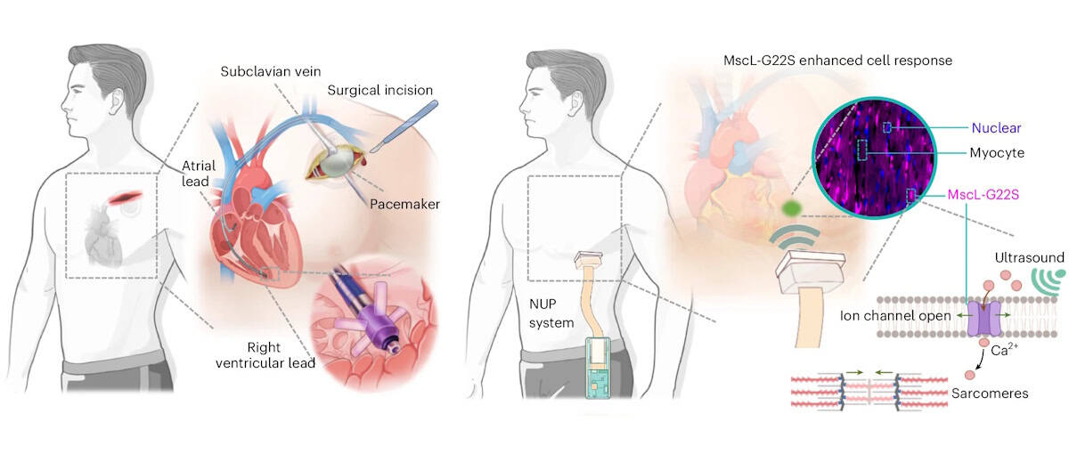
Traditional versus sonogenetic pacing Left: schematic illustration of the steps for traditional surgical implantation of a cardiac pacemaker. Right: operational workflow of the non-invasive ultrasound pacemaker with sonogenetics. Created in BioRender. (Courtesy: C Gong et al. Nat. Biomed. Eng. 10.1038/s41551-026-01673-z 2026, Springer Nature)
Cardiac arrhythmia – a disorder in which the heart beats too fast, too slowly or irregularly – affects millions of people worldwide. Currently, arrhythmias are treated using pacemakers to regulate the heart’s rhythm, but traditional pacemakers require invasive surgical implantation and intracardiac leads that risk infection and tissue injury. As an alternative, a research collaboration headed up at Massachusetts Institute of Technology (MIT) and the University of Southern California (USC) has developed a non-invasive pacemaker that’s worn like a sticker on the chest and stimulates the heart using ultrasound.
“Ultrasound offers a unique combination of deep tissue penetration, spatial focusing and non-invasive delivery,” explains first author Chen Gong from USC. Previous approaches for ultrasound-based cardiac pacing, however, lacked reliability or required the use of microbubbles. In this new study, Gong and collaborators employed a technique called sonogenetics, in which cardiac cells are genetically altered to increase their sensitivity to ultrasound. Once exposed to ultrasound waves, the engineered cells trigger the opening of ion channels that let in calcium, which signals the cells to squeeze and beat.
“Our central motivation was to address the limitations of conventional pacemakers, while preserving precise control of cardiac rhythm,” says Gong.
The team first examined whether ultrasound could modulate cardiac cells in vitro, using sonogenetically engineered human heart muscle cells. Ultrasound stimulation caused almost three-quarters of the engineered cells to beat in synchrony with the ultrasound waves. In contrast, unaltered cells did not exhibit such behaviour. Importantly, ultrasound exposure did not impact the cells’ expression of biomarkers for inflammation, myocardial injury or oxidative stress, demonstrating the safety of this approach.
The non-invasive ultrasound pacemaker
The researchers designed a sonogenetics-based non-invasive ultrasound pacemaker (NUP) capable of real-time imaging and steerable stimulation, describing the device in Nature Biomedical Engineering. The postage stamp-sized device, which sticks to the skin via a bioadhesive interface, incorporates a 64-channel phased-array transducer, as well as data acquisition, wireless transmission and power modules.
To assess whether this prototype NUP could achieve non-invasive cardiac pacing in sonogenetically engineered rats, they attached the device onto the rats’ chests and applied ultrasound stimulation through the chest wall to the heart.
At an acoustic pressure of 2 MPa and a 40 ms pulse duration, the NUP increased the rats’ heart rates from 240 to 360–540 bpm, with a higher pulse repetition frequency leading to increased heart rate. Upon stopping the ultrasound stimulation, the heart returned to its natural rhythm. In control rats without the genetic modification, NUP could not control the heart rate effectively.
The team demonstrated that the NUP could electrically steer the ultrasound beam focus with a spatial precision of less than 1 mm, enabling targeting of different regions of the heart to induce chamber-specific pacing. NUP also successfully treated cardiac arrhythmias induced in the engineered rats, returning their heart rates to normal levels. In contrast, ultrasound stimulation of control rats did not effectively treat their arrythmias.
“A major objective of this work was to move beyond proof-of-concept stimulation and demonstrate a wearable system that could realistically support daily use,” explains Qifa Zhou, group leader at USC. “The current prototype integrates imaging, stimulation, wireless communication and battery-powered operation into a wearable format.”
Clinical potential
For future clinical use, the NUP device needs to automatically detect the wearer’s heart rate, locate heart chambers and deliver stimuli. To achieve this, the researchers used a cloud-based, artificial intelligence (AI)-powered imaging feedback loop that determines the heart rate and stimulation coordinates, and steers the ultrasound beam to target sites.
To test its feasibility for human-scale applications, they assessed the acoustic energy penetration of the device. Using simulations and experiments on a pig heart beneath layers of tissue, they confirmed that, even after tissue attenuation, NUP could deliver sufficient ultrasound pressure (approximately 2 MPa) to a human heart to enable stimulation at clinically relevant pacemaker sites.
The team also assessed the biosafety of the NUP approach, finding that the delivered ultrasound energy remained below approved safety limits and generated minimal thermal effects during and after stimulation. They also confirmed that the sonogenetic engineering induced no off-target effects, immune responses or pathological changes in the rats.
“In the long term, we are optimistic about applying sonogenetics in humans,” explains co-author Gengxi Lu from MIT. “Sonogenetics is different from gene editing – it does not aim to rewrite a patient’s DNA sequence, but instead enables target cells to temporarily or controllably express ultrasound-responsive proteins. At the same time, extensive clinical validation will still be needed to establish safety and long-term effectiveness.”
The team is now focusing on clinical translation and validation of the NUP technology, which includes improving gene delivery strategies, validating the system in larger animal models, and developing closed-loop control approaches that combine real-time physiological sensing with adaptive stimulation. There’s also much engineering work to be done, such as device miniaturization, and improving battery life, skin coupling and motion robustness.
“For cardiac pacing, we envisage that the final goal of NUP technology is to be a permanent alternative to a long-term implanted pacemaker,” Xuanhe Zhao from MIT tells Physics World. “More broadly, we are interested in expanding ultrasound-enabled bioelectronic medicine beyond cardiac pacing toward other organs and therapeutic applications where non-invasive, spatially precise modulation could have clinical impact.”
Radiation therapy is an essential component of cancer care and is used in the treatment of roughly half of all cancer patients. To ensure that these treatments are both safe and effective, it’s critical that the linear accelerators, or linacs, that deliver the radiation are as reliable, efficient and accurate as possible.
The US-based medical physics consultancy One Physics aims to help hospitals and cancer centres meet these goals by bringing together a team of highly-trained expert medical physicists to provide a wide range of radiotherapy and diagnostic imaging physics services.
This episode of the Physics World Weekly podcast features James Giltz, director of commissioning services at One Physics. In a wide-ranging conversation with Physics World’s Tami Freeman, Giltz explains how the company provides comprehensive medical-physics support for radiotherapy – from planning new centres through to ongoing clinical operations.
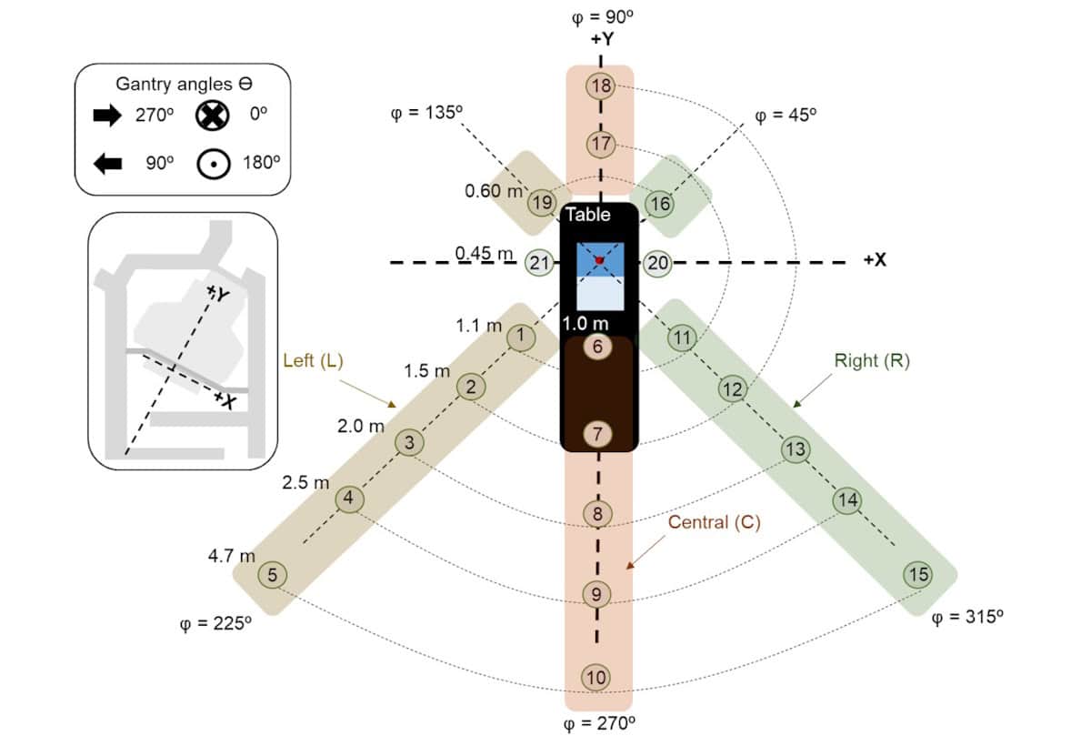
Experimental setup Neutron dose measurement points within the treatment room, showing gantry angles, measurement lines (φ) and distances from the isocentre. (Courtesy: Phys. Med. Biol. 10.1088/1361-6560/ae72e7)
Proton therapy plays an increasingly important role in cancer treatments, targeting tumours with high precision while sparing nearby healthy tissues. Proton irradiation can, however, also produce secondary neutrons due to nuclear interactions of the therapeutic beam. In modern pencil-beam scanning proton therapy, neutrons generated inside the patient are the main source of out-of-field dose – unwanted radiation that could potentially contribute to secondary cancer risks.
A research team headed up at Clínica Universidad de Navarra in Spain has experimentally characterized the neutron field in a proton therapy treatment room using a range of different detectors. They used their findings to create a practical Python-based calculation tool to estimate neutron dose for arbitrary irradiations.
“The tool provides a fast first-order estimate of neutron doses anywhere in the treatment room using information from the treatment plan,” explains medical physicist Verónica Morán. “It could support radiation protection studies, workplace dose assessments, research projects, and the evaluation of neutron exposure in situations where direct measurements are not available.”
Neutron field characterization
For their study, reported in Physics in Medicine & Biology, Morán and colleagues employed a Hitachi PROBEAT-CR proton therapy system with pencil-beam scanning. They measured neutron dose using a range of devices, including two ambient detectors and four types of personal dosimeter: thermoluminescent dosimeters (TLDs); track-etch detectors; bubble detectors (BDs); and electronic personal dosimeters (EPDs).
Using up to 21 measurement points within the treatment room, the team examined the dependence of out-of-field neutron dose on various beam and room parameters, including gantry angle, field size, proton energy and distance to isocentre.
At each measurement point, they assessed the neutron ambient dose equivalent, which is used to characterize the radiation field at a specific location, plus the personal dose equivalent, which estimates the dose that a person may receive while occupying that location. The ambient detectors performed the best, working as expected in a synchrotron-based facility, while the personal dosimeters exhibited clear variations in response.
To determine whether neutron doses measured on one side of the treatment room can predict doses at equivalent locations on the opposite side, the team investigated room symmetry. Measurements with the ambient detectors indicated that the treatment room was symmetric for the 270°–90° gantry pair, while for the 0°–180° pair, doses at 0° were on average 8% lower than at 180°.
Corresponding author Medical physicist Verónica Morán. (Courtesy: Clínica Universidad de Navarra)
“We found that the room was largely symmetric for certain gantry orientations,” notes Morán. “This reduces the number of measurements needed and helps extend the applicability of the dose calculation model.”
The measurements also showed that neutron doses created by a single spot field and a 10×10 cm field were similar (and can be regarded as interchangeable). The 20×20 and 30×30 cm fields differed by up to 22% relative to the single spot. Neutron dose dependence on proton energy followed the expected power law, with best fits for the ambient detectors, followed by the BDs and TLDs.
The researchers also delivered a clinical proton treatment comprising 27 energy layers (from 121.6 to 173.1 MeV) to a scattering phantom. They examined whether the total neutron dose from such an irradiation can be expressed as a weighted sum of contributions from the individual energy layers. If this assumption holds, the total neutron dose for an arbitrary plan could be reconstructed from measurements at discrete proton energies. Comparing calculated and measured neutron doses revealed that this linear superposition approach worked with the ambient detectors and the BDs, but not the EPDs.
A practical tool
The team then developed a Python-based tool to estimate neutron dose at any point in the treatment room for arbitrary irradiations and detectors (including ambient detectors, BDs and EPDs). As inputs, the tool requires the radiotherapy plan, detector data and calculation parameters including the gantry angle, and measurement distance and angle. It then outputs neutron dose estimates along with associated uncertainties.
The researchers verified the tool by assessing additional measurement points that weren’t used in its development. Comparisons of experimental and calculated dose values showed that the tool provided reliable and useful estimates for the ambient detectors and BDs, even at points where no prior measurements existed.
For the EPDs, however, the calculated intervals were often broad and the researchers suggest that EPD results should be interpreted with caution. They point out that such detectors were retained in the tool because “not all proton therapy centres have access to the same detector types, and approximate detector-specific estimates may still be of practical interest in such settings”.
The tool is thought to be the first to estimate out-of-field neutron dose based on treatment room measurements, and should also work at other clinical centres. “Because modern pencil-beam scanning proton therapy systems have been shown to generate similar neutron fields across different facilities, we believe the methodology behind the tool may be transferable to other centres using comparable technology,” says Morán.
The researchers are now extending the tool to include paediatric cases, and different proton energies, patient sizes and treatment configurations. “We are also investigating how these methods could be applied to estimate neutron doses received by patients, with the long-term goal of improving the characterization of out-of-field radiation exposure in proton therapy,” Morán tells Physics World.
Obstructive sleep apnoea (OSA) is a sleep disorder in which the throat muscles relax and block the airway during sleep, causing pauses in breathing. If left untreated, OSA can lead to serious health problems. A research team led by Mingjiang Wang at the Harbin Institute of Technology, Shenzhen in China, in collaboration with Huizhou University, has now come up with a new method for detecting OSA – based on recordings of the subject snoring.
“OSA is a common yet underdiagnosed sleep disorder,” explains first author Heng Li. “The clinical standard for diagnosis is polysomnography [PSG], which is expensive, time-consuming and unsuitable for large-scale or home-based screening.”
PSG collects multiple physiological signals via various sensors that patients wear while asleep, potentially disrupting their sleep patterns. “Therefore, we aim to develop a non-contact detection method using snoring, which is a typical symptom of OSA and can be recorded contactlessly using a microphone,” says Li.
The acoustic patterns of snoring, produced by the vibrations of upper airway, contain markers that reflect airway narrowing and collapse. However, developing a reliable snoring-based detection model is challenging, as labelled snoring datasets are scarce and snoring sounds vary greatly between individuals.
In this study, published in Physiological Measurement, the research team uses a pretrained audio model called Wav2vec 2.0 to address these challenges. By transferring acoustic knowledge learned from large-scale unlabelled speech and audio data, the model removes the need for labelled snoring datasets.
Wav2vec 2.0 was originally designed for speech recognition and is computationally intensive, making it unsuitable for routine OSA monitoring. Thus the team adapted it for snoring sounds, removing layers more relevant to speech recognition while retaining acoustic features relevant to snoring pathology, aiming to lower the computational cost.
“We do not use the pretrained model directly,” Li explains. “Instead, we prune higher speech-related Transformer layers and fuse low- and mid-level representations, so that the model emphasizes snoring-related acoustic information.”
Constructing a snoring dataset
In collaboration with the Shenzhen People’s Hospital, the team collected sleep recordings from 100 individuals with apnoea-hypopnea indices (AHI) ranging from 2.4 to 68.3 events/hour. AHI – defined as the average number of apnoea (where breathing stops completely) and hypopnoea (shallow breathing) events per hour of sleep – is used to classify OSA from normal (AHI of below 5) through to severe (30 or above).
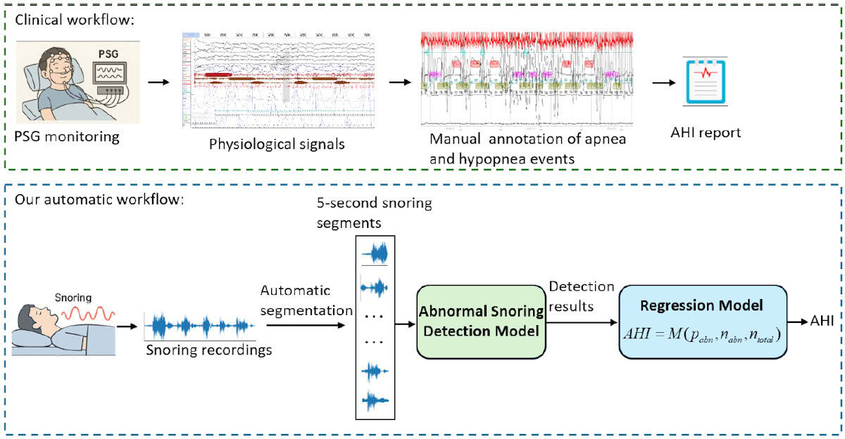
All subjects slept in hospital wards and were also monitored with a PSG device during sleeping. Sleep physicians then used the PSG data to annotate the audio recordings with apnoea and hypopnea events. Each recording was divided into 5-s audio segments and assigned as either abnormal, if they included apnoea/hypopnoea events, or normal.
Abnormal snoring detection Upper panel: the clinical workflow using PSG and physiological signals to determine AHI. Lower panel: the proposed automatic workflow, which estimates AHI directly from snoring recordings using segment-level abnormal snoring detection and regression models. (Courtesy: Physiol. Meas. 10.1088/1361-6579/ae6f6a)
To evaluate their framework’s ability to detect abnormal snoring, the researchers compared it against five other pretrained audio models. They first ran a subject-dependent evaluation, in which random audio segments from all subjects were used for training, validation and testing. All models performed well – as expected when the training and test data come from the same subjects – but the adapted Wav2vec 2.0 model achieved the highest accuracy and sensitivity.
The team then performed a subject-independent evaluation, which better reflects a real-world scenario, with each subject’s data randomly assigned to the training, validation or test set. Here, the existing methods exhibited a reduced accuracy of between 65% and 71%, while the new model retained an accuracy of 73.95%.
“In subject-independent testing, this adapted model achieved better performance than the compared baseline models, suggesting improved generalization to unseen subjects,” says Li. The team’s model was also significantly more computationally efficient, an important factor for OSA screening in home-based or resource-limited scenarios.
AHI estimation
Finally, the researchers designed a snoring-based AHI estimation framework, using their detection model to identify abnormal snoring segments and calculating a ratio-based statistic – the number of abnormal segments divided by the total number of segments – to quantify OSA severity. They then used four common linear regression models to estimate AHI for individual subjects.
All models demonstrated reasonable accuracy and good correlation between predicted and PSG-derived AHI values. The mean absolute error was roughly 11 events/h, which means that the estimated AHI may deviate from the true value by 11 events per hour of sleep and could (if near a threshold between categories) result in an incorrect severity diagnosis. However, while the current model is not yet precise enough for clinical diagnosis, it could prove ideal as a potential tool for home-based OSA screening or pre-screening.
The researchers are now investigating how to more effectively apply pretrained audio models to clinical snoring analysis. “We are focused on making these models lighter and more suitable for home-based screening, while validating their effectiveness across large, multicentre datasets,” Yun Lu, one of the corresponding authors from Huizhou University, tells Physics World. “Additionally, we plan to improve the accuracy of AHI estimation by integrating snoring-based predictions with additional non-contact physiological information.”
Ronan Smith, a postdoctoral research fellow at Adelaide University, has been chosen as the winner of this year’s Physics in Medicine & Biology (PMB) Early Career Researcher Award. The award is presented to the author of the “best paper” in PMB’s 2025 Early Career Researcher Focus Collection, as selected by the journal’s editorial board.
EBVs are one-way valves that are placed into the lung to help treat emphysema – a condition that damages air sacs in the lungs, causing air to get trapped inside and making breathing difficult. The EBV, which in some cases can be used in place of surgery, prevents airflow into damaged lung areas so that the rest of the lung can function more effectively.
Successful valve placement causes the targeted area of lung to collapse, which can be imaged using CT. Smith proposed that use of XV functional lung imaging to non-invasively measure regional and local changes in airflow could more accurately assess the clinical impact of EBV placement.
“The lungs are a dynamic organ, their job is to be constantly moving,” he explains. “Because X-ray velocimetry looks at lung motion, it lets us see exactly where the air is or isn’t flowing, so you can instantly see that airflow has changed within the lungs. CT only measures structural changes, which may not necessarily be correlated to changes in lung function.”
In vivo demonstration
To investigate this potential advantage, Smith and colleagues carried out a pilot study on healthy sheep, which have a similar lung size to humans. They performed XV imaging on two anaesthetized and ventilated animals, before and after placing EBVs in their lungs.
The XV scanning process involves recording fluoroscopic videos of individual breaths at various angles around the lung, with anatomic positioning provided by an accompanying breath-hold CT scan. To analyse the data, the researchers used XV LVAS software from 4DMedical, the MedTech company that developed and commercialized the XV technology.
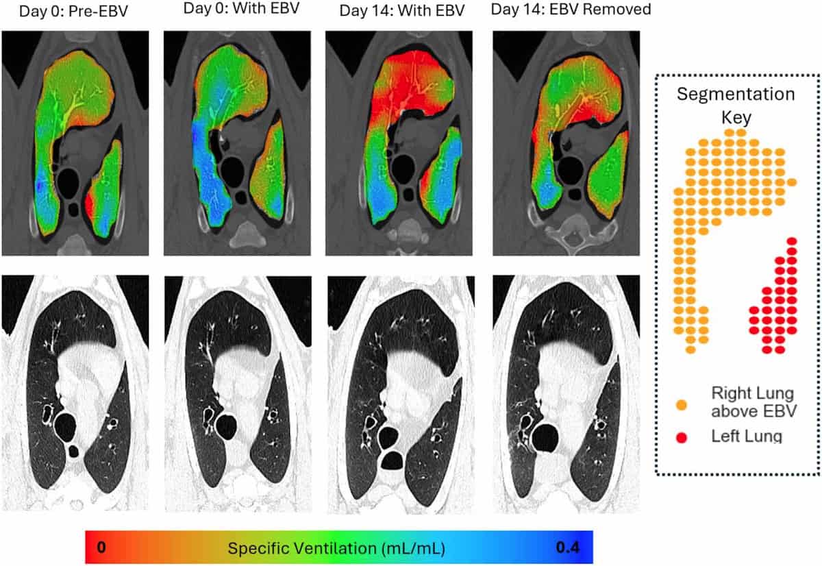
Visualizing ventilation Slices from XV and CT imaging taken from an area of lung above the EBV. XV shows clear changes in ventilation immediately after valve insertion. (Courtesy: Phys. Med. Biol. 10.1088/1361-6560/ade196)
The software correlates motion in the XV videos with CT data to measure the lung’s expansion and contraction during a breath cycle. It creates a 3D map of specific ventilation (the change in voxel volume during a breath, divided by its starting volume) in small voxels throughout the lungs. This map can then be used to calculate mean specific ventilation and ventilation heterogeneity across a given lung region.
As soon as the EBVs were inserted into the animals’ lungs, XV imaging could visualize and quantify a reduction in airflow to areas downstream of the valves. This effect was seen both in regions where collapse was visible in CT scans, as well as those where collapse could not be detected by CT. Ventilation changes were also clearly observed in the remainder of the lungs.
“The main finding of our study was that X-ray velocimetry imaging can detect airflow changes from endobronchial valve placement in the lungs,” says Smith. “Our research could be really important for people [with emphysema], as tools to help with better placement and verification will lead to improved treatment options.”
Future prospects
Since the publication of this paper, Smith has focused on further applications of pre-clinical and clinical XV imaging. “I’ve been working as part of a great interdisciplinary team looking at how lung function changes in a range of diseases, to both understand the diseases, and as an outcome measure when we test treatments,” he says.
This work includes the world’s first paediatric clinical trial of XV imaging, which is examining the feasibility of using the technology in children with cystic fibrosis. The researchers have imaged around 30 children to date and aim to publish their findings later this year. They are currently planning future studies to see how XV imaging could enhance clinical decision making and improve outcomes for these children, as well as looking at other childhood diseases where it could be of relevance.
“As an early-career researcher, I’m also focussing on developing my own research, looking at another novel X-ray imaging method called dark-field X-ray imaging,” Smith adds.
The perfect award
Smith tells Physics World that he was excited to receive the PMB Early Career Researcher Award, acknowledging the efforts of everyone involved in this “hugely collaborative project”, including clinicians, scientists, 4DMedical and the staff in the preclinical imaging facility where the study was performed.
Team effort Ronan Smith (back row, centre) and colleagues in the lab. (Courtesy: Women’s and Children’s Health Network South Australia)
“As a physicist working in medicine/biology, it feels like the perfect award to get,” he says. “It’s great to see interest in the work we are doing, and fantastic evidence we can use to convince the funding bodies it’s worth continuing this work.”
The PMB Early Career Researcher Focus Collection 2025 publishes papers from early-career researchers (defined for this collection as a postgraduate student or someone who completed their PhD in 2019 or later) in biomedical physics, with the aim of highlighting research excellence from emerging leaders. The Early Career Researcher Award was introduced to further recognise an outstanding contribution from one of the early-career authors, selected according to the quality of scientific content, number of citations and downloads, and peer review ratings.
Image of the year Left: 18F-GP1 PET/CT images showing multiple blood clots in the deep veins of the left leg, plus several clots in the right calf and clots in both lungs. Right: 18F-GP1 PET/CT images showing widespread blood clots throughout the body. (Courtesy: S Han et al. Asan Medical Center, Seoul, Korea)
A PET radiotracer that can detect deep vein thrombosis (DVT) in the legs and clots that have travelled to the lungs has been chosen as the “Image of the Year” at the Society of Nuclear Medicine and Molecular Imaging (SNMMI) 2026 annual meeting. Developed by Sangwon Han and colleagues at the Asan Medical Center, University of Ulsan College of Medicine, in Korea, the novel tracer enables whole‑body imaging of blood clots (thrombi) in the legs and lungs in a single scan.
A DVT is a blood clot that forms in a deep vein, usually in the legs. It’s a common condition, with an incidence of roughly half that of all cancers, and it can lead to serious complications. Clots can break off and travel to the lungs, which could cause a potentially life‑threatening pulmonary embolism (a blockage in the artery supplying blood to the lungs). Early detection of DVT is therefore critical for determining the most appropriate treatment for each patient.
Currently, the standard imaging method for diagnosing DVT is venous ultrasonography (VUS). But while this works well for detecting clots in the thigh-to-knee region, whole-leg VUS requires skilled operators and advanced machines, takes longer, and has lower diagnostic sensitivity in the calf. In addition, conventional imaging techniques such as VUS and CT rely on indirect structural changes rather than directly visualizing the clot.
Aiming to enable faster and more efficient DVT diagnosis, Han and his research team are studying fluorinated GP1 (18F-GP1) a novel thrombus-targeted PET tracer. The tracer selectively binds to specific receptors on activated platelets (the cell fragments that cause blood to clots), allowing direct visualization of active thrombus formation.
“In our Phase 1 study, 18F-GP1 PET/CT showed 100% detection rate in 20 patients with confirmed DVT or pulmonary embolism,” Han told the SNMMI delegates. “But that study was limited by its small sample size and an absence of negative groups so specificity could not be assessed at that time.”
So in this latest work, Han and his team performed a phase 2, non-randomized study investigating the ability of 18F-GP1 PET/CT to identify acute lower-extremity DVT in 46 symptomatic patients. This included 22 patients with proximal DVT and 24 with none or distal DVT, as diagnosed using VUS.
The researchers acquired chest-to-feet PET/CT scans approximately 2 h after intravenous administration of 250 MBq of the radiotracer. The images were assessed by three blinded nuclear medicine physicians from different institutions, who assigned focal 18F-GP1 uptake higher than background activity as positive for thrombosis. They classified proximal DVT as clots involving the iliac (pelvic), femoral (thigh) and popliteal veins (behind the knee), and distal DVT as clots confined to the calf veins.
“Our primary objective was to assess the sensitivity and specificity of qualitive 18F-GP1 PET/CT interpretation for proximal DVT,” Han explained. “Secondary objectives included assessing the agreement between PET/CT and VUS for distal DVT, inter-reader reproducibility, exploring the detection of pulmonary embolism and assessing safety.”
When evaluated against VUS as a reference standard, 18F-GP1 PET/CT exhibited high diagnostic accuracy for detecting clots, demonstrating a sensitivity of 95% and a specificity of 92% for proximal DVT. “For distal DVT, both positive and negative agreement between PET/CT and VUS were strong,” added Han. “Inter-reader agreement was also excellent.”
The scans also identified concomitant pulmonary emboli in some patients, as confirmed by CT pulmonary angiography, illustrating the advantage of simultaneously assessing DVT and pulmonary embolism in a single scan. The researchers noted that the radiotracer was well tolerated, with no drug-related adverse events observed.
Speaking in the plenary session when his award was announced, Han shared a “striking image” recorded using 18F-GP1 PET/CT, which showed extensive blood clots, not only in the leg and lungs, but also in many unusual sites, including cranial and spinal vessels, cardiac valves, and vessels in the pelvic region. “This image clearly shows the remarkability ability of fluorinated GP1 to visualize thrombi throughout the body,” he explained.
“We believe this represents an important step towards thrombus-specific imaging,” Han concluded. “The potential of GP1 PET can expand beyond DVTs to many other thrombotic diseases such as embolic stroke or other cardiovascular diseases.”
The SNMMI Image of the Year is the society’s highest award, and the most anticipated, given out in recognition of an image that’s truly cutting-edge and representative of the future of nuclear medicine. This year’s winning image was chosen from nearly 1500 abstracts submitted for the meeting.
“It is truly a great honour to receive the Image of the Year award,” said Han.
Two years ago, the ESTRO 2024 meeting in Glasgow dedicated a conference session to the discussion of upright radiotherapy. In particular, the speakers pondered whether this emerging technique – in which patients are treated sitting up rather than lying down – offers hope of increasing access to advanced radiotherapy, or whether it’s merely hype.
Things have moved on since then. Leo Cancer Care introduced its upright photon therapy system, Grace, and received commercial approval in the US and (just last week) Europe for its Marie upright positioning and CT system. Stanford Medicine recently unveiled the world’s first ultracompact proton therapy facility, pairing Mevion Medical Systems’ compact S250-FIT proton therapy system with the Marie platform. Meanwhile, the body of published research on the feasibility and patient experience of upright treatments continues to grow.
At this year’s ESTRO 2026 meeting in Stockholm, the theme was revisited by four experts in the field, who debated the motion that “Upright radiotherapy will be a mainstream and standard radiotherapy delivery option in 2035”.
The customary pre-debate vote revealed that just one quarter of the audience thought that photon-based upright radiotherapy would become mainstream, with the remainder believing that it would remain a niche technique. When it came to upright proton therapy, however, the vote was split roughly 50:50. So could the speakers persuade the attendees to change their minds?
Patient-centred care
The debate began with Tomas Kron from the Peter MacCallum Cancer Centre in Australia arguing the case for upright X-ray radiotherapy. He pointed out that upright positioning is not a new idea. “Historically, photons and upright have been around for a very long time. It has been, if not standard practice, widely used. But what role will it play in 2035?”
Not a new idea The first high-energy linear accelerator for medical use, known as LA-1, was developed at Stanford in the 1950s. Patients with head-and-neck or lung cancer could sit on an isocentrically mounted chair for treatment. The machine was decommissioned in 1972 and now resides in the Smithsonian Institution. (Courtesy: Stanford Medicine)
Kron described a clinical imaging trial underway at Peter Mac investigating upright cone-beam CT for planning lung cancer radiotherapy. The study showed that image quality was good enough for adaptive treatment planning, and that the lung was expanded and moved less in the upright position. Kron noted that patient setup and imaging was “really, really easy”, taking just a few minutes.
But what’s more important, he emphasized, is the patient experience. Patients treated while sitting up can maintain eye contact with the doctors throughout, they feel more involved and empowered, with one patient commenting: “My breathing was strong, I felt comfortable, the band around my chest was giving me a bear hug.”
“It’s really all about patient-centred care. Physical comfort and emotional wellbeing are top priorities,” Kron said. “Clearly, in an upright scenario this is much more likely to be the case.”
Upright radiotherapy offers many other unique features, including anatomical advantages and the ability to customize the chair, for example, for bariatric or paediatric patients. An upright treatment system is also more compact than a couch-based machine, requiring a smaller bunker. It could also be used as a mobile radiotherapy unit, said Kron – reducing the need for patient travel.
Kron’s team found that 80–90% of their patients could be treated just as well with upright radiotherapy as supine (lying down). “There are anatomical advantages with upright, there are patient preferences, there are economic benefits. What’s not to like,” he concluded.
The myth of mainstream
“Upright radiotherapy will not be mainstream and standard,” declared the second speaker, Livia Marrazzo from the University of Florence in Italy.
“Mainstream means widely adopted, used across the majority of radiotherapy centres, the default in clinical practice … and standard is even stronger, backed by clinical evidence, guideline-endorsed, reproducible and validated,” Marrazzo told the delegates. “It’s not ‘it works in some centres, is technically feasible, has early adopters, may have advantages for some patients’. But that is where we are with upright radiotherapy.”
The art of persuasion Livia Marrazzo argued that upright radiotherapy will not be mainstream by 2035. (Courtesy: Icro Meattini)
From a practical standpoint, most of the roughly 16,000 radiotherapy systems worldwide are linac-based recumbent machines with a typical lifecycle of 10 to15 years. Many were recently replaced with supine systems optimized for intensity-modulated and image-guided radiotherapy. “The installed base is locked into supine geometry for another full cycle,” Marrazzo explained.
She refuted many of the advantages proposed by Kron. “We have limited clinical evidence supporting comfort advantages,” she said. “It may benefit specific patient groups and conditions, but this doesn’t mean mainstream.” Overall, clinical experience is limited, with no comprehensive evaluations of plan quality and no comparative clinical studies.
She highlighted the particular challenges of breast cancer treatments, which account for 25-30% of cases in her radiotherapy department. “When we place a breast cancer patient upright, we lose the natural breast separation, so have much more difficulty in hitting the target and avoiding the contralateral breast,” she explained. “This exemplifies how upright is not a plug-and-play replacement for a conventional supine workflow.”
“Are we sure we would like to have upright as the standard radiotherapy delivery option by 2035 or do we want to push our efforts somewhere else?” Marrazzo concluded. She would prefer a focus on introducing technologies such as AI-driven planning and contouring, fully adaptive workflows, ultra-hypofractionation or biology-guided treatment adaptation. “These are all solutions that can be software-driven, scalable and compatible with existing supine infrastructure.”
The motion for protons
With half of the audience already agreeing that upright proton therapy will become mainstream, Petra Trnkova from Czech Technical University had perhaps a slightly easier task as she presented the case for upright protons. Nevertheless, she began by suggesting that her opponents are simply “scared of progress and won’t accept that, even without evidence, we can move forward in radiotherapy”.
Trnkova reiterated the benefits of upright radiotherapy cited by Kron: favourable patient anatomy, lower installation cost, improved sustainability, and patient-centric management. “For proton therapy, these improvements are much more significant,” she noted.
For starters, upright systems could help address the massive disparity in access to proton therapy around the globe. Sharing a map showing how proton therapy facilities are mostly distributed in wealthy countries, Trnkova noted: “My opponents may tell you that it’s not possible to do this by 2035, but when you look at this map, I ask you, can we wait any longer?”
Saving space The compact S250-FIT proton therapy system is small enough to fit in a standard linac vault. (Courtesy: Stanford Medicine)
Increasing access to proton facilities is enabled by the extreme size reduction when eliminating the need for a large rotating gantry, enabling proton therapy systems small enough to fit in a standard linac vault. Upright proton therapy can also reduce machine complexity, increase rotation speed and lower energy consumption – reducing costs, improving system upgradeability and increasing environmental sustainability.
“Another consequence of smaller facilities is we can really have patient-centred care,” Trnkova added. Recalling the 10 to 15 year linac lifetime mentioned by Marrazzo, she suggested another option: “You can replace your linac with proton therapy. Then you can have the full set of treatments available for each patient”.
Upright proton therapy could also ease the introduction of new treatment techniques, such as proton arc therapy, which offers dosimetric benefits over intensity-modulated proton therapy, but it is difficult to deliver with a gantry. It could also enable in vivo dosimetry, using shoot-through protons for range verification, or mixed-beam delivery of protons and photons.
“Upright positioning offers many opportunities, it’s the only way towards the democratization of proton therapy,” Trnkova concluded. “Stop asking what opportunities upright radiotherapy brings, start asking what you can do to bring it faster to clinical practice.”
The reality check
The final speaker, Carles Gomà from Clinic Barcelona in Spain, reflected upon what makes a good radiotherapy system. “In my view, it’s a three-legged stool: beam delivery, imaging and immobilization,” he said. “And progress comes with a combination of the three.”
For example, focusing too heavily on beam delivery and imaging can lead to immobilization being forgotten. “Immobilization means comfort, and if we are comfortable, we are still,” Gomà explained. “I cannot care less how many papers say patients are more comfortable in an upright position,” he added, pointing out that people will pay five times more to fly in business class where they can lie down.
The other reason cited for moving to upright proton therapy is its lower cost. “But is proton therapy expensive?” Gomà asked. He described the situation in Catalonia, which has a population of eight million and in 2018 spent Euro 42.2M on external-beam radiotherapy. “This is exactly the same cost as one immunotherapy drug for the same population,” he pointed out. “Proton therapy is not expensive; photon therapy is ridiculously cheap.”
Gomà also considered whether “suboptimal protons” are better than photons. “I’m going to answer no,” he said, describing two recent phase III, randomized trials comparing photons with protons for oropharyngeal cancer. The US trial concluded that proton therapy provides a new standard-of-care option, but the UK trial reported no difference between the two modalities.
“Let’s learn from history and not repeat the same mistakes,” he concluded. “True progress is improvement without compromise. If we want to make the stool higher, we have to work on all three legs at the same time.”
The final vote At the end of the debate, the ESTRO delegates strongly supported upright proton therapy. (Courtesy: Ye Zhang)
The debate concluded with decisive a final vote: while support for upright photon therapy reduced a little, over two-thirds of the audience believed that upright proton therapy will indeed become mainstream and standard by 2035.
Writing on LinkedIn, session co-chair Ye Zhang from the Paul Scherrer Institut noted: “The debate sparked an inspiring shift in perspective, with final voting showing slightly increased scepticism toward mainstream upright photon therapy (dropping from 23% to 18% support), but a dramatic surge in favour of upright proton therapy, which jumped from 47% to a 69% majority.”

 Login