
❌

Normal view

Dose calculations reveal how patient motion impacts proton minibeam therapy
31 August 2026 at 14:00
Calculated dose distributions
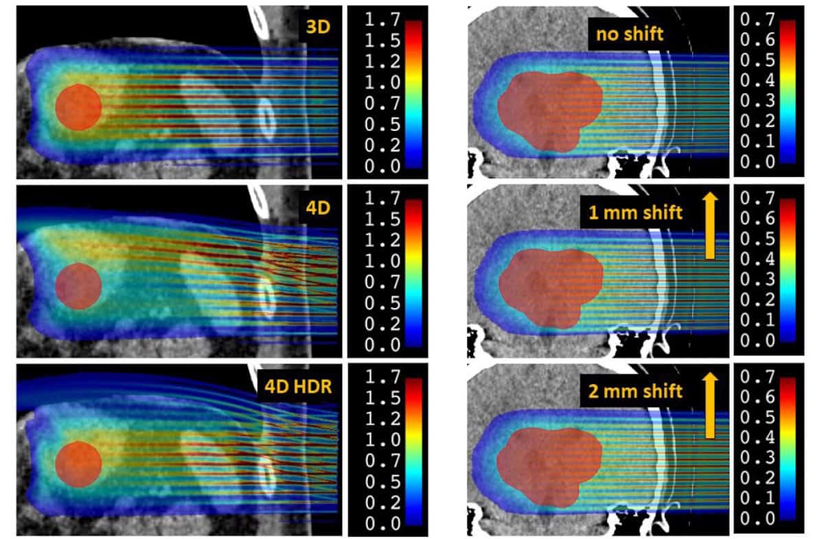
Dose calculations Left: dose distributions for thoracic pMBRT without motion (3D), with respiratory motion (4D) and with respiratory motion at high dose rate (4D HDR). Right: dose distributions for a brain treatment with zero shift, a 1 mm shift and a 2 mm shift. (Courtesy: Phys. Med. Biol. 10.1088/1361-6560/ae8ef8)

Proton minibeam radiotherapy (pMBRT) is an innovative new cancer treatment that uses an array of narrow proton beams to create alternating regions of high and low dose, before converging to a homogeneous dose distribution within the target volume. Numerous studies in small animals have shown that this spatially modulated dose distribution can destroy tumours while sparing healthy tissue and reducing side effects.

To implement pMBRT in the clinic, it’s important to first assess the impact of organ motion during treatment, which could lead to overlapping of the dose peaks and valleys, and potentially diminish the sparing effect. With this aim, a team at Institut Curie in France has developed a Monte Carlo (MC)-based framework to quantify the dose distribution for realistic clinical treatments, reporting their findings in Physics in Medicine & Biology.

To deliver pMBRT, a multi-slit collimator is used to create narrow beams, typically 0.3 to 1 mm wide, spaced 2 to 6 mm apart. As this collimator blocks much of the initial proton beam, longer treatments are needed to deliver the prescribed dose, increasing the likelihood of organ motion.

“Given the longer irradiation times, possible patient movement could deteriorate the heterogeneous spatial dose distribution of pMBRT,” explains senior author Ludovic De Marzi. “This is all the more problematic because it is this heterogeneous distribution that is believed to be responsible for the technique’s biological sparing effect.”

The dose reconstruction workflow

De Marzi and colleagues developed a 4D dose calculation workflow based on time-resolved MC simulations to determine how motion affects pMBRT dose distribution. The tool requires two main inputs: a 4D CT dataset capturing the patient’s anatomy and breathing motion over one respiratory cycle; and a proton pencil-beam scanning (PBS) treatment plan created on a reference CT.

To validate their workflow, the researchers irradiated a thoracic motion phantom with a conventional PBS plan and compared the measured dose with the simulations. For a 3%/3 mm gamma index (a standard quality check tool), the mean pass rate for nine phantom irradiations was 97.3%, confirming that the calculation performs reasonably well for 4D dose modelling in conventional PBS.

They then used the tool to assess the impact of motion in representative clinical cases, first examining a thoracic treatment with large breathing movements. They calculated 4D dose distributions for three scenarios: 3D pMBRT without motion; 4D pMBRT with 20 mm breathing motion; and 4D high-dose rate pMBRT, with the instantaneous dose rate increased tenfold to reduce beam-on time by a factor of ten.

The calculated dose distributions showed that breathing motion introduced significant variations in the patterns of peaks and valleys, impacting a crucial parameter: the peak-to-valley dose ratio (PVDR). At a depth of 5 mm, for example, the mean PVDR dropped from 11.9 for the 3D scenario to 6.7 in 4D. The high-dose rate scheme slightly mitigated this degradation, but not significantly.

“The PVDR is an index that takes into account both doses in the valleys (which are likely correlated with the biological sparing effect on healthy tissue) and the maximum delivered doses (which are likely responsible for the anti-tumour effect),” De Marzi explains. “It can also be used to help optimize the 3D dose distribution of the treatment plan.”

Breathing motion also degraded the dose delivered to the tumour, with the mean target dose and D95% (the minimum dose received by 95% of the target volume) reduced by 30% and 35%, respectively, for 4D pMBRT. Nearby organs-at-risk (OARs) were also impacted, with a decrease in dose to the stomach and a significant increase in mean dose to the spleen for the 4D cases.

The researchers next examined an intracranial treatment. While there is no intra-organ motion in this case, the patient’s head undergoes small rigid translations – generally limited to less than 1 mm using thermoplastic masks. As such, they modelled patient motion as a simple continuous 1 or 2 mm translation.

Motion did not significantly impact the mean dose or D95% for the target or OARs. A 1 mm shift was, however, enough to degrade the peak–valley patterns, reducing the mean PVDR by 10% from the static case (at 15 mm depth). For a 2 mm shift, this increased to a 24% reduction in mean PVDR.

A valuable tool

The researchers conclude that their 4D dose reconstruction workflow provides a useful tool for assessing interplay effects caused by organ motion during pMBRT. Without a specific motion management strategy, only intracranial treatment with movements below 1 mm was robust to interplay effects. A similar workflow could prove valuable for estimating the sparing effect of pMBRT while accounting for patient motion and adapting treatment plans accordingly.

Looking forward, it may be possible to minimize delivery times by combining pMBRT with the ultrahigh-dose rate delivery used for FLASH treatments.

“This idea is appealing because it could both solve the problem of reduced dose rate in pMBRT and add an additional biological effect (FLASH) to further spare healthy tissue,” says De Marzi. “However, its feasibility remains to be demonstrated, as does the value of developing a technique that combines such considerable complexities. A simple increase in dose rate – without going as far as FLASH – would already be of interest.”

In their next step towards clinical application, De Marzi and colleagues are working to validate calculation tools and measurement procedures tailored to this pMBRT technique. “Ones that are fast, accurate and usable by clinicians,” he says.

The post Dose calculations reveal how patient motion impacts proton minibeam therapy appeared first on Physics World.

Easy-to-use calculation tool estimates out-of-field neutron dose during proton therapy

16 June 2026 at 15:00
Neutron dose measurement points within a treatment room
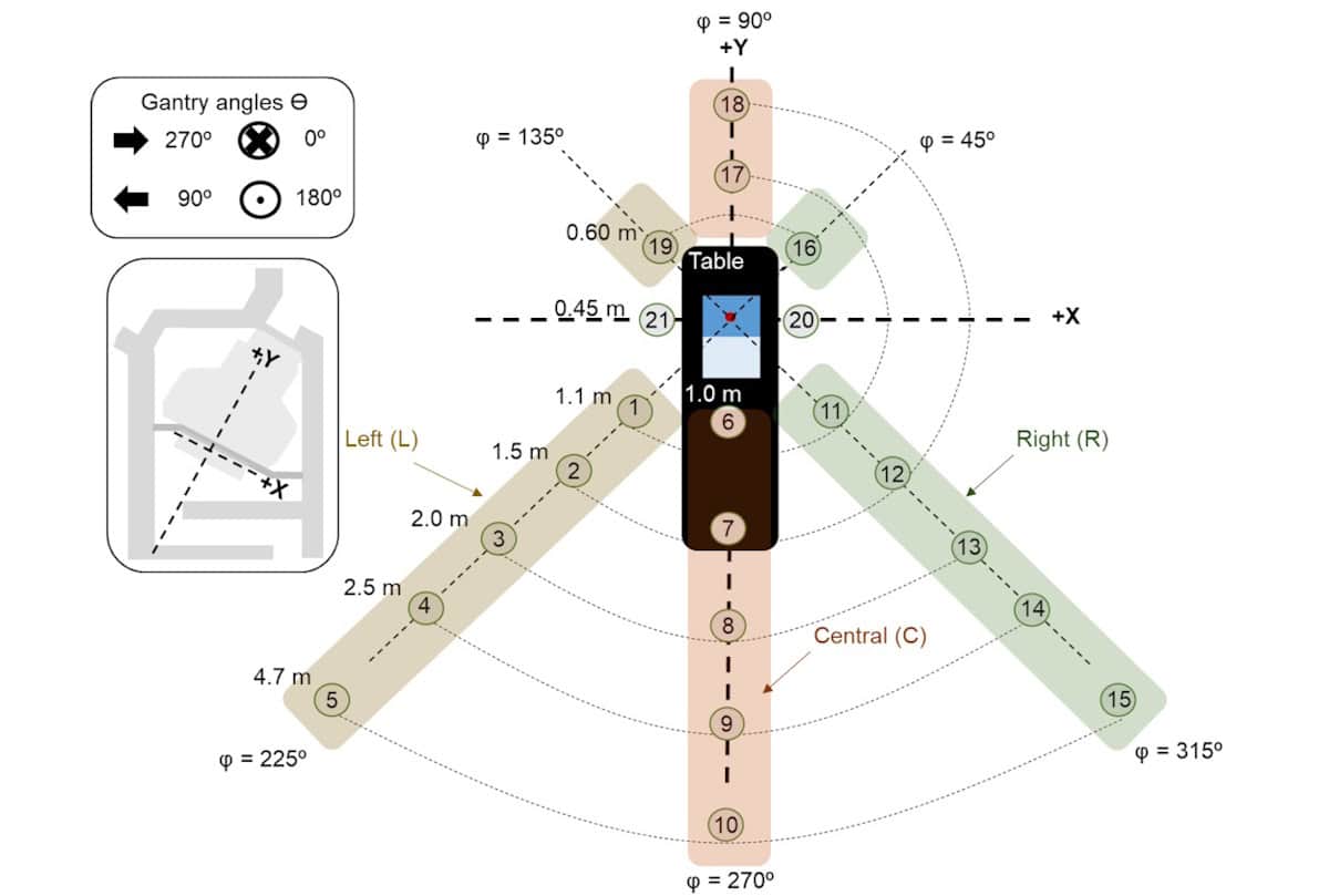
Experimental setup Neutron dose measurement points within the treatment room, showing gantry angles, measurement lines (φ) and distances from the isocentre. (Courtesy: Phys. Med. Biol. 10.1088/1361-6560/ae72e7)

Proton therapy plays an increasingly important role in cancer treatments, targeting tumours with high precision while sparing nearby healthy tissues. Proton irradiation can, however, also produce secondary neutrons due to nuclear interactions of the therapeutic beam. In modern pencil-beam scanning proton therapy, neutrons generated inside the patient are the main source of out-of-field dose – unwanted radiation that could potentially contribute to secondary cancer risks.

A research team headed up at Clínica Universidad de Navarra in Spain has experimentally characterized the neutron field in a proton therapy treatment room using a range of different detectors. They used their findings to create a practical Python-based calculation tool to estimate neutron dose for arbitrary irradiations.

“The tool provides a fast first-order estimate of neutron doses anywhere in the treatment room using information from the treatment plan,” explains medical physicist Verónica Morán. “It could support radiation protection studies, workplace dose assessments, research projects, and the evaluation of neutron exposure in situations where direct measurements are not available.”

Neutron field characterization

For their study, reported in Physics in Medicine & Biology, Morán and colleagues employed a Hitachi PROBEAT-CR proton therapy system with pencil-beam scanning. They measured neutron dose using a range of devices, including two ambient detectors and four types of personal dosimeter: thermoluminescent dosimeters (TLDs); track-etch detectors; bubble detectors (BDs); and electronic personal dosimeters (EPDs).

Using up to 21 measurement points within the treatment room, the team examined the dependence of out-of-field neutron dose on various beam and room parameters, including gantry angle, field size, proton energy and distance to isocentre.

At each measurement point, they assessed the neutron ambient dose equivalent, which is used to characterize the radiation field at a specific location, plus the personal dose equivalent, which estimates the dose that a person may receive while occupying that location. The ambient detectors performed the best, working as expected in a synchrotron-based facility, while the personal dosimeters exhibited clear variations in response.

To determine whether neutron doses measured on one side of the treatment room can predict doses at equivalent locations on the opposite side, the team investigated room symmetry. Measurements with the ambient detectors indicated that the treatment room was symmetric for the 270°–90° gantry pair, while for the 0°–180° pair, doses at 0° were on average 8% lower than at 180°.

Verónica Morán
Corresponding author Medical physicist Verónica Morán. (Courtesy: Clínica Universidad de Navarra)

“We found that the room was largely symmetric for certain gantry orientations,” notes Morán. “This reduces the number of measurements needed and helps extend the applicability of the dose calculation model.”

The measurements also showed that neutron doses created by a single spot field and a 10×10 cm field were similar (and can be regarded as interchangeable). The 20×20 and 30×30 cm fields differed by up to 22% relative to the single spot. Neutron dose dependence on proton energy followed the expected power law, with best fits for the ambient detectors, followed by the BDs and TLDs.

The researchers also delivered a clinical proton treatment comprising 27 energy layers (from 121.6 to 173.1 MeV) to a scattering phantom. They examined whether the total neutron dose from such an irradiation can be expressed as a weighted sum of contributions from the individual energy layers. If this assumption holds, the total neutron dose for an arbitrary plan could be reconstructed from measurements at discrete proton energies. Comparing calculated and measured neutron doses revealed that this linear superposition approach worked with the ambient detectors and the BDs, but not the EPDs.

A practical tool

The team then developed a Python-based tool to estimate neutron dose at any point in the treatment room for arbitrary irradiations and detectors (including ambient detectors, BDs and EPDs). As inputs, the tool requires the radiotherapy plan, detector data and calculation parameters including the gantry angle, and measurement distance and angle. It then outputs neutron dose estimates along with associated uncertainties.

The researchers verified the tool by assessing additional measurement points that weren’t used in its development. Comparisons of experimental and calculated dose values showed that the tool provided reliable and useful estimates for the ambient detectors and BDs, even at points where no prior measurements existed.

For the EPDs, however, the calculated intervals were often broad and the researchers suggest that EPD results should be interpreted with caution. They point out that such detectors were retained in the tool because “not all proton therapy centres have access to the same detector types, and approximate detector-specific estimates may still be of practical interest in such settings”.

The tool is thought to be the first to estimate out-of-field neutron dose based on treatment room measurements, and should also work at other clinical centres. “Because modern pencil-beam scanning proton therapy systems have been shown to generate similar neutron fields across different facilities, we believe the methodology behind the tool may be transferable to other centres using comparable technology,” says Morán.

The researchers are now extending the tool to include paediatric cases, and different proton energies, patient sizes and treatment configurations. “We are also investigating how these methods could be applied to estimate neutron doses received by patients, with the long-term goal of improving the characterization of out-of-field radiation exposure in proton therapy,” Morán tells Physics World.

The post Easy-to-use calculation tool estimates out-of-field neutron dose during proton therapy appeared first on Physics World.

❌