❌

Normal view

Optogenetic therapy shows promise for vision restoration

8 October 2026 at 16:00

Retinitis pigmentosa is an inherited degenerative condition that destroys photoreceptors in the retina and can eventually lead to irreversible blindness. In a clinical trial first, an international research team has demonstrated that optogenetic therapy is safe to use and shows potential for restoring visual function in patients with late stages of this disorder.

In a first-in-human study in 2021, the collaboration – led by José-Alain Sahel from the University of Pittsburgh School of Medicine and Botond Roska from the Institute of Molecular and Clinical Ophthalmology Basel (IOB) – demonstrated that optogenetic therapy led to partial recovery of visual function in a blind patient with retinitis pigmentosa. This latest work, reported in the New England Journal of Medicine, expands the cohort to 10 participants.

Optogenetics is a method that uses light to characterize and manipulate the activity of neurons in the brain – and which earlier this week won its inventors the 2026 Nobel Prize in Physiology or Medicine. The optogenetic therapy developed by Sahel, Roska and collaborators combines gene therapy that makes surviving ganglion cells (neurons in the retina) sensitive to light with special goggles that stimulate the altered cells.

“Although patients with retinitis pigmentosa lose their rod and cone cells, which normally detect light, many other cells within the retina can remain present for years after vision loss,” explains Sahel, director of the UPMC Vision Institute and first author on this study. “Optogenetics enables us to introduce a light-sensitive protein into these surviving cells, allowing them to respond to light and transmit visual information to the brain. The therapy bypasses the damaged photoreceptors and takes advantage of retinal circuits that remain intact.”

Sahel notes that retinitis pigmentosa can be caused by mutations in any one of hundreds of different genes, making optogenetic therapy a particularly suitable approach as it can potentially help regardless of the underlying mutation.

Safe to use

In the study, 10 participants with blindness due to advanced retinitis pigmentosa were injected in their worse-seeing eye with a gene that produces ChrimsonR – a light-sensitive protein with a peak response at around 590 nm (amber). The team divided the subjects into three dose-escalation groups, receiving 5.0 x 10¹⁰, 1.5 x 10¹¹ or 5.0 x 10¹¹ vector genomes per eye.

The primary aim was to evaluate the safety of the optogenetic therapy. The researchers observed 33 mild and moderate adverse ocular events in nine of the participants, such as temporary inflammation and increases in eye pressure, plus one severe event, which resolved within minutes after treatment. They concluded that, within the limits of the study, the treatment was safe.
Visual stimulation system
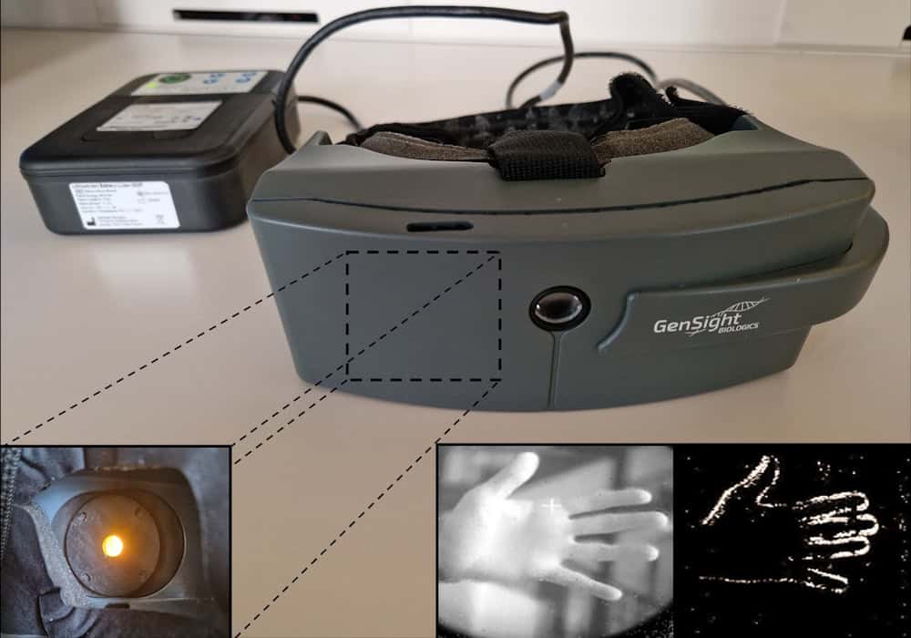
Visual stimulation system The specialized goggles (centre) send visual information to a processor (upper left) that converts the information into patterns of light. A projector within the glasses (lower left) sends these patterns back to the eye. Lower right: the system converts the image of a hand into a simplified light pattern that’s projected onto the retina. (Courtesy: Chloé Pagot, Streetlab, Paris)

Using the light-stimulating goggles, they then assessed the visual performance of treated eyes, before and after injection. The goggles contain a camera that detects changes in light intensities pixel-by-pixel and converts this visual data into pulses of amber light. This light is projected onto the eye to activate ChrimsonR in the modified retinal ganglion cells. Vision tests revealed that the treatment increased light sensitivity in seven of the 10 participants, by a factor of 2.0 to 62.3, with six patients showing a clinically meaningful improvement.

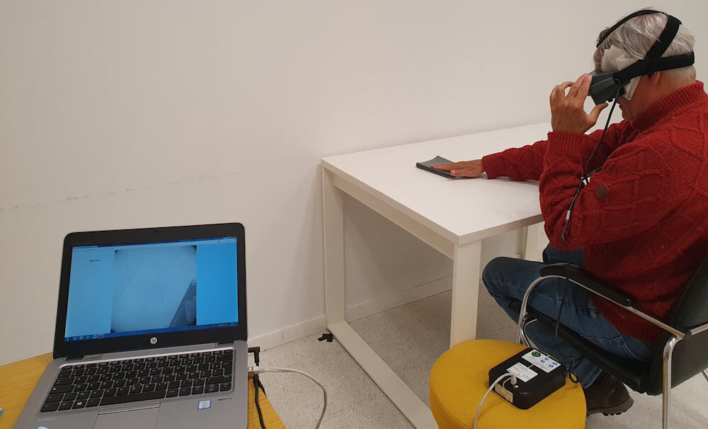
The patients also performed a series of tasks to detect, localize and touch a notebook and a staple-box. In the notebook task, five of eight participants had higher accuracy when wearing the goggles than when not wearing them, while four showed improved accuracy in the staple-box task. In a task determining the orientation of a single bar, accuracy was higher with the goggles in five of eight participants.

To provide an objective measurement of brain activity in response to light, the researchers obtained electroencephalographic (EEG) recordings in five patients performing two visual tests: detection of a tumbler placed in front of them (with the eyes open or closed); and passive viewing of periodic visual stimuli.

In the tumbler test, EEG decoding accuracy was higher with goggles than without when the eyes were open, but not when they were closed – implying a genuine improvement in visual processing rather than an artefact. The second test also showed increased decoding accuracy, which correlated with mean improvements in the notebook and staple-box tasks.
Visual behavioural test
Visual behavioural test After receiving optogenetic therapy, participants used a visual-stimulation system to complete tasks. Here, a participant reaches for a notebook. (Courtesy: Chloé Pagot, Streetlab, Paris)

A bright future

The changes in visual performance were consistent across multiple tasks and testing sessions, with responses observed in all three dose groups. Participants who spent longer being trained to use the goggles performed better in the visual behavioural tests. The researchers point out, however, that functional changes were modest: improving object detection and discrimination capabilities but not restoring normal sight. The small sample size also limited their ability to characterize treatment response.

Several projects are currently underway to further enhance the technology. This includes digital holographic goggles with an eye tracker being developed by the team of Valentina Emiliani at the Institut de la Vision in Paris. The system will project 20 µm-wide images that stimulate individual cells in the retina.

“With the eye tracker, we can make sure we are projecting exactly onto the area of the retina that we want. We’ve already developed a prototype, and it is moving forward,” says Sahel.

Elsewhere, University of Pittsburgh scientists are working with Roska’s group to develop cell-specific gene therapy vectors. “There are different types of cells: some respond to the onset of light, some to the offset of light, some to movement and direction,” Sahel explains. “We are trying to selectively stimulate the ‘on’ and the ‘off’, and discriminate the signal we are sending.”

“We are optimistic about advancing this approach toward future clinical trials,” he tells Physics World.

The post Optogenetic therapy shows promise for vision restoration appeared first on Physics World.
Tiny biodegradable stars enhance drug delivery to the skin

18 September 2026 at 09:30

Delivering drugs directly to the skin provides a targeted means of treating dermatological conditions while minimizing side effects. The approach is limited, however, by the stratum corneum – the skin’s outer layer – which acts as a barrier that can restrict the effectiveness of most topically applied drugs. To overcome this, a team at Georgia Tech has developed biodegradable “STAR particles” that use microscopic needles to painlessly puncture the skin and increase its absorption of medication.

“Very few drugs can be absorbed effectively into the skin, which means that many drugs in dermatology are given by mouth or injection,” explains team leader Mark Prausnitz. “This exposes the whole body to the drug, often causing side effects and reducing drug efficacy. STAR particles painlessly make micropores in the skin that allow drug that is rubbed on the skin to be absorbed. This targets drug delivery exactly to the site where it is needed.”

Previously, the researchers fabricated the STAR particles from a ceramic material (titania), which is safe to use on the skin, but not biodegradable, raising possible environmental concerns. In their latest work, reported in Advanced Healthcare Materials, they designed polymer STAR particles that dissolve or biodegrade after use, reducing their potential environmental impact.

STARs effectively create micropores

Prausnitz and colleagues created STAR particles from three polymers: water-soluble poly(vinyl alcohol) (PVA), enzyme-degradable cellulose acetate (CA) and hydrolysable polylactic acid (PLA). All three materials have previously been used to create microneedle patches, which enhance drug delivery but can generally only be used on small areas of skin.

“For dermatological conditions like eczema and psoriasis, patients need to treat skin with variable and sometimes large areas,” says Prausnitz. “STAR particles provide the power of a microneedle patch to increase skin permeability with the flexibility to apply them over large and variable areas by simply rubbing a gel or cream containing STAR particles on the skin.”

The researchers fabricated the polymer STAR particles using femtosecond laser micromachining to create star-shaped structures with sharp, well-defined microneedle tips and a tapered profile. They tested the ability of the various particles to puncture pig skin samples.

While PVA STAR particles cannot be applied using water-based vehicles, when suspended in non-aqueous formulations such as isopropyl palmitate (a non-toxic ingredient widely used in dermatology), they successfully punctured the skin. The STAR particles remained intact after use and the formulations worked equally well after storage for one week.

Similarly, CA and PLA STAR particles in water formulations demonstrated consistent skin-puncturing ability, even after a week’s storage, with no visible damage to the particles after application. The researchers note that titania STAR particles in water (examined as a control) generated more pores than the polymer particles, due to their higher hardness.

Drug delivery demonstration

The team next investigated how the STAR particles could enhance the delivery of three drugs – tacrolimus, methotrexate and copper tripeptide-1 – into pig skin samples.

Tacrolimus is used to treat inflammatory skin conditions and is soluble in non-aqueous solvents. After topical drug delivery, rubbing the skin with PVA STAR particles in isopropyl palmitate for 10 s or 30 s increased drug levels within the skin 1.7- or 3.2-fold, respectively, compared with control samples.

For CA STAR particles, the researchers examined methotrexate, which is commonly used for psoriasis treatment but cannot be delivered topically due to its very low skin permeability. Treatment with CA STAR particles for 10 or 30 s increased intradermal delivery of methotrexate 5.4- or 25.2-fold, respectively.

Finally, they used PLA STAR particles to deliver copper tripeptide-1, a skincare ingredient employed for anti-aging, wound healing and skin regeneration applications. The particles enhanced intradermal drug delivery 12.1- or 37-fold, after 10 or 30 s application, respectively.

Safety considerations

As the STAR particles are designed to create micropores in skin, it’s possible that they could unintentionally end up in other parts of the body, where the microneedles could cause damage. While this risk is likely small (as the STAR particles require forceful application to be effective), the water-soluble PVA STAR particles eliminate any potential risk as they rapidly dissolve upon contact with wet tissues.

The CA and PLA STAR particles will likely retain their structure immediately after use, but will eventually become blunt and weak upon enzymatic degradation or hydrolysis. Likewise, any potential environmental consequences of non-degradable STAR particles are reduced or eliminated with the water-soluble and biodegradable materials.

The team concludes that the biodegradable STAR particles address environmental and safety concerns while enhancing drug delivery to the skin. The approach paves the way for improved patient outcomes and broader applications, enabling delivery of hydrophilic drugs and larger molecules that are usually blocked by the stratum corneum barrier. Prausnitz tells Physics World that the research is primarily targeted at treatment of dermatological diseases that can spread over large areas of skin – such as psoriasis, eczema, vitiligo and allergic rashes – as well as cosmetic applications.

“We have licensed the STAR particle technology to a company that is preparing for a clinical trial next year using STAR particles to deliver siRNA as a novel eczema treatment,” he says. “At Georgia Tech, we are focused on advanced materials, manufacturing and formulations for the next generation of STAR particles.”

The post Tiny biodegradable stars enhance drug delivery to the skin appeared first on Physics World.

Breaking in: how ultrasound smuggles drugs past the blood–brain barrier

2 September 2026 at 09:00
Brain tumour MR images
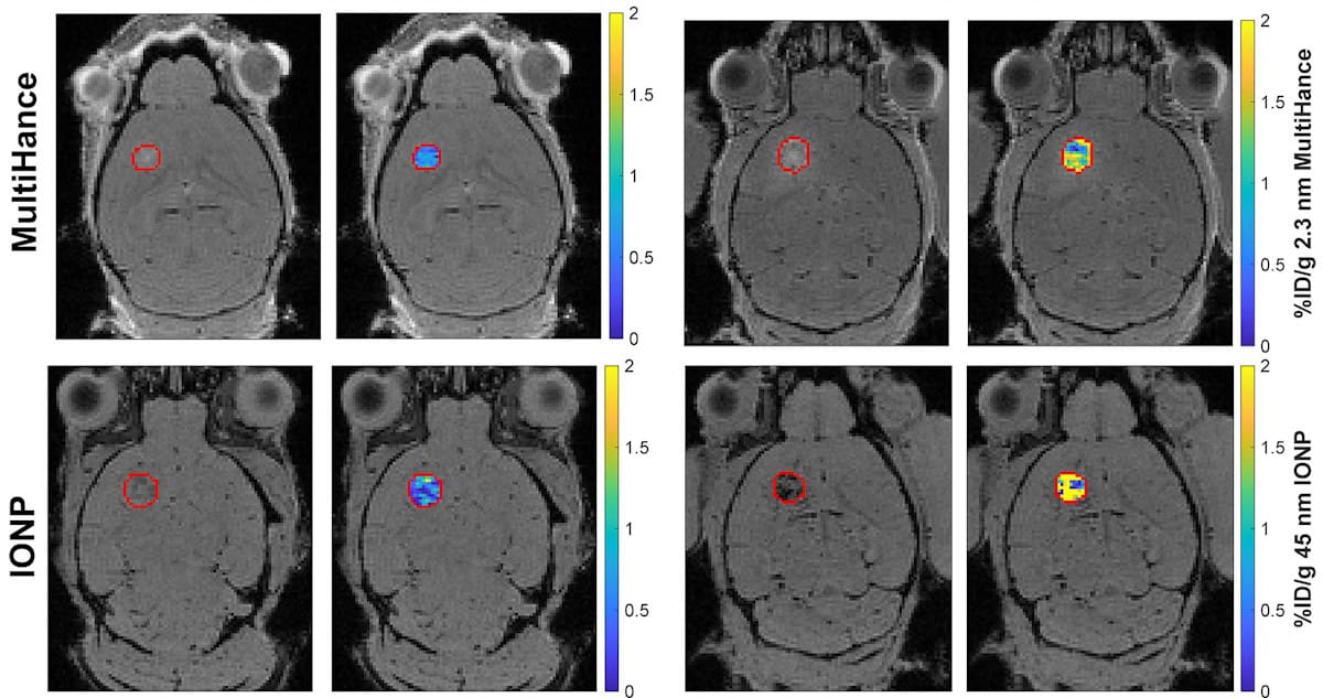
Brain tumour images Representative MR images of baseline (left panels) and FUS-treated (right panels) mouse brains. The top row shows administration of a 2.3 nm contrast agent (MultiHance), the bottom row shows administered iron oxide nanoparticles, 45 nm in size. Each pair of panels shows the anatomic image with the tumour outlined in red on the left, and the corresponding image superimposed with a concentration map showing the amount of nanoparticle retained within the tumour (as a percentage of the injected dose per gram) on the right. (Courtesy: CC BY 4.0/Radiology 10.1148/radiol.251005)

Drug delivery to the brain has been a longstanding challenge in medicine. One option is focused ultrasound (FUS), which can safely disrupt the blood–brain barrier for drug delivery. But how much of a drug gets into the brain depends heavily on its size. A long-standing assumption is that delivery decreases as drug size increases. But by using a clever spin on MRI physics, researcher Matthew Hoch and colleagues from the University of Virginia have shown that “the smaller the better” might not be entirely correct.

The blood–brain barrier is one of biology’s toughest engineering problems. Composed of a dense, tightly sealed layer of cells lining every blood vessel in the brain, the blood–brain barrier only lets nutrients and oxygen through and protects the central nervous system by blocking bloodborne pathogens. In doing so, however, it also blocks the delivery of drugs to the brain. Brain tumours compound this with their own blood–tumour barrier, further hindering drug delivery. For years, researchers have pondered how therapeutics could be delivered past these checkpoints; the answer may simply lie in the use of sound.

The process begins with the injection of gas-filled microbubbles into the bloodstream, where they circulate freely. Exposure to FUS then expands and contracts the volume of these microbubbles, creating a temporary, localized gap that pries the barrier open just long enough for drugs to slip through. Although the technique works, it is unclear how much of a drug actually comes through. In this new study, the team investigate how the size of the drug influences FUS-mediated drug delivery and introduce a novel technique to measure precisely how much drug is delivered.

The measurement problem

Many neurotherapies, with a range of sizes, could benefit from this drug delivery method. But measuring how much of the drug gets past the blood–brain barrier is challenging with current imaging methods. For example, fluorescence imaging is limited by its semi-quantitative nature, while PET scanning suffers from poor spatial resolution.

Standard MRI methods are not only time-consuming, but, in the form of T1-mapping, are typically limited by the size of commercially available contrast agents, which are small in comparison to larger neurotherapeutics used for immunotherapy and gene delivery. Other standard MRI methods sensitive to the presence of iron (T2-mapping) provide an alternative avenue to measure larger agents, but can again be limited by scan time for 3D visualization. Both approaches also have potential to suffer from low sensitivity at low concentrations.

Hoch and his team solve this measurement problem by introducing quantitative susceptibility mapping (QSM), an MRI technique with diverse applications in neuroimaging including evaluation of haemorrhage, iron deposition and calcification. QSM works by exploiting the distortion of local magnetic fields by different materials, a property called magnetic susceptibility. By injecting iron-based nanoparticles that induce measurable, spatially localized shifts in the MRI signal’s phase, QSM can generate a 3D map of magnetic susceptibility. This can then be used to calculate the actual concentration of iron-based nanoparticles within tissues.

By transforming an MRI scanner into a sensitive nanoparticle detector, the researchers present a novel way to quantify drug delivery down to fractions of a percent of the injected dose.

What did they find?

The team tested this approach using four different particles, with a 20-fold range in size. The first was a small gadolinium-based contrast agent, MultiHance (a proxy for small molecule therapeutics), at around 2.3 nm; the other three were iron oxide nanoparticles (IONPs) of 15, 23 and 45 nm, sizes that mirror the dimensions of therapeutic candidates used for immunotherapy and gene therapy.

Using healthy and glioma brain tumour-carrying mice, the researchers used FUS treatment to open the blood–brain barrier and deliver particles of varying sizes. They recorded MRI scans before and after delivery and used QSM to generate a precise measurement of how much of each particle made it into brain tissue.

Brain delivery by particle size
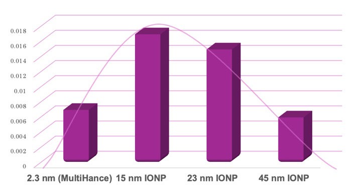
Brain delivery by particle size The amount of drug delivered, as a percentage of the initial injected dose, that has successfully travelled to the brain. (Courtesy: Adapted from Radiology 10.1148/radiol.251005/CC BY 4.0)

The results were unexpected. Drug delivery followed a bell-shaped curve in healthy mice. From the smallest (2.3 nm) particle to the 15 nm IONP, delivery increased 2.6-fold, then at 23 nm, remained steady. Interestingly, at 45 nm, delivery dropped by 2.5-fold compared with the 23 nm nanoparticle, comparable to that of the smallest agent.

This sweet spot, ranging from 15 to 23 nm, demonstrates that smaller isn’t always better. The researchers suspect that this trend in drug delivery is due to a balance between how easily a particle can squeeze through the barrier (so favouring smaller particles) and how long a particle stays in circulation, giving it more time to travel across the barrier (favouring larger particles).

In the mice with brain tumours, where there is an addition of the tumour’s own leaky, disorganized vasculature, the blood–tumour barrier, drug delivery can differ from that to healthy brain. Specifically, this barrier is thought to make drug delivery more difficult than in healthy tissue due to higher tissue pressures and inconsistent blood flow. But the researchers unexpectedly found that FUS boosted delivery of both small and large agents into tumours, with no real difference to the drug delivery levels seen in healthy brain tissue, despite the added presence of the blood–tumour barrier.

“The outcome is exciting because it means that focused ultrasound delivery performance is not expected to diminish in brain tumours,” says senior author Richard J Price in a press statement. “In fact, it may even be enhanced for some types of therapeutics.”

FUS-mediated barrier opening is currently in clinical trials for use in Alzheimer’s disease and glioblastoma, and this research has implications in both drug engineering and treatment planning. By introducing QSM, Hoch and his colleagues have provided a universal, precision technique to quantify contrast agent delivery. Taking existing tools, used for a specific physics measurement problem, and developing them to answer a completely different biological question is no small feat. Here, the researchers present an ingenious intersection of physics and biology, and its potential application in medicine.

The researchers report their findings in Radiology.

The post Breaking in: how ultrasound smuggles drugs past the blood–brain barrier appeared first on Physics World.

Encapsulated islet transplants restore blood sugar control in diabetic mice

27 August 2026 at 15:45
Uncoated and coated cell clusters
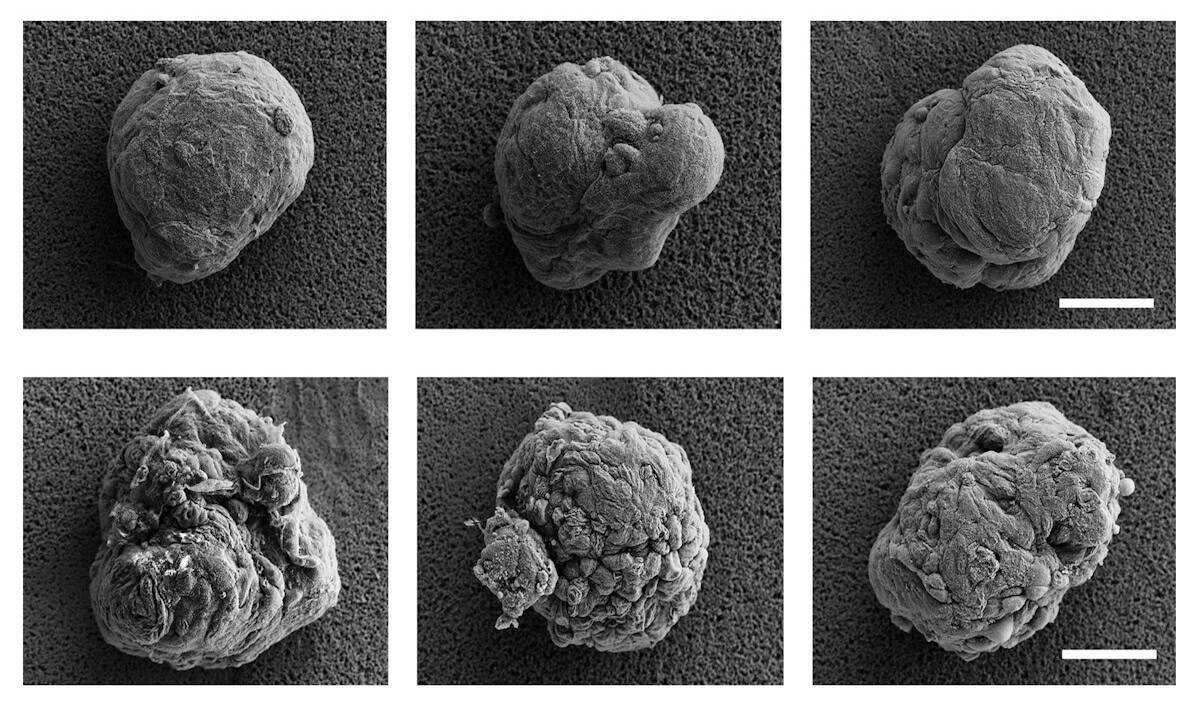
An “invisibility cloak” for cells The top row of photos, taken with a scanning electron microscope, shows uncoated cell clusters (islets). The bottom row shows a series of islets covered in a thin film that hides the donor cells from the body’s immune response, potentially offering a novel approach to treating diabetes. Scale bar: 0.05 mm. (Courtesy: Provided by Kyungsene Lee)

Islet transplantation, in which insulin-producing islet cells from a donor are implanted into a patient’s liver, is a promising therapy for type 1 diabetes. To prevent rejection of the donated cells, however, recipients need to continuously take immunosuppression drugs. Using a new type of cell encapsulation, researchers at The Pennsylvania State University demonstrated that diabetic mice could maintain normal blood glucose levels for 100 days, without requiring immunosuppressants. This represents a substantially longer time period than traditional cell therapies for diabetes.

The Penn State researchers developed their cell encapsulation process based on the molecular characteristics of the zona pellucida, a thin membrane that coats mammalian eggs. Once an egg is fertilized, biochemical reactions lead to hardening of the zona pellucida, which acts as a protective shield during embryo development. Here, biomimetic zona pellucida (BZP) encapsulation serves to hide the donor islets from the body’s immune system.

Pancreatic islets are clusters of five types of cells that work in tandem to regulate insulin and sugar levels in the body and prevent severe insulin deficiency. Islets harvested from healthy pancreases of deceased organ donors were first transplanted into Type 1 diabetes mellitus patients in 1974.

And in 2000, a landmark procedure increasing the mass of islets transferred and using steroid-free immunosuppression regimens was introduced by the University of Alberta in Edmonton, offering the hope of curing diabetes. But when implemented clinically, only 24% of recipients achieved insulin independence at 28 months with the Edmonton protocol.

Research on islet implantation continued unabated, particularly initiatives in islet encapsulation, using semi-permeable membranes to allow the entry of oxygen and nutrients and prevent the entrance of immune response cells. Most current work in this area uses large, 1.5 mm capsules. The Penn State team took an opposite tack: creating a thin 20-µm hydrogel capsule that spontaneously generates on the cell surface with 100% encapsulation efficiency.

Protection from immune attack

Principal investigator Yong Wang and colleagues spent eight years developing an ultrathin hydrogel layer that effectively lays against the curved edge of living cells or cell clusters without impacting their functionality. The islet encapsulation process takes place spontaneously in aqueous solutions under physiological conditions, without exposure to harsh physical, chemical or biological factors, ensuring that BZP development on the cell membrane does not cause any loss of cellular viability or function.

Yong Wang
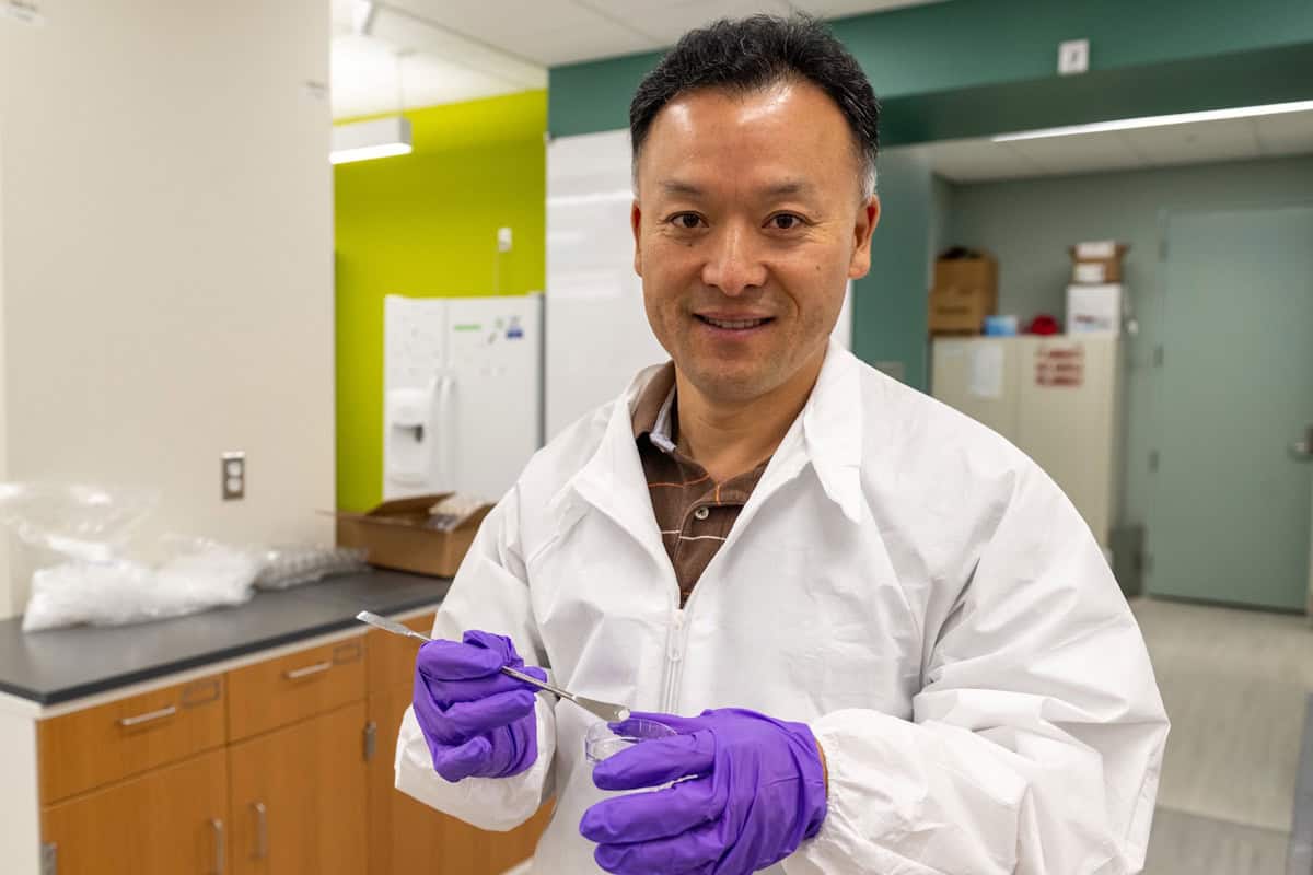
Corresponding author The research is led by Yong Wang, professor of biomedical engineering at Penn State. (Courtesy: Kate Myers/Penn State)

Writing in Nature Biomedical Engineering, the researchers explain that they emulated the biological processes that harden the zona pellucida on a fertilized egg (calcium ion-induced glycoprotein cleavage, assembly and crosslinking on the egg surface). In spite of its thinness, the BZP protects the cell as effectively as the much larger 1.5 mm diameter capsules.

The small capsule size significantly reduces the potential transplantation volume, improving an organ’s ability to receive and accommodate the large number of islets needed to be effective. When coating a 100-µm islet, for example, a 20-µm-thick BZP uses only 1/71 of the polymer volume of a 500-µm microcapsule. The researchers also suggest that the reduced polymer volume used for cell protection by the thin capsule may be a safety advantage of BZP compared to larger capsules.

In vivo assessment

The researchers first examined whether BZP could reduce the level of foreign body response to polystyrene microparticles injected into the peritoneal cavities of mice. Compared with uncoated microparticles, the BZP-coated particles induced a lower immune system response. They examined BZP synthesized using two types of alginate (involved in crosslinking reactions during BZP formation and hardening): chemically modified ultrapure alginate (BZP-U) and regular alginate (BZP-R). Tests in mice verified that BZP-U significantly mitigated the foreign body response compared with BZP-R encapsulation.

Immunocompetent diabetic mice treated with BZP-U-coated islets had their blood sugar restored to healthy levels within seven days. Additionally, the majority stayed diabetes free for over 100 days without continuously needing systemic immunosuppression. By comparison, mice treated with uncoated islets could not maintain healthy blood sugar levels for more than seven days.

In addition to effectively regulating blood glucose levels, one of the biggest advantages of the BZP approach is that it eliminates the need for long-term systemic immunosuppression. Lantidra, the only islet transplantation treatment for Type 1 diabetes cleared by the US Food and Drug Administration, requires continuous immunosuppressants to stop immune system attacks. Such long-term immunosuppressant use can lead to increased risk of infection and other serious health issues, including cancer.

Wang and colleagues plan to further study the BZP approach to better understand the specific duration of resistance each islet transplant may offer, as well as its effect in large animals.

“Large animal studies will be necessary to fully demonstrate their potential in cell transplantation,” the researchers write. “When large animals are used, one needs to examine not only glucose levels but also other key biomarkers. Moreover, the long-term immune responses to BZP and BZP-encapsulated cells warrant further investigation to yield deeper insights into BZP optimization and improvement.”

The post Encapsulated islet transplants restore blood sugar control in diabetic mice appeared first on Physics World.

Artificial blood vessels grow with the help of mechanical forces

12 August 2026 at 09:00

Tissue engineering offers a means to restore, maintain or replace biological tissues. But it is difficult to grow blood vessels using conventional fabrication techniques like 3D printing, as they are typically not precise enough to print fine capillaries and veins. A team of engineers from the Massachusetts Institute of Technology (MIT) has now developed a new approach to do just that, by using mechanical forces. Their blood-vessel-on-a-chip could be the first step in the scalable production of vascular networks that could then be implanted in the body to replace tissue that has been damaged by disease or injury.

The researchers, led by Ritu Raman, made their chip by filling a Petri dish with a gel containing nutrients and cell-growth factors. They then embedded a small magnet into the gel. Finally, they introduced a thin, hollow tube into the gel and coated the inside of the tube with live endothelial cells – the cells that naturally line blood vessels in the body.

Once anchored onto the inner walls of the tube, the endothelial cells began sprouting new, capillary-like vessels in the gel. And when the researchers moved the embedded magnet back and forth in different directions and by varying degrees, they found that the mechanical forces created by the moving magnet could stretch the vessels and increase the number of new capillaries that grew, as well as their length.

‘Steering’ blood vessel growth

“We also showed that blood vessel growth, or angiogenesis, could be directionally patterned using the mechanical forces created by the magnet and that these directions could change dynamically over time,” explains Raman. “This allows us to ‘steer’ blood vessel growth in all three spatial directions (x, y, z) and make complex geometries (like L-shaped branches) that would not be possible to fabricate using conventional methods.”

The researchers had previously demonstrated that biological cells are highly responsive to mechanical forces in the body, but they did not know how different types of forces – for example, stretching in different directions and at different degrees and frequencies – impacted the 3D structure of blood vessels. The main obstacle lay in developing a coupled magnet system to precisely stretch blood vessels in different directions. To achieve this, they designed a method for mechanically stimulating tissues, dubbed “magnetic matrix actuation”.

In their new work, which is detailed in PNAS, they went a step further and developed a method to accurately yet non-invasively stretch 3D blood vessel tissues they had built in the laboratory.

To better understand the effect of mechanical forces on angiogenesis, the MIT team decided to look into the role of a gene known as PIEZO1, which is just one of the many mechanically sensitive genes in the body. They built on the work of molecular biologist Ardem Patapoutian, who received the 2021 Nobel Prize in Physiology or Medicine for discovering that ion channels in cells open and close in response to mechanical pressure. One such ion channel is PIEZO1, which is regulated by the PIEZO1 gene.

By suppressing this PIEZO1 gene in the endothelial cells they studied, the researchers indeed observed that significantly fewer new blood vessels grew in the gel. This, they say, provides evidence that the applied mechanical forces in their experiment may have been opening the PIEZO1 ion channel in the endothelial cells, so triggering the new blood vessel growth.

“My group has always been interested in engineering artificial tissues that can be implanted in the body to repair damage caused by disease or traumatic injury, but such artificial tissues require integrated networks of blood vessels in order to function,” says Raman. “We hope that our method for precisely fabricating complex networks of blood vessels within engineered tissues will enable the development of such implants,” she tells Physics World.

The post Artificial blood vessels grow with the help of mechanical forces appeared first on Physics World.

❌