Normal view

Proton therapy could improve treatment options for dangerous heart rhythm disorder

5 May 2026 at 12:00

A first-in-human study from researchers at the Mayo Clinic has shown how proton therapy could provide a new treatment option for patients with hard-to-treat ventricular tachycardia (VT), a life-threatening heart rhythm disorder. In the small group of patients examined in this early feasibility study, the treatment led to a 79% reduction in VT episodes.

VT is a type of abnormal heartbeat in which faulty electrical signals in the ventricles cause the heart to beat too quickly, meaning that it can’t pump enough blood around the body. Treatments include antiarrhythmic drugs or the use of catheter ablation to destroy the areas of myocardium (cardiac muscle) responsible for the abnormal signals. Sufferers can also be fitted with an implantable cardioverter-defibrillator (ICD) that automatically delivers a shock to reset the heart’s rhythm during a VT attack.

Some patients, however, don’t respond to conventional therapies, including antiarrhythmic medications and catheter ablations, and ICD shocks can significantly impact quality-of-life. For these cases, cardiac radioablation – which uses external-beam radiotherapy to target the problematic myocardium – is under investigation as an alternative, catheter-free treatment for VT.

Previous clinical studies of cardiac radioablation have employed photon-based irradiation, which can expose surrounding cardiac tissue to low-to-moderate radiation doses. Beams of protons, on the other hand, deposit almost all dose at a defined depth (the Bragg peak) and could enable more precise targeting with reduced irradiation of nearby healthy tissue.

“The main motivation for investigating cardiac radioablation is to improve upon the limitations and suboptimal outcomes of catheter ablation of VT in some patients,” explains lead investigator Konstantinos Siontis. “The motivation specific to protons is the potential dosimetric advantage, allowing more precise myocardial targeting while minimizing radiation to surrounding cardiac and extracardiac structures compared with photons.”

In this new study, reported in Heart Rhythm, Siontis and colleagues used proton-based cardiac radioablation to treat seven patients with advanced cardiomyopathy (disease of the heart muscle) and recurrent VT despite drug treatment and previous catheter ablations.

First-in-human investigation

To define the target myocardium for radioablation, the team integrated data from multiple imaging modalities (primarily MRI, plus CT) with information from electrocardiogram (ECG) and electrophysiology mapping originating from the patient’s prior invasive ablation procedures. The CT images were then used to contour the target and organs-at-risk (OARs) and for treatment planning.

The researchers designed treatment plans to deliver a single 30 Gy fraction of expiration-gated intensity-modulated proton therapy to the cardiac internal target volume (ITV, the target myocardium expanded to include cardiac motion) while sparing surrounding OARs. They point out that, due to safety uncertainties in thisfirst-in-human study, they took a generally conservative approach to target definition. In all patients, at least 90% of the ITV received 100% of the prescription dose, while a median of 96.2% of the ITV received at least 95%. Importantly, only 4.3% of non-target myocardium received a dose of 20 Gy or above.

VT episodes before and after treatment
VT control A: VT episodes before and after treatment until each patient’s end of follow-up. B: the median number of VT events per patient in each time interval. (Courtesy: K C Siontis et al. Heart Rhythm 10.1016/j.hrthm.2026.04.021)

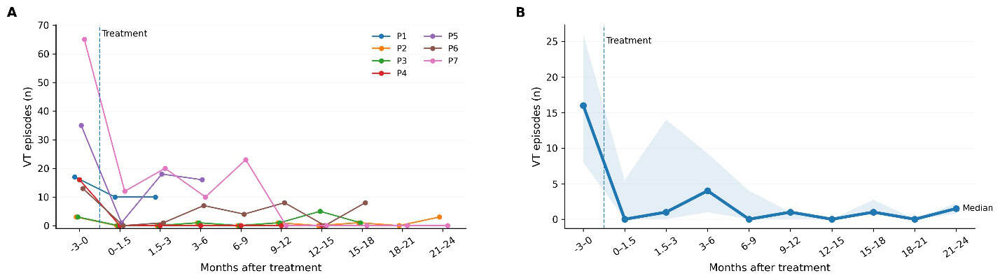
After treatment, the investigators performed follow-up evaluations for up to two years (median 514 days). Most patients experienced recurrent VT during this time, although less frequently than before the radioablation. Across all patients, the rate of VT events declined from 7.24 per patient-month in the three months before treatment to 1.52 per patient-month afterwards – corresponding to a 79% reduction in VT event rate. None of the group experienced serious treatment-related side effects and key heart function measures remained largely stable.

All patients in this study had advanced structural heart disease with severely reduced ventricular function and recurrent VT, putting them at high risk of both arrhythmic and heart failure-related mortality. In line with this profile, two patients required heart transplantation (at 66 and 514 days after treatment) and three died (at 155, 502 and 529 days), due to progressive heart failure.

“This early feasibility study demonstrates that proton cardiac radioablation for refractory VT can be safely planned and delivered with encouraging reductions in arrhythmic burden and no clear treatment-related toxicity,” the researchers conclude. “These findings support the feasibility of proton-based cardiac radioablation and justify further investigation,” they write.

Siontis notes that alongside the emergence of cardiac radioablation techniques, catheter ablation tools are also constantly improving. “Radioablation is unlikely to replace catheter ablation broadly, but it could become an important complementary or salvage option for patients with refractory VT who are poor candidates for invasive procedures,” he tells Physics World.

The team is now planning a larger prospective trial to better define the safety, efficacy and optimal targeting. “We are also investigating improved radiation delivery techniques, such as optimizing treatment planning around cardiac motion,” says Siontis. “In parallel, we continue to investigate photon radioablation in a pivotal randomized trial (RADIATE-VT), while we also offer proton therapy as a compassionate use option for patients in need in our clinical practice.”

The post Proton therapy could improve treatment options for dangerous heart rhythm disorder appeared first on Physics World.

Stanford Medicine unveils world’s first ultracompact proton therapy facility

10 April 2026 at 11:00

Stanford Medicine has opened a new proton therapy facility – featuring an ultracompact treatment system that’s small enough to fit in a room the size of a conventional linear accelerator vault.

Proton therapy is an advanced cancer treatment that offers precise tumour targeting while minimizing dose to healthy tissues. The technique is particularly beneficial for treating tumours located near critical structures and for treating cancers in children. Currently, however, access to proton therapy is limited by its high costs and substantial space requirements.

The new treatment facility – opened earlier this week at Stanford Medicine Cancer Center in Palo Alto, CA – incorporates the S250-FIT proton therapy system from Mevion Medical Systems, the most compact cyclotron in the industry. But even with a much small accelerator, proton therapy delivery usually requires a bulky gantry that rotates around the patient to aim the proton beams at the optimal treatment angles. As such, most proton facilities need a whole new multi-storey building to be built just to fit everything in.

To eliminate this obstacle, the Stanford facility is using a positioning system from Leo Cancer Care to deliver protons via a novel approach known as upright radiotherapy. Here, the patient is treated in an upright position (rather than lying down) and rotated in front of a static treatment beam, removing the need for a gantry and slashing space requirements and installation costs.

Installation of the compact cyclotron
Mafe to fit Installation of the compact cyclotron. (Courtesy: Stanford Medicine)

By combining these advanced technologies, the new equipment fits into a standard 1200 sq. ft linear accelerator vault (as used for standard X-ray-based radiotherapy) and was installed without having to construct a new building.

The advanced system also incorporates built-in CT scanning, enabling extremely precise targeting of tumours within patients with minimal collateral damage to the rest of the body.

“Developing this novel approach to proton therapy at Stanford Medicine, in collaboration with our industrial partners Mevion and Leo Cancer Care, gives us an important additional tool to treat our patients in a personalized, case-by-case way,” says Billy Loo, professor of radiation oncology and co-director of particle therapy at Stanford Medicine. “We are excited to pioneer this world’s first ultracompact and efficient technology that will benefit not only patients at Stanford but expand access to proton therapy worldwide and improve patient outcomes.”

“This milestone really marks the transition from concept and theory to clinical reality,” adds Leo Cancer Care’s CEO Stephen Towe. “Proton therapy installed inside a linac vault always felt like an impossible goal – our partnership with Stanford and Mevion has made that vision possible.”

Loo tells Physics World that patient treatments on the new proton therapy system are likely to start this summer. “As with any first-of-its-kind system in medicine, introducing this complex technology requires a rigorous process of testing and optimization to ensure it meets our high standards for patient safety and treatment quality,” he explains. “We are moving through these steps now.”

The Stanford Medicine team emphasize the particular advantages of proton therapy for children, not least that it can really decrease the radiation dose delivered to normal tissues. Minimizing irradiation of sensitive developing tissue can dramatically reduce the risk of long-term side effects. In addition, treating children while they are sitting up and actively engaged may be far less intimidating for them than having to lie down and have the treatment “happen to them”.

Ribbon-cutting ceremony at Stanford Medicine Cancer Center
Official launch The proton therapy facility at Stanford Medicine Cancer Center was opened on 7 April with a ribbon-cutting ceremony. (Courtesy: Steve Fisch, Stanford Medicine)

The first proton treatments will likely be “cranial and head-and-neck sites, for both adults and selected paediatric patients, for which we already have established patient positioning solutions,” says Loo. In parallel, the radiation oncology team will develop the workflows and immobilization solutions for all other anatomic sites.

The team also plans to investigate new ways to advance the technology and explore the clinical advantages of delivering upright radiotherapy. For example, evidence suggests that for some diseases, such as lung cancer, upright treatment puts the targeted organ in a more favourable position to irradiate safely. Upright positioning also provides greater flexibility to deliver radiation from many different angles. The team will also study the impact of upright positioning on FLASH treatments, in which radiation is delivered at ultrahigh dose rates.

Looking ahead, nine other medical centres are installing this new ultracompact proton therapy system, ultimately making proton therapy increasingly accessible to patients around the world.

“The clinical data to support the use of protons is stronger than ever before,” says Towe. “The strength of this data, combined with the cost reductions delivered by Leo’s technology, has sparked a new wave of growth for protons globally.”

The post Stanford Medicine unveils world’s first ultracompact proton therapy facility appeared first on Physics World.

❌