Normal view

Received today — 3 June 2026 Google News Content : ScienceAlert : The Best in Science News and Amazing Breakthroughs

Experimental Brain 'Pacemakers' May Rewire Circuits Linked to Depression

Every year, more than 2 million people in the United States are diagnosed with treatment-resistant depression.

Desperate for solutions, some brave patients are now volunteering to undergo surgery to place experimental 'pacemakers' into their brains.

These implanted electrodes are part of a treatment known as deep brain stimulation, which is currently used to address some cases of Parkinson's disease and epilepsy.

Now, clinical trials are starting to test if the therapy can treat severe cases of major depressive disorder, too.

The initial results are promising, albeit inconsistent.

In 2021, a patient treated with one of these brain pacemakers said that after the surgical procedure, her depressive symptoms disappeared abruptly.

"I wasn't sure if it would last," she reported at the time. "But it has… "

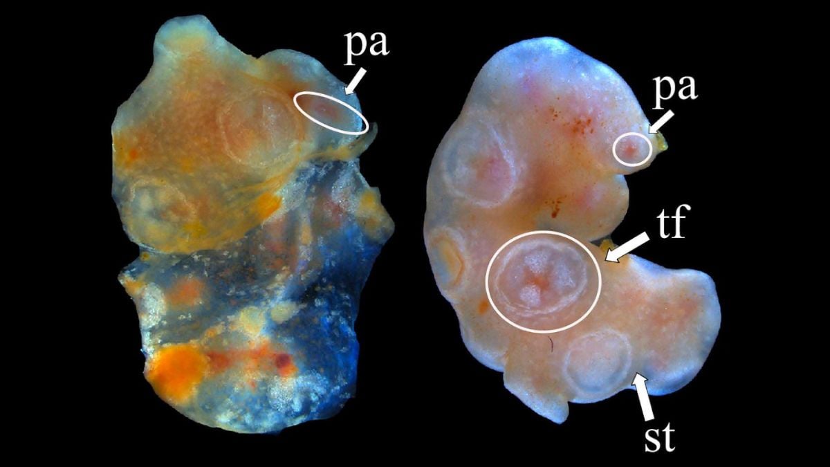
Now, neuroscientists at the Icahn School of Medicine at Mount Sinai have used the brains of three monkeys to show how this therapy might exert such lasting effects.

It appears to restructure key brain regions involved in depression.

"What is exciting about our findings is that they change how we think about deep brain stimulation," says neuroscientist Peter Rudebeck.

"For the first time, we show that deep brain stimulation does not simply alter electrical activity in the brain in the short term; it can actually remodel white matter structure, essentially rewiring brain circuits associated with depression."

YouTube Thumbnail

Whether deep brain stimulation can trigger similar white matter changes in human brains remains to be seen. But these signs in a close primate relative are telling.

White matter in the brain contains nerve fibers, the 'arms' of neurons, which are protected in a fatty sheath called myelin. This protective layer helps conduct electrical messages between brain cells more quickly and efficiently.

Patients with depression typically show a decay of white matter in their brains.

While it is unclear if this association between depression and white matter has anything to do with behavioral symptoms, the link keeps showing up in study after study.

In monkeys, researchers at Mount Sinai have found that deep brain stimulation increases myelination of brain cells in brain regions involved in mood regulation.

Long tubes with pink insides
Illustration of myelinated nerve fibers. (Science Photo Library/Canva)

The therapy also changes the way that neurons interact across various other brain networks, "most notably the default mode network that has been implicated in depression," the authors write in their published paper.

An overactive default mode network is linked to depression.

"Overall," the team concludes, "our data indicate that white matter remodeling as well as selective changes in multiple brain networks may contribute to deep brain stimulation's therapeutic efficacy."

To this day, no one knows why depression arises, or why its symptoms vary so widely from person to person, though there are some known risk factors.

Many standard treatments for depression are based on hypotheses about what causes the mental health disorder, such as a lack of serotonin in the brain.

For up to a third of patients with major depressive disorder, however, standard treatments like antidepressants or therapy don't seem to work.

Until recently, electroconvulsive therapy has been one of the only available alternatives.

This treatment involves electrically stimulating the brain to trigger controlled seizures under anesthesia, and it seems to be very effective at treating episodes of mental illness. But it is not necessarily a long-term solution.

It also comes with risks and negative side effects, such as nausea, headache, fatigue, confusion, and temporary memory loss, and it doesn't work for everyone.

That's why some researchers are turning to deep brain stimulation.
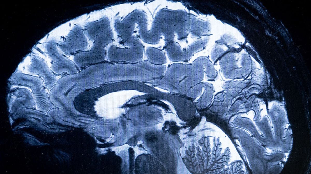
Subscribe to ScienceAlert's free fact-checked newsletter

A brain implant that works sort of like a neurological 'pacemaker' could be a more precise alternative to electroconvulsive therapy.

Once the device is implanted in the brain, it sends high-frequency electrical pulses, usually without the patient feeling the stimulation.

For cases of epilepsy or Parkinson's, deep brain stimulation targets gray matter, or the bodies of neurons, in parts of the brain involved with motor control.

But for depression, the best results in clinical trials so far tend to be when the implants target white matter.

One potential target has been white matter tracts adjacent to the subcallosal anterior cingulate cortex, an area implicated in mood regulation.

"Previously, it was not clear how deep brain stimulation affected brain structure and function," explains neurologist Helen Mayberg.

But research on monkeys is changing that.

"This study addresses a major gap in our understanding and points to an unappreciated mechanism contributing to sustained long-term recovery," adds Mayberg, "something we have observed in our deep brain stimulation depression clinical research over many years."

Related: A Common Arthritis Drug Appears to Work When Antidepressants Don't

Researchers at Mount Sinai were some of the first in the US to test how deep brain stimulation might treat depression.

Their follow-up research among monkeys is now digging deeper to figure out what may be driving these symptoms in the brain.

"Now that we know deep brain stimulation can drive structural plasticity in white matter, we can begin thinking about how to optimize stimulation approaches and potentially develop novel therapies that target these mechanisms through nonsurgical means," says Mayberg.

The study is published in Nature Neuroscience.

ScienceAlert stories are written, fact-checked, and edited by humans, never generated by AI. Don't miss a story, subscribe here.

Received yesterday — 2 June 2026 Google News Content : ScienceAlert : The Best in Science News and Amazing Breakthroughs

Thousands of Brain Scans Reveal A Worrying Consequence of Night Shifts

Night shift work is not for the weak.

Staying awake from dusk through to dawn, as many nurses, doctors, and emergency responders do, seems to take a toll on the body and mind.

But does it have an impact on the brain?

Neuroscientists in Singapore have now found evidence that shift work is tied to brain volume losses in key parts of the brain.

If shift work is stopped, however, those reductions are partially recovered within two and a half years, on average.

What those losses and gains actually mean for human health or behavior is unclear.

A secondary analysis revealed a negative correlation between volume loss and cognitive performance: Increasing volume loss was associated with poorer performance on some, but not all, cognitive tests.

But the effect size is "very small", the authors warn, so the results "should be interpreted cautiously."

That said, there's an important clue in the details. The brain regions that showed significant volume losses also help govern our sleep cycles.

What's more, they are involved in many of the symptoms of shift work, like poorer emotional regulation and memory performance.

Shift workers tend to work long hours. (Thomas Northcut/DigitalVision/Getty Images)

The study is the largest of its kind and finds a change in brain volume where most previous analyses of shift work have not.

It analyzed MRI and long-term health data from 14,198 middle- to older-age adults with no medical issues who took part in the UK BioBank.

Among 2,122 shift workers, the researchers noticed a symmetrical pattern of modest volume loss in the right thalamus, which is part of the brain's information relay 'hub' and is closely involved in memory retrieval.

They also noticed modest volume loss in the left amygdala, which regulates emotional responses.

Brain scans revealed small yet detectable volume loss and white matter degradation among shift workers. (Welton et al., NeuroImage, 2026)

This was after accounting for age, sex, chronotype, and skull volume, among other factors, in their analysis.

"The selective thalamic and amygdalar volume loss observed in healthy shift workers may represent an early, subclinical marker of neural vulnerability linked to chronic circadian disruption," the team concludes, led by neuroscientist Thomas Welton.

"These regions are central to sleep-wake regulation, emotion, and attention, functions that are commonly affected in shift work-related fatigue and mood disturbance."

Challenges with regulating emotions are often tied to poor sleep, and shift workers are known to face higher risks of both sleep disorders and mental health problems.

YouTube Thumbnail

Researchers have long speculated that a disrupted circadian rhythm is to blame.

Other factors that may contribute include a lack of sunlight or changes to eating times.

But just because some parts of the brain are shrinking does not mean they are necessarily dying. The brain is a flexible organ that can rewire itself to meet the challenges of the time.

Perhaps that is what it is doing for shift workers; maybe their brains are somehow compensating in a way that allows them to work through the night.

"It is possible," the authors note, "that individuals who fail to acquire these brain changes are unable to tolerate shift work and are therefore biased toward non-shift working roles."

Subscribe to ScienceAlert's free fact-checked newsletter

The study took place only among older adults, which means it's not clear how the brains of younger workers may cope with the demands of shift work.

Further studies are needed to fully understand how different people respond and are affected.

Today, full-time shift workers make up about 10 to 17 percent of the US population, but by some estimates, roughly a quarter of the adult workforce currently labors during non-traditional hours.

Related: Sleepless Nights Could Drive Half a Million Cases of Dementia in The US Each Year

If this work repeatedly disrupts the body's natural circadian rhythm, it could have a long-term and measurable impact on the brain, but we won't know until those changes are studied further.

"In the "era of longevity", it is critical to understand the relationship between shift work and structure of the middle-older aged brain," Welton and colleagues write.

"The apparent reversibility of these [observed] structural effects within two years of ceasing shift work highlights a potential therapeutic window for prevention and recovery," they add.

The study is published in NeuroImage.

ScienceAlert stories are written, fact-checked, and edited by humans, never generated by AI. Don't miss a story, subscribe here.

Received — 1 June 2026 Google News Content : ScienceAlert : The Best in Science News and Amazing Breakthroughs

A Sea Cucumber's Amputated Tissue Refuses To Die. Could It Live Forever?

The oceans are home to many of Earth's longest living creatures.

Glass sponges can survive for more than 10,000 years, and an individual quahog clam can thrive for more than 500.

A few jellyfish, jellies, and hydra are so good at regenerating themselves that they can theoretically live forever.

But the humble sea cucumber has a truly unique longevity trick.

Scientists in Canada have now discovered a sea cucumber species with tissue that may live 'indefinitely'.

When scientists amputated bits of a scarlet sea cucumber (Psolus fabricii), the tissues refused to die.

A scarlet sea cucumber (Psolus fabricii). (Nozères, Claude/CC-BY-NC-SA 4.0)

For three years and counting, the isolated tube feet and tentacles have sat all on their own in a tank of natural running seawater, without decaying away.

Not only are they not dead, but these tissues are biologically active and changing.

Many of their immune, metabolic, and cellular processes are still intact.

That's never been seen before – not from the tissue of any known animal on Earth.

"We haven't grown a new, complete sea cucumber yet, but we are seeing pretty stunning growth and diversification of cells literally years after this tissue was removed," explains marine biogeochemist Rachel Sipler from the Bigelow Laboratory for Ocean Science, a nonprofit research institute in the US.

"It's like a lizard that loses its tail. We know some lizards can grow new tails; we're talking about whether the tail can grow a new lizard."

Amputated Tissue Sea Cucumber
The amputated tissue of a sea cucumber's 'tube foot' one year after amputation (left) versus several years after (right). The change in overall color from red-orange to lighter white and pink reflect pigmented cells forming and consolidating transparent connective tissue. (Sara Jobson)

Like many lizards on land, the sea cucumber species, P. fabricii, is a bit of a klutz in the ocean. It regularly loses or injures its tube feet and tentacles, which means it has a potentially great capacity for regeneration.

To test that idea in the lab, Sipler and her colleagues at Memorial University of Newfoundland watched and waited to see what happened to excised bits of this wild-looking sea cucumber.

Soon enough, the tissue samples began showing signs of wound repair. Their immune cells appeared to spring into action, and any dead cells were removed.

Repair was then followed by regeneration. Over time, the tissues began to absorb dissolved nutrients from the seawater, growing and restructuring themselves.

Years on, the isolated tentacles can still respond to tactile stimuli, indicating the preservation of a neural network.

This is the first known case of a tissue 'explant' surviving and growing long-term in a natural setting, write Sipler and her colleagues.

"Our findings," they add, "challenge conventional perceptions of tissue immortality."

They also raise the question: What does it mean for tissue to be alive?

Six days after excision (c), the P. fabricii samples contained almost no dead tissue (stained blue), and further imaging more than a year later showed regeneration of tube feet (tf). DPE: days post-excision. (Jobson et al., Sci. Adv., 2026)

For centuries now, scientists have tried to keep the cells and tissues of living animals functional, even when they are removed from the rest of the body.

While researchers have managed to engineer immortal cell lines from animal and human stem cells, these self-proliferating units must be kept in highly controlled environments, where they are carefully guarded against pathogens.

Keeping a whole bunch of cells alive within a section of tissue is much harder to manage.

Animal tissue is a flexible yet delicate structure; it requires a complex scaffold of communicating cells and a robust nutrient delivery system to keep everything plump.

Subscribe to ScienceAlert's free fact-checked newsletter

Even when animal tissue is kept in a special solution to extend its longevity, it typically survives about 9 weeks in the laboratory.

But a bit of P. fabricii could live "indefinitely" in natural seawater, researchers speculate. In fact, it seems to thrive in the natural 'dirtiness'.

"Natural seawater is just about the most microbially diverse, least clean approach we could take experimentally," says Sipler.

"Yet, that rich environment full of bacteria and all this organic matter was actually feeding them and allowing this tissue to heal and grow."

Tube Foot Sea Cucumber
Microscopic image of the excised tube foot with fluorescent staining to show cell differentiation. The denser green colors reflect areas of more active cellular processes. (Sara Jobson)

The only other tissue culture that scientists have described as 'indefinite' was taken from a chicken embryo, and it did not show the same capacity for healing or survival as the scarlet sea cucumber.

In fact, P. fabricii may be unique even among sea cucumbers.

Sipler and her colleagues tested several other sea cucumbers, but none of their tissue explants survived more than 3.5 months.

"Here is this species that has this groundbreaking ability, and we had no idea," says Sipler.

"It's a reminder how much is yet to be discovered in the marine environment."

Related: Mammals May Have a Hidden Limb Regeneration Ability We Never Knew About

Andrea Bodnar, science director at the Gloucester Marine Genomics Institute, was not involved in the study, but she agrees with the paper's conclusions.

"The fact that tissue explants from a sea cucumber can heal, reorganize, and survive independently for years in natural seawater suggests an entirely new model for biological resilience and tissue regeneration," she says.

The study is published in Science Advances.

ScienceAlert stories are written, fact-checked, and edited by humans, never generated by AI. Don't miss a story, subscribe here.

Received — 31 May 2026 Google News Content : ScienceAlert : The Best in Science News and Amazing Breakthroughs

Giant Study Reveals The Secret to Heart Health Isn't Low-Carb or Low-Fat

The key to heart health isn't cutting down on pasta or potatoes, new evidence suggests; it's not even a low-fat diet.

The research suggests the focus of healthy eating shouldn't necessarily be on what's being excluded from your diet (for example, reduced carbs or lowered calories).

Instead, the emphasis should be on what you're actually putting into your body, and the quality of those ingredients.

A study that tracked nearly 200,000 men and women in the US for around 30 years found that some low-fat and low-carb diets are better for heart health than others.

What separates them?

The key was the quality of the food itself, not the quantity of carbs or fats.

The research, led by public health researchers at Harvard University, suggests that if a diet contains too many processed foods and animal proteins or fats, or if it otherwise lacks in adequate vegetables, fruits, whole grains, healthy fats, or essential macronutrients, it may not benefit cardiovascular health as much in the long run, even if it is low carb or low fat by definition.

"Our findings highlighted that it's not simply about cutting carbs or fat, but it's about the quality of foods people choose to construct those diets," concluded Harvard epidemiologist Zhiyuan Wu, who led the research, published in February.

"Focusing only on nutrient compositions but not food quality may not lead to health benefits."

Participants in the study who ate healthy, varied diets with adequate macronutrients showed higher levels of 'good' cholesterol in their blood, as well as lower levels of fats and inflammatory markers compared to those who ate diets lacking in those essentials.

They also had a significantly lower risk of developing coronary heart disease, the most common cause of heart attacks.

Cutting Carbs or Eating Low Fat
The research suggests the focus of healthy eating shouldn't be on what's being excluded from your diet. (fcafotodigital/Getty Images)

"These results suggest that healthy low-carbohydrate and low-fat diets may share common biological pathways that improve cardiovascular health," explained Wu.

"Focusing on overall diet quality may offer flexibility for individuals to choose eating patterns that align with their preferences while still supporting heart health."

YouTube Thumbnail

The findings are based on the self-reported diets of participants, who were all health professionals, so they may have had higher health awareness and better access to health care than the general population.

Related: This Diet Change Cuts Over 300 Calories a Day, Without Decreasing Meal Size

That's somewhat limiting; however, the length of follow-up in the study is impressive, amounting to more than 5.2 million person-years.

The findings join growing evidence suggesting that eating fewer processed foods and more whole grains and vegetables is generally best for a wide range of health outcomes.

Strict diets that count calories, carbs, or fats may not be necessary.

Subscribe to ScienceAlert's free fact-checked newsletter

"This study helps move the conversation beyond the long-standing debate over low-carbohydrate versus low-fat diets," said Yale University cardiologist Harlan Krumholz, editor-in-chief of the Journal of the American College of Cardiology.

"The findings show that what matters most for heart health is the quality of the foods people eat. Whether a diet is lower in carbohydrates or fat, emphasizing plant-based foods, whole grains, and healthy fats is associated with better cardiovascular outcomes."

The study was published in the Journal of the American College of Cardiology.

ScienceAlert stories are written, fact-checked, and edited by humans, never generated by AI. Don't miss a story, subscribe here.

New Wearable Ultrasound Device Could 'Transform' Pregnancy Care

It's hard to imagine pregnancy care without the ultrasound.

Since the 1950s, this incredible technology has provided an essential snapshot into the womb.

Now, scientists are taking it up a notch by trying to provide a continuous window of imaging.

In the coming years, new inventions may allow prospective parents and their physicians to monitor a developing fetus for hours on end, without the need for a traditional handheld ultrasound device or a sonographer standing by.

That sounds like sci-fi, but the proof of concept already exists.

Scientists at the University of California San Diego, Stanford, and Oxford have now invented a wearable ultrasound patch, called UPatch.

Mariana Tome, study co-author and obstetrics doctor at the University of Oxford, thinks the invention could "transform pregnancy care".

"This is the kind of technology obstetrics has been waiting for," she claims.

Like a handheld ultrasound, UPatch sends high-frequency sound waves inside the body to bounce off structures.

Ultrasound is used to periodically monitor fetal growth during pregnancy. A new device could provide continuous monitoring over longer periods. (Maskot/Getty Images)

The returning echoes are then read by special software to capture a real-time view of what's going on inside the body.

UPatch sticks to the skin of the abdomen, where it 'reads' the echoes of red blood cells deep within the vessels of a developing fetus.

It can even accurately measure anatomical features of the fetus, such as the head circumference, abdominal circumference, or femur length, thereby providing an estimated weight.

Most impressively, UPatch does all this autonomously, without the need for a trained sonographer on hand.

The patch needs to be connected to a bulky backend powering system, and it doesn't work when a mother is walking or moving too much, but it is technically hands-free.

"Babies in the womb still cannot be monitored reliably, which is a major gap in maternity care worldwide, with huge implications. Solutions are needed urgently," says Antoniya Georgieva, a reproductive health researcher at Oxford.

"The UPatch technology opens the possibility of monitoring the most important signals of fetal health over much longer periods, gain essential new knowledge of how babies' oxygen supply and wellbeing adapt inside the womb, and ultimately helping clinicians identify problems earlier."

New Ultrasound Patch Could "Transform Pregnancy Care"
An exploded view of the UPatch and how it works. (Park et al., Nat. Biotechnol., 2026)

When researchers tested the patch on 62 pregnancies within a clinical setting, it performed on par with current ultrasound devices.

For one participant, the patch even noticed a dangerous change in blood flow to the fetus, signaling preeclampsia.

"Following the detection of compromised fetal health using the UPatch, the preeclamptic participant underwent intensive monitoring and the baby was delivered by Cesarean section four days later," write the study authors.

During pregnancy, ultrasounds are regularly recommended to monitor the health of both the mother and the child.

In higher-risk pregnancies, where patients are kept in the clinic for longer periods of time, ultrasounds are done multiple times a week.

Each one of those scans, however, takes time and requires a sonographer to use a handheld device to focus on parts of the uterus.

UPatch allows patients to be monitored in bed for hours, without the need for a clinician to move the device's focus or interpret the results in real time.

Movements UPatch
UPatch can image the womb even when a mother moves around. (Park et al., Nat. Biotechnol., 2026)

If UPatch is used in conjunction with classical imaging techniques, then perhaps pregnancy outcomes could be greatly improved, its inventors argue.

"This technology could expand access to prenatal imaging in healthcare deserts and low-resource settings, where shortages of trained sonographers often delay care for high-risk pregnancies," says Tom Park, the main engineer who designed and fabricated UPatch.

After comparing the patch to current ultrasound devices, the researchers then tested the patch continuously for between 1 and 6 hours in 52 pregnant women, including those affected by preeclampsia, gestational diabetes, hypertension, or poor fetal growth.

The findings reveal differences between short-term fluctuations in ultrasound readings and longer-term changes that require closer monitoring.

Subscribe to ScienceAlert's free fact-checked newsletter

Researchers hope the device can help clinicians more readily detect signs of sustained fetal distress, so they can intervene sooner.

The flexible patch is designed with electrodes and an acoustic lens, so that when it wraps around an expectant mother's abdomen, it provides a window to the entire uterus while sitting, standing, or lying down.

Related: Yawning Is So Contagious You Can Catch It Before You're Born, Study Suggests

It can even provide details on how blood flow rates in the umbilical artery compare to those in the fetus's brain.

"This work shows how advances in soft electronics, ultrasound engineering, and clinical science can come together to address one of the most important unmet needs in pregnancy care," says senior author and engineer Sheng Xu from Stanford.

The study is published in Nature Biotechnology.

ScienceAlert stories are written, fact-checked, and edited by humans, never generated by AI. Don't miss a story, subscribe here.

❌