The Universe Should Be Empty — A Particle Physicists Ignored for Decades May Explain Why It Isn’t


Physicists have confirmed the two foundational postulates of Richard Feynman’s path-integral approach to quantum mechanics in a real-world experiment for the first time. This comes nearly 80 years after Feynman first published his path-integral formulation, which describes the time evolution of quantum systems.
Fundamental thought experiments, such as Schrödinger’s cat, the Einstein–Podolsky–Rosen paradox and the quantum double-slit experiment have always been important for developing the theories of quantum mechanics. As technology improves, some thought experiments have been realized in the lab, confirming many aspects of quantum theory. However, thought and real experiments thus far have not addressed the fundamental question: how do quantum systems evolve between initial and final states? This question, explains Shi-Liang Zhu of South China Normal University in Guangzhou, lies at the heart of Feynman’s path integral postulates for his famous propagator equation.
The postulates, first proposed in 1948, have never been directly tested – despite their foundational role in modern physics. Indeed, the path-integral formulation has been critical for the development of fields ranging from quantum field theory to cosmology and has provided a unified framework that connects quantum mechanics with classical physics through the principle of least action. The path integral’s predictions also underpin modern quantum science, from condensed matter physics to quantum statistical physics.
As quantum theory was developed, several “pictures” emerged of how a quantum system evolves with time – including the Schrödinger and Heisenberg pictures. The Schrödinger equation, the Heisenberg equation and the Feynman propagator equation are formally equivalent formulations of quantum mechanics, explains Zhu. “However, while the first two are typically treated as fundamental postulates, the Feynman propagator equation is derived from two underlying postulates. This derivational asymmetry makes experimental tests of Feynman’s postulates particularly compelling.”
The first postulate is that a quantum particle does not travel along a single trajectory when moving from point A to point B; and instead every probable path contributes to its trajectory. The second postulate is that all possible paths have the same probability (or equal-magnitude amplitudes) and differ only by a path-dependent phase factor. This is very different from simply summing all the probabilities as in classical physics.
At time ti, explains Zhu, a particle is localized at xi with its wave function denoted as ψ(xi, ti). The probability of the particle reaching xf at time tf is therefore determined through all possible paths connecting points A at (xi, ti) and B at (xf,tf). The j-th path contributes a probability amplitude ϕj.
Feynman postulated that the probability P of the particle reaching xf at time tf equals the absolute square of the sum of probability amplitudes ϕj for all paths. As mentioned, all these contribute equally in magnitude, with each contribution’s phase being given by the classical action (in units of the Planck constant, ħ).
“We have now directly measured the probability amplitudes ϕj of more than 1.4 million possible paths taken by single photons in an optical system,” says Zhu. “We did this by dividing the region between the starting point A and the end point B into a grid of points. We connected adjacent points by line segments and then measured the propagator for each segment. In total, there are 175 possible paths.”
The results from this experiment, which is detailed in Science Advances, validate the first postulate with a mean absolute percentage error of 4.45% and a fidelity of 94.9%. They also confirm the second postulate with 94.7% fidelity.
The paper’s first author Yong-Li Wen notes: “because there were so many possible paths that the photons could take, the primary challenge in our experiments was to achieve sufficient accuracy to reconstruct millions of path amplitudes, since even marginal errors in individual propagator measurements accumulate multiplicatively, destroying phase coherence and rendering the reconstructed path distribution nearly random.
“We overcame this problem through four key technical advancements: amplifying the signals; designing a customized high-precision imaging system; implementing real-time normalization via a reference beam to correct for photon fluctuation errors; and making sure the set-up was mechanically stable on the nanoscale.”
These innovations improved single-photon propagator fidelity from 87.6% to 98.5%, something that allowed the team to resolve the global structure of path amplitudes with unprecedented precision.
The work establishes a powerful experimental framework for investigating path integrals in quantum systems, Zhu tells Physics World. “By confirming that quantum probabilities arise from path interference and that phases are governed by classical action, we provide experimental evidence supporting the view that quantum paths reflect physical reality rather than being mere mathematical artefacts.”
The high-precision propagator methodology could also be used to study quantum-to-classical transitions and decoherence mechanisms, where understanding the role of interfering paths is essential, he adds. And it could be extended to probe fundamental phenomena such, as entangled histories and indefinite causal order across multiple space–time points.
Beyond fundamental physics, the technique may make quantum simulations of complex condensed matter and quantum field theory phenomena, including instantons and magnetic monopoles, easier. “Perhaps most excitingly, by enabling precise propagator measurements in diverse environments, this framework could eventually be extended to interacting systems and curved space–times, allowing for valuable experiments with artificial quantum systems,” says Zhu.
The post Single-photon measurements confirm Richard Feynman’s vision of quantum mechanics appeared first on Physics World.
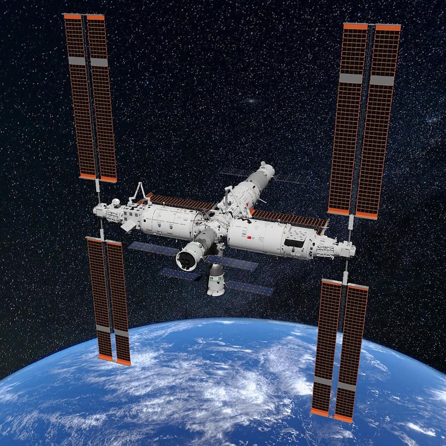
A new quantum test of the weak equivalence principle performed on the China Space Station using cold atom interferometry is the most precise to date, yielding an uncertainty of 2.8 x 10⁻⁸ and a test result of around –2.7 x 10⁻⁷. Meanwhile, a separate team in Israel, Germany and the UK has measured an object’s quantum phase in freefall in the laboratory and confirmed that Einstein’s equivalence principle holds here too.
The weak equivalence principle (WEP), or the “universality of freefall”, is an important pillar of Albert Einstein’s general theory of relativity, in which mass and gravity are intimately linked to the curvature of space–time. The WEP states that, in the absence of other forces, all objects fall with the same acceleration under the influence of gravity regardless of their mass or composition. In other words, gravitational mass (the m in F = GMm/r2, where F is the gravitational attraction between two masses placed a distance r apart and G is the gravitational constant) and inertial mass (the m in F = ma, where a is the acceleration produced by the force F) are the same.
Precisely measuring this principle is crucial since any detected violation could point to new physics. It is also important for continuing to corroborate the theory of general relativity, which may be incomplete because it appears to be fundamentally incompatible with another well-tested theory: quantum mechanics. Today, tests of the WEP using macroscopic bodies have reached extraordinary precision – at the 10−13 level on Earth and 10−15 in space. These latest studies investigate the principle in the quantum realm.
Conventional interferometry works by splitting a coherent beam of light and then recombining the different components at a detector. If the two components are in phase when they recombine, the interference is constructive and the two components reinforce each other. However, if the two components are out of phase, they cancel each other out, leading to a characteristic pattern of bright and dark fringes. Atomic interferometry is similar but relies on beams of atoms rather than beams of light.
Cold atom interferometry (CAI), which is atomic interferometry at ultracold temperatures of just above absolute zero, has found applications in inertial sensing and fundamental physics research. It also turns out to be a powerful technique for testing the WEP.
There is a problem with using CAI on Earth, however, because our planet’s gravity limits the interference time of the atom beams to just a few seconds. Experiments in microgravity allow this time to be extended to minutes, but because the resolution of a WEP test scales inversely with the square of the interference time, even longer interference times are needed to achieve the required precision (10−17 or more). This necessitates a permanent microgravity environment, something that is only possible in space.
In the new work, detailed in Science Advances, a team of physicists led by Mingsheng Zhan from the Chinese Academy of Sciences, Hefei National Laboratory and Wuhan Institute of Quantum Technology sent their experiments to be performed onboard the China Space Station, using the cold atom interferometer installed on its High Microgravity Level Research Rack. This instrument contains clouds of billions of 85Rb and 87Rb atoms that interfere when probed with counterpropagating Raman lasers reflected by a piezo tilt mirror.

By exciting and detecting the fluorescence of the isotopes sequentially at slightly different times, the researchers obtained two sets of symmetric interference images, one from the 85Rb atoms and one from the 87Rb atoms. Measuring the differential phase of these images – after suppressing the residual acceleration of the space station and effects from vibrations – allowed them to calculate the difference in the acceleration between the two isotopes in the vertical, freefalling direction.
After running their experiment for 280 days, during which time the researchers acquired more than 9700 pairs of interference fringes, they obtained a test result of around –2.7 x 10⁻⁷, after correcting for errors.
“We believe that our technique will have a significant impact,” says Zhan. “First, it demonstrates that an integrated interferometer meeting the requirements of in-orbit operation is possible, giving the community confidence to continue with even more technically demanding space-based interferometry experiments that aim for ever increasing precision. The technology developed in this project will also itself help advance atom-interferometry-based instruments, such asinertial navigation systems.”
Meanwhile, physicists at the Ben-Gurion University of the Negev, the University of Ulm and the University of Oxford have also used CAI, this time to measure an object’s quantum phase in freefall. They confirm that the effect is the same as predicted by Einstein’s equivalence principle (EP), which states that for an observer in freefall, gravity should locally disappear.
In their work, also detailed in Science Advances, the researchers used a completely novel version of the CAI technique that they call the Quantum Galileo Interferometer (QGI) – in honour of Galileo Galilei’s discoveries regarding the laws of freefall. The experiment, which was carried out at Ben-Gurion University, again makes use of clouds of 87Rb atoms cooled to just above absolute zero.
The researchers began by applying microwave pulses to put the ultracold atoms – which were placed 113 µm below the surface of an atom chip containing current-carrying wires that produce magnetic field gradients – into a quantum superposition. This meant that each atom effectively travels along two different paths at once. One part of the atomic wave responded to the magnetic fields such that an applied upward force exactly counteracted the downward pull of gravity and the wave was held stationary in the apparatus – that is, static relative to Earth. This part of the wave is referred to as the reference atomic wave (or wavepacket). The second part of the wave was sent upwards in a ballistic trajectory.
Realising this ballistic trajectory, affected only by gravity, required a “magnetic cannon” and a “magnetic parachute”, explains Ron Folman, who is one of the leaders of this study. “After the short-duration magnetic cannon, we switched this part of the wave into a state that is almost unaffected by the magnetic field so that it could fall freely under gravity. And at the end of the fall, we used another precisely controlled magnetic pulse to stop the relative motion between the two waves and reunite the two parts so that they interfered with each other.”
That interference allowed the researchers to measure the tiny difference in quantum phase accumulated while one was falling and the other was held still — two frames that may be referred to as the Einsteinian and the Newtonian frames, respectively. This means that when both parts of the wave have the same velocity, the transformation between the wavefunction in a Newtonian (laboratory) frame of reference and the wavefunction of the same object in an Einsteinian frame that freefalls with the same acceleration, involves a transformation phase known as a gauge phase.
According to their experiments, the gauge phase measured by the researchers is the same as the one predicted when Einstein’s EP is applied to such a quantum wave, so confirming the principle for these quantum objects and at these low masses and energies.
“Some members of our team,” notes Folman, “including Nobel laureate Sir Roger Penrose, expect this co-existence between general relativity and quantum mechanics to break for objects with higher masses. Indeed, the Ben-Gurion University group is now repeating the same experiment with nanodiamond particles to test this hypothesis,” he tells Physics World.
The observation, says study co-leader Vlatko Vedral at Oxford University’s department of physics, constitutes a fundamental test of the interface between quantum theory and gravity. “The connection between these two pillars of modern physics remains one of the most important open questions in physics, and it is thus of paramount importance to understand the phenomenon of freefall and the EP in the quantum domain.”
The post Einstein’s equivalence principle put to two new tests appeared first on Physics World.
Almost a hundred years ago, physicists theorized out what free fall should do to a quantum wave. If the solution they came up with is wrong, quantum mechanics and Einstein's theory of gravity flatly contradict each other. But testing it has been impossible because nobody has managed to build an interferometer that could perform the necessary measurement.
Now, a team led by Ron Folman, a physicist at Ben-Gurion University of the Negev, with collaborators in Germany, the UK, and the US, including Nobel laureate Roger Penrose, has done it. They built a new interferometer that gives a single atom two possible paths at once: one that involves a free fall, and another where it is held perfectly still. Both paths end at the same place at the same moment, allowing the team to measure what the fall does to a wave-like property of the atom.
Ever since Galileo, physicists have known how to describe a falling object—where it is, how fast it goes, or how quickly it accelerates. Quantum mechanics, though, insists that every object is also a wave. “Every particle, doesn't matter if it's a car or a spaceship or an atom, is a wave,” Folman says. “Everything that is a wave, like sea waves or sound waves, goes up and down. And if you're up or down, this is measured by something called a phase. A phase just tells you if you are at the top of the wave or at the bottom.”


© ALIOUI Mohammed Elamine
In a finding that could reshape how ophthalmologists think about one of the most delicate complications of corneal surgery, a team of computational engineers has built the first detailed computer model of a human eye in which the Descemet membrane—the paper-thin basement layer at the back of the cornea—has become detached. By simulating the fluid–structure interaction between the aqueous humor sloshing inside the eye’s anterior chamber and the flexible, detached membrane itself, the researchers have produced quantitative predictions about the mechanical stresses such a detachment imposes, and about how different transplanted corneas might alter those stresses during post-operative thermal therapy.
The study, published in the Annals of Biomedical Engineering, addresses a long-standing blind spot in ocular biomechanics. Detachment of the Descemet membrane is a recognized complication of cataract surgery, corneal transplantation and certain corneal dystrophies, and it can cause significant corneal swelling and vision loss if the membrane does not reattach. Yet most previous computational examinations of the anterior chamber have treated the eye’s interior as rigid-walled plumbing, focusing on fluid flow or heat transfer while excluding the complex two-way coupling between the intraocular fluid and the fragile ocular structures suspended within it. The new work changes that by modeling the detached Descemet membrane as a deformable, elastic solid that bends, stretches and deforms under the pressure and shear of the aqueous humor flowing around it.
The researchers, led by Ammar I. Alsabery of the Islamic University in Najaf, Iraq, together with colleagues from the University of Basrah, King Khalid University in Saudi Arabia, Saveetha School of Engineering in India, and Universiti Kebangsaan Malaysia, employed a coupled numerical scheme that solves simultaneously for the buoyancy-driven flow of aqueous humor and for the structural deformation of the membrane. The geometry represents the anterior chamber—the fluid-filled space between the cornea and the iris—with a warm region corresponding to the lens contact zone held at an isothermal hot temperature, mimicking the thermal gradients that arise in ocular warming therapies and in normal physiological conditions.
At the heart of the analysis is the Rayleigh number, a dimensionless parameter that governs the strength of natural convection: warmer, less dense fluid rises while cooler fluid sinks, generating circulating currents within the chamber. The team varied the Rayleigh number across three orders of magnitude, from 10³ to 10⁶, which corresponds to temperature differences spanning from a fraction of a degree to 250 Kelvin. This range deliberately encompasses both the mild thermal gradients present in the living eye and the much more aggressive temperature differences applied during clinical thermal therapy of the cornea, allowing the same model to speak to both resting physiology and active treatment.
Equally central to the study is the elasticity modulus of the detached membrane. The researchers swept this property across nine orders of magnitude, from 10⁸ to 10¹¹, a range designed to represent everything from highly compliant, freshly detached tissue to the much stiffer mechanical character of donor corneal tissue used in transplantation. Because the Descemet membrane’s stiffness varies with age, disease state and surgical history, this parametric approach lets the model answer a question that clinicians genuinely care about: does the type of cornea grafted into a patient change the mechanical load experienced by a detached membrane during recovery? The answer, according to the simulations, is nuanced. The cornea type does affect the stress imparted on the detached Descemet membrane, but it does not affect the thermal characteristics of the anterior chamber or the overall displacement behavior within it. In other words, stiffer transplanted tissue changes the mechanical environment at the membrane itself without appreciably altering the convective heat transfer patterns of the aqueous humor circulating beneath it.
The fluid dynamics of the anterior chamber turned out to be far more sensitive to geometry than to tissue stiffness. One of the most striking results concerns the iris, the pigmented diaphragm whose length in the model determines the extent of the hot, isothermal contact region associated with the lens. When the team simulated a shrinkage of the iris—effectively reducing the length of the contacting part of the lens held at the hot temperature—they found that the convective thermal characteristic of the anterior chamber rose by approximately 78 percent. This dramatic enhancement arises because the reduced iris length reshapes the buoyancy-driven circulation cells, allowing warmer fluid to travel more freely and exchange heat more efficiently with its surroundings. For clinicians, the implication is that the geometry of the anterior chamber, shaped in part by the iris and the lens, is a first-order determinant of how heat is distributed during thermal therapy, potentially more important than the material properties of the graft itself.
The temperature difference applied during thermal therapy also proved to be a powerful lever. Raising the applied thermal gradient from 2.5 Kelvin to 25 Kelvin—a tenfold increase—promoted heat transfer within the anterior chamber by 47.6 percent. While the scaling is sublinear rather than directly proportional to the temperature difference, the result confirms that modest increases in therapeutic temperature can yield meaningful gains in convective heat delivery without requiring a hundredfold jump in applied thermal load. The finding offers a quantitative framework for calibrating thermal treatments of the anterior segment: therapy protocols could, in principle, be tuned to achieve target heat transfer rates while respecting the narrow thermal safety margins of living ocular tissue.
Technically, the simulations belong to a class of problems known as fluid–structure interaction, or FSI, in which the fluid equations and the solid mechanics equations are coupled and solved together rather than in isolation. In this case, the aqueous humor—an essentially water-like fluid—drives natural convection under the influence of gravity, while the detached Descemet membrane responds to the pressure and viscous stresses exerted by that flow. The membrane’s deformation, in turn, modifies the shape of the fluid domain, feeding back into the flow field. This two-way coupling is computationally demanding but essential, because a rigid-wall approximation would entirely miss the bending and displacement of the detached membrane, along with the localized stress concentrations that could determine whether the membrane reattaches, tears further, or remains stable during healing. The methodology builds on the team’s earlier FSI studies of flexible fins, baffles and partitions in convective systems, as well as their prior work applying FSI modeling to blood flow in abdominal aortic aneurysms under thermal treatment, adapting those engineering tools to the peculiar geometry and physiology of the human eye.
The choice to represent the detached Descemet membrane as a flexible structure rather than a fixed boundary is what gives the study its clinical bite. Prior models of aqueous humor flow, including foundational work on fluid flow in the anterior chamber of the human eye published decades ago, captured the buoyancy-driven currents but assumed static, impermeable walls. By letting the membrane move, the new simulations capture the interplay between hemodynamics-like fluid loading and tissue compliance—precisely the interplay that governs how a detached membrane flutters against the aqueous humor, how stresses concentrate at its edges and attachments, and how different graft stiffnesses redistribute those stresses across the corneal structure. The team’s results suggest that surgeons and biomaterials scientists selecting corneal grafts for patients prone to Descemet membrane detachment could use stress predictions of this kind to weigh graft stiffness as part of the decision, even though the overall thermal environment of the chamber will remain largely unchanged.
Beyond transplantation, the model has direct relevance to thermal therapy of the eye, a class of treatments in which controlled warming is applied to manage conditions of the anterior segment. Because the simulations connect an easily controlled clinical variable—the applied temperature difference—to a measurable outcome in convective heat transfer, and because they quantify the role of iris geometry in shaping the thermal flow field, they provide a template for patient-specific treatment planning. A clinician armed with imaging of a patient’s anterior chamber geometry could, in principle, use models of this type to predict how a planned thermal protocol will distribute heat in the presence of a detached membrane, and whether the resulting stresses on the membrane fall within tolerable limits.
The work is not without its simplifications, as the authors themselves frame the study as an evaluation of corneal transplantation processes rather than a complete clinical simulator. The anterior chamber geometry is idealized, the aqueous humor is treated with standard transport properties, and the elasticity modulus range, while broad, samples the problem in parametric steps rather than matching every individual patient’s tissue. Real eyes also feature aqueous production and drainage, blinking, saccadic motion and ocular pulse effects that a steady natural-convection FSI model does not capture. Nonetheless, the paper’s central contribution—demonstrating that corneal stiffness governs membrane stress while iris geometry and thermal gradient govern heat transfer—stands as a clean separation of effects that future, more detailed models will need to reproduce.
The research was supported by the Universiti Kebangsaan Malaysia Research Grant GP-2024-K006388 and by the Deanship of Research and Graduate Studies at King Khalid University through a large Research Group Project. As computational biomechanics continues to migrate from engineering journals into clinical planning, studies like this one mark a shift toward treating the eye not as a static optical instrument but as a living, deformable, fluid-filled machine—one in which a membrane thinner than a human hair can dominate the mechanical conversation, and where the difference between a compliant and a stiff cornea may one day inform the surgeon’s graft choice.
Article Title: Fluid–Structure Interaction Analysis of Human Eye with Descemet Membrane Detachment
Article References: Alsabery, A. I., Ismael, M. A., Raizah, Z., Ghalambaz, M., & Hashim, I. (2026). Fluid–Structure Interaction Analysis of Human Eye with Descemet Membrane Detachment. Annals of Biomedical Engineering. https://doi.org/10.1007/s10439-026-04317-0
Image Credits: AI Generated
DOI: 10.1007/s10439-026-04317-0
Keywords: Descemet membrane detachment, anterior chamber flow, fluid–structure interaction, human eye, cornea transplantation, natural convection, thermal therapy, Rayleigh number, elasticity modulus, aqueous humor, corneal biomechanics, numerical simulation
In a development that could reshape how scientists study the mechanics of human movement, a team of biomedical engineers and computer scientists has unveiled an open-source software tool capable of tracking the tiny, intricate bones of the wrist as they glide and rotate during motion. The new tool, called Hierarchical 3D Registration, or 3DH, is a module within the SlicerAutoscoperM extension of the widely used 3DSlicer computing environment. Described in a study published in BioMedical Engineering OnLine, the software was validated against previously established methods using both sequential three-dimensional computed tomography, or 3DCT, scans of living participants performing thumb tasks and dynamic four-dimensional CT, or 4DCT, images of cadaveric wrists moving through flexion and extension. The results demonstrate that the open-source approach can match the accuracy of independent, previously published methodologies while offering researchers everywhere a free and reproducible path to quantifying skeletal kinematics.
The wrist has long been one of the most challenging joints for biomechanists to study. It contains eight small carpal bones, including the scaphoid and lunate, along with the radius of the forearm and the metacarpal bones of the hand, all articulating in a tightly packed arrangement that allows the remarkable range of motion humans rely on every day. Understanding how these bones move relative to one another, a field of study known as arthrokinematics, is essential for unraveling the causes of wrist instability, osteoarthritis, ligament injuries and the long-term outcomes of surgical repairs. Yet measuring those movements in living, moving joints has historically required either invasive techniques, labor-intensive manual analysis or proprietary software that few laboratories can access or verify.
Quantitative tracking of skeletal structures in vivo, with both efficiency and high temporal resolution, matters to a wide range of questions in biomechanical research, and the authors of the new study argue that the field has been held back by a shortage of accessible, semi-automated registration tools. Image registration, the computational process of aligning one three-dimensional image volume to another, lies at the heart of motion tracking from CT data. When a bone moves between two scans, its position and orientation change, and a registration algorithm must find the transformation that best overlays the bone’s image from one time point onto the next. Done well, this yields precise measurements of rotation and translation. Done poorly or inconsistently, it introduces errors that can swamp the small movements of carpal bones, which may shift by only a few millimeters and a few degrees during normal function.
The new 3DH module addresses this problem with a hierarchical strategy for registering bones across sequences of image volumes. The researchers presented and evaluated the software using two distinct classes of data drawn from previous studies. The first consisted of sequential 3DCT datasets of human participants performing various thumb tasks, in which the radius, the first metacarpal and the trapezium were the target bones. The second consisted of dynamic 4DCT datasets capturing simulated flexion-extension motion in cadaveric wrists, providing time-resolved, three-dimensional image volumes of the scaphoid and lunate in motion. By testing the tool on both static sequential scans and true dynamic imaging, the team was able to demonstrate that 3DH is not limited to a single imaging paradigm but can serve as a unified tracking solution across modalities.
To determine whether the new open-source tool could be trusted, the investigators compared the arthrokinematics computed with 3DH against values that had been previously calculated using independent approaches for the same 3DCT and 4DCT datasets. The statistical framework for this comparison was the Bland-Altman analysis, a standard method for assessing agreement between two measurement techniques. Rather than simply correlating the two sets of results, Bland-Altman analysis quantifies the mean bias, the systematic difference between methods, and the 95 percent limits of agreement, the range within which most individual differences between the methods are expected to fall. This approach is particularly well suited to method-comparison studies because it reveals whether a new technique introduces systematic error and how widely individual measurements might deviate from those of the established gold standard.
The performance figures reported in the study are striking. For the 3DCT data, in which the radius, first metacarpal and trapezium were tracked, the mean bias for the helical angle, a single-axis description of three-dimensional rotation, was just minus 0.06 degrees, with 95 percent limits of agreement ranging between minus 1.87 degrees and 1.74 degrees. Translation measurements fared nearly as well: the mean bias was 0.12 millimeters, with limits of agreement spanning from minus 2.23 millimeters to 2.00 millimeters. In practical terms, the open-source tool reproduced the rotational and translational measurements of the earlier methods to within roughly two degrees and two millimeters at the extremes, differences small enough to preserve the biological signal in most wrist kinematics experiments.
The dynamic 4DCT results told a similar story for the carpal bones. Tracking the scaphoid and lunate through simulated flexion-extension, 3DH produced mean biases of the Euler angle rotations, which decompose three-dimensional rotation into three orthogonal components, of less than 0.77 degrees about any direction. The widest 95 percent limits of agreement ranged between minus 7.11 degrees and 8.65 degrees. For translations, mean biases were less than 0.5 millimeters in any direction, with the widest limits of agreement ranging between minus 2.36 millimeters and 1.43 millimeters. The wider limits of agreement for rotations in the dynamic data likely reflect the added challenges of time-resolved imaging, including motion blur and the smaller, more complexly shaped carpal bones, but the near-zero mean biases indicate that the tool does not systematically drift away from established values.
Crucially, all target bones were successfully tracked in both the 3DCT and 4DCT datasets, meaning the hierarchical registration algorithm converged on correct solutions in every case tested. This robustness matters because registration algorithms can occasionally fail, locking onto incorrect alignments or diverging entirely, and such failures typically demand time-consuming manual intervention. A semi-automated tool that reliably succeeds across diverse bones, motion tasks and imaging protocols removes a significant bottleneck. Manual bone tracking in CT volume sequences can take hours per bone per trial, and the labor multiplies rapidly in studies involving multiple participants, multiple bones and dozens of motion cycles. By automating this process within a validated framework, 3DH promises to accelerate the pace at which wrist biomechanics data can be collected and analyzed.
The choice to build the module inside 3DSlicer, and specifically within the SlicerAutoscoperM extension, is a deliberate strategic decision with implications well beyond the wrist. 3DSlicer is a free, open-source platform for medical image computing and visualization with a large international user base spanning research, clinical and educational communities. By embedding 3DH in this ecosystem, the developers have made the tool immediately available to any laboratory already working with 3DSlicer, complete with the platform’s established infrastructure for loading DICOM data, segmenting anatomical structures and visualizing results. The authors note that the ability to track bones from both 3DCT and 4DCT data sources, with agreement to prior methodologies, demonstrates that the 3DH approach is a capable tool that could promote collaboration and consistency of results through open-source software.
Consistency is a quietly urgent issue in biomechanics. When different laboratories use different proprietary tools with undisclosed algorithms, it becomes difficult to distinguish true biological differences between study populations from mere methodological discrepancies. Open-source registration changes that calculus: the code can be inspected, verified, extended and reused, and results from different institutions become directly comparable because they are produced by identical algorithms. This transparency also lowers the barrier to entry for research groups in settings that cannot afford commercial licenses, potentially broadening the global community capable of conducting rigorous skeletal kinematics research.
The study itself emerged from a collaboration spanning several institutions. The team included Joseph J. Crisco, Amy M. Morton and John D. Holtgrewe of the Department of Orthopedics at The Warren Alpert Medical School of Brown University and Rhode Island Hospital; Cesar Lopez, Andrew Thoreson and Kristin D. Zhao of the Assistive and Restorative Technology Laboratory and the Department of Physical Medicine and Rehabilitation at the Mayo Clinic in Rochester, Minnesota; and Jean-Christophe Fillion-Robin and Beatriz Paniagua of Kitware Inc. in Clifton Park, New York. The involvement of Kitware, a company known for its leadership in open-source scientific computing, underscores the blend of academic rigor and software engineering expertise required to produce a research tool robust enough for widespread distribution. Funding was provided by the National Institute of Arthritis and Musculoskeletal and Skin Diseases of the National Institutes of Health under award numbers AR059185, AR078924 and AR071338.
The implications of this work extend naturally to the future of musculoskeletal imaging. As newer CT technologies, including photon-counting detector systems, deliver ever-finer spatial resolution and as dynamic 4DCT becomes more widely available, the volume of image data available for kinematic analysis will grow dramatically. Tools like 3DH, which are fast, semi-automated and validated across modalities, will be essential for converting that imaging bounty into quantitative biomechanical knowledge. For patients with wrist disorders, the downstream promise is equally tangible: better measurements of how carpal bones actually move in living joints can inform more accurate diagnoses, improved surgical planning, more realistic computational models of joint loading and ultimately better-designed treatments for conditions ranging from scapholunate instability to thumb-base arthritis. With the tool now openly available in the 3DSlicer environment, researchers around the world can begin putting it to work.
Article Title: Kinematic tracking of the small bones of the wrist in sequential 3DCT and dynamic 4DCT volume images using open-source Hierarchical 3D Registration, a module within SlicerAutoscoperM
Article References: Crisco, J. J., Morton, A. M., Lopez, C., Thoreson, A., Zhao, K. D., Holtgrewe, J. D., Fillion-Robin, J.-C., & Paniagua, B. (2026). Kinematic tracking of the small bones of the wrist in sequential 3DCT and dynamic 4DCT volume images using open-source Hierarchical 3D Registration, a module within SlicerAutoscoperM. BioMedical Engineering OnLine. https://doi.org/10.1186/s12938-026-01601-x
Image Credits: AI Generated
DOI: 10.1186/s12938-026-01601-x
Keywords: Computed tomography, 3DCT, 4DCT, Bone tracking, 3DH, 3DSlicer, Image registration, Wrist kinematics, Arthrokinematics, Open-source software, SlicerAutoscoperM, Bland-Altman analysis

For the first time, physicists in Japan have carried out an atomic-scale version of the double-slit experiment. Using an ultraprecise electron beam channelled along two adjacent columns of atoms in a crystal of silicon, Naoya Shibata and colleagues at the University of Tokyo observed clear interference fringes in electrons scattered by the atoms: fringes that persisted even at high temperature, thanks to correlations in the atoms’ thermal vibrations.
The double-slit experiment is perhaps the most iconic demonstration in quantum mechanics: showing how a single particle’s wavefunction interferes with itself as it passes through two slits at once, with a tangible impact on where the particle is ultimately observed.
In practical research, this effect is now routinely harnessed in electron, neutron and atom interferometers. By observing the interference fringes that it creates, researchers can extract valuable information about both the quantum matter wave and whatever object is doing the diffracting.
Recently, physicists have attempted to push double-slit interferometry all the way down to the atomic scale, replacing conventional slits with crystal lattices. When a moving electron encounters an atom in a crystal, it scatters across a range of angles – producing an outgoing spherical wave that’s mathematically identical to the diffraction pattern from a classical slit.
Since atoms in a crystal sit at such well-defined separations, any neighbouring pair of them could, in principle, act as a double-slit interferometer. In reality, however, the many repeated unit cells in a crystal mean the resulting interference fringes are washed out, making it difficult to isolate the signal from a single atomic pair.
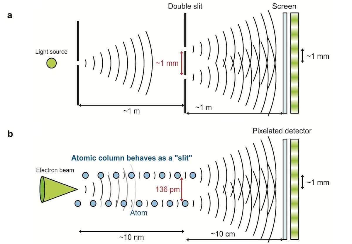
To address this challenge, Shibata’s team used the ultraprecise probe of a 4D scanning transmission electron microscope: an instrument designed to capture electron diffraction patterns with a pixellated detector. Fired into a pure silicon crystal at just the right orientation, the microscope’s precision let the researchers target two adjacent columns of aligned silicon atoms, separated by just 136 pm. As a result, the electron wave overlapped both columns, and no others.
“Our key idea was to use these two atomic columns as the two slits,” describes team member Takehito Seki. “By channelling the probe along both columns at once, we turned them into two coherent scattering sources – effectively making the crystal itself into an atomic-scale double-slit interferometer.”
At this scale, Shibata and Seki’s team created electron interference fringes some seven orders of magnitude smaller than in Thomas Young’s original double-slit experiment with light, first carried out in 1801.
Just as in a larger-scale experiment, these patterns encoded information about the crystal and the electron beam. But at this scale, the researchers could also detect the signature of individual phonon modes in the columns. Rather than destroying coherence as uncorrelated atomic motion would in a classical picture, these correlated vibrations between the two atomic columns kept them moving in sync, preserving the interference.
“In a which-path picture, relative motion of the two atomic columns makes the two electron pathways more distinguishable and reduces the interference, whereas correlated in-phase motion preserves coherence,” Shibata explains. As a result, distinct interference fringes remained visible across temperatures ranging from 300 to 900 K.
The team’s results already point to how an atomic-scale double-slit experiment could offer new ways to measure phonon correlations directly between single pairs of atoms. But as Shibata explains, the possible applications could stretch much further.
“It also opens a route to probing local lattice dynamics at the scale of individual atomic bonds, including at interfaces and defects, where local lattice dynamics can strongly influence thermal transport,” he says. “In the long run, such atomic-scale insight could help inform better thermal management strategies for future chip technologies.”
The study is published in Nature.
The post Researchers shrink double-slit experiment to atomic scale appeared first on Physics World.
It's that time of year again, when we learn which lucky scientists are among the winners of the Ig Nobel Prizes. This year, the prizes honor research on designing the perfect splash-free urinal; using mosquito proboscises to "necroprint" tiny nozzles; studying composition rates of buried cotton underwear; and the aerodynamics of a healthy nose-blow, among other highlights.
Established in 1991, the Ig Nobels are a good-natured parody of the Nobel Prizes; they honor “achievements that first make people laugh and then make them think.” The unapologetically campy awards ceremony features miniature operas, scientific demos, and "24/7 lectures," whereby experts must explain their work twice: once in 24 seconds and the second in just seven words.
Acceptance speeches are limited to 60 seconds. And as the motto implies, the research being honored might seem ridiculous at first glance, but that doesn’t mean it’s devoid of scientific merit. In the weeks following the ceremony, the winners will also give free public talks, which will be posted on the Improbable Research website.


© YouTube/Improbable Research
An international research team has announced the first-ever successful observation of gravity’s effect on a falling quantum object, demonstrating a connection between gravity and the quantum world first predicted by Albert Einstein.
The research team said the result does not unify quantum mechanics and gravity, nor does it prove that gravity is a quantum phenomenon.
However, the researchers said their study offers experimental support that Einstein’s equivalence principle, a fundamental concept at the heart of the famed scientist’s theory of gravity, remains consistent with matter in the quantum world.
When physicists want to describe nature, they rely on two distinct sets of rules. Einstein’s theory of gravity explains the mechanics of large-scale phenomena like stars and galaxies. Conversely, quantum mechanics helps scientists characterize the behavior of extremely small objects. Still, finding a scientific bridge uniting the two sets of rules has proven elusive.
“These two pillars of modern physics have so far eluded all attempts at a unified theoretical framework,” explained Professor Ron Folman from Ben-Gurion University of the Negev and the lead author of the new study.
Einstein did, however, identify one mechanism where the two worlds meet. Called the equivalence principle, the concept says that gravity should effectively disappear for an observer in free fall. For example, NASA simulates zero-gravity environments by flying astronauts in a plane, making successive rising and falling parabolic arcs. During the short periods of falling, the astronauts briefly experience ‘weightlessness” due to the equivalence principle.

Curious if the same phenomena would apply to the quantum world, the research team, which included Nobel Prize-winning physicist Professor Sir Roger Penrose, designed experiments. According to a statement announcing the research, the team hoped their setup would allow them to measure a “distinctive change in the quantum properties of atoms as they fell under gravity.”
To search for gravity’s effect in the quantum world, the researchers created clouds of rubidium atoms cooled to just above absolute zero. The researchers used a specially designed atom chip that allowed the team to manipulate the ultracold rubidium atoms.
Next, PhD student Or Dobkowski and colleagues used microwave pulses to place the atoms in quantum superposition. This unique quantum state lets each atom travel down two paths at once.
Using tiny electrical wires embedded on the specialized chip, the researchers generated tunable magnetic fields. The researchers said the field’s precision tunability allowed them to apply an upward force to one half of the atomic wave. For this experiment, the team created a magnetic field at the precise strength needed to counter gravity, effectively holding that half of the wave stationary relative to Earth.
After suspending the first half of the wave, the team used a magnetic pulse to lift the second part. Then, the team switched to a state almost unaffected by the magnetic field, allowing it to fall under gravity.
Once the second half completed its fall, the team used magnetic fields to reunite the two halves. When reunited, the two halves of the atomic wave interfered with each other. The researchers said that interaction allowed them to “measure the tiny difference in quantum phase accumulated while one was falling and the other was held still.”
“The phase measured in the new experiment is the same as the one predicted when Einstein’s principle is applied to such a quantum wave,” they explained.
When discussing the results, the research team said their result should not be construed as proof that gravity is quantum. The results also do not unite gravity and quantum mechanics. However, Folman noted, “this complex experiment gives more hints as to how such a unification may be achieved.”
Study co-author Professor Vlatko Vedral from the University of Oxford’s Department of Physics agreed, while also noting that their experimental results successfully demonstrated Einstein’s equivalence principle’s consistency with quantum mechanics, which alone provides an experimental connection between quantum physics and Einstein’s theory of gravity.
“This experiment pushes quantum mechanics into one of its most intriguing frontiers, gravity, and shows that, once again, its predictions hold,” Professor Vedral explained.
The professor also noted the experiment’s combination of hard experimental data with “a far-reaching theoretical interpretation, about one of the most fundamental questions in physics: How can gravity (described by Einstein’s theory of relativity) and quantum theory be unified into one understanding of the universe?”
The study “Observation of the quantum phase of free fall and the consistency with the equivalence principle” was published in Science Advances.
Christopher Plain has spent the last six years as Associate News Editor and Head Science Reporter at The Debrief. Follow and connect with him on X, learn about his novels at plainfiction.com, or email him at christopher@thedebrief.org.
D-Wave is a bit of an oddity in the quantum computing space, having been founded back in the last century. And its initial offering wasn't a quantum computer like those being developed by IBM or Google. Instead, the company built what's now called a quantum annealer, a machine that isn't general-purpose but can solve a large class of optimization problems. While the hardware shares some similarities with the qubits used in gate-based quantum computers, it operates in a fundamentally different way.
But a few years back, D-Wave started working on gate-based hardware, apparently choosing a somewhat unusual qubit technology called fluxonium. And this year, the company acquired a startup called Quantum Circuits that spun out of Yale University and has been developing what's called a dual-rail qubit (the same technology used by Amazon), which promises to make most errors very easy to detect, simplifying error correction.
On Wednesday, the company is publishing a paper in Nature that describes a key step in validating this dual-rail technology, showing that two of the qubits can be entangled without altering their best feature: Most are a single type that is easy to detect.


© D-Wave

There are many algorithms for which it has been mathematically proven that a quantum computer can generate results that would take a classical computer an unreasonable amount of time to generate. Unfortunately, today's quantum computers either can't run those algorithms or can only run simplified versions that classical computers can also handle. This has left the field facing a challenging question: Can we demonstrate the promise of quantum computers on today's noisy, limited hardware?
That's a more difficult question than it may first appear. If you generate a result that's out of reach of today's regular computers, it may not be possible to verify that you got the right result. And given that today's quantum computers are somewhat error-prone, getting the wrong result is a distinct possibility. Further, in the absence of a mathematical proof of the capabilities of quantum hardware, it's possible that a better classical algorithm could outperform the quantum hardware.
These issues inspired IBM to launch a quantum advantage tracker. On Thursday, the company announced three new entries that it says clearly show a quantum advantage, each using a different approach to overcoming errors and validating quantum results. "Trusted computing when you can do classical simulations is irrelevant," IBM's Jay Gambetta told Ars. "Trusted computing when you can't do classical simulations is a big deal."


© IBM