 Login

Anthropic and Blackstone Launch Ode, a $1.5 Billion AI Implementation Company
Anthropic Bets on Deployment AI models keep getting better. What enterprises actually do with them is still an


Many observations of solar flares suffer from saturation and blooming, causing crucial information to be lost. Researchers recently validated a technique for retrieving information from saturated flare images, unlocking a treasure trove of powerful solar flares for examination.
Since 2010, the Atmospheric Imaging Assembly (AIA) on the Solar Dynamics Observatory (SDO) has been capturing nearly continuous extreme-ultraviolet images of the Sun’s disk, revealing our star’s dramatic high-energy behavior. Though SDO/AIA’s observations have transformed our understanding of the Sun’s activity, they have limitations: powerful solar flares can saturate the detector, spilling charge into nearby pixels.
This saturation and overflow compromises our ability to study the most energetic solar flares — but the “lost” information may actually be recoverable. When light from an intense flare enters SDO’s instruments, not all of it pools in the saturated region of the detector: some of the light is diffracted by the optics, and by mathematically inverting this pattern of diffracted light, researchers can attempt to extract the encoded information about the saturated region. Researchers have used this technique for more than a decade, but options for validating the results were limited until now.
Launched almost 10 years to the day after SDO, the Solar Orbiter spacecraft provides an excellent opportunity to test inversion techniques for recovering information from saturated solar images. Solar Orbiter’s Extreme Ultraviolet Imager snaps brief photos of the Sun at a rapid cadence, and its images remain unsaturated even when faced with energetic solar flares.
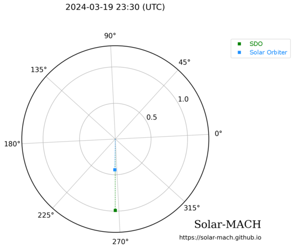
Positions of SDO (green) and Solar Orbiter (blue) during a solar flare on 19 March 2024. Click to enlarge. [Guastavino et al. 2026]
Guastavino’s team aligned and reprojected the data, accounting for the slightly different viewing angles of the two spacecraft, then applied an algorithm called Adaptive SE-DESAT to reconstruct the flux within the saturated portion of the SDO image. The team compared the desaturated SDO images to the unsaturated Solar Orbiter images over the course of the flare, finding the same flare morphology and similar time evolution between the two data sets.
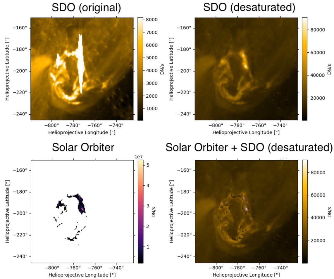
Example of unprocessed SDO data (top left), desaturated SDO data (top right), Solar Orbiter data (bottom left), and a combination of the two data sources (bottom right). Note that the Solar Orbiter observations have been clipped so that only the highest flare intensities were recorded. Click to enlarge. [Adapted from Guastavino et al. 2026]
Overall, these results demonstrate the effectiveness of the inversion technique, though Guastavino and coauthors noted that improvements could be made during the impulsive phase of the flare, when the emission is increasing. The team identified several reasons why the results may not match during this phase, including rapid evolution of the flare during individual exposures.
Despite these areas that require further attention, the validation of this technique unlocks 16 years of solar flare observations from SDO. This especially enhances our sample of powerful solar flares, increasing the available records of the most energetic solar flares by orders of magnitude.
“Validation of an Extreme-Ultraviolet Desaturation Technique for the Atmospheric Imaging Assembly on Board the Solar Dynamics Observatory Using Observations from the Extreme Ultraviolet Imager on Board Solar Orbiter,” Sabrina Guastavino et al 2026 ApJL 1005 L50. doi:10.3847/2041-8213/ae7d2e
The post When the Spacecraft Align: Rescuing Saturated Images of Solar Flares appeared first on AAS Nova.
Neuroscientists have long believed that language processing was only found in regions of the brain’s left hemisphere, including the well-known Broca’s area. However, new evidence suggests the brain’s language network is far more widespread than previously thought.
Researchers at the Massachusetts Institute of Technology (MIT) analyzed functional magnetic resonance imaging (fMRI) scans from 772 participants and identified 17 additional brain regions involved in language comprehension. These newly discovered areas are distributed across the brain, including the cerebellum, hippocampus, amygdala, and several regions of the cerebral cortex. In total, this accounts for five percent of the adult brain volume (roughly the size of a large strawberry).
“Even though there are all these distant components, it’s pretty restricted in terms of volume. You don’t need that much of the brain to do language,” says senior author Evelina Fedorenko, an MIT associate professor of brain and cognitive sciences and a member of MIT’s McGovern Institute for Brain Research, in a statement.
To find these hidden language-related regions in the brain, the researchers revisited years of brain imaging data collected since 2013. Additionally, participants completed a standard “language localizer” task, during which they read or listened to meaningful sentences and sequences of meaningless nonwords. By comparing brain activity between these two conditions, the research team identified regions that responded more strongly to language.
“When we initially started looking at language, in the first couple of papers, I tried to be comprehensive and include anything that seemed consistent across participants, and there was a huge amount of resistance,” said Fedorenko. “People would say things like, ‘Well, we know those are not language areas, so please focus on the language areas.’”
The team decided to adopt a more flexible statistical approach to detect weaker signals and identify previously overlooked language-sensitive regions beyond the classical language areas. They then compared these regions’ responses during a spatial working memory task to distinguish language-specific areas from those involved in what would be considered general cognitive functions.
Interestingly, five of the newly identified language regions are located in the cerebellum, a structure traditionally associated with movement and coordination. Some of these cerebellar regions were also active during non-language tasks, suggesting they may help integrate information across different brain systems rather than process language alone.
“It’s a very simple paradigm that lets you identify this core language system in individual brains,” says MIT postdoc Agata Wolna, the lead author of the paper.
Additional language-sensitive regions were identified in the hippocampus and amygdala, areas better known for their roles in memory and emotion. Their involvement suggests that understanding language may rely more on integrating memory, emotional context, and higher-order cognitive functions than previously understood in science.
“We always see this frontal temporal network, but there’s quite a lot of evidence that there are other regions that are also critical for language processing,” said Wolna. “By using a laxer threshold and zooming in on areas with weak MRI signal, we tried to maximize the chances of finding small and weakly responsive regions outside of this left frontal temporal system.”
The findings challenge long-held assumptions about the brain’s organization and can open new avenues for research into how language processing is understood in neuroscience.
The recent study was published in the Journal of Neuroscience.
Chrissy Newton is a PR professional and the founder of VOCAB Communications. She currently appears on The Discovery Channel and Max and hosts the Rebelliously Curious podcast, which can be found on YouTube and on all audio podcast streaming platforms. Follow her on X: @ChrissyNewton, Instagram: @BeingChrissyNewton, and chrissynewton.com. To contact Chrissy with a story, please email chrissy @ thedebrief.org.
Enzymes and other biomolecules are often anchored onto solid materials so they can be reused, purified more easily, and made more stable, a strategy biotechnology has relied on since industrial-scale immobilized enzymes first appeared in chemical manufacturing in the mid-twentieth century. Immobilized enzymes today drive processes from food and pharmaceutical production to biosensors and medical diagnostics. Fixing a biomolecule in place, however, is not as simple as gluing it down. The exact spot where it attaches, the direction it faces, and how much freedom it retains to move can all change how well it works. Testing every possible arrangement in the laboratory is slow and costly, which is why computer simulation has become such a valuable complement to experiments.
One of the main tools for this kind of study is molecular dynamics simulation, which calculates how atoms move over time by applying the basic laws of physics. Simulating every single atom of a protein, its surrounding water, and a supporting material gives rich detail, but is extremely demanding on computing power, especially when many attachment geometries need comparing. A widely used shortcut is coarse-grained modeling, in which small clusters of atoms are grouped into single, simplified particles. This keeps the essential physical behavior of the system while making the calculations far cheaper. The most established coarse-grained approach for biological molecules is the Martini force field, first developed in 2004 to describe fatty, membrane-forming molecules and later extended to proteins and, more recently, to a more accurate third generation of the model. Martini has since become a standard tool for simulating large biological assemblies that would otherwise be out of reach.
A team of researchers now introduces MartiniSurf 1, an open-source software package that automates the preparation of these coarse-grained simulations for biomolecules attached to surfaces. Building such a system by hand normally involves several separate programs and many manual steps, which makes it hard to reproduce results or compare different setups fairly. MartiniSurf instead combines the whole process into a single, streamlined workflow. Starting from the structure of a protein or a DNA strand, it converts the molecule into its coarse-grained form, builds a model of the supporting surface, positions the molecule in a chosen orientation, adds water and dissolved ions, and produces all the files needed to run the simulation with GROMACS, a molecular dynamics program that has been developed and refined for decades. Because every step is controlled by explicit settings rather than manual adjustments, two researchers using the same settings should obtain the same starting system, making comparisons across studies far more reliable.
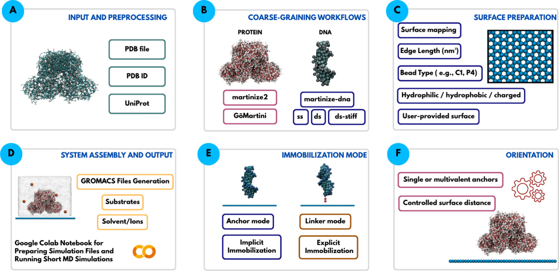
A central feature of the software is its flexibility in representing how a biomolecule is attached. In one mode, selected regions of the molecule are held near the surface through simple restraining forces, without modeling the physical connector itself. In another mode, an explicit molecular linker is placed between the biomolecule and the surface, so that the linker’s own length and flexibility can influence the simulation. Because the properties of such linkers are known to affect how active and stable an attached enzyme remains, this second option offers a more realistic picture whenever the necessary coarse-grained description of the linker is available.
The supporting surfaces themselves can also be varied widely. The software can generate simple flat reference surfaces or draw on previously developed coarse-grained models of real materials such as graphene, graphite, carbon nanotubes, and agarose-like gels, the type of porous material commonly used to immobilize enzymes in the laboratory. Properties such as surface chemistry, electric charge, particle spacing, and added chemical groups can all be adjusted. Recent studies have shown that surfaces built without care can cause water molecules near the interface to line up in unrealistic, almost frozen patterns, an artifact rather than genuine physical behavior. The software includes options that let users introduce controlled irregularity into the surface to reduce this problem, while staying consistent with the wider Martini modeling framework.
To demonstrate how the approach works in practice, the study presents examples involving both proteins and DNA. For an enzyme attached to an agarose-like surface, the software reproduces several different attachment arrangements, including a complete system containing the enzyme together with its natural helper molecule and reaction substrate. These examples show how changing the attachment point or adding chemical groups to the surface can be explored systematically, without needing to reconstruct every model from scratch by hand.
The approach is also extended to DNA tethered to a graphene-like surface, comparing an electrically neutral surface with a positively charged one. The coarse-grained simulations reproduce the same general behavior previously reported in much more computationally demanding all-atom studies: the DNA strand remains mostly upright above the neutral surface but bends progressively toward the surface when it carries a positive charge, drawn in by electrostatic attraction. This comparison is not meant to reproduce exact experimental values, but rather to confirm that the automated setup captures the right physical trends, providing a check on the reliability of the workflow.
Every simulation generated by the software automatically records the settings, software versions, and modeling choices used to build it, so that another researcher can repeat or directly compare a given calculation later. Importantly, the software does not introduce new physical models or new force-field parameters of its own. Instead, it draws on already validated Martini tools and organizes them into a consistent, automated pipeline.
Overall, MartiniSurf addresses a practical bottleneck in computational chemistry rather than proposing a new simulation method. By automating the construction of surface-bound protein and DNA systems, it makes coarse-grained molecular dynamics studies faster to set up, easier to reproduce, and simpler to scale across many attachment geometries. This kind of tool should make it easier to systematically study how surface properties, attachment geometry, and molecular flexibility shape the behavior of immobilized biomolecules, supporting the design of better catalysts, biosensors, and other biotechnology applications.
Author: César Tomé López is a science writer and the editor of Mapping Ignorance
Disclaimer: Parts of this article may have been copied verbatim or almost verbatim from the referenced research paper/s.
The post Overcoming bottlenecks in bio-interface simulations: The MartiniSurf Approach appeared first on Mapping Ignorance.

