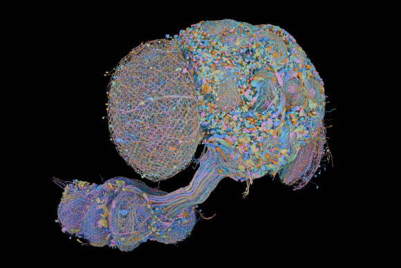
After nearly two decades of work, scientists have completed a wiring diagram of the central nervous system of an adult male fruit fly (Drosophila melanogaster).
Interest in psychedelic medicine has increased rapidly, with several compounds now being tested in clinical trials for conditions such as depression, PTSD, and addiction. Most previous studies have focused on individual drugs, leaving it unclear how the effects of different psychedelics actually overlap. To address this, researchers at the University of Southern Denmark designed a study to directly compare the effects of different psychedelics side by side.
The recent researchers relied on PET scans to monitor brain metabolism and connectivity in rats after giving them LSD, psilocybin, or a synthetic psychedelic known as 2C-B. They recorded changes during the immediate effects of each drug and again one week later, allowing a direct comparison of the resulting brain activity patterns.
Different Drugs, Different Fingerprints
Each drug produced its own distinct pattern of brain activity, and only a handful of changes showed up across all three.
“Psychedelic drugs are often discussed as if they work in the same way. But when we look at the brain, the differences are clear,” said Mikael Palner, associate professor at the University of Southern Denmark’s Department of Clinical Research and senior author of the study. “Each drug appears to affect its own networks, and that is new knowledge.”
2C-B and LSD drove the most changes while the drugs were active, but the more striking result showed up after the drugs had worn off. At the one-week mark, 2C-B primarily affected regions of the brain associated with reward, while LSD showed effects in areas involved in habits and motivation. Meanwhile, psilocybin primarily affected regions associated with emotion.
“What is interesting is that the differences do not only appear while the drugs are active. We can also see different traces in the brain afterwards,” said Frederik Gudmundsen, the study’s first author, who completed his PhD at the University of Southern Denmark and now works as a postdoc at Aarhus University’s Translational Neuropsychiatry Unit.
Not Better, Just Different: and Still a Long Way From the Clinic
None of this means one drug is better than another, but it does suggest psychedelics may not be one interchangeable category when it comes to developing them for medical practice.
“If the drugs affect the brain differently, it is also possible that they may eventually prove better suited to different disorders,” Palner said. “We do not know that yet, but our study provides a biological starting point for investigating it.”
Despite these intriguing findings, the research does not determine if these distinct patterns of brain activity from different psychedelic substances apply to human patients. The researchers acknowledge this limitation, noting that the results are based on animal models and that further studies in humans are necessary to determine if the same differences apply.
The authors also disclosed that Mikael Palner holds shares in the psychedelic drug companies Compass Pathways and AtaiBeckley, but neither company was involved in the research.
The study still offers a detailed biological picture of how different psychedelic substances alter brain metabolism and network activity and could help researchers investigate whether specific compounds might eventually prove better suited to particular disorders.
For now, researchers have yet to determine whether LSD, psilocybin, and 2C-B will ultimately have distinct roles in medicine or whether the differences between them are less important in humans.
Austin Burgess is a writer and researcher with a background in sales, marketing, and data analytics. He holds an MBA, a Bachelor of Science in Business Administration, and a data analytics certification. His work focuses on breaking scientific developments, with an emphasis on emerging biology, cognitive neuroscience, and archaeological discoveries.
Cluster headache causes extreme pain, but its origins remain unclear. New research links the disorder to genetics, inflammation, and tobacco smoke exposure. Unlike an ordinary headache, a cluster attack typically erupts without warning and causes excruciating pain around one eye. Episodes can last from 15 minutes to three hours and often return at similar times [...]
Depression in later life may be an early warning from the brain, appearing years before Alzheimer’s begins to affect memory and thinking A new study in JNeurosci found that faster accumulation of tau, a protein closely tied to Alzheimer’s, was associated with worsening depressive symptoms in older adults who remained cognitively healthy. Teodora Markova of [...]
Si le cerveau, capable de se réinventer tout au long de la vie, détenait les clés des prochaines avancées médicales ? Entre grandes tables rondes et rencontres en petit comité, la 10e édition de Futurapolis Santé 2026 permettra à tous les curieux de décortiquer ce que la science sait, et ne sait...
A provocative new paper suggests consciousness may not be unique to Earth and could arise in alien life forms far stranger than we can imagine. A new philosophical paper argues that consciousness may not be exclusive to life as we know it. According to Eric Schwitzgebel, a distinguished professor of philosophy at the University of [...]
Whether consciousness arises entirely within the brain or involves processes beyond it has long been debated in scientific and philosophical circles. Historically, most academics have associated consciousness with neurons firing and communicating information; however, precisely how neural activity gives rise to subjective experience remains an open question.
Neuroscientist and consciousness researcher Christof Koch, a former faculty member at institutions including MIT and Caltech, challenges academics to take a closer look at what philosophers call the “hard problem” of consciousness: how the physical processes of the brain could produce subjective experiences in humans.
Earlier this year, Koch presented his arguments at the “Behind and Beyond the Brain” Symposium organized by the Bial Foundation, where he argued that the nature of consciousness, as well as phenomena like near-death experiences, may require a broader scientific framework.
Koch focused on three areas. First is the physical brain itself; second is the question of how we can truly agree on what is “real.” What does blue actually look like to you? What does pain feel like? These questions are difficult to answer scientifically as universal truths.
Third are unexplained experiences that may not fit neatly within a materialistic scientific framework, such as near-death experiences.
Revisiting an old approach
Looking at older philosophical models might lead researchers toward new understandings or modern ways of approaching consciousness. Koch points to concepts such as idealism and panpsychism.
Idealism holds that consciousness or mind is fundamental to reality—mind comes first, matter second—while panpsychism holds that consciousness is a fundamental feature of the universe.
If mainstream science adopted such a shift, it could profoundly change how we understand ourselves. A paradigm shift of sorts may be coming for humanity, potentially through the integration of these ideas and a move away from strictly materialistic approaches to consciousness.
Koch points to these concepts as potential frameworks in which consciousness remains a fundamental aspect of reality. He also supports the controversial Integrated Information Theory (IIT), which proposes that systems possessing sufficiently integrated information may have some form of subjective experience.
Consciousness in Perspective
For additional perspectives, The Debrief reached out to Julia Mossbridge, a neuroscientist and Sr Distinguished Fellow in AI, Mind and Society at Florida Atlantic University, and co-author of Transcendent Mind: Rethinking the Science of Consciousness (2017), the first American Psychological Association textbook on consciousness to not assume consciousness is reducible to brain activity.
“To many neuroscientists, the brain is starting to seem more like a radio receiver and potentially also a transmitter of consciousness,” Mossbridge said in an email to The Debrief. “The idea that’s losing more ground with every new credible report of so-called ‘anomalous cognition’ is the old one—that the brain creates consciousness like the gallbladder creates bile. Maybe it’s more apt to compare the brain to the heart—it doesn’t create blood, it circulates it. But because you can use a machine to circulate the blood, it’s very clear the heart is important, but in modern times, not essential for circulation.”
Mossbridge points to reports of what she calls “veridical conscious observations” that occur when a patient’s brain shows little or no measurable activity to researchers, or what is described as “brain death,” as proof that consciousness may not be produced entirely by the brain.
These experiences led Mossbridge to consider the brain as a “transformer of consciousness rather than a creator of it.” She compares the relationship to a broken radio: “If you have a disruption in your brain, like a broken radio, you might have a disruption of consciousness.”
To test this hypothesis, Mossbridge says researchers could investigate whether people can perceive and later accurately recall “veridical information” while in deep sleep or under general anesthesia. Although she acknowledges that even positive findings might have another explanation, noting that they “could be explained by basic psychic functioning using some unknown physical mechanism.”
“So if that kind of experiment produces a positive result, and is replicated, you either have to argue that psychic ability is real but is something the brain does nonlocally (like maybe through entanglement) or that consciousness can wander from the body and observe things, or both,” Mossbridge concluded.
“The physical but nonlocal explanation is a bit like a heart pumping the blood, the brain guiding consciousness. The consciousness-wandering explanation is a bit like a heart being fully separated from the blood–and another method is used to circulate consciousness.”
“Either way, the standard model fails,” she added.
Taken together, Koch and Mossbridge’s perspectives point to a provocative possibility: that understanding consciousness may ultimately require science to reconsider some of its most fundamental assumptions about the relationship between mind, brain, and reality.
The team, which included researchers from Dartmouth College, said the study builds on earlier theories linking fire and language to the extra hours it added to the day.
However, their findings suggest that the biological changes caused by fire use may have enabled complex language as much as fire-associated behavioral changes, accelerating the evolution of verbal communication away from the ape-like systems still used by modern apes toward more complex, “uniquely human” interactions.
Researchers Wondered if Firelight Reprogrammed the Human Brain
Catherine Hobaiter, a Professor of the School of Psychology and Neuroscience at the University of St Andrews and the lead author of a paper detailing the team’s idea, said she has always been fascinated by the origins of language.
“What was the first word?” the professor asked. “The first story?”
Unfortunately, Professor Hobaiter noted, many questions about the origin of humanity’s complex and diverse forms of verbal communication remain unanswered.
Some earlier research has explored a possible link between the first spoken words and the advent of controlled fire use. However, the team notes that most studies have focused primarily on the extra hours of practical light added to the end of each day.
Curious whether other biological mechanisms also contributed to the evolution of language and complex storytelling behavior, Professor Hobaiter and colleagues examined the effects of sitting around a campfire to see whether the emitted light could reprogram the human brain.
Potential Effects of Red Wavelengths and Flickering Lights
First, the team noted that one potential catalyst emitted by firelight that could help reprogram the human brain for complex language is the color of the light wavelengths emitted by campfires. Specifically, Hobaiter and colleagues noted that campfire light emits almost no blue wavelengths but instead mostly in the visible red spectrum.
Many modern studies have focused on the potentially adverse effects of exposure to blue light wavelengths from modern technology on sleep patterns and overall health. Conversely, the paper’s authors described the red wavelengths emitted from fire as ‘uniquely suited’ for human socializing, “allowing an extension to the day without negatively impacting sleep.”
Another component of firelight the study authors suggest could have reprogrammed the human brain for speech is the way a campfire behaves. Unlike a modern, steady light source, the authors noted that firelight “flickers.”
Hobaiter and colleagues said that neurons in the human brain synchronize firing patterns with external flickering lights. For example, flickering lights in movies and TV shows often come with warnings against possible neurological effects on vulnerable viewers. The research team said this mechanism might also help synchronize the behavior of those seated around the campfire, resulting in a much deeper feeling of connection.
Finally, the researchers noted that firelight emits infrared wavelengths, which may have also contributed to helping firelight reprogram the human brain for complex verbal communication.
Are We the Storytelling Ape?
When discussing the potential impact of their proposal, study co-author Nathaniel Dominy, the Charles Hansen Professor of Anthropology at Dartmouth College, said many of the chemical properties have been well researched. However, Professor Dominy also notes that understanding the properties of light that capture our attention and focus, thereby instilling “harmony and cooperation,” has received far less attention.
“Measuring these features is an exciting part of this project,” the study author added.
Professor Hobaiter agreed, while also noting the conundrum scientists face when explaining why humans speak, when “decades of research with other species has shown us that you don’t need language to learn from each other, to organize where and when to forage, to learn cultural knowledge about tools and songs, to co-ordinate hunting, or navigate social politics.”
“While other species share all these skills, there is a fundamental puzzle: we struggle to find evidence of key differences between human and ape communication,” Professor Hobaiter explained. “And yet, we are surrounded by evidence of human distinctiveness. For all their incredible, rich communication and cultures, no other ape is inventing algebra, writing King Lear, or heading out into interstellar space.”
“So, it’s especially exciting to be able to generate testable predictions, to be able to ask if, at the end of the day, we are the storytelling ape,” the professor added.
Christopher Plain has spent the last six years as Associate News Editor and Head Science Reporter at The Debrief. Follow and connect with him on X, learn about his novels at plainfiction.com, or email him at christopher@thedebrief.org.
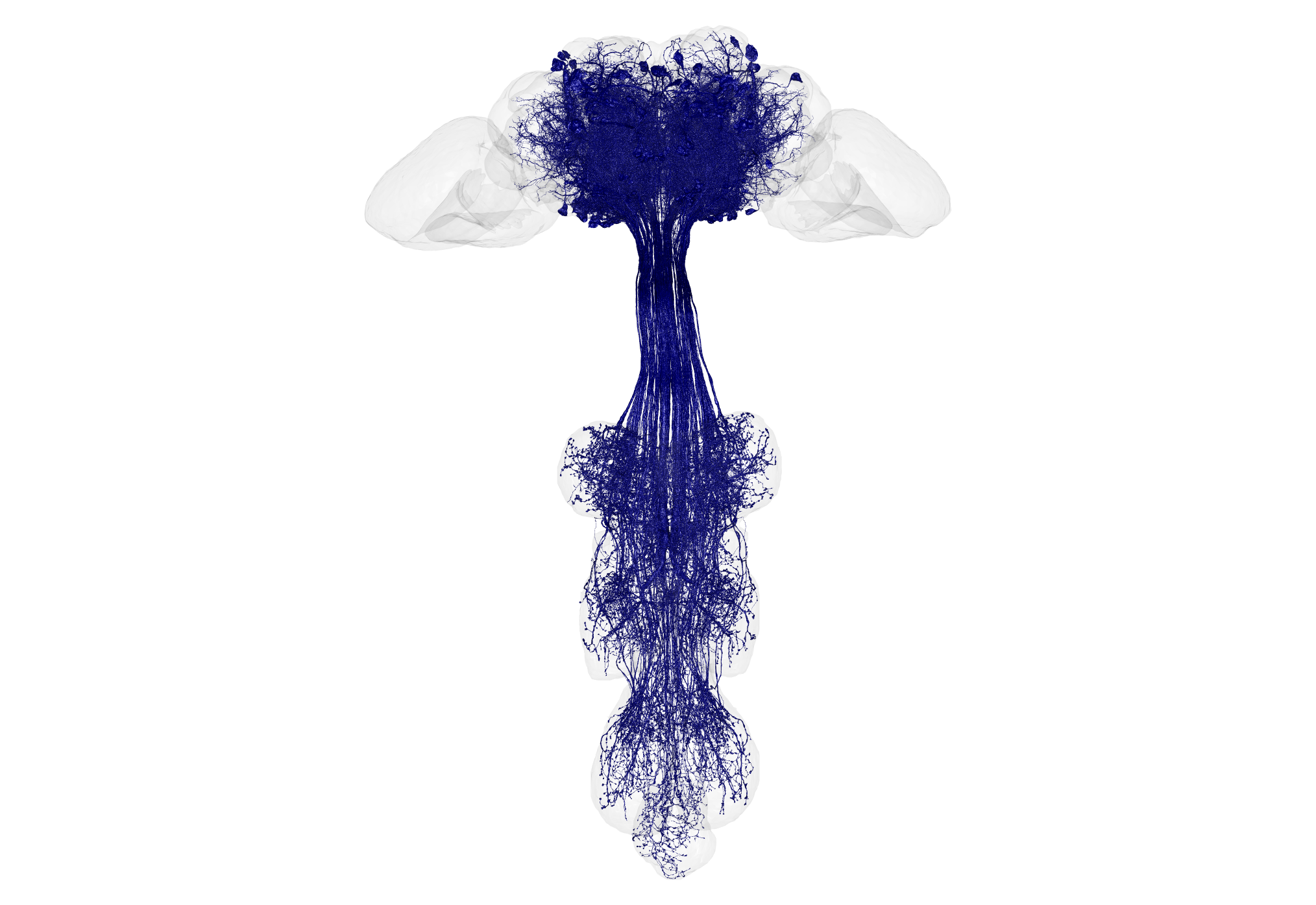
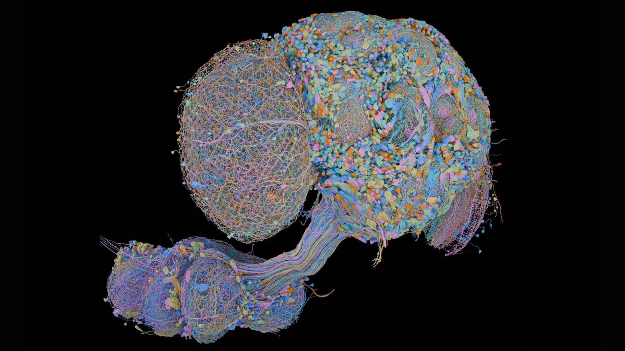
A fruit fly's brain is roughly the size of a poppy seed — and yet that tiny package contains over 100,000 neurons. A new map charts every single neuron in the male fruit fly brain, as well as the insect's equivalent of a spinal cord, totaling more than 166,000 neurons.
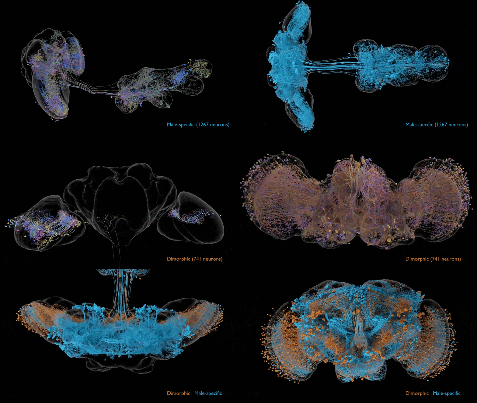
This new map joins a map of a female fruit fly brain that was unveiled in 2024 and covers about 140,000 neurons. These two wiring diagrams, also called "connectomes," can now be compared to see if there are differences between the sexes' brains that help to explain behavioral differences reflected during mating or in aggressive actions, including sex-specific fighting moves.
"It is the first time we can compare both sexes of an animal with complex social behavior," study co-author Gerry Rubin, head of biology and a senior group leader of the Howard Hughes Medical Institute's Janelia Research Campus, said in a statement. "Male and female flies have a lot of differences in their behavior, and neuroscientists want to understand how the brain controls those behaviors. This now allows us to easily home in on the neurons that are causing those differences."
This animation shows neurons that are specific to the male connectome, as well as neurons that are present in both males and females but differ between them. The male connectome will allow researchers to compare male and female fly brains and understand complex social behaviors — like mating and aggression — that can vary by sex. (Image credit: Data acquired and analyzed by the FlyEM Project Team at HHMI’s Janelia Research Campus, the Cambridge Connectomics Group, and Google Research. Video by Philip Hubbard/HHMI Janelia Research Campus)
Initially released as a preprint, the new fly brain map was published in the journal Cell and Current Biology Thursday (Sept. 3). The study describing the map was published alongside three other papers, each of which uses the new data to explore a specific aspect of fruit fly neurobiology.
"The fly nervous system performs remarkably sophisticated computations with relatively few neurons and little energy, and its architecture could suggest principles for designing more efficient artificial systems," said Carlos Ribeiro, a principal investigator at the Champalimaud Foundation in Lisbon, Portugal, whose team contributed to the brain map and led one of the related studies.
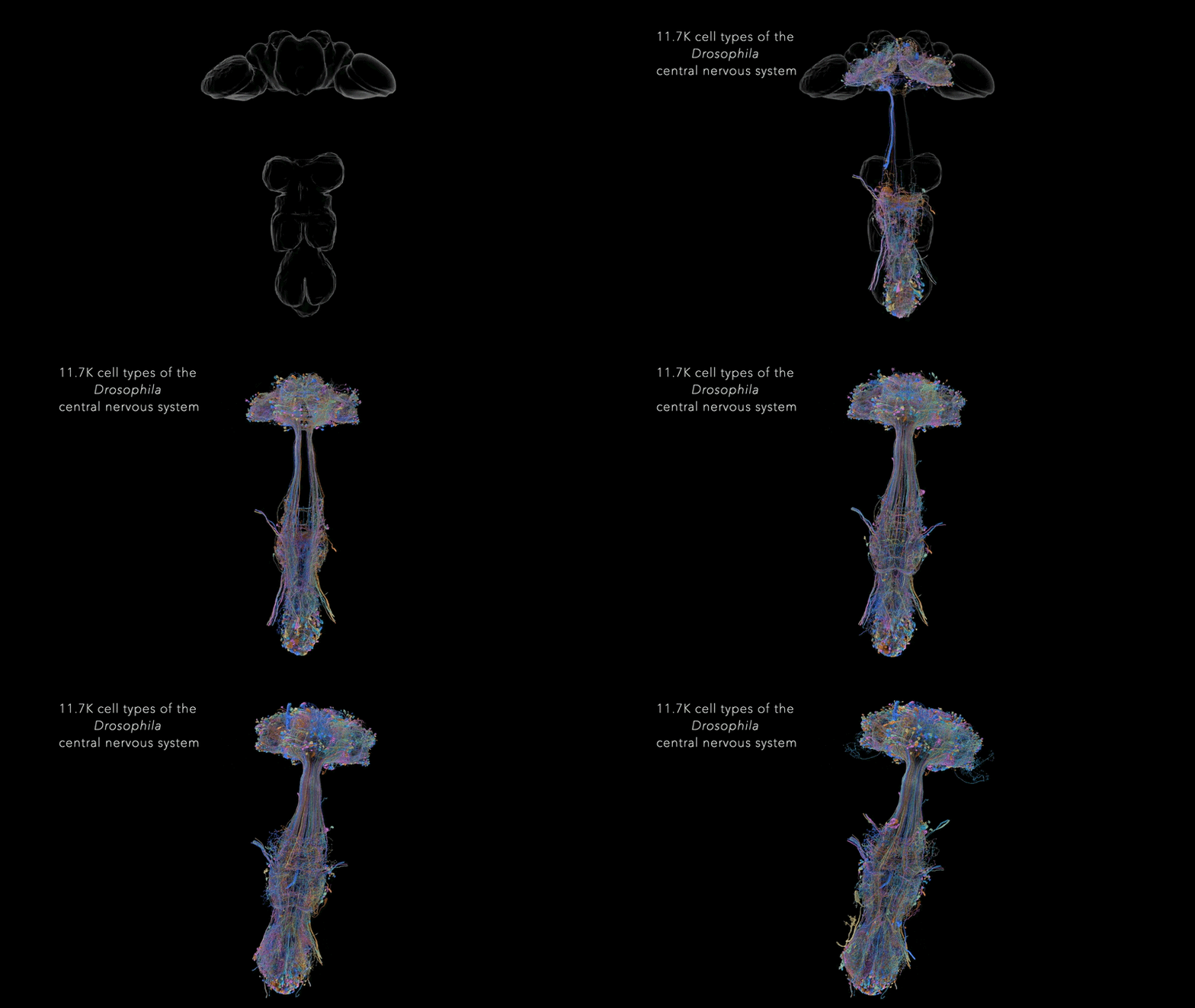
This animation shows all the types of neurons in the central nervous system of the male fruit fly. The newly completed connectome is the first map of an entire male fruit fly central nervous system, comprising all the neurons in the brain, both optic lobes and the ventral nerve cord. (Image credit: Data acquired and analyzed by the FlyEM Project Team at HHMI’s Janelia Research Campus, the Cambridge Connectomics Group, and Google Research. Video by Philip Hubbard/HHMI Janelia Research Campus)
"This work also provides a technical roadmap for more ambitious connectomics projects in the future, such as those for mice and humans," Ribeiro said in a separate statement from the Champalimaud Foundation.
In the near term, the scientists aim to map the brains of larval zebrafish (Danio rerio) and adult danionin fish (Danionella). In the long run, the goal of this research is to understand how vertebrates' brains enable complex behaviors, and then use that knowledge to help unravel the basis of neurological and psychiatric disorders in humans.
These neurons are responsible for allowing flies to process and react to tastes. (Image credit: Inês de Haan Vicente)
The study led by Ribeiro and other Champalimaud Foundation scientists aimed to map the circuits in the fly brain dedicated to the insect's sense of taste. Fruit flies have taste receptors in many body parts, including their legs, wings, mouthparts and inside of the throat. The researchers identified these taste receptors and traced their connections back to the fly brain. From there, they examined how those circuits interacted with those that govern behaviors such as swallowing or walking. When it comes to eating behaviors, this circuitry helps the fly determine whether a given morsel is safe or harmful and ultimately choose whether to eat it, the researchers concluded.
This diagram of taste processing is a "hypothesis-generation tool," study co-author Inês de Haan Vicente, a research technician in Ribeiro's lab, said in the Champalimaud statement. "Suppose you are interested in how taste controls locomotion. Now you can go to the map and ask: which sensory neurons are connected to the neurons controlling locomotion? Which intermediate neurons should I manipulate? It gives you a place to start."
The other two papers published alongside the map respectively explore the circuits behind vision processing in the fly brain and sex-specific differences in male and female fly brains. The first showed that visual processing extends deep into the brain, involving more than half of the 11,000 or so types of neurons identified in the map. The latter study found a network of cells that's specific to the male brain and appears to coordinate male-specific behaviors, such as specific aspects of fly courtship and physical aggression. (For example, female flies tend to headbutt while males lunge at their targets.)
While there were some networks specific to each sex, the circuits for sensation and movement are largely shared between males and females, the research found. At times, specific switches within those circuits reroute signals to different destinations in the male and female brain; the consequences of that rerouting will be a focus of future investigations.
See how much you know about the most complex organ in the human body with our brain quiz!
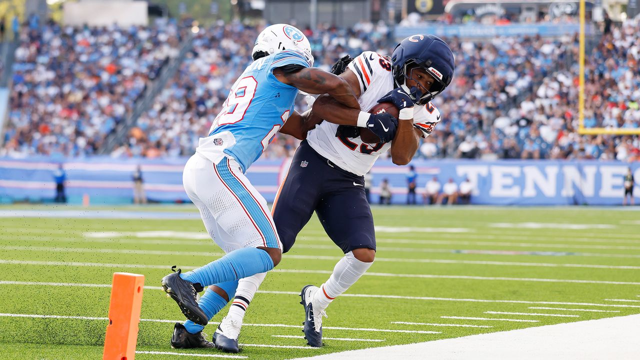
Football is a high-impact sport. A player may take hundreds of hits over the course of their career, including some that cause concussions. Other impacts may seem minor at the time, but there are concerns that repeated head blows can negatively impact long-term brain health.
Those worries gained new attention after a recent study estimated that at least 25% of recently deceased NFL players had chronic traumatic encephalopathy (CTE). CTE is a degenerative brain disease linked to repeated head impacts. Although the study doesn't necessarily mean that 25% of current NFL players have CTE, it does add to the growing evidence that repeated hits may carry serious consequences.
The NFL has taken steps to reduce those risks, including strengthening concussion protocols and investing in safer equipment, like Guardian Caps. But are those measures enough? With new research continuing to shed light on CTE, we want to know what you think. Weigh in via our poll below, and let us know your thoughts in the comments.
Of the former National Football League (NFL) players who died in recent years, at least 25% had brain changes related to chronic traumatic encephalopathy (CTE), a new study suggests.
The study, published Aug. 25 in the journal The BMJ, examined donated brain tissue from 338 former NFL players who died between 2008 and 2021. A total of 1,712 former players died in that time frame, so the analysis included only about 20% of the players' brains that could theoretically have been examined. Then, the researchers used that subset of brains to estimate the likely prevalence of CTE in the overall group.
Among the donated brains, over 93% showed physical signs of CTE, meaning an accumulation of abnormal proteins in specific places within the tissue. About 30% had stage IV CTE, the most severe form that comes with the most extensive brain changes.
These physical signs of CTE aren't always associated with overt symptoms, such as cognitive decline, mood and behavioral changes, or dementia. Only about 60% of the individuals with CTE-related brain changes were also diagnosed with dementia in this study. That said, those with stage IV CTE were more likely to have dementia, with 90% having a diagnosis.
Across the full study time frame, the researchers estimated that between 18.5% and 98.7% of the deceased players likely had CTE. Zooming in on the period with the highest number of brain donations — 2016 to 2021 — they estimated the rate to be between 24.5% and 97.7%. That latter time frame was likely the more representative sample, the researchers determined.
What can this study tell us about the CTE risk that current football players face, from youth sports to the professional level? To put the research into context, Live Science spoke with Thayne Munce, an associate professor and director of the Jayhawk Athletic Performance Laboratory at the University of Kansas, who was not involved in the BMJ study. Munce researches brain health and concussion in contact sports, primarily among young athletes. He cautioned that the BMJ study's findings shouldn't be extrapolated to all levels and ages of play, or even to today's NFL players.
Nicoletta Lanese: This study estimated the rate of CTE among this era of NFL players. Was the prevalence surprising to you, or in line with your expectations?
Thayne Munce: It's not surprising to me. This group and their colleagues have published previous work from donors — so these are former players who have donated their brains for research. They have reported even higher rates than 25% among just their donor sample [in this study]. This really high rate — over 90% of CTE cases in the donor group — is in line with what previous research has shown for these brain banks. So that's not terribly surprising.
This is a different era of football. So we should not be making any estimations about what current CTE rates are based on this study, because a lot has changed in 50 years.
What's different in this study is they compared the donors over a certain period of time versus all of the deaths among NFL players during that period of time and estimated the range based on how representative that donor sample was to the overall group of NFL players.
For example, if players just randomly selected to donate their brain to the brain bank and then they found that 98% of all of their samples had CTE — if that's representative of all the players from that era, then that's where you get the higher estimate. But if the majority of players that actually had CTE were the only ones who donated their brains because maybe they were showing outward signs or symptoms of dementia or cognitive decline or behavioral changes while they were living, then it becomes a very highly biased sample.
So that's where you get this big range. [The true prevalence] really depends on how representative that donor sample was to the peer group of players from that era.
NL: Does that broad range of estimates make it difficult to pin down the risk for current players?
TM: I think even more problematic than that big range is to understand that this is a historical study.
So these were players who died between 2008 and 2021, and the average age, I believe, was a little over 70 years of age. So if you extrapolate backwards, they probably played in the NFL starting around age 21 or 22 or 23 — that puts their playing days back in the '60s and '70s. So these were players who played in the NFL predominantly in the '60s and '70s, and would have played college and high school football in the '60s and '50s.
This is a different era of football. So we should not be making any estimations about what current CTE rates are based on this study, because a lot has changed in 50 years. It [the CTE rate] could be the same. It could be greater. It could be less. We really don't know.
I suspect, based on improvements in protective equipment and rules and advances in sports medicine, that the risk is likely lower. I don't have evidence that that's the case, but nobody does. We don't really know how the rates today compare to in the past, since CTE can't be diagnosed in the living. We can't take a sample of current players and look to see how many of them have CTE.
But that is the challenge of this study. It's interesting [in that] it gives us a range of what CTE may be in that group of former players. But it doesn't give us a lot of information about what rates are in current players in the NFL. And even more so, whatever the CTE rate is in professional NFL players does not translate to former collegiate, high school or youth football players, because CTE is an exposure-related disease. For players who have less exposure to these repetitive head impacts, it should follow that they have a considerably lower risk of CTE.
We don't want to confuse a range of CTE prevalence from players who played 50 years ago with contemporary NFL players and definitely don't want to conflate that with what CTE rates may be for current high school and youth players, who have far less levels of exposure.
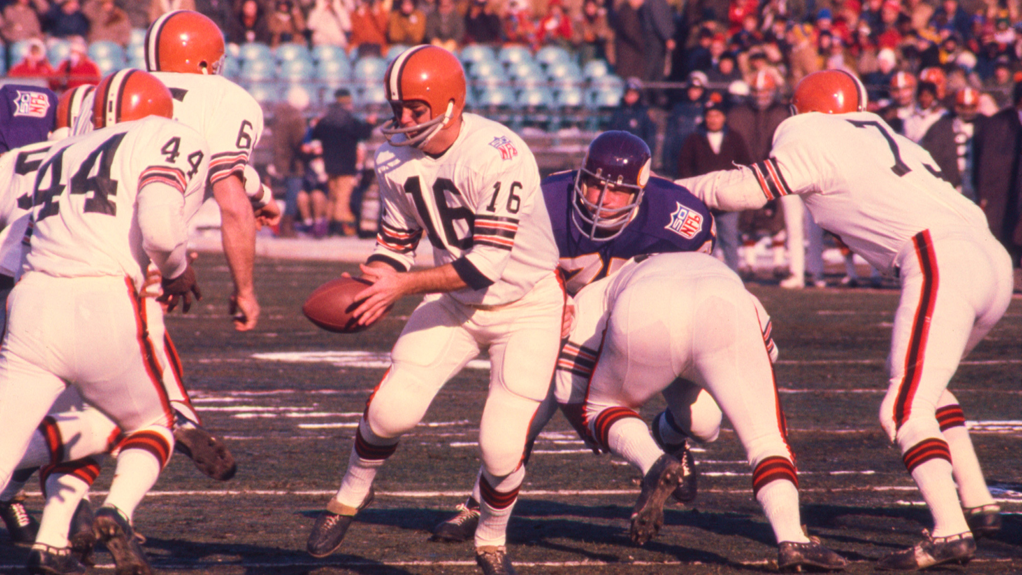
Cleveland Browns quarterback Bill Nelson hands off to running back Leroy Kelly during the 1969 NFL Championship Game against the Minnesota Vikings on January 4, 1970. Many safety precautions that are applied to today's NFL players did not exist at that time. (Image credit: James Drake via Getty Images)
NL: You mentioned that football has become safer over time. Could you spell out how the game has changed?
TM: We can look at different categories: With the rules back in that era, spearing — hitting with the crown of the head — was allowed, and oftentimes encouraged, as a style of play. That's now penalized and not taught, or shouldn't be taught.
There are changes in equipment, for sure. Modern football helmets — the material, the padding, the way in which these manufacturers are using head impact data to construct position-specific helmets and materials that are designed now to not just prevent catastrophic injuries, like skull fracture, but also to minimize the force from having repetitive head impacts — that's come a long way.
There's the Guardian Caps; you may have seen the NFL has adapted those. So they wear these soft-shell hats on their helmets during the preseason and during practices. That's now required for many of the position groups.
Virginia Tech has a helmet rating system where they test football helmets, and every year, the helmets score better on these tests. They now have star ratings for youth helmets, so manufacturers of youth football helmets are now being held to account, as well.
Maybe the most important thing is just the education around and recognition of concussion and brain injury in the sport. Now, when an athlete has a concussion, they are more likely to be recognized for having a concussion and be pulled from play and have an opportunity to recover before they return to the game. You hear anecdotal stories of former NFL players from that era, like the '60s and '70s, that suffered multiple concussions, oftentimes in the same game, and were not removed from play and continued to practice under those conditions. That medical recognition and treatment management of concussion and brain injury has moved ahead.
You look at the way in which practice was conducted, from full contact, two-a-day practices, which were common even in high school 15, 20 years ago. Now it's really not allowed at any level of play. So the amount of hitting that they do, the amount of contact that players are exposed to in practice and in the preseason is far less.
NL: When players weren't able to recover from concussions, did that likely increase their brain-injury risk?
TM: Theoretically, yes. There's evidence that if the brain is injured and then it's exposed to a second insult or injury before it's properly recovered, that makes the condition even worse.
There's actually a term called "second-impact syndrome," and it's very rare, so I don't want to scare people. But there have been instances where players — it's often, unfortunately, young players, high school players — who have had a concussion which, in retrospect, went undiagnosed and then there was a second concussive blow that led to brain swelling and death. That's the extreme, but there are cases of that [repetitive damage] happening even on a lower scale.
Even if you don't get another concussion, just those nonconcussive repetitive head impacts on top of an injured brain would not be good. It certainly is only going to delay recovery, likely is going to compound your inflammation and all of the downstream consequences of that initial injury.
I suspect that that was fairly common in years past, mainly because concussions generally weren't diagnosed. No one was looking for them, and players certainly weren't encouraged to report if they had the symptoms. So it's very likely that many of these players, including the players that were in the [brain donor] sample, had concussions and played through them and continued to receive additional trauma on top of a brain injury.
Now whether that leads to CTE, we can't really say. But I think it's reasonable to suspect that it probably would increase that risk.
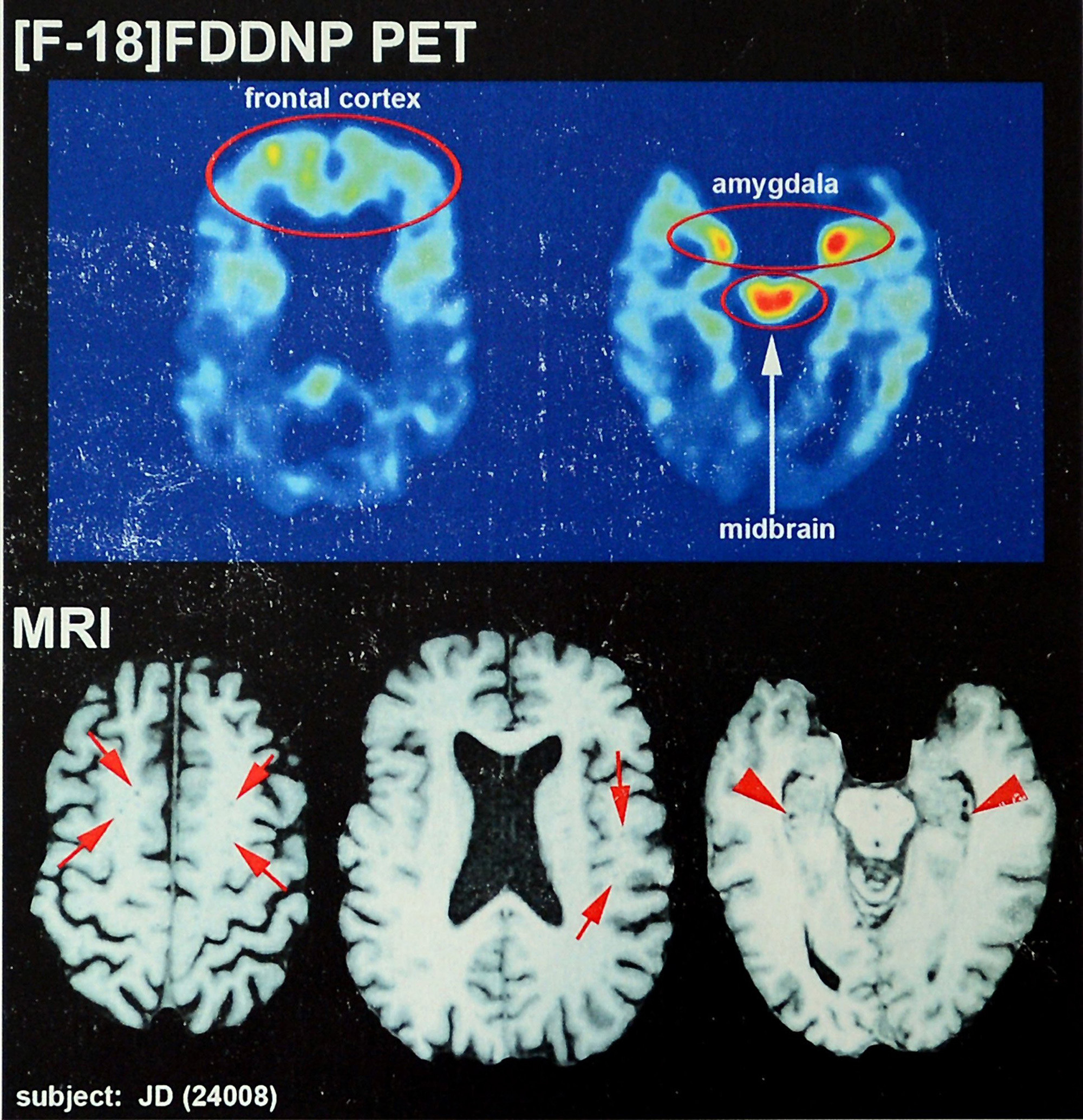
Recent brain scans of NFL hall-of-fame football player Joe DeLamielleure, who was diagnosed with CTE. (These are not from the new study.) (Image credit: Charlotte Observer via Getty Images)
NL: Could you explain whether the physical signs of CTE in the brain tend to come with symptoms, like cognitive decline? Or is the link more nuanced?
TM: So CTE is a neurodegenerative disease that can only be diagnosed by neuropathologists — people that look at slices of the brain and look for particular proteins that are present in certain regions of the brain. [This examination is done after death.] It's a pathological marker. It means that there are changes in the structure of the brain.
The clinical side of that is changes in mood, cognition, behavior, things like dementia or cognitive decline.
Even in the study, they showed that they [the CTE brain changes and symptoms] weren't necessarily linked. Now I believe they showed that for those who had more severe CTE — like the stage IV — that there was a pretty high rate of those individuals who also had subsequently diagnosed dementia. They had these clinicians that looked at the records and diagnosed them. That's a little bit of a limitation of the study: that a lot of the dementia cases were diagnosed after the person had passed away, based on medical records.
To your more immediate point, the presence of CTE: So the presence of these markers in your brain does not necessarily translate into a clinical disease or a clinical diagnosis. There can be signs or symptoms that somebody may have — let's say memory loss or changes in behavior — that may lead someone, like a former football player, to suspect that, "Oh, maybe I have CTE because I played football and I've heard that that's linked with dementia."
That may be true, but it may also be that it's due to old age, or it could be due to genetics or some other condition.
Many of these neurological diseases and conditions can be managed clinically, and there's treatment. There's therapy; there's pharmacological treatments. There's a whole toolbox available for people to seek help. That's, I think, an important message for former athletes or individuals who may be concerned about developing CTE, is to recognize that there is a disconnect between clinical disease and the pathological disease. Just because you have some outward signs or symptoms that doesn't mean that you have CTE.
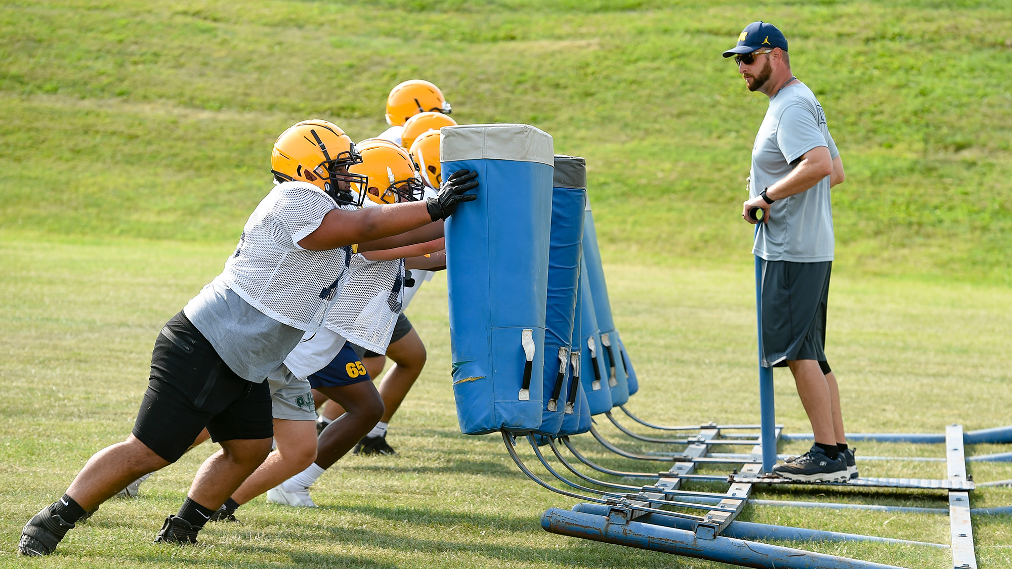
Football players at Muhlenberg High School in Pennsylvania do drills during practice. These young players likely don't have the same degree of risk as professional players when it comes to CTE. (Image credit: MediaNews Group/Reading Eagle via Getty Images via Getty Images)
NL: Since there was a higher likelihood of dementia in the stage IV cases, it seems like there's a stronger link between the pathological and clinical diagnoses at advanced stages. Is that a fair read?
TM: Again, from this study and other studies, just the presence of CTE does not necessarily mean that somebody's mood, behavior, cognition is altered. It seems to be more likely — it seems to be a tighter relationship at the more severe stages, which would kind of speak to what you're talking about. A certain threshold of disease progression seems to be more likely to manifest itself in clinical disease [with overt symptoms].
NL: What should current NFL players take from this study?
TM: I would go back to what we discussed earlier: just understanding the time frame in which the study was performed and when those players were in the NFL. I think we need to be cautious about projecting those rates; it's a pretty wide range of estimates, and it has to do with how representative the sample is. We need to use caution if we are trying to interpret those rates for the current players.
If anything, I think it probably validates all of these efforts that we discussed to try to improve brain health and minimize risk of exposure.
Even if the prevalence is on the low end — that 25% of players who played in the '60s and '70s developed CTE — that's pretty alarming. That in and of itself should get the NFL's attention. Players shouldn't have a 1-in-4 chance or greater of developing this disease.
So let's take steps to reduce that risk — and I think it validates all of the changes that have taken place over the last 50 years. I mean, a lot of them have taken place over the last 10 to 15 years. That's probably the biggest message: to say we were right to do these things and we need to continue to see if there are further opportunities to make the game safer.
Hopefully we've made a lot of progress and current-day rates are much lower than they were in the past. We don't know that, but let's not be idle. Let's continue to push forward to reduce as much risk as possible in a game where you can't completely eliminate the risk. That's something that I think everyone acknowledges; every player has to acknowledge that there is some risk.
NL: Looking forward, what further improvements could be made?
TM: There's a couple of areas that are really in the infancy stages right now. If you think about brain health as a continuum where you would have a disease like CTE on one end and then optimal health on the other end — let's say repetitive head impacts take away from your brain health and tip the scale in favor of developing disease, what are things we can do to tip the scale back in favor of improving brain health?
We know that sleep is really powerful and could learn more about that in and of itself. And what about nutrition? What about pharmacological interventions? Wouldn't it be great if we could develop a medicine, a drug at some point that could be taken preventatively to reduce athletes' inflammation? Really looking into ways in which we can optimally improve brain health as a countermeasure to the damage or the trauma that is occurring.
It's not just football where these treatments could be helpful; it's also with soccer, hockey, other sports and occupations where there is exposure to [head] trauma. I think that's a promising area.
And then, certainly, diagnostic techniques. We talked about CTE only being able to be diagnosed at death. It would certainly be a game changer if we could diagnose it convincingly in the living so it could be recognized early. People are going to have different risks, and some of that is probably genetically determined; some people are going to have the same level of exposure and develop CTE while some aren't. If we could identify individuals who are at higher risk or in the earliest stages of the development of the disease, you can then change lifestyle, behavior, exposure — that would be huge as well.
When I do talk to players and coaches and parents, I often like to remind them that, generally, the benefits of playing sports, including tackle football, outweigh the risk.
NL: To shift gears slightly, could you describe your own research in youth sports?
TM: Most of my work over the last 15 years has been focused on youth football players. That's been an interest in measuring repetitive head impacts and neurologic function in youth football players and some work in high-school-age players, as well. So a lot of that is focused around subconcussive injury [head trauma that does not trigger overt symptoms of concussion, like nausea, light sensitivity or thinking issues]. We're looking at the effects of these nonconcussive head impacts on brain health.
That ties into the CTE question in the sense that CTE is thought to be due to cumulative exposure to brain trauma — repetitive head impacts — and certainly most of the work, like in this study, has been done in NFL players. The question for a lot of the general population is, "What about youth players, high school players, that make up 90% of all football players?" So from a public health perspective, it's those younger players that I think we really need to be studying and concerned about.
That's not to say this study isn't really important. It certainly is, but my focus has been on that younger population. And then I do have some experience working more in a hospital background [on] clinical concussion management and [developing] objective pictures of concussion.
NL: With recent research, do we better understand how those nonconcussive impacts affect brain health?
TM: Going back maybe 15 to 20 years, people started to recognize that there were some cumulative effects of concussion. Those were some of the earlier studies showing that former NFL players who had multiple diagnosed concussions had worse outcomes later in life — that kind of put the spotlight on concussion. Concussion went to the forefront, in terms of how we thought about brain injury in football.
And then, over time, this recognition of what has been termed subconcussive or nonconcussive brain injury has emerged, as there's been some imaging studies in collegiate athletes, and even high school athletes, that have found that players who don't get concussed sometimes have changes in their brain, even over the course of the season.
That dovetails with some of the CTE research which has shown that history of concussions isn't necessarily predictive of who developed CTE. It's more of a cumulative effect of repetitive brain trauma. So the CTE researchers have essentially concluded that it's history of repetitive brain trauma, rather than history of concussion, per se, that's a leading risk factor for CTE.
With CTE, it's always looking backwards, because it's something that's diagnosed at death. For researchers like myself who study concussion, or study active football players, that has also then been a reaction of saying, "Well, if it is repetitive trauma, if it's repetitive head impacts, that is a risk factor for neurodegenerative disease such as CTE, then that makes it even more important to study it in active players." What does that head impact exposure look like at different ages, and how can we reduce that exposure? How can we reduce the number and severity of head impacts that players are experiencing?
So there's both the CTE angle, which is, again, retrospective — I'm looking at former players. And the contemporary issues of players who are currently playing and what's the risk for concussion, but also what's their exposure to repetitive head impacts? So it's really tied together.
That's why there's so much concern about repetitive head impacts and why there have been rule changes at the collegiate and high school level about the amount of contact and limiting those types of activities, with the idea of reducing that overall head-impact burden.
NL: When you talk about repetitive head impacts broadly, I assume that covers both concussive and nonconcussive impacts?
TM: Yep — so every hit would be considered a head impact, and then certain impacts are strong enough, or hit you in the right spot, to cause a concussion. The vast majority of impacts are nonconcussive.
NL: How do you speak to these athletes' families about the risks of concussion and CTE?
TM: When I do talk to players and coaches and parents, I often like to remind them that, generally, the benefits of playing sports, including tackle football, outweigh the risk. Oftentimes we get so concerned about the risk, which is real, that we forget about the benefits and about the alternatives of not playing. Almost always, the benefits — from the physical, the psychological and social components, the mental health components — are going to outweigh the risks. That's one thing to keep in mind.
Second, the risk of injury — in this case, CTE — is likely going to be far less in those younger players than older players, NFL players because their exposure, the number of impacts, the severity of impacts is far less. With my research, I've measured those head impacts and compared them to high school players and collegiate players, and extrapolated to NFL players because that data isn't available. And you see this big stepwise increase in exposure from the youth, the high school, the collegiate, to the NFL players.
[With parents], I'm reassuring them that the risk is far less for the younger players than what they hear about in this type of study. We're talking about an apples-to-oranges comparison.
I also like to remind them that, in my opinion, the game of football has never been safer. I've got a bit of a personal perspective on this as well, because I played football myself through college.
TM: I played for a Division II school in South Dakota called — it's Augustana University now; it was Augustana College when I played there.
So I played 12 years of tackle football, and I can compare what tackle football was like when I was in fifth grade versus now — with the training that coaches have and the type of equipment they have and the way the practices are structured. It's completely night and day.
Even at the collegiate level, the way in which collegiate practices are conducted and the safety measures that are in place for current collegiate players is nothing like what we had when I was playing. So I like to reassure players that this is probably the safest time to be playing the sport of football.
At the same time, I acknowledge that this is an individual decision that parents need to make with their kids and evaluate that risk. One of my goals as a researcher is that I want to provide more empirical evidence so that they can make an informed decision, so that a parent would be able to say, "My son's risk of getting a concussion while playing youth football is 1 in whatever" — you know, 1 in 10, 1 in 5, or 1 in 100, whatever that case may be. And his risk of getting CTE by playing four years of youth football or four years of high school football would be this. Then you make an informed decision. Is that risk worth the benefit of participation?
Unfortunately, we don't know what that risk is [currently]. We can't tell parents, "Here's objectively what your risk of injury is going to be." It makes it tough for parents. So I like to reassure them that a lot of the concerns that are expressed for NFL players are greater than they should have as parents of youth players.
This interview has been condensed and edited lightly for clarity. It is for informational purposes only and is not meant to offer medical advice.
See how much you know about the most complex organ in the human body with our brain quiz!
Diseases that cause nerve cells in the brain to die over time are known as neurodegenerative diseases. These cells, known as neurons, are vulnerable to DNA damage because they are highly active cells that require large amounts of energy. Neural DNA damage is a normal part of aging as changes to the DNA sequence, called mutations, build up over time.
To answer these questions, researchers at Boston Children’s Hospital and Harvard Medical School analyzed brain tissue from people who died with and without AD, FTD, or ALS. They compared the processes that cause DNA mutations in the 3 diseases by first accessing the DNA inside individual neurons from healthy and diseased postmortem brain tissue.
To do this, they broke apart the brain tissue and added a fluorescent marker that identified the center of each neuronal cell where DNA is housed, called its nucleus. They used a sorting machine to separate the nuclei from the rest of the cell material to access the neuron’s DNA. Using a DNA sequencing method called single-cell whole genome sequencing, the team then sequenced each neuron’s DNA to identify mutations.
They analyzed 159 neurons from people with AD, 61 neurons from people with FTD, 77 neurons from people with ALS, and 232 neurons from healthy individuals. This allowed the team to compare the number and types of DNA mutations between diseased and control (non-diseased) neurons. The researchers found that compared to the control neurons, which typically accumulate only 200 to 300 mutations over an 80-year lifespan, neurons from all 3 diseases contained an unusually high number of 2-base-pair deletions, often exceeding 1,000 mutations per cell.
When a biological process damages DNA, it leaves behind its own unique signature. By identifying these signatures, scientists can identify the biological process that likely caused the damage. Since these 2-base-pair deletions were so frequent in diseased neurons, the researchers wanted to know whether they were left behind by a biological process. The researchers used a machine learning algorithm to scan DNA sequences for recurring mutation patterns and identified a signature in neuronal DNA, which they named ID-4.
When the researchers compared the ID-4 signature with a database of previously characterized signatures, they found that it was associated with a DNA repair protein called TOP1. TOP1 temporarily cuts and rejoins DNA to relieve the tension that builds up during normal cellular activity. TOP1 typically detaches from the DNA after completing this process. However, if TOP1 fails to detach, it breaks one of the two DNA strands.
To test whether abnormal TOP1 activity was breaking the DNA, the researchers isolated DNA from control and diseased neurons. They visualized the DNA on a gel, where intact DNA forms bands while broken DNA spreads out into smears. The more fragmented the DNA is, the more intense the smear appears. Using a machine that quantified the intensity of smears, the researchers found that DNA from diseased neurons produced smears that were up to 3.2 times more intense than those from control neurons. They also found that neurons with more DNA fragmentation had more ID-4 signatures.
Based on these findings, the researchers concluded that abnormal TOP1 activity created frequent single-strand DNA breaks. They proposed that some of these breaks are repaired incorrectly, eventually leading to widespread DNA breaks in all 3 neurodegenerative diseases.
Overall, the researchers found that neurons from these 3 neurodegenerative diseases accumulated excessive DNA mutations despite differences in the disease symptoms and underlying causes. Their findings also provided evidence that abnormal TOP1 activity contributes to this mutation accumulation. Based on these findings, they stated that preventing TOP1 from becoming trapped on DNA, or improving the DNA repair process that removes trapped TOP1, could be a promising way to treat a wide range of neurodegenerative diseases.
New research from Indiana University suggests that when your brain makes a decision, it may not actually “make” it in the way neuroscientists have traditionally assumed.
Conventionally, decision-making has been described as a sequence: you have options to consider, you choose one, and then you act. However, neuroscientist Tom James argues that this sequence may not accurately reflect what actually happens in the brain.
Traditional models suggest that the brain has centralized processes responsible for making decisions. James argues that what people experience as “decision-making” may instead arise from ongoing interactions among perception, movement, the body, and the environment. If correct, this perspective could significantly change how scientists understand human behavior.
The Sandwich Model Debated
James describes one traditional approach to cognition as the “sandwich model”: sensory systems take in information, cognitive processes evaluate it and determine what to do, and motor systems then carry out the chosen action.
The problem, James argues, is that while scientists can identify many of the physical mechanisms involved in sensation and movement, it is much harder to locate a central point in the brain where a “decision” actually occurs.
Instead, James proposes thinking in terms of “action selection,” in which sensory, sensorimotor, and motor processes continually interact to produce behavior. But does that mean humans don’t actually make decisions?
“Of course they do. We use this language all the time, and it’s very helpful in terms of describing behavior,” James said in a statement. “The leap, I think, is to say that the brain works by having decision-making or control processes. It produces behavior that is well described in that way. But it doesn’t need a process that does that to make it look that way.”
In his view, “decision-making” may therefore be a useful way of describing human behavior rather than something the brain accomplishes through a specific, centralized process or system.
“As mental phenomena, they are defined on too abstract a level for the goals of cognitive neuroscience,” adds James.
Decisions are not “things”
Bridging neuroscience and philosophy, James draws on a physicalist framework associated with philosopher Daniel Dennett. Consider an object’s center of mass: it helps us describe and predict how the object behaves, but it isn’t a separate physical component causing the object to move.
James suggests decisions may work similarly. We can say someone “decided” to pick up a glass, but that does not necessarily mean there is a central location in the brain where that decision was made. Instead, the action could emerge from multiple brain and bodily processes working together.
As an analogy, James describes a robot programmed to follow a wall. Someone watching it might interpret its movements as a series of decisions, yet the robot does not require a centralized decision-making system. Instead, its sensors continually interact with its surroundings to guide its behavior.
James suggests that aspects of human behavior could be understood in much the same way.
A philosophical challenge
James’s argument also addresses a longstanding philosophical problem. If the brain contains a central system that observes information and decides how we should act, what controls that controller?
This resembles what Dennett famously called the “Cartesian Theater”: the idea of an internal observer somehow watching and interpreting what happens inside the brain. But proposing such an observer only pushes the problem back another step—who, then, observes the observer?
Rather than placing a hypothetical decision-maker somewhere inside the brain, James argues that researchers should focus on the interacting physical systems that produce behavior.
That could mean studying cognition as something that emerges through the ongoing relationship between the brain, body, and environment, rather than as a sequence of isolated stages.
The feeling that “I made a decision” may remain perfectly real and useful as a description of human experience, even if there is no single place or process in the brain where that decision occurs. From James’s perspective, what we call a decision may instead be the outcome of interactions among the brain, body, and surrounding world.
Chrissy Newton is a PR professional and the founder of VOCAB Communications. She currently appears on The Discovery Channel and Max and hosts the Rebelliously Curious podcast, which can be found on YouTube and on all audio podcast streaming platforms. Follow her on X: @ChrissyNewton, Instagram: @BeingChrissyNewton, and chrissynewton.com. To contact Chrissy with a story, please email chrissy @ thedebrief.org.
The human brain is remarkably adept at juggling different kinds of information, allowing us to switch rapidly between tasks and adapt our behavior as circumstances change. A new study by MIT neuroscientists takes a closer look at how the brain accomplishes this, providing evidence for flexible neural modules that may help support this cognitive versatility.
Using mice, scientists found that a group of neurons in the prefrontal cortex could switch between retaining sensory information and maintaining an action plan in working memory.
“We found that the brain doesn’t dedicate a separate group of neurons for every type of information,” says Yuma Osako, an MIT postdoc and the lead author of the new study. “Instead, it uses the same populations of neurons to perform the same computation on different kinds of information, which means the same subset of neurons can hold both an action and a sensory stimulus in working memory,”
The findings also lend support to a longstanding theory known as compositionality—the idea that cognition can be constructed by combining and reusing neural components.
“One of the solutions that’s always been proposed has been this idea of compositionality — that you can take pieces of cognition that perform part of a task and reuse them in another task,” says Timothy Buschman, a professor at the Princeton Neuroscience Institute, and the senior author of the paper, in a statement.
Another way to see this is to view it as “cognitive Legos,” Buschman suggests. Instead of building a new neural circuit when a new task starts, the brain reuses existing circuits and combines them in different ways.
The Test
To see how this neural network operates, the scientists trained mice to compare two tones and respond depending on whether the sounds matched. As the mice performed the tasks, the scientists recorded electrical activity from thousands of neurons.
“When mice do tasks that test whether memory computations can be reused, the answer is they are,” notes Mriganka Sur, the Newton Professor of Neuroscience at MIT’s Picower Institute for Learning and Memory. “There are subspaces of functional activity in the prefrontal cortex that can be the substrate of mixing and matching toward flexible cognition.”
Two important components of the task allowed the researchers to examine how different kinds of information were maintained. First, the mice had to remember the initial tone for a short period. They then had to retain their decision and form an action plan.
In the parietal cortex, neurons were dedicated to storing information about the tone. However, researchers found something different in the prefrontal cortex: a specific group of neurons could switch roles.
During one stage of the task, the circuit helped maintain a sensory memory. During another, the same population was involved in maintaining an action plan. This flexibility may help explain how a finite number of neurons can support an enormous range of behaviors.
“Our everyday life requires us to temporarily hold many different kinds of information. One big question is how the brain can represent an unlimited variability of information using only a finite number of neurons,” Osako says.
The researchers view these reusable neural circuits as potential building blocks for flexible cognition. More broadly, the findings suggest that the brain may be less like a machine filled with components dedicated to individual tasks and more like an interconnected system that continually repurposes its existing neural resources.
Chrissy Newton is a PR professional and the founder of VOCAB Communications. She currently appears on The Discovery Channel and Max and hosts the Rebelliously Curious podcast, which can be found on YouTube and on all audio podcast streaming platforms. Follow her on X: @ChrissyNewton, Instagram: @BeingChrissyNewton, and chrissynewton.com. To contact Chrissy with a story, please email chrissy @ thedebrief.org.
Scientists have uncovered new details about how the brain’s “information hub” responds to uncertainty, revealing a remarkably flexible system that rapidly changes how it communicates with other brain regions as we make decisions.
Every second of our lives, the human brain sorts through enormous amounts of information. From remembering a driving route and handling daily tasks to making decisions, the brain must continually determine what information matters and how to respond. Researchers at the University of Iowa wanted to better understand how the human brain manages and processes this information and found that it may be more flexible than previously thought.
At the center of this sorting process is an area of the brain known as the frontoparietal cortex. This region supports decision-making and behavioral control and can function as a kind of “information hub” for the brain.
The Study
Using MRI scans, behavioral experiments, and computational modeling, researchers took a deeper look at how the frontoparietal cortex responds when people are uncertain about what they should do next.
“That’s the main contribution,” says Kai Hwang, associate professor in the Department of Psychological and Brain Sciences and the study’s corresponding author, in a statement.
“Our study shows in more detail how the frontoparietal cortex operates — what kind of information it extracts from other systems and how it uses its connectivity pattern to integrate information that is coming in from different areas of the brain,” Hwang adds.
The research team recruited 38 participants between the ages of 18 and 35 and had each person learn associations between combinations of colors, faces, and scenes and particular actions—for example, pressing a button with the index or middle finger of either hand. The researchers then intentionally changed those associations.
When an action or rule changes, the brain has to determine what went wrong.
Did the situation change? Was the information unclear? Or did the person simply make a mistake? This uncertainty allowed researchers to observe how the brain’s “information hub” responds under different circumstances.
“It’s like where other areas of the brain don’t have all the information, so they send what they have to the frontoparietal cortex for guidance,” Hwang explains.
University of Iowa researchers have detailed the workings of the brain’s information hub, called the frontoparietal cortex. The colored areas in this illustration show frontoparietal regions tracking uncertainty when participants encounter changes to associations they previously learned. Photo Credit: Kai Hwang lab, University of Iowa.
The results from the team’s research suggest the frontoparietal cortex does more than become more active when a task becomes difficult. Instead, the regionchanges how it communicates with other brain regions.This also depends on what information is needed at different decision-making stages.
Hwang notes, “If they always get it right, they know they’ve made the correct association, but once they start doing it wrong, they will have to guess, ‘Oh, did the context change, or did I not see the color clearly?’ That creates uncertainty.”
Your Brain Has an Air Traffic Controller
Hwang describes the brain’s “information hub” as a system that gathers information from different regions, determines what is relevant, and integrates those signals into a broader picture. One way to think of this function is as an air traffic controller, monitoring incoming information and adjusting communications as circumstances change.
“These are situations where people struggle with regulating their behavior. That, to me, is an integration problem. If that integration function is not working properly, then that could very likely mean they didn’t use the right context to regulate their behavior,” Hwang adds.
The study does not address how neurodiversity might affect this information-sorting process, although the findings could provide a foundation for researchers to investigate that question.
“It remains an open question how neurodiversity might affect frontoparietal integration. It is a very interesting question that, as far as I know, has not been studied directly, so I don’t have a definitive answer. One of our goals in conducting this study is to lay the foundation for future clinical investigations,” Hwang tells The Debrief in an email.
The findings suggest that the frontoparietal cortex can rapidly integrate and update information as circumstances change. Understanding this flexibility could eventually improve researchers’ understanding of neurological and psychiatric conditions involving behavioral regulation and decision-making. In particular, studying these communication patterns could help inform future investigations of conditions such as ADHD and schizophrenia, in which behavioral regulation and the use of contextual information can be impaired.
Chrissy Newton is a PR professional and the founder of VOCAB Communications. She currently appears on The Discovery Channel and Max and hosts the Rebelliously Curious podcast, which can be found on YouTube and on all audio podcast streaming platforms. Follow her on X: @ChrissyNewton, Instagram: @BeingChrissyNewton, and chrissynewton.com. To contact Chrissy with a story, please email chrissy @ thedebrief.org.

 Login