 Login

Soft optical sensor offers new way to map the heart and brain
A team of biomedical engineers have developed a fully flexible light sensor that precisely detects changes in the

By controlling a type of polarized light widely used in quantum optics, researchers have transformed subtle optical patterns in our peripheral vision into vibrant, easily perceptible shapes. Led by Dusan Sarenac at the University of Waterloo, Canada, the team showed for the first time that the “Boehm’s brushes” perceived by participants can be altered by adjusting the topology of spin-orbit coupled light. Their results, reported in Proceedings of the National Academy of Sciences, could lead to promising new techniques for checking our eye health.
For the most part, human eyes are incapable of detecting polarization in visible light. But in some rare cases, structures inside the eye itself can scatter light in different ways depending on its polarization, creating subtle yet perceivable “entoptic” patterns. Perhaps the best-known examples of these are “Haidinger’s brushes”: hourglass-shaped patterns that can form in our central vision, but only when viewing linearly polarized light at blue wavelengths.
In 1940, Swiss biophysicist Gundo Boehm discovered another example: a two-lobed, bowtie-shaped entoptic pattern that forms when viewing linearly polarized light in the peripheral vision as it scatters from subcellular structures in the inner retina. Unlike Haidinger’s brushes, this lesser-known effect is driven by angular variations in scattering strength relative to the polarization direction.
But as Sarenac explains, these ethereal shapes are more than just a curiosity. “Because retinal disease may alter a person’s ability to perceive the pattern, researchers have long considered whether Boehm’s brushes could serve as a biomarker of retinal health,” he says. “However, their weak visibility has limited practical use.”
Whereas previous studies simply asked participants to passively observe the effect, Sarenac’s team considered what would happen if the light’s polarization was constantly varied. To explore this possibility, the researchers created a beam of spin-orbit coupled light, in which polarization is linked to the light’s orbital angular momentum – a property describing how the wavefront twists as the beam travels – so that the polarization direction rotates steadily across the beam.
In quantum optics, these structured waveforms are already being widely explored for their possibilities in imaging, communication and information processing. But for the first time, Sarenac’s team considered how our eyes could actually perceive this light. “We engineered the spatial polarization of the light to match the symmetry of the retinal scattering response,” Sarenac describes. “This allowed many weak local contributions to reinforce one another.”
The researchers then projected this structured light through a ring-shaped aperture, targeting specific regions in the periphery of participants’ retinas, which could also be adjusted to target varying distances from the centre of vision. Using an automated test, they measured how much contrast participants needed to reliably distinguish a Boehm’s brush from the background at these varying distances.
Based on the subject’s responses, Sarenac’s team determined that the use of spin-orbit coupled light not only made Boehm’s brushes appear far more vivid to the participants, it also transformed the bowtie shape into a multi-lobed pattern, with the number of lobes varying depending on the topology of the light’s twisting polarization.
“Across 11 participants, detection thresholds improved exponentially away from the centre of vision, and the pattern became robustly visible at approximately one degree of retinal angular distance,” Sarenac describes. These results reveal a fascinating link between human vision and quantum mechanics, which the team hope could be harnessed in practical healthcare. With further studies involving patients with retinal disease, they now envisage eye tests where retinal conditions could be diagnosed in people who can’t clearly perceive vibrant, multi-lobed Boehm’s brushes in their peripheral vision.
“In the longer term, these patterns may provide a non-invasive functional probe of retinal integrity because their shape and visibility can be measured across different retinal locations,” Sarenac says.
The post Structured light could deliver a new method for diagnosing eye disease appeared first on Physics World.
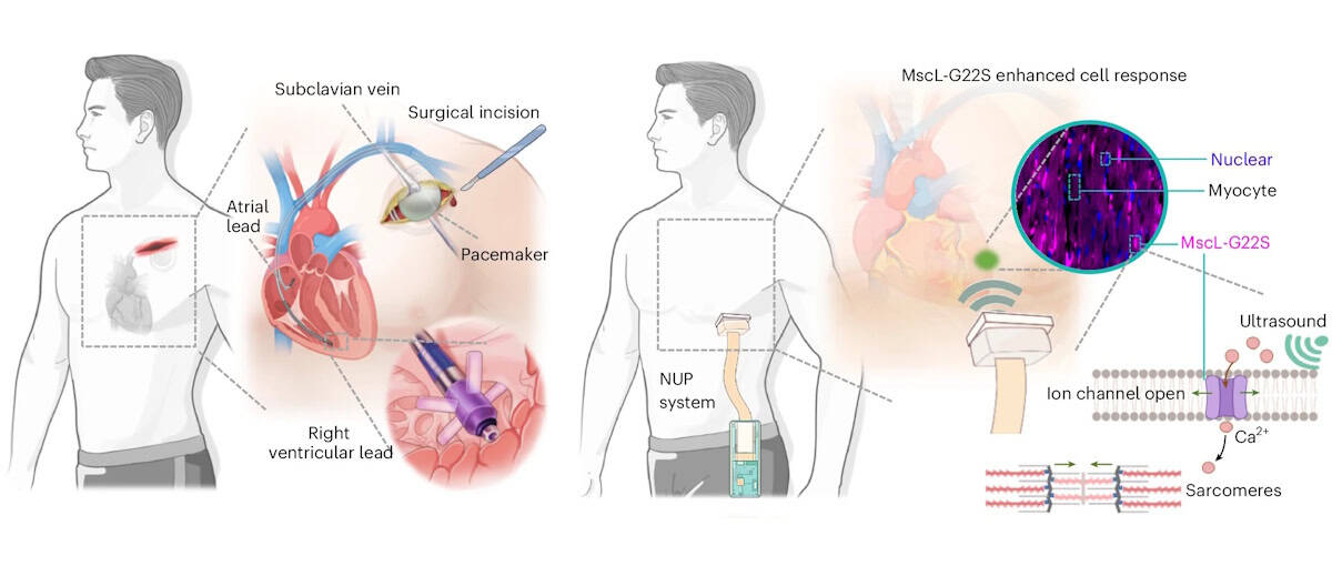
Cardiac arrhythmia – a disorder in which the heart beats too fast, too slowly or irregularly – affects millions of people worldwide. Currently, arrhythmias are treated using pacemakers to regulate the heart’s rhythm, but traditional pacemakers require invasive surgical implantation and intracardiac leads that risk infection and tissue injury. As an alternative, a research collaboration headed up at Massachusetts Institute of Technology (MIT) and the University of Southern California (USC) has developed a non-invasive pacemaker that’s worn like a sticker on the chest and stimulates the heart using ultrasound.
“Ultrasound offers a unique combination of deep tissue penetration, spatial focusing and non-invasive delivery,” explains first author Chen Gong from USC. Previous approaches for ultrasound-based cardiac pacing, however, lacked reliability or required the use of microbubbles. In this new study, Gong and collaborators employed a technique called sonogenetics, in which cardiac cells are genetically altered to increase their sensitivity to ultrasound. Once exposed to ultrasound waves, the engineered cells trigger the opening of ion channels that let in calcium, which signals the cells to squeeze and beat.
“Our central motivation was to address the limitations of conventional pacemakers, while preserving precise control of cardiac rhythm,” says Gong.
The team first examined whether ultrasound could modulate cardiac cells in vitro, using sonogenetically engineered human heart muscle cells. Ultrasound stimulation caused almost three-quarters of the engineered cells to beat in synchrony with the ultrasound waves. In contrast, unaltered cells did not exhibit such behaviour. Importantly, ultrasound exposure did not impact the cells’ expression of biomarkers for inflammation, myocardial injury or oxidative stress, demonstrating the safety of this approach.
The researchers designed a sonogenetics-based non-invasive ultrasound pacemaker (NUP) capable of real-time imaging and steerable stimulation, describing the device in Nature Biomedical Engineering. The postage stamp-sized device, which sticks to the skin via a bioadhesive interface, incorporates a 64-channel phased-array transducer, as well as data acquisition, wireless transmission and power modules.
To assess whether this prototype NUP could achieve non-invasive cardiac pacing in sonogenetically engineered rats, they attached the device onto the rats’ chests and applied ultrasound stimulation through the chest wall to the heart.
At an acoustic pressure of 2 MPa and a 40 ms pulse duration, the NUP increased the rats’ heart rates from 240 to 360–540 bpm, with a higher pulse repetition frequency leading to increased heart rate. Upon stopping the ultrasound stimulation, the heart returned to its natural rhythm. In control rats without the genetic modification, NUP could not control the heart rate effectively.
The team demonstrated that the NUP could electrically steer the ultrasound beam focus with a spatial precision of less than 1 mm, enabling targeting of different regions of the heart to induce chamber-specific pacing. NUP also successfully treated cardiac arrhythmias induced in the engineered rats, returning their heart rates to normal levels. In contrast, ultrasound stimulation of control rats did not effectively treat their arrythmias.
“A major objective of this work was to move beyond proof-of-concept stimulation and demonstrate a wearable system that could realistically support daily use,” explains Qifa Zhou, group leader at USC. “The current prototype integrates imaging, stimulation, wireless communication and battery-powered operation into a wearable format.”
For future clinical use, the NUP device needs to automatically detect the wearer’s heart rate, locate heart chambers and deliver stimuli. To achieve this, the researchers used a cloud-based, artificial intelligence (AI)-powered imaging feedback loop that determines the heart rate and stimulation coordinates, and steers the ultrasound beam to target sites.
To test its feasibility for human-scale applications, they assessed the acoustic energy penetration of the device. Using simulations and experiments on a pig heart beneath layers of tissue, they confirmed that, even after tissue attenuation, NUP could deliver sufficient ultrasound pressure (approximately 2 MPa) to a human heart to enable stimulation at clinically relevant pacemaker sites.
The team also assessed the biosafety of the NUP approach, finding that the delivered ultrasound energy remained below approved safety limits and generated minimal thermal effects during and after stimulation. They also confirmed that the sonogenetic engineering induced no off-target effects, immune responses or pathological changes in the rats.
“In the long term, we are optimistic about applying sonogenetics in humans,” explains co-author Gengxi Lu from MIT. “Sonogenetics is different from gene editing – it does not aim to rewrite a patient’s DNA sequence, but instead enables target cells to temporarily or controllably express ultrasound-responsive proteins. At the same time, extensive clinical validation will still be needed to establish safety and long-term effectiveness.”
The team is now focusing on clinical translation and validation of the NUP technology, which includes improving gene delivery strategies, validating the system in larger animal models, and developing closed-loop control approaches that combine real-time physiological sensing with adaptive stimulation. There’s also much engineering work to be done, such as device miniaturization, and improving battery life, skin coupling and motion robustness.
“For cardiac pacing, we envisage that the final goal of NUP technology is to be a permanent alternative to a long-term implanted pacemaker,” Xuanhe Zhao from MIT tells Physics World. “More broadly, we are interested in expanding ultrasound-enabled bioelectronic medicine beyond cardiac pacing toward other organs and therapeutic applications where non-invasive, spatially precise modulation could have clinical impact.”
The post Wearable pacemaker uses ultrasound to control heart rhythm appeared first on Physics World.
A new contact-free way of rotating delicate samples, such as biological cells, undergoing three-dimensional (3D) optical microscopy increases the image resolution and could reveal more details about microscale structures and processes.
Current optical microscopes provide extremely good image resolution in a single plane, but since depth information is often lacking, samples are routinely rotated and imaged at multiple different angles to create a 3D picture. Because rotating via mechanical microtools such as tiny grippers, needles or pipettes can damage samples, contact-free approaches such as optical tweezers, magnetic manipulation or optothermal forces are often used instead. However, these methods rely on the sample being birefringent, magnetic, light-absorbing or specially shaped.
By contrast, the laser-based technique developed by a team led by Moritz Kreysing and Fan Nan from the Institute of Biological and Chemical Systems at the Karlsruher Institut für Technologie (KIT) in Germany can rotate and spin floating microscopic objects no matter what their shape or material properties.
Their method involves focusing a 1455 nm infrared laser beam on a small region of a highly viscous liquid – initially at 25°C – in which the sample will be suspended. This gently heats that region of the liquid by a few degrees. But rather than focusing on one spot, an acousto-optic deflector steers the beam rapidly along a carefully designed two-dimensional (2D) pattern.
“Because the viscosity of the liquid changes with temperature, this moving heat pattern generates tiny fluid flows, known as thermoviscous flows. By choosing the scan geometry and timing correctly, these flows become three-dimensional and helical, like microscopic corkscrew-shaped currents,” explain Kreysing and Nan. Once a small object is floated freely in these 3D helical flows, it can be rotated, spun, transported or stabilized solely via software-defined scan patterns, with different symmetries controlling the movement of the surrounding liquid.
The KIT technique allows stacks of high-resolution images focused at different depths to be acquired along multiple different angles by re-orienting the sample, a feature that “can help overcome the anisotropic resolution of conventional 3D microscopy”, explain the researchers. Using a high-viscosity liquid to suspend the samples not only enables kinematic stop-and-go actuation, it also suppresses Brownian motion (the jiggling around of tiny particles in fluids), which otherwise decreases image resolution and makes the study of cellular transport processes tricky.
Since previous methods of laser-driving fluid flows could only create motion in one plane, the researchers found it “particularly exciting” when they first observed their laser scanning in a 2D plane generating 3D flow fields. “Another key moment was the observation of opto-hydrodynamic focusing. The particles did not just rotate; their spiral motion converged toward a stable height and position. This was significant because it meant that the optofluid could stabilize the sample, rather than requiring mechanical confinement or complex feedback control.”
As detailed in their recent Light: Science & Applications paper, the KIT team demonstrated rotation and spinning of various samples including stained biological cells, nano-printed micro-tiles and perfectly spherical beads. “We see immediate potential in advanced optical microscopy, especially multi-view imaging of suspended cells, cell clusters, organelles, microstructures or soft materials,” Kreysing and Nan tell Physics World.
Since the basic hardware components of their system will be familiar to many optics and microscopy labs, they are hoping for early adoption by specialized research groups. But to optimize the technique for biological applications, they will now investigate the use of different liquids and operating conditions “that better preserve the viability of live mammalian cells while still providing sufficient stability for precise rotation and imaging”.
The researchers suggest that future areas of application could include microfluidics, microrobotics, colloidal assembly, materials science and 3D microfabrication. As such, they also want to develop more advanced scan patterns that can generate curved, programmable or multi-axis helical flows. “This could allow more complex 3D manipulation, such as controlled orientation of irregular objects, coordinated actuation of multiple particles, or integration with microfabrication workflows,” they explain.
The post Laser-based rotation of floating samples enables high-resolution 3D microscopy appeared first on Physics World.
Obstructive sleep apnoea (OSA) is a sleep disorder in which the throat muscles relax and block the airway during sleep, causing pauses in breathing. If left untreated, OSA can lead to serious health problems. A research team led by Mingjiang Wang at the Harbin Institute of Technology, Shenzhen in China, in collaboration with Huizhou University, has now come up with a new method for detecting OSA – based on recordings of the subject snoring.
“OSA is a common yet underdiagnosed sleep disorder,” explains first author Heng Li. “The clinical standard for diagnosis is polysomnography [PSG], which is expensive, time-consuming and unsuitable for large-scale or home-based screening.”
PSG collects multiple physiological signals via various sensors that patients wear while asleep, potentially disrupting their sleep patterns. “Therefore, we aim to develop a non-contact detection method using snoring, which is a typical symptom of OSA and can be recorded contactlessly using a microphone,” says Li.
The acoustic patterns of snoring, produced by the vibrations of upper airway, contain markers that reflect airway narrowing and collapse. However, developing a reliable snoring-based detection model is challenging, as labelled snoring datasets are scarce and snoring sounds vary greatly between individuals.
In this study, published in Physiological Measurement, the research team uses a pretrained audio model called Wav2vec 2.0 to address these challenges. By transferring acoustic knowledge learned from large-scale unlabelled speech and audio data, the model removes the need for labelled snoring datasets.
Wav2vec 2.0 was originally designed for speech recognition and is computationally intensive, making it unsuitable for routine OSA monitoring. Thus the team adapted it for snoring sounds, removing layers more relevant to speech recognition while retaining acoustic features relevant to snoring pathology, aiming to lower the computational cost.
“We do not use the pretrained model directly,” Li explains. “Instead, we prune higher speech-related Transformer layers and fuse low- and mid-level representations, so that the model emphasizes snoring-related acoustic information.”
In collaboration with the Shenzhen People’s Hospital, the team collected sleep recordings from 100 individuals with apnoea-hypopnea indices (AHI) ranging from 2.4 to 68.3 events/hour. AHI – defined as the average number of apnoea (where breathing stops completely) and hypopnoea (shallow breathing) events per hour of sleep – is used to classify OSA from normal (AHI of below 5) through to severe (30 or above).
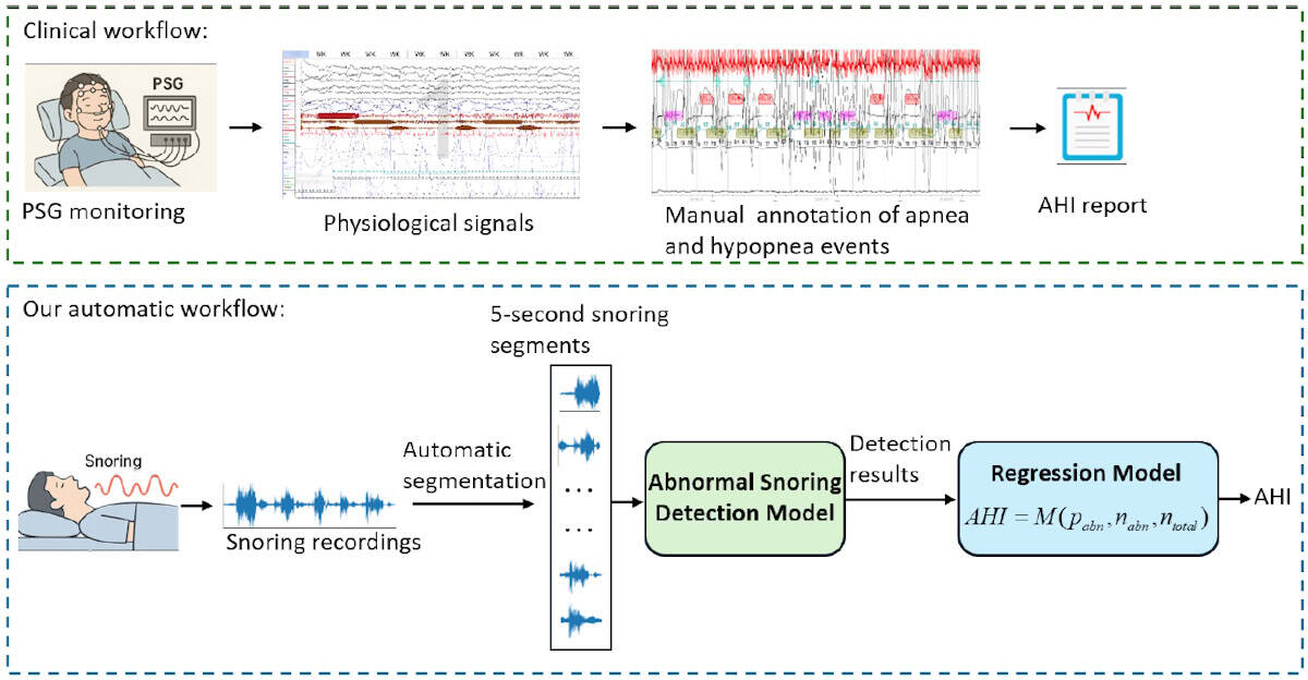
All subjects slept in hospital wards and were also monitored with a PSG device during sleeping. Sleep physicians then used the PSG data to annotate the audio recordings with apnoea and hypopnea events. Each recording was divided into 5-s audio segments and assigned as either abnormal, if they included apnoea/hypopnoea events, or normal.
To evaluate their framework’s ability to detect abnormal snoring, the researchers compared it against five other pretrained audio models. They first ran a subject-dependent evaluation, in which random audio segments from all subjects were used for training, validation and testing. All models performed well – as expected when the training and test data come from the same subjects – but the adapted Wav2vec 2.0 model achieved the highest accuracy and sensitivity.
The team then performed a subject-independent evaluation, which better reflects a real-world scenario, with each subject’s data randomly assigned to the training, validation or test set. Here, the existing methods exhibited a reduced accuracy of between 65% and 71%, while the new model retained an accuracy of 73.95%.
“In subject-independent testing, this adapted model achieved better performance than the compared baseline models, suggesting improved generalization to unseen subjects,” says Li. The team’s model was also significantly more computationally efficient, an important factor for OSA screening in home-based or resource-limited scenarios.
Finally, the researchers designed a snoring-based AHI estimation framework, using their detection model to identify abnormal snoring segments and calculating a ratio-based statistic – the number of abnormal segments divided by the total number of segments – to quantify OSA severity. They then used four common linear regression models to estimate AHI for individual subjects.
All models demonstrated reasonable accuracy and good correlation between predicted and PSG-derived AHI values. The mean absolute error was roughly 11 events/h, which means that the estimated AHI may deviate from the true value by 11 events per hour of sleep and could (if near a threshold between categories) result in an incorrect severity diagnosis. However, while the current model is not yet precise enough for clinical diagnosis, it could prove ideal as a potential tool for home-based OSA screening or pre-screening.
The researchers are now investigating how to more effectively apply pretrained audio models to clinical snoring analysis. “We are focused on making these models lighter and more suitable for home-based screening, while validating their effectiveness across large, multicentre datasets,” Yun Lu, one of the corresponding authors from Huizhou University, tells Physics World. “Additionally, we plan to improve the accuracy of AHI estimation by integrating snoring-based predictions with additional non-contact physiological information.”
The post Abnormal snoring sounds help diagnose common sleep disorder appeared first on Physics World.
Researchers in Japan have succeeded in measuring the temperature inside living cells with high precision using a new class of biocompatible quantum nanosensor – something that has been difficult to do until now even. If improved, the nanosensor could be used to characterize a wide range of biological phenomena and so help in disease diagnosis, they say.
Recent years have seen the advent of a new generation of nanoscale quantum sensors that can detect the tiny magnetic fields of biological systems. Some of these sensors rely on photons and others on electrons or spin defects – typically diamond specially engineered with nitrogen–vacancy (NV) defects. This material is made by removing two carbon atoms from the diamond lattice and replacing one with a nitrogen atom. The other “hole” is left empty, thereby creating a vacancy or defect. The spin state of the defect is influenced by the local magnetic field that can be “read out” from the way it fluoresces.
While a powerful tool, and biocompatible, this type of quantum sensor does suffer from certain limits. For one, it can be structurally inhomogeneous, which affects how it detects temperature and other physical or chemical parameters inside biological cells.
Even though the new molecular quantum nanosensor (MoQN) works in the same way as these conventional devices, it does not suffer from this problem, explain Nobuhiro Yanai of the University of Tokyo and Hitoshi Ishiwata of the National Institutes for Quantum Science and Technology (QST), who led this research effort. This is because it has a more homogenous structure and does not contain any defects. Instead, it is made by embedding molecular spin qubits, in this case fabricated from pentacene, in nanocrystals of para-terphenyl. This design makes the structure uniform on a molecular scale and preserves the quantum coherence of the spin qubits. It is then coated with Pluronic F127, which is a biocompatible surfactant.
By detecting the spin direction of the “excited triplet state” of the pentacene qubits using a technique known as optically detected magnetic resonance (OMDR), the researchers can precisely determine the temperature of the qubits’ surroundings from the OMDR peak position. When they tested their method inside the cytoplasm of cancer cells in vivo, they found that the intracellular temperature was consistently higher than the surrounding medium.
Yanai says he embarked on this study after reading about the work of Sam Bayliss’ group at the UK’s University of Glasgow, and Ashok Ajoy’s group at the University of California, Berkeley in the US on OMDR in pentacene-doped para-terphenyl crystals. He says he immediately got the idea that nanocrystals of this material could be used for quantum sensing inside cells. This was because his group had already developed such nanocrystals for a different purpose in previous research.
“I then spoke with Hitoshi Ishiwata, who is an expert in quantum sensing using NV centres,” he recalls. “While many molecular qubits have been developed to date, there had been no examples demonstrating their sensing ability within living cells.”
The project required materials science expertise, he tells Physics World, and in particular, finding out how to reduce the material to the nanoscale and ensuring it was biocompatible.
“We already knew that nanodiamonds are good quantum sensors for temperature measurements, but I had noticed a practical limitation: their ODMR spectra often vary significantly from particle to particle,” he says. “This spectral dispersion can introduce errors, especially when trying to perform precise measurements at the single-particle level.”
The researchers thought they had overcome this problem during the first run of their experiments because they found that different particles showed identical OMDR spectra. However, their joy quickly waned when they observed that the spectra were still broadened by hyperfine interactions between the pentacene-doped para-terphenyl molecules’ electron spins and hydrogen nuclear spins.
To improve the spectral resolution, Ishiwata says he suggested chemically modifying the molecule by replacing the hydrogen in it with deuterium. And the technique worked: “the hyperfine broadening was strongly suppressed, allowing us to determine the OMDR spectra much more precisely.”
These findings, which are detailed in Science Advances, show that MoQNs are a chemically versatile platform for quantum sensing in living cells and that they can operate directly inside them while maintaining the precision needed for absolute thermometry, he says. Their appeal also lies in in the fact that their structures can be easily modified.
It will not all be plain sailing, however, adds Yanai. MoQNs cannot yet target specific organelles within cells, so endowing them with this targeting capability is an important future challenge. “What is more, their size has been limited to around 200 nm so far, so creating smaller MoQN particles will be crucial,” he says.
The post Molecular spin sensor takes the temperature of cancer cells appeared first on Physics World.
In a groundbreaking advancement that promises to reshape the landscape of wearable technology and soft robotics, researchers at the Korea Advanced Institute of Science and Technology (KAIST) have developed a pioneering self-powered piezoelectric fiber sensor exhibiting unprecedented stretchability and resilience. This novel device, capable of enduring elongations up to 668%, heralds a new era of flexible sensors that can reliably monitor physiological signals over extended periods without relying on external power sources such as batteries.
Wearable medical devices designed to continuously track vital signs—heart rate, respiration, and joint movement—have long been limited by the mechanical and electrical degradation of their sensing components under repeated deformations. Traditional piezoelectric fiber sensors, which convert mechanical forces into electrical signals, often suffer from diminished performance as their thin electrode layers and fragile piezoelectric materials falter during routine bending and stretching. Overcoming this barrier, the KAIST research team led by Professor Miso Kim has introduced a transformative approach that stabilizes sensor function even after thousands of mechanical cycles.
Central to this breakthrough is the adoption of a meticulously engineered “Hierarchical Resilient Design,” wherein resilience is embedded across multiple structural levels of the sensor. This concept mirrors the elasticity of a rubber band, allowing the piezoelectric fibers to recover their original form and function after extensive stretching. The research team achieved this by dispersing elastic polymer microparticles within the piezoelectric nanofibers, creating a Velcro-like interlocking microstructure that enhances the sensor’s mechanical robustness and self-restorative capabilities.
The interface engineering between the sensor’s electrical components also proved critical. By enhancing the bonding between the piezoelectric layers and the electrodes, the team prevented delamination—a common failure mode under deformation—that typically degrades signal integrity. This strong adhesion at the material interfaces ensures continuous charge transfer and maintains stable electrical outputs even when the sensor is twisted, pressed, or stretched significantly.
One of the most remarkable aspects of the sensor’s design is its coil and knot structures. By coiling the piezoelectric fibers, the researchers capitalized on geometric extensions, enabling the sensor to stretch over six times its initial length without losing sensitivity. Furthermore, knot configurations were tested to validate sensor durability under complex and dynamic mechanical stresses such as sudden impacts or localized pressure, demonstrating stable electrical signals regardless of deformation mode.
Beyond mechanical resilience, the KAIST team integrated artificial intelligence techniques to interpret the electrical outputs from the sensor. Utilizing machine learning algorithms, they could accurately distinguish between distinct mechanical stimuli—such as bending, stretching, and pressing—offering a valuable platform for intuitive and precise biosignal monitoring in real-world environments. This capability opens new horizons for dynamic human-machine interfacing, where nuanced movement detection is critical.
What sets this breakthrough apart is its self-powered nature. Unlike conventional wearable sensors that depend on finite battery life, the piezoelectric polymer fibers harvest mechanical energy directly from body movements, converting it into usable electrical signals. This sustainable mechanism not only eliminates maintenance concerns but also allows for ultra-lightweight, unobtrusive devices suitable for long-term wear.
The implications of this technology extend well beyond healthcare monitoring devices. Electronic skins capable of mimicking human sensory reception, soft robotic actuators with sensitive feedback loops, and next-generation digital health tools could all benefit from the combination of stretchability, mechanical resilience, and stable self-generated electrical outputs demonstrated by this platform. Such multifunctional usability places this development firmly at the forefront of flexible electronics innovation.
Professor Miso Kim emphasized the significance of their approach: “By simultaneously achieving mechanical resilience and electrical reliability through fiber structure design combined with precise electrode interface engineering, we have laid the foundation for wearable devices capable of long-term operation under strenuous conditions,” she remarked. This robust design paradigm, she asserts, will empower a new generation of devices that continuously monitor biosignals with unprecedented accuracy and durability.
Published in the prestigious journal ACS Nano, the team’s research showcases both fundamental materials science and applied engineering excellence. The paper titled “Mechanically and Functionally Resilient Piezoelectric Fiber Coils and Knots for Reliable Self-Powered Sensing” details the synthesis of the nanofiber composites, the meticulous fabrication of coil and knot geometries, and a comprehensive analysis of the sensor’s electromechanical performance over extensive cyclical tests.
The scientific community has lauded this work for addressing long-standing issues in flexible sensor design. By merging nanoscale material innovations with macrostructural strategies, the KAIST team has created a versatile sensing platform that bridges the gap between laboratory prototypes and real-world applications. Their work sets a precedent for future interdisciplinary efforts aimed at wearable electronics that demand both high performance and durability.
Funded by multiple National Research Foundation of Korea initiatives, this research underscores Korea’s growing leadership in advanced materials and smart sensor development. The integration of elastic polymer microparticles within piezoelectric nanofibers, combined with interface science and architectural engineering, exemplifies the kind of holistic approach necessary to push boundaries in next-generation electronics.
Looking ahead, this resilient, self-powered fiber sensor technology could transform how we collect and interpret physiological data in ambulatory settings, enable more sophisticated prosthetics with tactile feedback, and empower soft robots with human-like sensory capabilities. As devices become lighter, more adaptable, and energy-autonomous, the convergence of materials science and artificial intelligence will accelerate innovations that make previously impossible applications a reality.
In conclusion, the KAIST team’s pioneering piezoelectric fiber sensor offers a robust and reliable solution to historic challenges in wearable sensory technology. Its remarkable combination of extreme stretchability, stable electricity generation without batteries, and intelligent signal analysis marks a significant leap forward that is poised to impact multiple sectors, from healthcare to robotics and beyond.
Subject of Research: Development of highly stretchable, mechanically resilient, self-powered piezoelectric fiber sensors for wearable devices and soft robotics.
Article Title: Mechanically and Functionally Resilient Piezoelectric Fiber Coils and Knots for Reliable Self-Powered Sensing
News Publication Date: June 18, 2026
Web References: https://doi.org/10.1021/acsnano.5c19628
References: Choi, Y. J., Park, J., Nam, J., Sim, G.-D., Kim, M.-G., & Kim, M. (2026). Mechanically and Functionally Resilient Piezoelectric Fiber Coils and Knots for Reliable Self-Powered Sensing. ACS Nano.
Image Credits: KAIST
Self-powered sensor, piezoelectric polymer, wearable medical devices, stretchable electronics, fiber coil sensor, interface engineering, mechanical resilience, electrical stability, nanofiber composites, artificial intelligence, biosignal monitoring, flexible sensors.
In a remarkable leap forward for medical technology, a team of researchers led by Zou, X., Zhang, J., and Chan, C.P.Y. have unveiled a groundbreaking advancement in haptic feedback systems designed specifically for endoscopic teleoperation and remote palpation. Detailed in their recent publication in npj Flexible Electronics, this innovative approach employs dense elastomeric liquid metal microcoil-based interfaces to transform how surgeons and medical professionals interact with remote internal environments. The intersection of flexible electronics, smart materials, and precision control opens new vistas for minimally invasive procedures and telemedicine.
At the heart of this breakthrough lies the integration of liquid metal microcoils encapsulated within elastomeric substrates, forming a highly sensitive yet mechanically robust haptic interface. These microcoils are capable of generating precise electromagnetic fields which can be finely tuned to mimic various tactile sensations encountered during palpation of organic tissues. The elastomeric medium ensures that the device remains soft and compliant, paralleling the flexibility of biological tissues, a critical characteristic for interfacing with delicate anatomical structures during endoscopic maneuvers.
Traditionally, the lack of tactile feedback in endoscopic teleoperation has been a significant barrier limiting the surgeon’s ability to perform nuanced examinations and manipulations remotely. Without real-time haptic cues, operators must rely heavily on visual data, often constraining them to less precise interventions. The dense microcoil architecture introduced here circumvents this limitation, delivering graded force feedback that enhances situational awareness and operational dexterity. This certainly augurs well for improved patient outcomes and expanded applicability of robotic-assisted minimally invasive surgeries.
The engineering challenge of embedding electrically conductive liquid metals within stretchable matrices is particularly formidable due to factors such as metal leakage, durability under repeated mechanical deformation, and signal fidelity. By leveraging advanced microfabrication techniques, the research team has managed to arrange multiple liquid metal microcoils in closely packed networks, maintaining electrical continuity while allowing for substantial skin-like elasticity. The resulting elastomeric platform simultaneously exhibits excellent mechanical endurance and electromagnetic responsiveness, making it highly suitable for continuous use in clinical settings.
Further advancing the technology, the microcoils’ electromagnetic emissions can be dynamically modulated to emulate various textures and firmness levels characteristic of different tissue types. Thus, the interface can replicate sensations ranging from firm nodules to soft cystic formations, invaluable during diagnostic remote palpation. This capacity to differentiate subtle differences in tissue compliance could revolutionize tele-diagnostic methodologies and enable the virtual presence of expert clinicians regardless of their physical location.
Additionally, the researchers demonstrated that the compactness and biocompatibility of their design allow seamless integration with existing endoscopic instruments. Since these haptic interfaces are fabricated on flexible substrates that conform to complex geometries, retrofitting current devices for enhanced tactile perception becomes feasible without significant hardware overhaul. This compatibility introduces a pragmatic pathway for rapid clinical adoption and accelerates the translation from lab prototype to bedside tool.
Significantly, the device’s exceptional spatial resolution stems from the microcoil density that surpasses conventional haptic arrays by an order of magnitude. This density enables highly localized force feedback, empowering surgeons to discern minute anatomical features and manipulative pressures, akin to natural touch. Enhanced spatial acuity not only minimizes inadvertent tissue damage but also refines the precision of surgical gestures performed remotely, a vital enhancement for robotic endoscopy.
The durability tests conducted reveal that the elastomeric microcoil arrays withstand thousands of mechanical cycles without degradation in electrical or tactile function. This endurance is crucial for clinical reliability, as surgical procedures often extend over hours and necessitate consistent tactile feedback. The elastomer’s resistance to bodily fluids and sterilization processes further underscores its practicality for repeated use within sterile operating room environments.
Beyond endoscopy, the principles underpinning this technology have vast implications across medical robotics and wearable health devices. From remote rehabilitation to advanced prosthetics, densely packed liquid metal microcoil arrays embedded in soft elastomers could deliver highly intuitive haptic sensations that bridge the gap between man and machine. Their ability to accurately simulate touch and force presents a paradigm shift in direct human-machine interaction technologies.
Expanding the scope of teleoperation, this innovation paves the way for real-time remote palpation performed by specialists thousands of miles away. The combination of high-fidelity haptic feedback with tactile sensation mapping can enable remote diagnosis with unprecedented accuracy. This is particularly transformative for geographically isolated healthcare facilities, where access to expert practitioners is limited, enhancing healthcare equity on a global scale.
Further insights from the study highlight that the electromagnetic microcoils can operate efficiently with low power consumption, vital for portable and battery-operated surgical tools. Efficient energy usage not only extends operational time but reduces heat generation, an important factor in preserving tissue safety and user comfort. The smart material approach balances functionality with operational pragmatism, addressing key limitations in current haptic technologies.
From a materials science perspective, the marriage of liquid metal and elastomer illustrates a masterclass in multifunctional composite design. The liquid metal component, inherently conductive and deformable, harmonizes with the elastomer’s resilience to achieve electro-mechanical transduction without compromising softness. This synergy redefines the possibilities of flexible electronics by providing a multi-physics platform capable of sensing, actuating, and adapting simultaneously.
Another notable aspect lies in the scalability of the fabrication method. This enables mass production of the microcoil arrays with reproducible quality and design flexibility tailored to varied endoscopic devices. Scalability is essential for widespread clinical deployment and cost-effectiveness. By providing a reliable route for industrial manufacturing, the researchers have addressed one of the critical barriers to translating cutting-edge soft electronics into real-world medical accessories.
In conclusion, Zou et al.’s pioneering contribution in developing dense elastomeric liquid metal microcoil-based haptic interfaces represents a landmark achievement with far-reaching consequences for telemedicine and minimally invasive surgery. Their meticulous integration of fluid mechanics, materials engineering, and electromagnetic control offers a new standard for tactile interaction in remote medical procedures. This breakthrough invigorates the field of flexible electronics and sets a blueprint for next-generation human-machine interfaces enhancing surgical precision and patient care worldwide.
As the medical community faces the continuing challenge of providing expert care in increasingly complex and constrained environments, innovations such as these provide a beacon for safely extending human touch beyond physical boundaries. Dense elastomeric liquid metal microcoil haptics are poised to redefine surgical telepresence, promising a future where distance no longer limits the healing power of human touch.
Subject of Research: Dense elastomeric liquid metal microcoil-based haptic interfaces for improving tactile feedback in endoscopic teleoperation and remote palpation.
Article Title: Dense elastomeric liquid metal microcoil-based haptic interfaces for endoscopic teleoperation and remote palpation.
Article References:
Zou, X., Zhang, J., Chan, C.P.Y. et al. Dense elastomeric liquid metal microcoil-based haptic interfaces for endoscopic teleoperation and remote palpation. npj Flex Electron (2026). https://doi.org/10.1038/s41528-026-00608-7
Image Credits: AI Generated
