❌

Reading view

Study Finds: Scientists Built Cancer Kill Switch That Turns On With Flash Of Light

Photonics influences cell behavior by using light. Technocrats have discovered that many cells, or genetically modified cells, express light-sensitive proteins (opsins, rhodopsins) that open or close ion channels when illuminated, changing membrane potential and firing patterns. In neurons, this directly controls excitability, synaptic transmission, and network dynamics on millisecond timescales. ⁃ Patrick Wood, Editor.

Cancer has a dirty trick: it can put itself to sleep. When tumor cells slip into a kind of biological hibernation, they become hard to kill, shrugging off treatment and lying low until conditions improve, then waking up and bringing the disease back. For decades, researchers have struggled to shut down this hiding strategy without causing serious harm elsewhere in the body. A team in Switzerland has now built a molecule that flips on and off with flashes of light, giving scientists a precise new way to probe, and possibly disrupt, the way sleeping cancer cells hide.

Behind this cellular sleep state, at least in certain cancers, sits a protein called the glucocorticoid receptor, a sensor inside cells that reacts to stress hormones. When it switches on, it can push cancer cells, especially in some solid tumors such as lung cancer, into a drug-resistant, dormant state. The obvious fix would be to destroy the receptor outright, but there is a catch: the same receptor does important jobs all over the body, including calming inflammation. Removing it everywhere would cause real damage. What was needed was a way to hit the receptor inside a tumor and leave the rest of the body alone.

That is what the team set out to build. Their solution, described in the journal Proceedings of the National Academy of Sciences, is a class of molecules called photoPROTACs: drug-like compounds engineered to destroy the stress hormone receptor, but only when a specific color of light switches them on. Shine the right light on them, and they go to work. Keep them in the dark, and they stay idle.

A Kill Switch for a Stress Hormone Receptor

It helps to know what a PROTAC is. A PROTAC is a molecule that grabs onto a specific protein inside a cell and tags it for destruction by the cell’s own waste-disposal system. It works like slapping a “delete me” sticky note on an unwanted protein. PROTACs have stirred huge excitement in medicine because they can eliminate proteins that ordinary drugs cannot touch.

Conventional PROTACs have a problem: they are always on. Once taken, they work throughout the entire body, destroying their target in cancer cells and healthy cells alike. For a receptor this important to normal health, that is a dealbreaker.

To get around it, the team embedded a light-sensitive part directly into the PROTAC molecule. By adding a chemical structure that physically changes shape depending on the wavelength of light hitting it, they created a molecular switch. In one shape, called the E-form, the molecule has the right geometry to do its job and destroy the receptor. In the other, the Z-form, it bends into a configuration that leaves it far less active. The switch runs both ways: one wavelength turns the molecule on, another turns it off.

Building the Light-Activated Cancer Switch

Researchers built and tested several versions of these light-switchable molecules, hunting for the best balance of stability and responsiveness. Two lead compounds, KH-5-306 and KH-5-309, stood out. Both flipped almost completely between their active and inactive shapes under different wavelengths of light and showed no signs of wearing out after repeated exposure.

Inside cells, both compounds destroyed the stress hormone receptor in their active E-form at very low doses. Switched to the inactive Z-form, the effect dropped sharply, though the inactive form still held on to some activity. Because the Z-form naturally drifts back toward the active E-form at body temperature, researchers kept it locked in place during experiments by hitting the cells with brief bursts of ultraviolet light every 30 minutes, a lab-only workaround they confirmed did not harm the cells or disturb the biology under study, and not a method meant for patients.

Additional tests confirmed the receptor destruction ran through the cell’s normal disposal machinery: when a separate inhibitor blocked that machinery, the photoPROTACs stopped working. The compounds also left a closely related receptor alone, even at higher doses, a sign the molecules were hitting their intended target and not damaging look-alikes. And the effect proved temporary. When the active compound was washed away after 12 hours, receptor levels started climbing back within 24.

Infographic by StudyFinds

Waking Up Sleeping Lung Cancer Cells

With the chemistry checked, researchers moved to a test closer to real medicine. They took human lung cancer cells and treated them with dexamethasone, a common steroid that switches on the glucocorticoid receptor and nudges these cells into a dormancy-like state. That setup mirrors a real problem: dexamethasone is routinely given to cancer patients to manage inflammation and treatment side effects, yet it may quietly push tumor cells toward this drug-resistant sleep.

After confirming that dexamethasone triggered the expected dormancy response, the team tested whether the lead photoPROTAC, KH-5-309, could reverse it. Cells treated with the active E-form saw the dormancy-linked gene activity roll back toward normal, and a sweep across thousands of genes confirmed the shift toward a more active state. By contrast, the inactive Z-form left those genes essentially untouched, behaving in this experiment as a neutral control on par with no treatment at all.

What’s Next for Light-Controlled Cancer Treatment

Researchers are candid about the limits. The biggest obstacle to reaching real patients is the kind of light involved. The wavelengths that switch these compounds on do not travel far into living tissue, so the current version could not reach a tumor buried deep in the body. Developing compounds that respond to longer, redder wavelengths, such as near-infrared light that penetrates tissue more deeply, is a key goal for future work rather than a solved problem.

For now, the strongest promise is in the lab: studying cancer biology in cell cultures, tissue slices, and lab-grown miniature organs called organoids, where light can be aimed precisely and repeatedly. Those tools give scientists a way to study how stress hormone signaling drives cancer behavior that existing drugs cannot match.

Cancer’s ability to play dead has long been one of the most frustrating barriers to lasting cures. This is early-stage work, years from any use in the clinic, but a well-aimed flash of light that disrupts cancer’s hiding strategy is the kind of proof-of-concept result that hands researchers a fresh grip on a stubborn problem.

Read full story here…

  •  

SpudCells: Scientists Build Fully Synthetic Life Form That Can Eat And Reproduce

Professor Kate Adamala is an expert in synthetic life at the University of Minnesota and is clearly pursuing alternative life forms from biochemistry. As I have written in The Evil Twins of Technocracy and Transhumanism, Technocrats and Transhumans share the same fundamental mechanistic worldview of life. If you can follow the video below, you will see how crazy ideas morph into crazy projects and then into crazy outcomes. Note that all of this so-called science is performed at public expense. ⁃ Patrick Wood, Editor.

Scientists claim that for the first time in history, they have constructed a synthetic cell that feeds, grows, and replicates — just like a natural one — out of non-living chemical components.

The team from the University of Minnesota says their “SpudCell” is the “first synthetic cell with a complete cell cycle,” and can “grow, replicate its genome, divide, and undergo selection and competition across multiple generations.”

The research, which is being hailed as a major breakthrough, could usher in a new biotech era defined by synthetic organisms that can be grown from scratch and programmed to complete specific functions. It’s a futuristic approach that could open many doors for our efforts to solve a wide variety of human problems, from fighting cancer to capturing carbon.

It could also challenge our preconceived notions of what’s considered “life,” while exploring the essential functions of a living cell.

“We’ve replicated in chemistry what only used to be possible in biology: the complete set of behaviors of a cell,” said team lead and University of Minnesota synthetic biologist Kate Adamala in a statement about the work. “It proves that the most fundamental functions of life, like growth and replication, do not need a mysterious magical spark.”

Yet the research is still in its infancy. SpudCell is still primitive, most closely resembling a bacterium. But since it’s built from scratch, the team says it has some key advantages over natural cells.

“I know the full ingredient list of the cell, I know exactly what chemicals, what molecules at what concentrations,” Adamala told CNN. “It is fully defined, which means we can engineer it.”

“We’re hoping we’re really starting the true age of bioeconomy, enabling technology that will let people engineer biology,” she added.

As detailed in a preprint paper that’ll be submitted for publication this week, SpudCell is made up of anywhere from 150 to 200 molecules. While it can feed, grow and replicate across roughly five generations, it’s orders of magnitude simpler than a natural cell, which can hold billions of molecules.

Adamala called the construct an “incredibly wimpy organism that right now basically does nothing other than to eat and occasionally make a daughter cell.”

To get it to replicate once every 12 hours or so, scientists have to feed it externally while holding it at 86 degrees Fahrenheit. That’s a lot slower than natural bacteria, like E. coli, which divides every 30 minutes, as CNN points out.

It also can’t produce its own proteins, meaning that scientists have to feed it regularly.

Put simply, SpudCell is far from self-sufficient.

“It’s just the beginning,” Adamala told CNN. “It’s a chassis that we’re hoping to build on, and that’s significant, because now we actually can have some reasonable idea of how to build on it.”

Whether SpudCell actually constitutes what we refer to as “life” remains hotly debated, with experts arguing it can’t evolve on its own without the laborious care of its makers.

“I would say Kate has constructed a cell,” Stanford University associate professor of bioengineering Drew Endy, who founded a public-benefit institution alongside Adamala, told CNN. “I don’t think she’s created life.”

“However, does it promise a future where more people will be able to build cells?” he added. “Yes.”

Others were more open to the idea that the researchers had made a major step towards creating synthetic life.

“Kate Adamala’s team designed and built a nonliving synthetic cell that is much closer to being ‘alive’ than anything else produced by the bottom-up synthetic cell field,” J. Craig Venter Institute synthetic cell researcher John Glass, who was not involved in the research, told the New York Times.

Read full story here…

💾

In this episode of The Cognitive Revolution, University of Minnesota Professor of Genetics and Synthetic Biologist, Kate Adamala, offers insights into synthe...
  •  

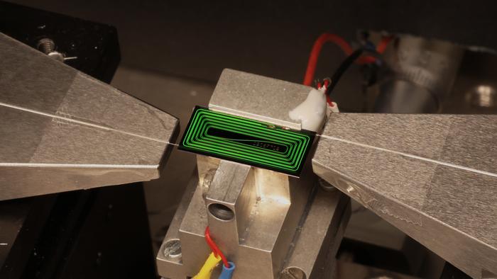
Scientists figured out how to shrink huge ultrafast lasers so they fit on a tiny chip ‪‪—‬ the 'holy grail' of the field

A breakthrough in photonic chips could make large, costly, ultrafast lasers dramatically smaller, leading to portable and affordable imaging, diagnostic and information-processing devices, researchers say.

By using a decades-old overlooked laser architecture, scientists managed to fit an ultrafast laser onto a tiny photonic chip — a chip that uses light, rather than electricity, for computing operations.

In a new study published June 3 in the journal Nature, the team demonstrated that a tiny laser on the photonic chip could deliver 1.05 nanojoules of energy in 147-femtosecond (147 quadrillionths of a second) bursts — thereby competing with the output of laboratory-class ultrafast lasers.

Ultrafast lasers are used in a variety of applications, from precision manufacturing and eye surgery to biological imaging and atomic clocks, but the systems needed to power them tend to take up whole tabletops in labs or factories. Yet the powerful output of these laser pulses made them difficult to miniaturise onto photonic chips.

"For more than twenty years, a high-pulse-energy femtosecond laser on chip was widely regarded as a holy grail of integrated photonics," Tobias Kippenberg, a photonics professor at the Swiss Federal Institute of Technology(EPFL), said in a statement.

"Our result shows that it is not only possible, but that it can be achieved with a surprisingly elegant architecture that the integrated-photonics community had overlooked."

Forward-thinking breakthrough comes from looking back

Photonic chips manipulate light by using microscopic structures called waveguides — usually in the form of optical fibers or etched cavities — to carry information. They aren't particularly novel, and can be found in fiber-optic communications, medical sensors and lidar systems.

But photonic chips have previously struggled when handling high-powered, ultrafast lasers. That's because they need to contain light to extremely small waveguides, leading the light to interact strongly with itself and destabilizing the laser pulses.

To tackle this problem, the researchers looked at a laser architecture called the Mamyshev oscillator, created in 1998 by Pavel V. Mamyshev, a physicist and engineer at Bell Labs.

A close up of a chip on a metal platform.

EPFL's chip-based ultrafast laser operates in a testing set up. (Image credit: Zheru Qiu/EPFL)

This oscillator, which has received little attention in the world of photonic chips, works by placing a nonlinear waveguide between two optical filters. This causes a high-intensity laser pulse to expand into a broader range of colors that can then pass through both filters while weaker light, which can cause laser destabilization, is blocked out. This technique essentially means that a high-intensity laser pulse can be maintained.

Because the Mamyshev oscillator doesn't require extra components to manufacture on a chip, it presents an attractive design for use on photonic chips. And although the laser cavity needed to direct an ultrafast laser is 16.5 inches (42 centimeters) long, it can be folded to occupy around the same area as a match head. This can't be done with conventional fiber-optic-based lasers, often used in photonic chips.

That takes care of the size, but the cost of ultrafast laser systems is another challenge. But because photonic chips can be fabricated using silicon wafers in the same fashion as computer chips, more than 1,000 laser cavities could potentially be produced in a single batch, the researchers said. As such, photonic chips with ultrafast laser capabilities could be produced at scale, in turn reducing manufacturing costs and even expanding their use.

Photonic chips capable of handling ultrafast lasers could, in the future, lead to portable tools for tasks like detecting pollutants or performing advanced medical diagnostics in the field, the researchers noted in the study. The technology also opens the door to smaller atomic clocks that can benefit navigation and future communications.

  •  

Cutting Through Optical Noise: A Clearer Method to Image the Eye

In the realm of modern ophthalmology, Optical Coherence Tomography (OCT) has revolutionized the way eye diseases are diagnosed and monitored. This non-invasive imaging technology allows clinicians to peer through the layers of the retina, producing detailed cross-sectional images that reveal the structural integrity of this delicate tissue without the need for surgical intervention. However, despite OCT’s widespread adoption and profound clinical impact, the technology is inherently limited by physical constraints that occasionally obscure or degrade the crucial information contained within these images. The newly developed Spatio-Temporal Optical Coherence Tomography (STOC-T) promises to address these limitations by fundamentally altering data acquisition methods, thereby enhancing image quality and diagnostic precision at the earliest possible stage.

OCT functions by directing light into the eye and interpreting the backscattered photons to construct images of internal structures like the retina and choroid. But the journey of photons through tissue is fraught with challenges. When light encounters complex biological matrices, it scatters unpredictably, producing signals that blend with those carrying useful information. This phenomenon, known scientifically as “optical crosstalk,” causes image blur and loss of contrast, making it difficult to discern fine detail that could be critical for early disease detection. Optical crosstalk is essentially a breakdown of coherence within the returning light, as photons from a single spatial point arrive dispersed across multiple detector pixels. For ophthalmologists and patients alike, this translates to potentially missed or delayed diagnosis of conditions where subtle changes herald disease onset.

The breakthrough reported by Professor Maciej Wojtkowski and his team from the International Centre for Translational Eye Research (ICTER) introduces STOC-T, an innovative approach that goes beyond conventional image enhancement techniques. Unlike software filters applied post-hoc to clean up images, STOC-T reforms the optics of data collection itself—imposing controlled, repetitive phase modulations onto the illuminating light during scanning. By employing distinct spatial phase masks that alter the wavefront of the illuminating beam, the system captures a series of images, each with a differently patterned illumination. Scattered photons, which respond chaotically to these phase shifts, become decorrelated and diminish upon averaging multiple frames. Conversely, photons faithfully reflecting true tissue architecture maintain coherent behavior, thus strengthening their visibility in the final reconstructed image.

This methodology can be likened to differentiating a single voice in a bustling crowd. Just as a consistent voice can be isolated amid random background chatter using adaptive recording strategies, STOC-T distinguishes meaningful optical signals from noise in real-time. This preemptive discrimination obviates the need for computational post-processing to “clean” images after data acquisition, which historically cannot fully recover information lost to scattering. Defensive design of the imaging process ensures that interfering signals are suppressed from the outset, preserving delicate cellular-level details of the retina and choroid that are indispensable for early diagnosis.

STOC-T’s performance has been rigorously tested both in laboratory settings and on living tissue. In one compelling demonstration, a standard imaging target obscured by a highly scattering artificial medium and even by rat skin rendered virtually invisible under conventional OCT. Upon integrating STOC-T’s phase modulation technique, the obscured structural features resurfaced with remarkable clarity. This experiment underscores the scale of the optical crosstalk problem and the potency of STOC-T, since the objects remained physically present but were previously hidden by noise.

The true clinical significance comes from STOC-T’s application to human retinal imaging, where it achieves a lateral resolution near five micrometers—a scale sufficiently fine to resolve individual photoreceptors and ganglion cells as well as intricate vasculature within the choroid. This extraordinary level of detail enables prospective monitoring of microscopic disease processes with an acuity far surpassing existing OCT methods. Additionally, STOC-T provides new insights through optoretinography (ORG), capturing functional responses of photoreceptors to flickering light stimuli at frequencies up to 45 Hz. These functional measurements resemble electrophysiological studies conducted via invasive patch-clamp techniques, suggesting that STOC-T could non-invasively monitor cellular function—a significant leap for early detection of retinal dysfunction before structural damage occurs.

The potential clinical impact of this technology cannot be overstated. Visual impairment affects more than 2.2 billion individuals worldwide, with over a billion cases attributable to conditions amenable to early diagnosis and intervention. Diseases such as glaucoma, diabetic retinopathy, and age-related macular degeneration often become irreversibly severe due to delayed detection. STOC-T’s capacity to enhance diagnostic accuracy at the cellular and functional levels augments the ophthalmologist’s ability to initiate timely therapies, potentially halting or reversing vision loss before clinical symptoms manifest.

Despite these promises, STOC-T remains an experimental technique, currently limited by technical demands. The system requires cutting-edge hardware, including a high-speed CMOS camera capable of capturing quarter-million frames per second and a tunable near-infrared laser source spanning 800 to 870 nanometers in wavelength. The immense data volume generated—exceeding 8.5 gigabytes per acquisition—also presents significant computational challenges for real-time processing and analysis. These hurdles explain why STOC-T is not yet in widespread clinical use, although ongoing advances in photonic hardware and data science are likely to mitigate these barriers.

Looking forward, the research team envisions enhancing the system’s flexibility using multimode optical fibers for phase modulation. Such fibers, with diameters around 50 micrometers and lengths extending to hundreds of meters, support hundreds of propagation modes. They offer the theoretical potential to reduce optical crosstalk noise by a factor approaching 30 without complex electronic controls, simplifying implementation while preserving image quality improvements.

Professor Wojtkowski emphasizes that STOC-T represents a transformative conceptual advance rather than a final product. The roadmap to widespread application involves optimizing speed, minimizing data volume, refining phase encoding strategies, and automating image reconstruction workflows. The foundational principle—shaping the acquisition process to preempt noise contamination—opens avenues not only for ophthalmology but also for diverse biomedical imaging fields where light scattering impairs image fidelity. This innovation exemplifies how deep physical understanding combined with technical ingenuity can reshape diagnostic imaging, making previously invisible biological details accessible and advancing patient care.

In summary, STOC-T addresses a critical bottleneck in OCT imaging by employing spatio-temporal phase modulation to separate meaningful tissue signals from scattered noise at the data collection stage. This technique enhances resolution, contrast, and functional imaging capability, holding significant promise for earlier and more accurate diagnosis of a broad spectrum of vision-threatening diseases. Although currently laboratory-bound due to technical demands, STOC-T’s theoretical and experimental foundations herald a new era in optical imaging, where noise is never blindly accepted but actively prevented from degrading our view of living tissue.


Subject of Research: Human tissue samples

Article Title: Spatio-temporal optical coherence imaging and tomography for in vivo applications

News Publication Date: 25-May-2026

Web References: DOI: 10.1117/1.JBO.31.11.113504

Image Credits: Optica

Keywords

Optical coherence tomography, STOC-T, ophthalmology, retinal imaging, optical crosstalk, phase modulation, optoretinography, photoreceptors, biomedical optics, imaging noise reduction, high-resolution microscopy, optical imaging innovations

  •  

Photoreforming Solid Waste with Single-Source Co-Catalysts

In a groundbreaking development poised to redefine waste management and sustainable energy production, researchers have unveiled a revolutionary method for photoreforming solid waste on an unprecedented 1-square-meter scale. This innovative advance, detailed in the latest issue of Nature Chemical Engineering, leverages single-source precursor-derived co-catalyst films to convert ubiquitous solid waste into valuable chemical fuels, heralding a new era in environmental remediation coupled with renewable energy generation.

At the heart of this innovation lies the principle of photoreforming, a process by which sunlight catalyzes the chemical transformation of organic materials into hydrogen and other energy-rich molecules. Historically limited to small-scale demonstrations and powdered catalysts, this method struggled with scalability and practical application outside controlled laboratory settings. The reported technique shatters these limitations by fabricating robust co-catalyst films derived from a unified precursor source, enabling efficient photoconversion across large surface areas with enhanced stability and performance.

The researchers crafted these functional films through an ingenious synthesis route, where a single molecular precursor simultaneously yields both the active catalytic sites and the supporting matrix. This contrasts with conventional multi-step fabrication approaches that often result in inconsistent catalyst dispersion and energy losses. By integrating the catalyst components at the molecular level, the team ensured homogeneity, maximized photon absorption, and optimized charge separation dynamics, all critical parameters for sustained photocatalytic activity.

Transforming real-world solid waste – encompassing plastics, biomass residues, and mixed refuse – into clean fuels presents a formidable challenge due to their complex chemical compositions and structural heterogeneity. The co-catalyst films demonstrated remarkable versatility and adaptability, efficiently processing these diverse substrates under simulated sunlight without requiring extensive pre-treatment. This robustness signals a significant leap toward practical deployment in municipal waste processing facilities and industrial settings.

The experimental setup encompassed a square meter of coated substrate exposed to controlled illumination, mirroring natural sunlight intensity conditions. Over extended operation, the system consistently yielded high rates of hydrogen and other value-added chemicals, outperforming benchmark photocatalysts by a considerable margin. Importantly, the films manifested remarkable photostability and mechanical adhesion, demonstrating resilience against degradation mechanisms like photo-corrosion and mechanical abrasion that typically afflict photocatalytic layers.

At the nanoscale, characterization techniques revealed uniform distribution of nanosized catalytic domains embedded within a conductive, photoactive matrix. This architecture ensures rapid electron-hole separation and transport, minimizing recombination losses which commonly plague photocatalytic systems and limit hydrogen evolution rates. Spectroscopic analyses corroborated enhanced visible-light absorption, attributed to tailored bandgap engineering achieved during precursor design, broadening the usable solar spectrum beyond ultraviolet wavelengths.

The process design also embraced mass transport optimization, incorporating porous film structures that facilitated effective diffusion of reactants and removal of gaseous products. This morphologic control prevented stagnation zones and concentration gradients, enhancing catalytic turnover and ensuring stable long-term performance. Additionally, the modular film fabrication approach promises scalability and integration into various reactor geometries without compromising catalytic efficiency.

Beyond hydrogen generation, the system also showcased the capacity to produce liquid fuels and chemical feedstocks, capitalizing on selective reaction pathways induced by co-catalyst composition tuning. This selectivity allows tailored conversion routes matching industrial chemical demands, moving beyond mere waste disposal toward circular chemical economies. The dual benefit of environmental waste mitigation coupled with clean energy and chemical synthesis embodies transformative potential for sustainable industrial practices.

Critically, the team emphasized the environmental and economic implications of adopting such technology at scale. By converting problematic solid waste streams into valuable resources using sunlight – a free and abundant energy source – this approach diminishes reliance on fossil fuels and reduces landfill burden. Cost analyses suggested that, once scaled, the technique could rival established catalytic processes in operational expenditure, thus offering an attractive proposition for policymakers and industry leaders aiming to meet stringent environmental targets.

The interdisciplinary collaboration instrumental in achieving this advance integrated expertise across materials chemistry, photophysics, environmental engineering, and nanofabrication. Such synthesis of disciplines underscored the inherent complexity of developing scalable photocatalytic platforms capable of handling real-world waste complexities while maintaining high efficiency and durability.

Looking ahead, the researchers are exploring further enhancements including tandem catalyst layers, optimized co-catalyst configurations, and hybrid photochemical-electrochemical systems to elevate the energy conversion efficiency and broaden substrate compatibility. In parallel, pilot-scale demonstrations are underway to validate system performance in outdoor environments subject to variable weather conditions, pivotal for transitioning laboratory innovation into field applications.

This pioneering work sets a new benchmark in photoreforming science, illustrating how precise molecular engineering and thoughtful system design can transform a pressing global challenge—solid waste accumulation—into a renewable energy opportunity. Its implications resonate strongly with global sustainability aspirations, promising an economically feasible and environmentally benign pathway to simultaneously address climate change mitigation, waste reduction, and clean energy production.

As society grapples with mounting waste generation paired with escalating energy demands, innovations such as these underscore the invaluable role of scientific ingenuity in crafting solutions that are as elegant as they are practical. By harnessing the synergy of sunlight and advanced material chemistry, the future of waste management is illuminated—not as a burden, but as a wellspring of renewable chemical energy.

In summary, this major scientific milestone demonstrates that photoreforming solid waste at a 1 m² scale using single-source precursor-derived co-catalyst films is no longer a theoretical possibility but a tangible technological reality. It unequivocally paves the way for deploying solar-driven catalytic systems in addressing environmental and energy crises through smart design and scalable engineering.

Subject of Research:
Photoreforming of solid waste using single-source precursor-derived co-catalyst films.

Article Title:
Photoreforming of solid waste on 1 m² scale using single-source precursor-derived co-catalyst films.

Article References:
Bin Mohamad Annuar, A., Liu, Y., Bhattacharjee, S. et al. Photoreforming of solid waste on 1 m² scale using single-source precursor-derived co-catalyst films. Nat Chem Eng 3, 351–362 (2026). https://doi.org/10.1038/s44286-026-00406-y

Image Credits:
AI Generated

DOI:
10.1038/s44286-026-00406-y

Keywords:
Photoreforming, solid waste conversion, co-catalyst films, solar energy, hydrogen production, photocatalysis, renewable energy, waste-to-fuel, sustainable chemistry, scalable catalysis

  •  

Scientists discover hidden ‘charge trap’ that could slow next-generation solar and electronic materials

A team of researchers at Cornell University has uncovered a surprising problem in a promising class of materials known as hybrid perovskites. The discovery could help scientists build better materials for future technologies, including solar cells, energy-harvesting devices, and advanced electronics. Hybrid perovskites have attracted enormous interest in recent years because they are efficient at […]

The post Scientists discover hidden ‘charge trap’ that could slow next-generation solar and electronic materials appeared first on Knowridge Science Report.

  •  

New electronic skin can be made onsite, bringing smarter robots and wearables closer to reality

Researchers in South Korea have developed a new type of electronic skin that can be made directly where it is needed, potentially opening the door to smarter robots, advanced wearable devices, and more interactive technologies. Electronic skin, often called e-skin, is a thin and flexible material filled with sensors that can detect pressure, touch, and […]

The post New electronic skin can be made onsite, bringing smarter robots and wearables closer to reality appeared first on Knowridge Science Report.

  •  

Ice cream ingredient could help turn dirt into 3D-printed homes

An ingredient commonly used in ice cream may help scientists transform ordinary dirt into a new generation of environmentally friendly building materials. Researchers at the University of Colorado Boulder have discovered that sodium alginate, a substance made from seaweed and often used to improve the texture of foods, can make natural materials like clay and […]

The post Ice cream ingredient could help turn dirt into 3D-printed homes appeared first on Knowridge Science Report.

  •  

New Technology Uncovers How the Heart Produces Cells with Regenerative Potential

In a groundbreaking advancement for cardiovascular science, researchers at the Centro Nacional de Investigaciones Cardiovasculares Carlos III (CNIC) in Spain, in collaboration with the Karolinska Institute in Sweden, have devised a cutting-edge technique to analyze the proteome of individual cardiomyocytes. These cells, essential for heart contraction, have long eluded comprehensive single-cell proteomic scrutiny due to technological limitations. This novel approach now allows for a detailed protein-level characterization of single heart muscle cells, opening unprecedented avenues for understanding heart biology and regeneration.

The heart has traditionally been viewed as an organ comprised of relatively homogeneous cardiomyocytes. However, recent findings highlight remarkable heterogeneity even within this single cell type. Variability in protein expression among cardiomyocytes may underpin differences in their functional capacity and regenerative potential. This study, published in Genome Biology, leverages an innovative integration of optimized cell isolation methods, advanced mass spectrometry, and sophisticated bioinformatic algorithms to dissect this proteomic diversity with extraordinary precision.

Central to their investigation was Myc, a transcription factor already noted for its role in regenerative strategies for heart repair. Despite its promise, until now the specific mechanisms by which Myc modulates protein expression in individual cardiomyocytes remained elusive. Applying their novel single-cell proteomics technology, the researchers uncovered that Myc expression differentially alters metabolic enzyme levels across single cardiomyocytes. This heterogeneity results in a subset of cells exhibiting distinct immature states, which in turn possess enhanced regenerative potential—a finding that transforms our comprehension of how cardiac regeneration might be selectively targeted.

The technical feat achieved by these scientists hinges on the fusion of multiple cutting-edge methodologies. First, the meticulous isolation of adult mouse cardiomyocytes ensures viable, intact cells reflecting true physiological states. These cells then undergo mass spectrometry procedures tailored for minute sample volumes, capable of identifying thousands of proteins with high sensitivity and quantitative accuracy at the single-cell level. The ensuing data are processed with bespoke computational pipelines designed to handle the intrinsic variability and noise characteristic of single-cell omics measurements.

By revealing the proteomic fingerprints that distinguish a pro-regenerative subpopulation of cardiomyocytes, this research elucidates a previously uncharted layer of cardiac cellular complexity. Understanding how Myc drives metabolic remodeling at the protein level provides a molecular explanation for its regenerative effects and establishes a foundation for engineering therapeutic approaches that harness or mimic these cellular states.

Regeneration in the adult mammalian heart remains a formidable challenge, owing largely to the low intrinsic capacity of cardiomyocytes to proliferate or replace damaged tissue after injury such as myocardial infarction. Previous models relying largely on bulk tissue analyses have masked the nuanced cellular dynamics at play. This single-cell approach offers resolution sufficient to detect rare but critical cell subsets that might hold the key to effective myocardial repair.

Moreover, this study underscores the transformative impact of integrating proteomics with single-cell biology, moving beyond transcriptomic analyses that often fail to predict functional protein abundance accurately. Proteins, as the primary effectors of cellular function, are the crucial link between gene regulation and phenotype. Hence, this proteomic lens provides a direct readout of the biochemical state of cardiomyocytes under regenerative cues.

The implications of this work extend beyond cardiac biology. It exemplifies the broader potential of single-cell proteomics to deepen our grasp of cellular heterogeneity in health and disease, from cancer biology to neurodegeneration. The refinement and dissemination of such methodologies will empower researchers across biomedical fields to uncover subtle but impactful cellular states previously hidden in ensemble measurements.

Miguel Torres and Jesús Vázquez, who co-led the CNIC teams responsible for this study, emphasize the critical novel insights gained. According to them, dissecting Myc’s action at the single-cell level illuminates pathways and protein networks that govern cellular immaturity and regeneration, which are masked in population-level studies. This knowledge fundamentally shifts how regenerative medicine strategies might be designed, focusing on targeted modulation of key proteins identified within pro-regenerative cardiomyocyte subgroups.

Consuelo Marín-Vicente, the study’s first author, highlights the heterogeneous response to Myc induction in adult cardiomyocytes. Instead of a uniform effect, Myc induces diverse proteomic profiles, driving some cells toward a state primed for regeneration. This discovery points to the existence of intrinsic regulatory mechanisms that govern cardiomyocyte plasticity, with possible manipulation routes to expand these favorable subpopulations therapeutically.

This advancement also signifies a leap toward personalized regenerative therapies. By characterizing the protein signatures specific to regenerative-competent cardiomyocytes, future interventions could selectively enhance these signatures in patient hearts, enabling more effective recovery post-injury. It also offers biomarkers to monitor therapeutic efficacy or to stratify patients likely to benefit from Myc-based or related regenerative treatments.

The CNIC, recognized as a Severo Ochoa Center of Excellence and funded by a unique public-private partnership, remains at the forefront of cardiovascular research. Under the leadership of Dr. Valentín Fuster, CNIC’s mission to translate scientific discoveries into clinical innovations is exemplified by this study’s promise. Collaboration with leading international centers like the Karolinska Institute further underscores the global effort to unravel heart regeneration’s complexity.

In sum, by pioneering a technique for single-cell proteomic analysis of adult cardiomyocytes, this research opens transformative possibilities for cardiovascular medicine. It elucidates how transcription factors like Myc can orchestrate heterogeneous proteomic landscapes within single cells, generating subpopulations equipped with regenerative capabilities. These insights herald a new era in designing precision therapies that could one day restore heart function after injury, dramatically improving outcomes for millions suffering from cardiovascular diseases worldwide.


Subject of Research: Cells

Article Title: Pro-regenerative fingerprints identified in a sub-population of adult mouse cardiomyocytes by integrative single-cell proteomics

News Publication Date: 22-May-2026

Web References:
10.1186/s13059-026-04110-1

Image Credits: CNIC

Keywords

Cardiomyocytes, Single-cell proteomics, Myc transcription factor, Heart regeneration, Mass spectrometry, Cellular heterogeneity, Cardiovascular disease, Protein expression, Regenerative medicine, Proteome, Metabolic enzymes, Cardiovascular research

  •  

This flying solar-powered platform could deliver better internet from the air

As soon as August, a giant silver bullet will cut its way through the dry air of the southwestern US and cross the Pacific to reach the coast of Japan. 

Once there, the roughly 200-foot-long craft, built by the New Mexico–based company Sceye, will park some 18 kilometers above the ocean’s surface, in a wispy-thin layer known as the stratosphere. Then it will use a custom-built antenna to supplement Softbank’s 5G network, a test that will include beaming data straight to devices. 

Sceye (pronounced “sky”) is one of several firms building a class of airborne craft called HAPS, or high-altitude platform stations or systems. Such a platform can be a plane or a balloon or, yes, an oblong craft filled with helium and outfitted with solar panels. HAPS companies, including the Airbus subsidiary Aalto, envision them serving a variety of lofty purposes, such as delivering internet service to disaster sites and observing Earth’s surface. 

a silver blimp-like aircraft hovering over a field
Hovering over Roswell, the high-altitude system is sheathed in lightweight, reflective fabric.
COURTESY OF SCEYE

The stratosphere is a good place to be if you want to cover a large area. It’s also much closer to the ground than even the lowest-orbiting satellites, which means sending down a signal takes far less energy. “What we ultimately offer is space-like conditions, without the cost of going to space and without the complexity of being in orbit,” says Mikkel Vestergaard Frandsen, Sceye’s CEO and founder.

But it’s also not so easy to stay there. Sceye’s aircraft, Frandsen says, has to be light enough to stay aloft but also strong enough to carry the necessary systems. It must soak up and store enough solar energy during the day to provide around-the-clock power to an electric fan that can maneuver the HAPS back into place when winds knock it out of position—mettle it proved in a 2024 test flight.

Since then, Sceye has been preparing for its big Japan test. In the flight pictured here from this spring, for example, the craft stayed aloft for 12 days as it flew to the coast of Brazil and spent more than 88 hours “parked” in various locations. Eventually, the company expects its platform could help satellite operators better serve densely populated areas.

Someday, Frandsen says, spotting a HAPS may be as common as seeing ships at port or trains on the tracks.

  •  

A Plastic Motor Just Defied a Century of Engineering Assumptions

Electricity Sparks Outside Concept ArtElectrostatic forces enable low-voltage actuation without relying on rare earth materials. Nearly every electric motor in modern life, from the fan on your desk to the motor in an electric vehicle, relies on magnetism. For more than a century, magnetic fields have dominated motor design because electrostatic forces were considered too weak for practical machines. [...]
  •  

AI gives building inspection photos the location data they were missing

When a building inspector takes a photo of a cracked wall, a leaking pipe or a faulty ceiling panel, that image carries almost no information about where exactly it was taken. There's no GPS signal indoors, and manually recording locations is slow, error-prone and easily forgotten. As a result, thousands of inspection photos sit in folders with no spatial context, making it hard to track problems over time or link them to the correct part of a building's maintenance records.

  •  

Infrared filter could help detect pollution and disease

Researchers have developed a tiny, electrically tunable infrared filter that could help shrink bulky thermal sensing systems onto portable chips—a technology that could lead to handheld pollution detectors, compact multispectral cameras and next-generation chemical sensing devices.

  •  

Brewing sustainable power from used coffee grounds

What if your daily coffee routine could help power the world around you? 

Scientists are exploring ways to transform everyday waste, like spent coffee grounds, into materials for greener energy production. One promising method, called triboelectrification, uses electricity generated from motion. With this approach, materials that gain positive and negative charges are rubbed together to generate an electrical charge that can be harnessed by devices called triboelectric nanogenerators, or TENGs. TENGs are a greener alternative to fossil fuels because they generate electricity from everyday movement, such as pedestrian or vehicle motion. 

However, many negatively charged materials used in these devices are made from nonbiodegradable plastics, like PTFE, that persist as long-term waste in the environment. Over time, TENGs are also easily damaged by environmental conditions outside the laboratory. To create sustainable TENGs for real-world applications, scientists need to develop biodegradable and durable designs. 

To address this problem, researchers from Guangxi University in China developed a TENG that uses coffee grounds as its negatively charged material. Their design repurposes coffee waste to create a robust, low-cost device that can harvest energy.

To create the coffee-based material, the researchers mixed coffee grounds with a biodegradable glue-like plastic called polycaprolactone (PCL). They heated this mixture to 80°C (176°F) and molded it into cylindrical pellets. Then, they used a biodegradable plastic called polylactic acid (PLA) to 3D-print a fully enclosed box-like TENG containing vertical channels arranged like the parallel slots of a file organizer to house the pellets. 

Their TENG worked by shaking the coffee ground pellets within the channels, causing them to repeatedly rub against the PLA surfaces. Because coffee grounds strongly attract electrons, the pellets become negatively charged while the PLA surfaces become positively charged. This repeated pellet movement drove the electrons back and forth, generating electricity that was harvested through the TENG’s external circuit. 

To evaluate their TENG’s electrical performance, the researchers used an instrument that measures small amounts of electricity, called an electrometer. The electrometer measures electrical output based on how strongly the device pushes electrons through a circuit, called its voltage, how quickly electrons flow through it, called its current, and how many electrons flow through it, called its transferred charge.

The researchers verified that coffee ground pellets could serve as a viable TENG material by comparing their performance with commercial plastic pellets. The coffee ground pellet system produced a voltage of 0.6 Volts (V), a current of 18 nanoamperes (nA), and a charge transfer of 0.25 nanocoulombs (nC). This output was more than twice that of pure PCL pellets and over half that of nonbiodegradable PTFE. The researchers suggested that the negatively charged components of the coffee grounds altered the surface of the PCL to increase its charge generation and transfer. 

To optimize the TENG, the researchers experimented with different coffee grounds to PCL mass ratios, pellet lengths, and device vibrational frequencies. They found that their device achieved the best electrical performance at a 3:1 ratio of coffee grounds to PCL, with 8 millimeter long pellets, at a vibrational frequency of 2.6 Hertz. These conditions boosted the device’s electrical output to 4.7 V, 75 nA, and 1.3 nC, high enough to power tiny environmental sensors. 

The team also tested their device’s long-term durability by comparing its electrical performance before and after 6 months of storage. They observed only a 6% drop in voltage after storage, demonstrating that it could maintain stable long-term electrical performance for at least 6 months. 

Finally, to explore real-world applications, the researchers integrated 4 TENGs into a wind-energy harvesting system designed for remote island settings. The system used a wheel of rotating cups to capture wind energy and a sliding mechanism to convert it into repeated shaking of the TENGs. As the TENGs shook, the moving pellets inside generated enough electricity to power an LED light and a small weather-monitoring device. 

The researchers concluded that spent coffee grounds can function as biodegradable TENG materials while reducing dependence on fossil fuel-derived materials. By combining coffee waste with biodegradable plastics, the researchers created a TENG with good electrical performance, sustainability, and long-term stability. They suggested that with further development, the device could someday power road warning lights and provide sound-based alerts in remote areas.

The post Brewing sustainable power from used coffee grounds appeared first on Sciworthy.

  •  

Inflammation and Metabolism Trigger Early Puberty in Obese Kids

In a groundbreaking study published in Pediatric Research on June 21, 2026, researchers led by Wang, Yan, and Liu have unveiled critical insights into how metabolic, hormonal, and inflammatory pathways intertwine to accelerate precocious puberty in children suffering from obesity. This pioneering research uncovers the complex biological dialogue that propels early pubertal onset among obese youth, a phenomenon that has raised alarm bells worldwide due to its profound health implications.

Puberty, the process marking the transition from childhood to sexual maturity, is classically regulated by a symphony of endocrine signals, with the hypothalamic-pituitary-gonadal (HPG) axis as the conductor. However, the timing of this process is increasingly disrupted in obese children, leading to precocious puberty—defined as puberty occurring before age 8 in girls and 9 in boys. The new study delves deeply into the serum profiles of obese children to unravel the mechanistic underpinnings of this hastened developmental timeline.

Central to the pathophysiology elucidated by the researchers is the role of systemic inflammation, a hallmark of obesity. The team identified a distinctive inflammatory signature characterized by elevated levels of cytokines and acute phase reactants that converge on the HPG axis. This pro-inflammatory milieu is postulated to trigger neuroendocrine alterations that prematurely activate gonadotropin-releasing hormone (GnRH) neurons, thereby advancing pubertal initiation.

Complementing the inflammatory dimension are metabolic dysregulations that obesity engenders. The study meticulously profiles alterations in insulin and leptin dynamics—two pivotal hormones intimately linked to energy homeostasis and reproductive function. Elevated insulin levels, common in obese states due to insulin resistance, were found to potentiate GnRH neuronal activity. Meanwhile, hyperleptinemia, reflective of adipose tissue abundance, appears to modulate hypothalamic circuits governing puberty, providing a metabolic ‘green light’ for sexual maturation.

The research team employed high-throughput serum profiling techniques alongside advanced statistical modeling to choreograph the intricate interplay of metabolic and inflammatory mediators. This comprehensive approach allowed them to construct a predictive biochemical tableau correlating specific serum markers with rapid pubertal progression. Crucially, these findings bridge the gap between clinical observations of precocious puberty and the molecular pathways that orchestrate it in obesity.

Beyond identifying biomarkers, the study explores potential mechanistic pathways. It proposes that chronic low-grade inflammation potentiates neuronal sensitization within the hypothalamus, thereby lowering the threshold for GnRH secretion. Concurrently, metabolic hormones such as leptin may amplify neuroendocrine responsiveness, creating a synergistic environment favoring early puberty onset. This dual-hit model of inflammation and metabolic disturbance advances our understanding of the biological complexity underlying pubertal acceleration.

Furthermore, the research highlights the nuanced hormonal crosstalk influencing gonadal maturation. Upregulation of adrenal and gonadal steroidogenesis appears to be downstream of the altered serum milieu, fostering a hormonal cascade that further catalyzes secondary sexual characteristic development. This insight underscores the multifactorial nature of obesity-driven puberty alterations, extending beyond central neuroendocrine changes to peripheral endocrine feedback loops.

One of the notable technical innovations in this study is the longitudinal analysis of serum samples from a diverse pediatric cohort. This enabled the researchers to pinpoint temporal shifts in inflammatory and metabolic markers relative to pubertal milestones. The temporal dynamics revealed suggest potential windows for therapeutic intervention that could recalibrate aberrant biological signals before irreversible pubertal advancement occurs.

The implications of these findings are profound, particularly as precocious puberty is linked to increased risks of psychosocial stress, metabolic syndrome, and certain cancers later in life. By elucidating the biological signatures that accelerate puberty in obese children, the study opens avenues for early diagnostic screening and targeted therapeutics aimed at mitigating long-term health impacts.

Medical professionals and endocrinologists stand to gain new tools for assessing pubertal risk by integrating serum inflammatory and metabolic profiles into routine evaluations of obese pediatric patients. This could transform clinical practice by enabling proactive management strategies tailored to individual biochemical risk landscapes, rather than relying solely on phenotypic assessments.

From a public health perspective, the study amplifies the urgency of addressing childhood obesity not only for its well-known metabolic complications but also for its disruptive influence on normal developmental timing. Interventions that reduce systemic inflammation and restore metabolic balance might delay unwanted precocious puberty, improving both physical and mental health outcomes.

Looking forward, the researchers advocate for expanded studies to explore therapeutic modulation of the identified pathways. Immunomodulatory agents, metabolic enhancers, or lifestyle interventions targeting systemic inflammation and hormone regulation could serve as novel strategies to realign pubertal timing in susceptible children. Such approaches hold promise for personalized medicine frameworks in pediatric endocrinology.

This investigation also raises important questions about environmental and genetic factors that may interact with inflammatory and metabolic signals to influence puberty. Understanding how external exposures or inherited susceptibilities modulate these biochemical networks could refine predictive models and prevention efforts in diverse populations.

In conclusion, Wang, Yan, Liu, and colleagues have charted an unprecedented map of the intersecting biological forces accelerating precocious puberty in obese children. Their integrative analysis of inflammatory and metabolic serum markers fundamentally shifts our comprehension of childhood development, offering hope for interventions that could recalibrate the endocrine clock disrupted by obesity. As the obesity epidemic continues to burden global health, studies like this provide crucial scientific foundations to mitigate its far-reaching consequences on human growth and maturity.


Subject of Research: The interplay of metabolic, hormonal, and inflammatory factors in precocious puberty and obesity in children.

Article Title: Serum inflammatory and metabolic signatures accelerate precocious puberty in obese children.

Article References:

Wang, L., Yan, H., Liu, J. et al. Serum inflammatory and metabolic signatures accelerate precocious puberty in obese children.
Pediatr Res (2026). https://doi.org/10.1038/s41390-026-05214-x

Image Credits: AI Generated

DOI: 21 June 2026

  •  

Pediatric Genomic Medicine: Rapid Progress, Measured Pace

In an era marked by extraordinary advances in genomic science, pediatric medicine stands on the brink of a transformative revolution. Recent research, as highlighted in the 2026 John Howland Award Lecture by B.D. Gelb, reveals both the breathtaking pace of progress in pediatric genomic medicine and the imperative for measured, responsible integration. This evolving field is redefining the management and understanding of childhood diseases through the power of genomic technologies, yet it also confronts significant challenges that necessitate careful consideration.

At the core of pediatric genomic medicine is the ability to decode the human genome with unprecedented speed and resolution. Next-generation sequencing technologies have dramatically accelerated the identification of genetic variants associated with pediatric conditions, facilitating earlier diagnoses and more targeted therapies. This acceleration not only shortens diagnostic odysseys that often span years but also opens a window into personalized interventions that enhance efficacy and safety in young patients.

However, the speed of these advances demands caution. As Gelb emphasizes, the clinical application of genomic findings must be tempered by rigorous validation and ethical oversight. The rush to implement novel genomic tests and treatments without comprehensive understanding risks unintended consequences—ranging from misinterpretation of data to exacerbating health inequities. The pediatric population, uniquely vulnerable due to developmental considerations, requires a thoughtful approach that balances innovation with prudence.

One remarkable aspect of pediatric genomic medicine is its ability to elucidate rare genetic disorders that were previously enigmatic. Genomic sequencing has illuminated the molecular basis of numerous syndromes, enabling not only precise diagnosis but also shedding light on underlying biological pathways. This knowledge accelerates drug discovery and paves the way for novel therapeutic strategies tailored to the genetic makeup of each child, transitioning from a one-size-fits-all paradigm to precision medicine.

Data integration emerges as a crucial enabling factor in this field. Beyond raw sequencing data, combining clinical phenotypes, family history, and environmental factors enhances diagnostic accuracy. Advanced bioinformatics tools and artificial intelligence algorithms play integral roles in interpreting complex genomic datasets, filtering noise from clinically relevant signals. These computational advancements elevate pediatric genomic medicine from a purely descriptive science to a predictive and proactive discipline.

Yet, the ethical dimensions of pediatric genomics loom large. Issues surrounding informed consent, privacy, and data sharing become particularly complex when dealing with minors. Parents and guardians must navigate decisions that can affect the child’s present and future health, often mediated by uncertainties inherent to genomic interpretations. Responsible stewardship of genomic information necessitates frameworks that safeguard patient autonomy while promoting equitable access to advances.

The potential for genomic screening in newborns represents both an opportunity and a quandary. Early detection of genetic abnormalities could revolutionize preventive care and improve long-term outcomes. Nonetheless, the broader implications of widespread genomic screening require careful examination, including considerations of psychological impact, clinical utility, and cost-effectiveness. Balancing these factors will define the responsible trajectory of pediatric genomic screening programs.

An essential component of progress is the education and training of healthcare providers. Clinicians must be equipped with not only the technical understanding of genomic technologies but also the skills to counsel families effectively. Multidisciplinary teams integrating geneticists, bioinformaticians, ethicists, and pediatricians form the backbone of successful genomic medicine services, ensuring holistic patient care that accounts for biological and psychosocial dimensions.

The expanding landscape of pediatric genomic medicine also intersects with burgeoning fields such as gene editing and regenerative therapies. CRISPR-Cas9 and other tools offer the tantalizing prospect of correcting deleterious mutations at their source. Nonetheless, these approaches are nascent and fraught with scientific and ethical complexities, highlighting the necessity for robust preclinical studies and stringent regulatory oversight before clinical adoption.

International collaboration represents a powerful avenue for accelerating pediatric genomic research and equitable benefit. Shared databases, standardized protocols, and cross-border consortia facilitate the aggregation of genetic data from diverse populations—an essential factor in elucidating variant pathogenicity and expanding therapeutic horizons. Global efforts can help overcome the limitations of fragmented data and enhance the generalizability of genomic discoveries.

Importantly, the pediatric genomic medicine community recognizes the risk of exacerbating healthcare disparities. Technological advances tend to concentrate in well-resourced centers, potentially leaving underserved populations behind. Addressing these disparities requires concerted policy initiatives, public engagement, and infrastructure development to ensure that genomic medicine is accessible to all children, regardless of socioeconomic or geographic status.

Looking ahead, the integration of real-world data and longitudinal studies will be critical in refining the understanding of genomic variants’ penetrance and expressivity in pediatric populations. Such comprehensive datasets will inform dynamic risk models and adaptive therapeutic approaches, further personalizing pediatric care and optimizing outcomes across diverse clinical contexts.

In sum, the landscape of pediatric genomic medicine is one of breathtaking promise intertwined with considerable complexity. The rapid progression in genomic technologies propels the field into uncharted territory, demanding a measured pace that safeguards patient welfare while nurturing innovation. The insights from Gelb’s 2026 John Howland Award Lecture resonate as a clarion call to balance enthusiasm with responsibility, ensuring that the promise of genomics translates into tangible, equitable benefits for children worldwide.

As pediatric genomic medicine continues to evolve, the collaborative engagement of scientists, clinicians, ethicists, policymakers, and patient communities will be paramount. This multidisciplinary synergy will chart the course for harnessing genomic insights with precision and compassion, transforming pediatric healthcare in profound and enduring ways. The future, while bright, must be navigated with vigilance, wisdom, and inclusivity to realize the full potential of genomic medicine for the youngest and most vulnerable members of society.


Subject of Research: Pediatric Genomic Medicine and its clinical and ethical advancement.

Article Title: Pediatric Genomic Medicine—rapid progress, but not too fast: American Pediatric Society’s 2026 John Howland Award Lecture.

Article References:
Gelb, B.D. Pediatric Genomic Medicine—rapid progress, but not too fast: American Pediatric Society’s 2026 John Howland Award Lecture. Pediatr Res (2026). https://doi.org/10.1038/s41390-026-05233-8

Image Credits: AI Generated

DOI: https://doi.org/10.1038/s41390-026-05233-8

  •  

Cutaneous Risks in Advanced Pediatric IBD Therapies

In a groundbreaking study published in Pediatric Research, researchers have shed new light on the risk factors associated with cutaneous complications arising from advanced therapies in pediatric patients suffering from inflammatory bowel disease (IBD). This research is poised to revolutionize the way clinicians approach dermatological side effects linked to cutting-edge treatments, potentially transforming patient care and outcomes.

Pediatric inflammatory bowel disease, comprising Crohn’s disease and ulcerative colitis, has seen an upsurge in prevalence worldwide, presenting unique challenges due to its chronicity and systemic nature. Advanced therapies, including biologics and small molecule inhibitors, have significantly improved disease management, yet their use is not without adverse effects. Among these, skin complications have emerged as a concerning issue, sometimes compromising treatment adherence and quality of life for young patients.

This study meticulously analyzed a cohort of pediatric IBD patients undergoing advanced therapeutic regimens to identify predisposing factors for cutaneous side effects. The researchers employed a multidisciplinary approach, integrating clinical data with immunological and genetic profiling, to unravel the complex interplay leading to dermatological manifestations.

One of the pivotal findings highlighted the differential risk profiles between various classes of advanced therapies. For instance, tumor necrosis factor-alpha (TNF-α) inhibitors were more frequently associated with psoriasiform lesions, a paradoxical reaction where treatments aimed at immune suppression triggered immune hyperactivity in the skin. This paradox underscores the inherent complexity of immune modulation in pediatric patients.

Additionally, the investigation illuminated the role of individual patient factors such as genetic predispositions and baseline immune status. Specific polymorphisms in genes regulating cytokine production appeared to influence susceptibility to skin complications, revealing a potential avenue for personalized risk assessment before initiating therapy.

Environmental and lifestyle factors were also evaluated, revealing that patients exposed to certain external triggers, including ultraviolet radiation and specific microbiota profiles on the skin, manifested increased vulnerability to therapy-induced cutaneous conditions. These insights pave the way for adjunct preventive strategies alongside pharmacologic interventions.

The temporal dynamics of cutaneous adverse events emerged as another crucial aspect. Most skin complications manifested within the initial months of therapy commencement, emphasizing the need for vigilant dermatological monitoring during this critical window. However, late-onset reactions were not uncommon, suggesting that longitudinal follow-up remains essential throughout the treatment course.

The study further elucidated the pathogenic mechanisms underpinning these complications. Immune dysregulation triggered by therapy was shown to alter skin barrier function, promoting inflammatory cascades that translated into clinical manifestations. Understanding these pathways at a cellular and molecular level provides a framework for developing targeted therapeutics to mitigate side effects without compromising efficacy.

Importantly, the management strategies for cutaneous complications were explored. The researchers advocated for integrated care models involving dermatologists, gastroenterologists, and immunologists to tailor interventions. Topical therapies, dose adjustments, and, in some cases, switching to alternative agents were recommended based on severity and patient response.

This research also calls attention to the psychosocial impact of skin complications in pediatric IBD patients. Given the visibility of dermatological issues, children often experience emotional distress and stigma, highlighting the necessity for holistic care approaches that address both physical and mental health dimensions.

Moreover, the study underscores the potential of predictive biomarkers in clinical practice. By identifying patients at heightened risk, clinicians can preemptively adapt therapeutic regimens or introduce prophylactic measures, thereby reducing morbidity associated with cutaneous adverse events.

From a broader perspective, these findings resonate beyond pediatrics, offering insights applicable to adult IBD populations and other autoimmune conditions treated with similar modalities. The interplay between systemic immunity and cutaneous manifestations represents a fertile ground for future translational research.

In conclusion, the elaborate dissection of risk factors for cutaneous complications in pediatric IBD patients receiving advanced therapies marks a significant stride toward personalized medicine. As the therapeutic landscape evolves, integrating such nuanced knowledge will be crucial in optimizing outcomes and enhancing quality of life for young patients grappling with chronic inflammatory diseases.

This study not only deepens our understanding of adverse effects but also stimulates a paradigm shift in patient monitoring and management strategies. It advocates for a proactive approach, blending precision medicine with interdisciplinary collaboration, to navigate the complexities of advanced therapeutic interventions safely.

With inflammatory bowel disease continuing to impose a heavy burden on pediatric populations globally, these insights arrive at a timely juncture. They equip clinicians with evidence-based guidance to balance efficacy and safety, ensuring that advancements in treatment translate into tangible benefits without unintended harm.

The publication of this research invites further investigations into innovative solutions, such as novel drug formulations with reduced dermatological toxicity and development of comprehensive screening tools. As science progresses, the integration of immunogenetics, microbiome analysis, and environmental assessment promises to refine risk stratification models.

Ultimately, this work epitomizes the convergence of clinical acumen and scientific rigor, spotlighting the imperative to address all facets of patient health in the era of precision medicine. By illuminating the dermatological dimension of pediatric IBD therapy, it inspires a more empathetic, informed, and effective clinical practice moving forward.


Subject of Research: Risk factors for cutaneous complications associated with advanced therapies in pediatric inflammatory bowel disease patients.

Article Title: Risk factors for cutaneous complications of advanced therapies in paediatric inflammatory bowel disease patients.

Article References:
Kazeka, D., Zarubova, K., Copova, I. et al. Risk factors for cutaneous complications of advanced therapies in paediatric inflammatory bowel disease patients. Pediatr Res (2026). https://doi.org/10.1038/s41390-026-05195-x

Image Credits: AI Generated

DOI: 10.1038/s41390-026-05195-x

  •  
❌