Login

When the ability to smell goes away
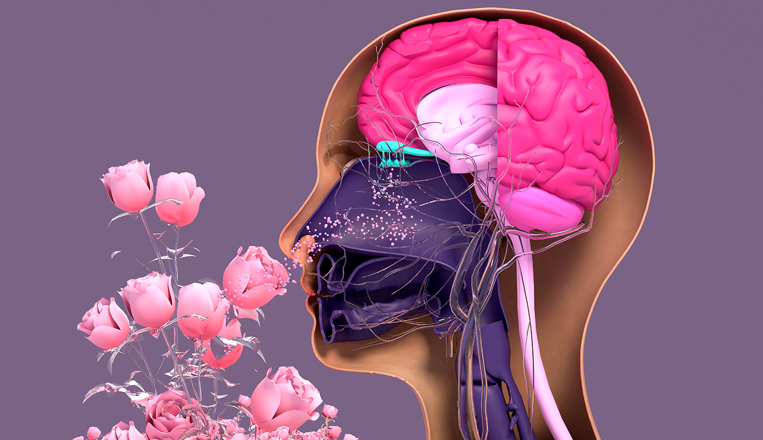
Disturbances in this critical sense are often linked to problems with brain health, researchers are learning

Disturbances in this critical sense are often linked to problems with brain health, researchers are learning

Most people know the ketogenic diet as a popular way to lose weight. Celebrities, athletes, and people trying to improve their health have embraced the low-carbohydrate eating plan over the past decade. But scientists are increasingly discovering that the keto diet may have effects far beyond the bathroom scale. According to a new review published […]
The post This Weight-Loss Diet May Also Fight Alzheimer’s and Parkinson’s Diseases appeared first on Knowridge Science Report.

Alzheimer’s disease gradually steals memories, independence, and quality of life from millions of people worldwide. Despite decades of research, scientists still struggle to find treatments that can stop the disease from progressing. Most available medications provide only modest benefits and cannot fully prevent the loss of brain cells that characterizes the condition. A new study […]
The post Scientists Find a New Way to Fight Alzheimer’s After 20 Years of Research appeared first on Knowridge Science Report.
 Compound 10 slowed dementia-like decline in mice.

The saying that the eyes are the window to the soul may contain more truth than people realize. Scientists now believe that our eyes may also provide valuable information about the health of our brains. A new study suggests that simple photographs of the retina, the light-sensitive tissue at the back of the eye, may […]
The post Your Eyes May Reveal Your Future Risk of Alzheimer’s Disease appeared first on Knowridge Science Report.
In a groundbreaking advance poised to transform the landscape of age-related immune decline, University College London (UCL) researchers are spearheading a pioneering clinical trial aimed at rejuvenating worn-out T cells through an innovative immune restoration therapy. This Phase 1 first-in-human study will explore the therapeutic potential of metabolically resetting exhausted or senescent T cells, a subset of immune cells that accumulate with age and chronic disease and lose their robustness in coordinating immune responses. The initiative promises to reopen avenues for combating a spectrum of diseases, including cancer, HIV, and neurodegenerative disorders like dementia, by profoundly modifying the immune system’s intrinsic capacity to defend and repair.
Central to the research is the recognition that immune ageing, characterized by the progressive dysfunction and decline in immune cell performance, is a key factor leading to increased vulnerability in the elderly and those with chronic infections. T cells, particularly the CD4+ subtype—often regarded as the conductors of the immune orchestra—become less effective with time, diminishing the immune system’s ability to coordinate defenses accurately. These cells, rendered senescent or exhausted, exhibit impaired proliferation, cytokine production, and cytotoxic activity, undermining responses to pathogens, malignancies, and cellular damage.
The novel therapy, developed by SenTcell, a biotech startup founded by Dr. Alessio Lanna from UCL Medicine, employs a liquid formulation administered intramuscularly akin to conventional vaccines. This delivery method facilitates patient-friendly administration while targeting key metabolic pathways within dysfunctional T cells. By reprogramming the intracellular environment of these immune cells, the treatment aims to restore youthful functional properties, including enhanced proliferative capacity and efficient pathogen recognition, thereby revitalizing the immune system’s broad protective functions.
Dr. Lanna explains that although antiretroviral advancements have significantly improved lifespans for individuals living with HIV, many still experience premature immune ageing. Similar patterns of immune exhaustion are also detected in cancer patients and other chronic disease cohorts. The imminent clinical trial represents a critical stepping stone towards verifying whether exhausted immune cells can be safely and effectively rejuvenated in human subjects, with the potential to redefine therapeutic paradigms for diseases rooted in immune dysregulation.
At the molecular level, one of the intriguing aspects under investigation involves the role of telomeres—protective caps on chromosome ends whose shortening serves as a hallmark of biological ageing. Laboratory studies have suggested that rejuvenated CD4+ T cells may release telomere-containing extracellular structures, whimsically termed “telomere Rivers,” into the bloodstream. The presence and behavior of these structures might offer novel insights into how restored immune cells influence systemic health, extending their benefits beyond immediate immune niches to broader tissue health. Although this phenomenon has yet to be validated in human clinical contexts, it highlights the sophisticated interplay between immune rejuvenation and organismal aging.
The clinical trial enjoys regulatory support from the UK’s Medicines and Healthcare products Regulatory Agency (MHRA) through its Innovative Licensing and Access Pathway (ILAP), underscoring the therapy’s potential to meet the pressing unmet medical needs associated with immune senescence. This endorsement accelerates development timelines and affirms the significance of targeting immune metabolic dysfunction as a viable therapeutic strategy.
Participants in the upcoming Phase 1 trial will be meticulously selected adults exhibiting characteristics of immune dysfunction, including evidence of premature immune ageing or persistent viral infections. Comprehensive immune profiling will be conducted before and after therapeutic administration to discern immunological changes and safety outcomes. Although the initial focus is on evaluating biological activity and tolerability, successful results could pave the way for advanced trials assessing clinical efficacy.
Unlike conventional treatments that combat diseases by targeting individual pathogens or tumor cells directly, this therapy seeks to recalibrate the immune system itself—restoring its intrinsic resilience and regenerative functionality. Such a paradigm shift holds profound implications; by enhancing the immune system’s natural defensive repertoire, it may afford broad-spectrum protection and facilitate healthier ageing trajectories.
Beyond chronic infections like HIV and cancer, this approach could ultimately apply to autoimmune disorders characterized by immune exhaustion, wherein overly active yet dysfunctional immune cells drive pathology. Immune rejuvenation strategies offer a novel means of restoring balance without broadly suppressing immune activity, potentially mitigating disease progression while improving quality of life.
This research aligns with a growing global focus on understanding the cellular and molecular underpinnings of ageing and immune decline. By intervening at the level of T cell metabolism and function, scientists hope to extend not only lifespan but healthspan—preserving cognitive and physical function while reducing susceptibility to infections, cancer, and degenerative diseases.
As UCL celebrates two centuries of academic excellence and innovation, this project embodies the institution’s commitment to translating bold scientific ideas into therapies capable of reshaping medicine. If validated through clinical trials, this immune rejuvenation therapy could herald a transformative era in treating age-associated diseases, offering hope for millions affected by immune dysfunction worldwide.
Subject of Research: People
Article Title: Immune Rejuvenation: UCL’s Pioneering Trial Targets T Cell Exhaustion to Combat Age-Related Immune Decline
News Publication Date: Not provided
Web References: www.ucl.ac.uk
References: Not provided
Image Credits: Not provided
Keywords: Immune ageing, T cell exhaustion, CD4+ T cells, immune rejuvenation, senescent immune cells, telomeres, immune dysfunction, chronic disease, HIV, cancer, metabolic resetting, Phase 1 clinical trial
In an unprecedented advancement for neurodegenerative disease research, a team led by Busquets, Li, Syed, and colleagues has unveiled iSCORE-PD, an innovative isogenic stem cell repository designed explicitly to accelerate Parkinson’s disease studies. Published in Nature Communications in 2026, this comprehensive collection represents a pivotal leap toward resolving the complex pathophysiology of Parkinson’s, a condition affecting millions worldwide yet still eluding curative therapies. The iSCORE-PD platform promises to redefine experimental paradigms by providing scientists with genetically matched cellular models that mirror the intricate genetic underpinnings of Parkinson’s disease.
Parkinson’s disease is characterized primarily by the progressive loss of dopaminergic neurons in the substantia nigra pars compacta, leading to debilitating motor symptoms such as tremors, rigidity, and bradykinesia, alongside a spectrum of non-motor complications. Despite extensive research, there remains a conspicuous gap between clinical observations and mechanistic understanding at the cellular and molecular levels. Existing animal models and conventional stem cell lines often fall short in recapitulating the exact human pathology. The iSCORE-PD initiative addresses this deficiency head-on by harnessing isogenic stem cell lines, which are genetically identical except for defined Parkinson’s-relevant mutations, thus enabling unprecedented control over genetic variables.
The technical foundation of iSCORE-PD relies on advanced genome editing methodologies, most notably CRISPR/Cas9 technology. By precisely introducing or correcting mutations in key genes implicated in Parkinson’s pathogenesis such as SNCA, LRRK2, PINK1, and PARKIN, researchers have generated isogenic pairs of induced pluripotent stem cells (iPSCs). These iPSC lines can subsequently be differentiated into dopaminergic neurons or other relevant cell types, facilitating side-by-side comparisons that isolate the effects of specific genetic alterations without the confounding noise of inter-individual genetic variability.
One of the remarkable features of the iSCORE-PD collection is its extensive genomic validation pipeline. Whole genome sequencing and transcriptomic analyses are systematically employed to ensure the integrity and authenticity of the edited lines, confirming that off-target effects are minimized and that the genetic landscape conforms precisely to intended edits. This meticulous validation is crucial for downstream experiments intending to dissect subtle cellular phenotypes attributable to Parkinson’s mutations with a high degree of confidence.
The availability of these isogenic cell lines dramatically enhances the fidelity of disease modeling. Scientists can now observe, in real time and within a human cellular context, the cascade of pathological events triggered by individual mutations. For instance, iSCORE-PD lines harboring the G2019S mutation in LRRK2 have already demonstrated distinct lysosomal dysfunction and mitochondrial impairment, hallmarks of Parkinson’s-related neurodegeneration. These insights provide vital clues about the molecular mechanisms that underpin neuronal vulnerability and death.
Beyond mechanistic studies, iSCORE-PD serves as an invaluable platform for drug discovery and therapeutic screening. High-throughput assays conducted on these standardized cellular models allow for robust evaluation of candidate compounds’ efficacy and toxicity. The isogenic background ensures that drug responses can be directly attributed to genetic contributors and not extraneous genomic differences, thereby accelerating the identification of promising therapeutic leads with increased translational potential.
Moreover, the iSCORE-PD repository supports integrative omics approaches. By coupling proteomics, metabolomics, and epigenomic profiling with the genetically defined cell lines, researchers can generate comprehensive molecular atlases elucidating the multifaceted alterations engendered by Parkinson’s-associated mutations. Such data-rich resources augment our understanding of disease heterogeneity and may reveal novel biomarkers for early diagnosis and progression monitoring.
The initiative also pioneers robust methodologies for modeling non-cell autonomous effects in Parkinson’s pathology. By co-culturing isogenic dopaminergic neurons with glial cells or other brain cell types derived from similarly engineered iPSC lines, investigators can explore the cell–cell interactions and inflammatory pathways that contribute to disease propagation. These complex in vitro systems increasingly resemble the in vivo brain microenvironment, bridging translational gaps.
In addition to its scientific hinge, iSCORE-PD represents a significant step forward in reproducibility and collaborative science. By providing open-access to a rigorously characterized and genetically standardized stem cell collection, the project promotes harmonization of experimental models across laboratories worldwide. This standardization mitigates discrepancies often observed in Parkinson’s research due to genetic heterogeneity of patient-derived cells and disparate culture conditions.
Ethical considerations have been front and center in the development and dissemination of the iSCORE-PD lines. The team has implemented stringent protocols to ensure donor anonymity, informed consent, and compliance with international guidelines for stem cell research. This ethical diligence is fundamental to fostering public trust and enabling broad scientific utilization with responsibility.
The iSCORE-PD research consortium envisions future expansion of the collection to encompass additional mutations, gene variants, and epigenetic modifications implicated in Parkinson’s disease, thereby creating an ever-more comprehensive toolkit for the field. This evolving repository will facilitate the study of gene–environment interactions and provide models for the sporadic forms of Parkinson’s that constitute the majority of cases.
Importantly, the development of iSCORE-PD aligns synergistically with advances in single-cell sequencing and live-cell imaging technologies. These combined approaches empower real-time monitoring of neuronal dynamics, synaptic connectivity, and intracellular trafficking in disease-relevant contexts. Such capabilities promise to unravel the temporal progression of neuronal dysfunction in unprecedented detail.
The translational impact of iSCORE-PD extends beyond fundamental research, holding profound implications for personalized medicine. Patient-specific isogenic lines can be derived, enabling evaluation of individualized drug responses and prediction of disease progression trajectories. This precision medicine paradigm could revolutionize clinical management and therapeutic development for Parkinson’s patients.
Finally, iSCORE-PD serves as a testament to the power of interdisciplinary collaboration, integrating expertise in stem cell biology, genome editing, neuroscience, bioinformatics, and clinical neurology. This concerted effort exemplifies how cutting-edge technologies, when harnessed collectively, can illuminate complex diseases and hasten the arrival of effective treatments.
In sum, the iSCORE-PD isogenic stem cell collection stands as a groundbreaking resource that transforms the landscape of Parkinson’s disease research. By providing genetically precise, highly validated cellular models, it enables unparalleled mechanistic insights, drug discovery opportunities, and personalized medicine applications. As the Parkinson’s field grapples with its most urgent challenges, iSCORE-PD promises to be a beacon guiding innovative interventions and ultimately improving patient outcomes worldwide.
Subject of Research: Parkinson’s disease; isogenic stem cell models; neurodegeneration; genome editing; induced pluripotent stem cells.
Article Title: iSCORE-PD: an isogenic stem cell collection to research Parkinson’s disease.
Article References:
Busquets, O., Li, H., Syed, K.M. et al. iSCORE-PD: an isogenic stem cell collection to research Parkinson’s disease.
Nat Commun (2026). https://doi.org/10.1038/s41467-026-74355-8
Image Credits: AI Generated
Microplastics: Unseen Threats to Liver Health Revealed Through Cutting-Edge Spatial Transcriptomics
In recent years, microplastics have emerged as pervasive environmental contaminants, infiltrating virtually every corner of the globe. These minuscule plastic particles, derived from the breakdown of larger plastic debris, have invaded air, water, and soil, exposing humans to continuous contact via inhalation, ingestion, and dermal absorption. Despite the ubiquity of microplastics, understanding their direct impact on biological systems has remained a formidable challenge within the scientific community. A groundbreaking study by researchers at the University of Oklahoma, recently published in the journal Science Advances, delves into this critical area by examining how microplastics particularly affect liver health under dietary stress conditions.
Tae Gyu Oh, Ph.D., an assistant professor of oncology science at the University of Oklahoma College of Medicine and lead author of the study, highlights the pressing concern: “Exposure to microplastics is inevitable. Their presence in human tissues has been confirmed across multiple studies. However, we wanted to investigate how microplastic exposure interacts with a high-fat, high-cholesterol diet, known to independently induce liver damage.” The study offers compelling evidence that the combination of a typical Western diet and microplastic exposure exacerbates liver injury, potentially accelerating the progression of metabolic liver diseases.
Central to the study is the focus on polyethylene, the most prevalent plastic polymer globally, commonly found in everyday items such as plastic bags and milk containers. The research team administered polyethylene microplastics to mice over eight weeks, with one cohort receiving a standard diet and another subjected to a diet mimicking metabolic dysfunction-associated steatohepatitis (MASH). This severe form of fatty liver disease is characterized by inflammation and liver cell damage, often culminating in cirrhosis and liver failure if untreated.
The findings were striking: mice consuming the high-fat diet alongside microplastic exposure exhibited blood markers indicating liver injury more than twice as elevated as those on a standard diet experiencing similar exposure. This synergistic effect underscores the intricate interplay between environmental pollutants and diet-induced metabolic stressors, intensifying hepatic damage beyond what each factor causes independently.
To unravel the molecular and cellular underpinnings of this phenomenon, the research employed an array of sophisticated analytical techniques, culminating in the use of spatial transcriptomics. Unlike conventional bulk transcriptomic approaches, which average gene expression across millions of cells and can obscure localized responses, spatial transcriptomics enables researchers to map transcriptional activity within intact tissue sections at near single-cell resolution. This technique revealed precise “hot spots” of inflammation and tissue injury within the liver, a breakthrough insight unattainable by earlier methodologies.
Analysis of gene regulatory networks through spatial transcriptomics indicated a pivotal role for PPAR-alpha (peroxisome proliferator-activated receptor-alpha), a nuclear receptor that orchestrates fat metabolism and energy homeostasis in liver cells. PPAR-alpha appears to engage in cross-talk with Anxa2, a gene implicated in tissue repair and membrane dynamics. The altered activity of this axis in microplastic-exposed livers suggests that microplastics may disrupt the liver’s natural defense and regenerative processes, impairing its capacity to recover from metabolic insults.
This discovery has profound implications for understanding the mechanistic pathways by which environmental contaminants like microplastics contribute to liver pathology. The perturbation of PPAR-alpha and Anxa2 signaling potentially links microplastic exposure with the dysregulation of lipid metabolism and compromised repair, exacerbating the severity of conditions such as nonalcoholic fatty liver disease (NAFLD) and MASH.
While these findings were generated in a murine model, they establish an essential framework that informs potential human health risks. Given the parallels between murine and human liver physiology, it is plausible that microplastic exposure combined with high-fat diets could similarly predispose humans to aggravated liver damage. However, the researchers caution that further studies are necessary to confirm this translation and to elucidate the long-term implications for populations worldwide.
Dr. Oh emphasizes the broader relevance of this research: “Microplastics are now inextricably linked to daily life, yet their biological impact is only beginning to be understood. Through advanced spatial transcriptomic mapping, we have visualized the precise loci of hepatic damage induced by microplastics, revealing a novel environmental dimension to liver disease pathogenesis.” This nuanced comprehension paves the way for future investigations targeting environmental and dietary risk factors in liver health.
Moreover, these insights open avenues for therapeutic targeting. Modulating the PPAR-alpha-Anxa2 pathway could become a strategy to mitigate microplastic-induced liver injury or fortify the liver’s resilience against environmental toxins. Understanding such molecular crosstalk also facilitates improved diagnostic markers sensitive to environmental damage, allowing for earlier intervention in vulnerable populations.
This pioneering study exemplifies the intersection of environmental health, genomics, and hepatology, demonstrating how innovative technologies can elucidate complex biological interactions. It underscores the urgent need to address microplastic pollution not only as an ecological crisis but as a public health priority, particularly in societies where high-fat diets are prevalent.
As humanity grapples with escalating plastic waste and its fragmentary descent into invisible pollutants, research such as this serves as a clarion call for comprehensive strategies. Reducing plastic production, enhancing waste management, and fostering healthier dietary practices collectively form the cornerstone of mitigating hidden dangers to liver health and overall well-being.
The University of Oklahoma study, titled “Spatial Transcriptome Mapping Identifies Ppara-Anxa2 Crosstalk in Microplastic-Induced Hepatotoxicity,” stands as a seminal contribution offering unprecedented mechanistic clarity. Through employing spatial transcriptomics, the researchers have achieved a level of resolution that redefines how environmental toxicology and metabolic disease research can coalesce to confront emergent health threats posed by our plastic-saturated environment.
Subject of Research: Animals
Article Title: Spatial transcriptome mapping identifies Ppara-Anxa2 cross-talk in microplastic-induced hepatotoxicity
News Publication Date: 17-Jun-2026
Web References: https://doi.org/10.1126/sciadv.aec8681
References: Oh, T.G., Jung, W., Joshi, A.D., et al. Spatial Transcriptome Mapping Identifies Ppara-Anxa2 Crosstalk in Microplastic-Induced Hepatotoxicity. Science Advances, 2026.
Image Credits: University of Oklahoma
Keywords: Microplastics, Liver Disease, Fatty Liver Disease, Polyethylene, High-Fat Diets, Spatial Transcriptomics, PPAR-alpha, Anxa2, Hepatotoxicity, Environmental Health, Metabolic Dysfunction, Inflammation
Ebola Virus Finds Sanctuary in the Brain: Breaking Ground with Human Cerebral Organoids
In a landmark study published in Nature Microbiology, researchers from the Icahn School of Medicine at Mount Sinai and the Bernhard Nocht Institute for Tropical Medicine (BNITM) have uncovered how the deadly Ebola virus can persist and replicate stealthily within the human central nervous system. By leveraging cutting-edge cerebral organoid models—three-dimensional brain-like structures derived from human pluripotent stem cells—the team has shed light on the viral mechanisms that allow long-term survival, raising critical implications for treatment and prevention strategies of Ebola virus disease (EVD).
It has long been a harrowing clinical observation that survivors of acute Ebola infections sometimes suffer relapses or develop inflammatory syndromes months or even years after recovery. This enduring threat is attributed to the virus’s ability to hide out in “immune-privileged” regions of the body—such as the central nervous system (CNS)—where the immune surveillance is deliberately restrained to protect vulnerable tissue from damaging inflammation. This latest research elucidates the ways Ebola virus exploits this sanctuary and endures prison-like conditions inside cerebral tissue without fully succumbing to immune clearance.
The team engineered cerebral organoids by coaxing human induced pluripotent stem cells (iPSCs) into forming spherical, differentiated structures composed of multiple neural cell types: neurons, astrocytes, and microglia alike. These organoids mimic the cellular environment of the human brain closely enough to serve as a viable model to explore viral persistence with unprecedented resolution and relevance. Unlike traditional animal models, these human-derived organoids enable direct examination of host-virus interactions in a context that recapitulates human neuroanatomy and immunology.
Longitudinal infection experiments revealed that Ebola virus and related filoviruses—including Sudan, Reston, and Marburg viruses—could sustain active replication within these cerebral organoids for up to 120 days. More intriguingly, Ebola virus infected not just neurons but also astrocytes and microglia, the brain’s resident immune cells. The virus exhibited two spreading modalities: direct cell-to-cell contact transmission as well as classical budding from host cells. This productive persistence clearly places the virus in an infectious, replicative state rather than a latent or inactive one.
Despite the robust production of pro-inflammatory cytokines by infected organoids, the innate immune responses orchestrated within this model failed to clear the infection entirely. The study observed an upregulation of immune and inflammatory markers at later stages, mirroring clinical presentations seen in Ebola virus disease survivors who develop delayed meningoencephalitis and other inflammatory sequelae. The data affirm that persistent filovirus infection in immune-privileged tissues contributes to localized inflammation, fostering neurological disease conditions even after primary viremia subsides.
At the molecular level, whole-genome sequencing of viral populations recovered from persistently infected organoids uncovered the presence of defective viral genomes (DVGs) and a spectrum of mutations emerging during chronic replication. DVGs are truncated or mutated forms of viral genomes known to modulate replication dynamics and immune evasion in other viral families. These defective genomes may allow the Ebola virus to dampen replication while maintaining a foothold inside host tissue, facilitating long-term survival without triggering overwhelming immune destruction.
Several mutations identified in the viral sequences corresponded to variants previously reported to attenuate replication in natural infections, while others were novel and uncharacterized. This emerging mutational landscape hints at an ongoing evolutionary arms race as the virus adapts to the CNS environment and immune pressures. Such adaptations could be critical determinants in the capacity of Ebola virus to establish persistent reservoirs and cause relapses.
This cerebral organoid model’s faithful recapitulation of human Ebola virus infection patterns underscores its utility as a powerful platform for mechanistic studies of viral persistence. Unlike animal models, which often fail to mimic intricate human neural immunology adequately, organoids offer a nearer-to-human experimental system. This breakthrough has potential ramifications for testing and optimizing targeted antiviral therapies aimed specifically at reservoirs residing in immune-privileged compartments.
Moreover, cerebral organoids provide an ethically favorable and scalable alternative to animal experiments, fostering more humane and potentially faster progress in infectious disease research. This innovation aligns with global efforts to reduce animal use in biomedical studies and to increase translational fidelity to human conditions.
Looking ahead, the research team emphasizes expanding their investigations to include other lesser-studied filoviruses such as Taï Forest, Bombali, and Bundibugyo viruses. Comprehensive deciphering of host–virus dynamics in such a model may unravel universal persistence mechanisms across filovirus species, informing cross-cutting therapeutic strategies. Crucially, this work also lays groundwork for exploring how viral persistence in the CNS intersects with immune modulation, neuroinflammation, and long-term neurological sequelae in EVD survivors.
Persistent Ebola infection in the brain highlights a formidable challenge for public health, given the potential for virus reactivation or even transmission from immune-privileged sanctuaries. This study delivers a compelling window into that hidden battleground, leveraging state-of-the-art human cellular models to confront a deadly pathogen’s stealth strategies. Unlocking the biology of Ebola’s cerebral persistence may ultimately pave the way for interventions that not only save lives during acute infection but also circumvent post-recovery complications and community outbreaks.
As our understanding deepens, harnessing cerebral organoids as investigative tools promises to revolutionize the study of neurotropic viral infections. The Mount Sinai and BNITM collaboration exemplifies how multidisciplinary, translational research can illuminate the dark corners of viral pathogenesis and guide clinical innovation for diseases once deemed intractable.
Subject of Research: Animals (via human-derived cerebral organoids)
Article Title: Host–virus determinants of Ebola virus persistence in a human cerebral organoid model
News Publication Date: 12-Jun-2026
Web References:
https://www.nature.com/articles/s41564-026-02388-2
http://dx.doi.org/10.1038/s41564-026-02388-2
Keywords: Ebola virus, cerebral organoids, central nervous system, viral persistence, filoviruses, neuroinflammation, antiviral therapy, immune evasion, defective viral genomes, virus-host interaction
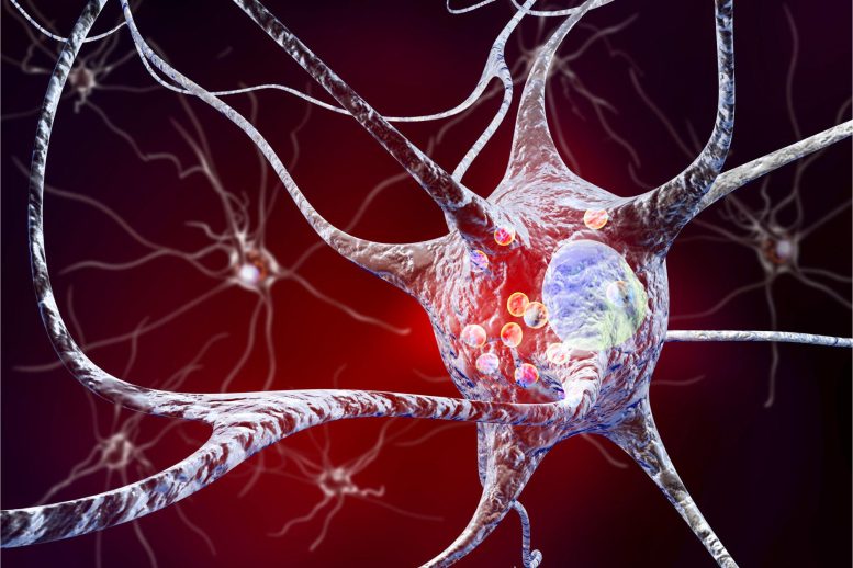 A naturally occurring anti-inflammatory protein fragment is showing promise in early Parkinson’s disease research. Parkinson’s disease affects millions of people worldwide, yet current treatments mainly address symptoms rather than the underlying damage occurring in the brain. Now, researchers in Brazil have identified a promising new approach that targets a different aspect of the disease: inflammation. [...]
 A copper-based drug helped the brain flush out Alzheimer’s toxins, cutting plaque buildup and improving memory in a promising new study. Scientists at Monash University have identified a promising new approach to tackling Alzheimer’s disease. In laboratory studies, a copper-delivering drug significantly lowered levels of toxic proteins linked to the disease while also improving long-term [...]
In a groundbreaking study published in Nature Communications, scientists have unveiled how a mitochondrial protein known as OPA3 plays a pivotal role in sustaining cardiac function by regulating calcium handling in male mice. This discovery sheds new light on the intricate cellular mechanisms that underpin heart performance and opens up promising avenues for treating cardiovascular diseases, which remain a leading cause of mortality worldwide. The findings emphasize the critical interplay between mitochondrial dynamics and calcium signaling within cardiac muscle cells, providing a fresh perspective on how cellular energy production influences heart health.
The heart’s ability to maintain rhythmic and efficient contractions is fundamentally dependent on the precise regulation of intracellular calcium levels. Calcium ions serve as essential second messengers in cardiac myocytes, triggering contraction through complex signaling cascades. However, the interplay between calcium dynamics and mitochondrial proteins, especially those involved in mitochondrial morphology and function, has remained elusive until now. The current study identifies the mitochondrial protein OPA3 as a key modulator in this regulatory network, maintaining both mitochondrial integrity and calcium homeostasis crucial for cardiac contractility.
OPA3 is traditionally recognized for its role in maintaining mitochondrial morphology, particularly in mitochondrial fission and fusion processes. These processes are vital for preserving the mitochondrial network’s dynamic nature, which is essential for cellular energy metabolism. Mitochondria not only produce ATP but also buffer intracellular calcium, thereby influencing calcium signaling within cardiac cells. By elucidating how OPA3 influences calcium handling, researchers have bridged an important gap between mitochondrial structural proteins and the functional output of cardiac cells.
Experimental models using male mice engineered for OPA3 depletion revealed significant impairments in cardiac function, demonstrating that loss of OPA3 disrupts calcium cycling in cardiac myocytes. Impaired calcium handling directly correlates with diminished contractile force and defective cardiac output. This suggests that OPA3’s influence extends beyond structural maintenance and into functional regulation of calcium channels or transporters. The dysregulation of such a critical ion pathway can lead to arrhythmias and cardiac failure, underscoring the potential clinical significance of these findings.
At the molecular level, the study highlights that OPA3 affects the expression and activity of key calcium regulators including the sarcoplasmic reticulum Ca2+ ATPase (SERCA) and ryanodine receptors (RyR). These proteins govern calcium reuptake and release in cardiac cells, orchestrating contraction and relaxation cycles. Through advanced imaging and electrophysiological techniques, the research team demonstrated that OPA3 deficiency impairs SERCA function and causes aberrant RyR-mediated calcium leak, leading to calcium overload or deficits. Such pathophysiological alterations are known contributors to cardiac hypertrophy and heart failure in humans.
The investigation also explores the bioenergetic consequences of OPA3 modulation. Mitochondrial bioenergetics in cardiac muscle cells are highly tuned to meet the energy demands of continuous contractions. OPA3 deficiency was found to compromise mitochondrial ATP production by disrupting inner membrane potential and electron transport chain efficiency. These mitochondrial defects culminate in inadequate energy supply for calcium pumps and ion exchangers, further exacerbating contractile dysfunction. This uncovers a feedback loop where structural mitochondrial proteins like OPA3 indirectly regulate calcium handling by maintaining energetic homeostasis.
A remarkable aspect of the study lies in its use of state-of-the-art imaging techniques such as live-cell confocal microscopy and super-resolution electron microscopy, which revealed morphological abnormalities in mitochondria lacking OPA3. Fragmented and swollen mitochondria with disrupted cristae architecture were prevalent in OPA3-deficient cardiomyocytes. These structural derangements are likely responsible for the observed bioenergetic failures and altered calcium buffering capacity, collectively impairing the cardiomyocyte’s functional integrity.
Furthermore, the team utilized optogenetics-based calcium sensors to monitor real-time calcium flux in isolated cardiomyocytes, providing unprecedented insight into how OPA3 influences calcium transients during excitation-contraction coupling. Data revealed that OPA3 loss leads to prolonged calcium decay time and increased diastolic calcium levels, conditions that predispose cardiac cells to arrhythmic events and contractile inefficiency. These calcium handling anomalies could explain the pathophysiology of certain forms of cardiomyopathy linked to mitochondrial dysfunction.
Importantly, the study addresses the potential sex-specific roles of OPA3, noting that experiments were conducted exclusively in male mice to control for hormonal influences on cardiac physiology. This raises intriguing questions about whether OPA3 functions similarly in female models and whether sex hormones modulate its expression or activity. Such considerations are critical as sex differences in cardiovascular disease outcomes are well-documented, emphasizing the need for further research in this domain.
The translational implications of these findings are profound. Therapeutic strategies aimed at restoring or enhancing OPA3 function could stabilize mitochondrial morphology and bioenergetics, thereby normalizing calcium handling and improving cardiac contractility. Such interventions might hold promise for treating heart failure syndromes characterized by mitochondrial and calcium dysregulation. Moreover, identifying small molecules or gene therapies targeting OPA3 pathways might complement existing treatments that primarily focus on calcium channels but neglect mitochondrial contributions.
Beyond the heart, the role of OPA3 in mitochondrial dynamics suggests potential relevance in other tissues with high energetic demand, such as skeletal muscle and the brain. Defects in mitochondrial morphology and calcium handling have been implicated in neurodegenerative diseases and metabolic disorders. Thus, understanding OPA3’s function could have broad implications in diverse biomedical fields, making it a protein of significant interest in physiology and pathophysiology.
This study also raises several compelling scientific questions for future research. How does OPA3 interact with other mitochondrial fission and fusion proteins, such as OPA1, MFN1/2, and DRP1, in regulating calcium signaling? What are the upstream regulators of OPA3 expression in cardiac tissue under physiological and pathological conditions? Additionally, can OPA3 modulation reverse established cardiac dysfunction or is it mainly preventive? Addressing these queries will deepen our understanding of cardiac mitochondrial biology and pave the way for novel therapeutic innovations.
Addressing cardiac diseases from a mitochondrial perspective represents a paradigm shift in cardiovascular medicine. Historically, calcium handling has been studied primarily through its direct regulators, but this research highlights the necessity of considering mitochondrial proteins like OPA3 as integral components of intracellular calcium homeostasis. This refined viewpoint may revolutionize how clinicians and scientists approach diagnosis, prognosis, and treatment of cardiac ailments.
In conclusion, the identification of mitochondrial protein OPA3 as a fundamental regulator of cardiac calcium handling marks a significant advance in cardiovascular biology. By elucidating the link between mitochondrial morphology, energy metabolism, and calcium signaling, this work provides critical insights into the molecular basis of cardiac function and dysfunction. It offers hope for innovative interventions that could alleviate the burden of heart disease, fostering improved patient outcomes and advancing the frontier of precision medicine in cardiology.
Subject of Research:
Mitochondrial protein OPA3 and its role in cardiac function via regulation of intracellular calcium handling mechanisms.
Article Title:
Mitochondrial protein OPA3 sustains cardiac function by regulating calcium handling in male mice.
Article References:
Geng, N., Chen, T., Li, H. et al. Mitochondrial protein OPA3 sustains cardiac function by regulating calcium handling in male mice. Nat Commun (2026). https://doi.org/10.1038/s41467-026-73991-4
Image Credits: AI Generated
 Small trial suggests diet could support gum disease care, but not replace cleaning.

The human brain has its own cleaning system. Every day, waste products and damaged proteins are removed to keep brain cells healthy and functioning properly. When this cleaning system breaks down, harmful materials can accumulate and damage the brain. Scientists believe this failure may play an important role in Alzheimer’s disease. Alzheimer’s disease is the […]
The post Can a Copper-Based Medicine Help the Brain Take Out the Trash in Alzheimer’s Disease? appeared first on Knowridge Science Report.
The patient: A 28-year-old woman in New Hampshire
The symptoms: In March 2020, the woman developed a high fever, chest tightness, shortness of breath and diarrhea, and she lost her sense of smell and taste. She also experienced coughing fits so intense that they caused her to faint. Based on these symptoms, her primary care provider diagnosed her with COVID-19, although she was not formally tested for the illness due to the lack of diagnostic testing available at the time.
After her diagnosis, she did not seek further medical care due to concerns about out-of-pocket costs, according to a report of the case.
What happened next: Three weeks after her symptoms started, the patient felt well enough to work from home. However, four weeks after that, many of her symptoms returned. She also noticed new feelings of disorientation and felt that "something was off with faces." Her primary care doctor suggested she go to an emergency room, where a CT scan was taken of her brain. The scan didn't show any active bleeding, the report notes, and she was discharged.
In June 2020, the patient spent time with family for the first time since catching COVID-19 and found she could not recognize her father or visually distinguish him from her uncle. She still recognized people by their voices, though.
The patient worked part time as a portrait artist; she used to be able to check her reference photos every 15 to 30 minutes but found she now struggled to hold the details in her mind. She told doctors that "faces are like water in my head," according to the case report.
The patient also had trouble doing everyday tasks, such as navigating her local grocery store or finding her way back to her car in a parking lot. In November 2020, she also started having balance issues and frequent migraines.
The diagnosis: Based on her wide-ranging and relapsing symptoms and her suspected history of COVID-19, doctors diagnosed the patient with post-acute sequelae of COVID-19 (PASC), now known as long COVID. They diagnosed her difficulties with recognizing faces as "face blindness," or prosopagnosia.
Roughly 2.5% of the general population is thought to be born with some degree of face blindness, while others can acquire the condition later in life. The exact number of people with acquired prosopagnosia is unclear, but some estimates suggest roughly 1 in 30,000 people in the U.S. are affected.
Many people with acquired prosopagnosia also have issues navigating familiar spaces. That was also the case with this patient, likely due to the "proximity of brain regions critical for scene and face processing," the case report authors noted.
To assess the extent of the patient's prosopagnosia, doctors had her complete several tests of facial memory, which asked her either to memorize strangers' faces or recognize celebrities' faces. The patient performed markedly worse on these tests than 10 women of similar age who were used as a comparison group.
However, she performed as well as the comparison group on other types of cognitive tests, suggesting her deficits were fairly specific to faces. She also got normal scores on tests of facial perception — the ability to recognize that a face is a face — so her face blindness seemed tied more to memory deficits for faces, the report authors said.
The treatment: The case report does not detail a specific course of treatment, and in general, few treatments exist for face blindness. If there's a clear cause for the deficit — such as a tumor in a key part of the brain for facial processing — addressing that underlying issue can help reverse acquired face blindness.
If the condition ends up being permanent, there are various strategies that can help patients cope. For instance, "perceptual training" can improve patients' ability to identify and remember the features of a person's face. Additional strategies include training people to use other cues, like voices and social context, to identify people.
What makes the case unique: In this patient's case, the report authors suspect that her COVID-19 infection triggered her face blindness either acutely or in the aftermath of the infection.
Various infections have been tied to face blindness; both bacterial meningitis and Whipple disease (caused by the bacterium Tropheryma whipplei) have been flagged as culprits, although such cases are uncommon. In a second known case involving COVID-19, a different person developed face blindness after an infection but had also had a stroke in the right hemisphere of their brain, a known cause of prosopagnosia.
The 28-year-old woman, by contrast, wasn't thought to have suffered a stroke, and it's unclear exactly how COVID-19 led to her facial processing issues.
The case report authors surveyed more than 50 other people with long COVID, to see if they also had signs of face blindness. While many showed general declines in visual recognition and navigational abilities, the group didn't have a specific problem with faces. So while long COVID is known to cause a range of neurological issues, it appears that face blindness is a rare consequence.
For more intriguing medical cases, check out our Diagnostic Dilemma archives.
This article is for informational purposes only and is not meant to offer medical advice.
Can you guess the diagnosis in these strange medical cases? Find out with our diagnostic dilemma quiz!

A groundbreaking discovery at the University of California, Los Angeles (UCLA) has unveiled a promising new therapeutic avenue for kidney regeneration, leveraging a drug initially designed to repair heart tissue following myocardial infarction. This drug, known as AD-NP1, targets a specific protein—ENPP1—that plays a critical role in impeding the body’s natural healing processes in injured tissues. The revelation that AD-NP1 can also accelerate kidney repair heralds a significant advancement in regenerative medicine, especially for patients suffering from chronic kidney disease (CKD), a condition that affects millions globally.
The heart of this research revolves around a protein named ectonucleotide pyrophosphatase/phosphodiesterase 1 (ENPP1). When tissues such as the heart or kidney sustain injury, ENPP1 production escalates, initiating a cascade of metabolic disturbances that hinder energy synthesis within cells. This disruption compromises cellular function and ultimately stalls effective tissue regeneration. In the context of the kidney, which is vital for maintaining systemic homeostasis, such impediments can lead to progressive scarring and functional decline, culminating in kidney failure.
Through years of meticulous experiments, the UCLA team delineated the detrimental impact of ENPP1 on the kidney’s reparative capacity. They observed that injured renal tissue upregulates ENPP1 expression, which in turn resets the metabolic landscape of cells in the damaged microenvironment. This altered metabolic signaling disrupts mitochondrial function, thereby curtailing ATP production essential for cell proliferation and repair. By employing genetic knockout models in mice, where ENPP1 expression was inhibited, the researchers documented a striking enhancement in renal recovery post-injury. These mice demonstrated reduced fibrotic scarring and improved renal biomarkers, signaling restored kidney function.
Central to translating these findings into potential clinical interventions is AD-NP1, a sophisticated monoclonal antibody engineered to specifically neutralize ENPP1. Unlike broad-spectrum immunosuppressants or anti-inflammatory agents, AD-NP1 offers targeted therapeutic precision by binding solely to human ENPP1, circumventing off-target effects. Initially conceptualized to mitigate cardiac fibrosis and promote myocardial regeneration following heart attacks, AD-NP1’s mechanism exerts a similar rejuvenating influence on kidney tissues. The antibody effectively halts the ENPP1-induced metabolic sabotage, facilitating an environment conducive to cellular proliferation and tissue repair.
In experimental paradigms, the administration of AD-NP1 in mice subjected to nephrotoxic diets and chemically induced renal injuries yielded compelling outcomes. Within merely seven days of treatment, subjects exhibited marked improvements in kidney function tests and histological assessments showed diminished collagen deposition and scarring. These findings underscore the drug’s potential to transform the management of acute and chronic kidney injuries by reinstating the organ’s intrinsic regenerative programs, which are often stifled in disease states.
Moreover, the team’s translational approach integrated analyses of human kidney biopsies acquired from individuals with chronic kidney disease. These samples consistently manifested elevated ENPP1 levels relative to healthy controls, substantiating the protein’s pathological role in human renal disorders. This correlation not only reinforces the relevance of the preclinical models used but also paves the way for clinical evaluation of AD-NP1’s efficacy in human subjects suffering from renal ailments.
The broader implications of this discovery extend into the metabolic regulation of tissue repair. ENPP1’s role appears multifaceted, orchestrating cellular energy dynamics and intercellular communication networks that dictate regenerative outcomes. Inhibiting this protein intervenes in maladaptive signaling cascades, thereby restoring bioenergetic balance and enabling clonal expansion of healthy cells adjacent to injury sites. This paradigm shift in understanding organ regeneration emphasizes metabolic modulation as a cornerstone of effective healing strategies.
Prior to this research, the therapeutic targeting of ENPP1 was primarily confined to cardiac medicine. The successful repurposing of AD-NP1 for kidney injury illustrates the concept of cross-organ regenerative mechanisms, where molecular pathways governing tissue healing are conserved albeit contextually modulated. This discovery propels the field into a new frontier, advocating for the exploration of shared regenerative targets that can be manipulated across diverse organ systems.
AD-NP1’s journey into clinical trials commenced recently with FDA approval of a Phase 1 safety study in human patients recovering from heart attacks. This milestone reflects the rigorous preclinical validation of the drug’s pharmacodynamics and safety profile. Building on these milestones, ongoing efforts at UCLA aim to extend these trials to include patients with progressive kidney disease, setting the stage for comprehensive evaluation and potential future clinical application.
Underlying this innovative research are substantial investments from the National Institutes of Health, California Institute of Regenerative Medicine, and the U.S. Department of Defense. This underscores the strategic importance and translational potential of regenerative medicine therapies in addressing chronic diseases that impose heavy societal burdens. The interdisciplinary collaboration among cardiovascular scientists, nephrologists, and molecular biologists at UCLA exemplifies the synergy needed to unlock complex biological processes for therapeutic gain.
In summation, the identification of ENPP1 as a metabolic gatekeeper that impedes tissue repair, combined with the development of AD-NP1 to neutralize its effects, represents a significant leap forward in regenerative medicine. The drug’s ability to foster organ regeneration by restoring metabolic and cellular homeostasis offers hope for millions afflicted by kidney disease and opens novel avenues for organ repair strategies. As clinical trials progress, the scientific community eagerly anticipates further validation of these findings, potentially heralding a new era where molecularly targeted therapies can substantially reverse organ damage and restore quality of life.
Subject of Research: Kidney regeneration, metabolic regulation of tissue repair, ENPP1 protein inhibition, monoclonal antibody therapy
Article Title: UCLA Researchers Discover Novel Role for ENPP1 Inhibition in Enhancing Kidney Repair via AD-NP1
News Publication Date: Not specified (based on provided content)
Web References: Cell Stem Cell Journal Article
Keywords: ENPP1, kidney injury, chronic kidney disease, tissue regeneration, monoclonal antibody, AD-NP1, metabolic signaling, renal repair, fibrosis, UCLA, regenerative medicine, Phase 1 clinical trial
In a remarkable acknowledgment of his contributions to nephrology, Dr. Sushrut S. Waikar has received the American Society of Nephrology (ASN) Distinguished Mentor Award. This prestigious accolade honors individuals who have profoundly impacted the kidney health community through their dedication to mentorship, fostering the careers of emerging clinicians and researchers. Dr. Waikar’s influence extends far beyond routine clinical practice; he has been instrumental in inspiring trainees to dedicate themselves to nephrology, emphasizing innovation in both research and healthcare practice.
Dr. Waikar currently holds the position of Chief of Nephrology at Boston Medical Center and serves as the Norman G. Levinsky Professor of Medicine at Boston University’s Chobanian & Avedisian School of Medicine. As a clinician-scientist, Dr. Waikar has maintained continuous funding from the National Institutes of Health (NIH) for over two decades, highlighting his sustained commitment to advancing kidney disease research. His expansive research agenda incorporates epidemiologic methodologies, translational science, and interventional clinical trials designed to address urgent clinical challenges.
Central to Dr. Waikar’s research portfolio is the refinement of diagnostic tools for kidney diseases, an area critical for early detection and improved patient outcomes. His laboratory has developed novel biomarkers and testing algorithms that aim to enhance diagnostic accuracy, thereby allowing for more timely and precise therapeutic interventions. Such advancements are vital in a field where early-stage kidney dysfunction often goes undetected until it progresses to irreversible damage.
Another significant thrust of Dr. Waikar’s research involves kidney biopsy studies, which offer invaluable insights into the pathophysiological mechanisms underlying disease progression within the renal parenchyma. By analyzing tissue samples, his team seeks to elucidate the cellular and molecular pathways that drive kidney deterioration, providing a foundation for the future design of targeted therapies. This level of detailed study bridges the gap between clinical observation and basic science, enabling a more comprehensive understanding of renal pathology.
Dr. Waikar’s investigations also spotlights a particularly devastating public health concern known as Chronic Kidney Disease of uncertain etiology (CKDu). This disease has emerged as a lethal epidemic primarily afflicting young agricultural workers in Central America and South Asia. Unlike traditional forms of chronic kidney disease associated with diabetes or hypertension, CKDu’s etiology remains elusive and multifactorial. His research endeavors aim to clarify environmental, occupational, and genetic risk factors contributing to this silent crisis, with hopes of informing preventative strategies and policy interventions.
Within the realm of precision medicine, Dr. Waikar acts as principal investigator for the NIH Kidney Precision Medicine Project. This ambitious initiative is engineered to leverage cutting-edge genomic, proteomic, and proteomic technologies to tailor kidney disease treatment at an individual level. By integrating these high-throughput techniques with clinical data, this project aspires to revolutionize management paradigms for kidney disorders, ultimately improving personalized patient care outcomes.
In addition to his leadership in precision medicine, Dr. Waikar coordinates the CKD of Unknown Etiology Consortium, a collaborative network of investigators dedicated to elucidating CKDu. This consortium underscores the importance of multidisciplinary cooperation in tackling complex health challenges, combining epidemiology, environmental sciences, and nephrology to develop holistic solutions to kidney health crises.
Dr. Waikar’s intellectual journey began with an undergraduate education in English and neuroscience at Amherst College, fostering a broad intellectual foundation that supports his interdisciplinary approach. He then earned his medical degree at Yale University, further complementing his scientific expertise with clinical acumen. His postgraduate training in internal medicine and nephrology at highly regarded institutions such as the University of California, San Francisco, and the Mass General Brigham system was distinguished by leadership roles, including chief medical resident, reflecting his early commitment to medical education and mentorship.
His academic trajectory also includes earning a Master’s degree in Public Health from Harvard University, which underpins his rigorous approach to epidemiology and population-based studies. This public health background allows Dr. Waikar to navigate and address the systemic factors impacting kidney health globally, marrying clinical insight with health policy and preventive medicine.
The American Society of Nephrology, founded in 1966, stands as the largest professional society devoted to kidney disease research and education worldwide. To further acknowledge the critical contributions of midcareer professionals, ASN established the Midcareer Awards Program in 2019, celebrating individuals like Dr. Waikar who demonstrate exceptional clinical expertise, innovative research, and dedicated service to the nephrology field and the society’s mission.
Dr. Waikar’s recognition by the ASN Distinguished Mentor Award not only celebrates his individual achievements but also highlights the critical role that mentorship plays in shaping the future of nephrology. His ability to guide, inspire, and cultivate the development of emerging leaders ensures that advances in kidney research and patient care continue to accelerate, ultimately benefiting countless individuals suffering from kidney disease globally.
Through the integration of basic science, clinical application, and public health strategies, Dr. Waikar exemplifies the modern physician-scientist who transforms knowledge into meaningful biomedical progress. His contributions serve as a catalyst for ongoing discoveries and innovations that hold the promise of reducing kidney disease burden worldwide.
Subject of Research:
Kidney disease diagnostics and research, Chronic Kidney Disease of uncertain etiology (CKDu), kidney biopsy studies, precision medicine in nephrology.
Article Title:
Not provided.
News Publication Date:
Not provided.
Web References:
Not provided.
References:
Not provided.
Image Credits:
Not provided.
Keywords:
Nephrology, kidney disease, chronic kidney disease, CKDu, NIH Kidney Precision Medicine Project, kidney biopsy, biomarkers, epidemiology, translational research, mentorship, American Society of Nephrology, public health.

Alzheimer’s disease is the most common cause of dementia and one of the biggest health challenges facing the world today. It gradually damages memory, thinking abilities, and the capacity to perform everyday tasks. In the early stages, people may forget recent conversations or misplace objects more often. As the disease progresses, many struggle to recognize […]
The post New Copper Drug Could Clean Alzheimer’s Waste and Improving Memory appeared first on Knowridge Science Report.

Copper diacetyl bis(4-methyl-3-thiosemicarbazone), or Cu(ATSM), restored a key waste-removal system in the brain, reducing toxic amyloid-beta buildup and improving spatial memory in lab models of Alzheimer’s disease, according to a team of researchers at Monash University.
The post Experimental Copper-Based Drug Clears Alzheimer’s Plaques and Boosts Memory in Mice appeared first on Sci.News: Breaking Science News.
From Ebola to Nipah, the combined death toll since Covid is under 1,000 worldwide. So why the endless panic? Follow the money – pandemic fearmongering is a growth industry, says Dr David Bell.
The post Follow the Money: Pandemic Fearmongering is a Growth Industry appeared first on The Daily Sceptic.