❌

Reading view

Siloton secures £8.4m to speed development of portable retinal imaging system

UK medical technology start-up Siloton has been awarded an £8.4 million contract to accelerate the development of its portable eye imaging system. Based on optical coherence tomography (OCT), the company’s GiraffeOCT device generates detailed retinal images for detection and monitoring of a range of treatable eye diseases.

OCT is a non-invasive optical technique that creates high-resolution, depth-resolved images of biological tissue. While OCT is the clinical gold standard for diagnosing retinal diseases, existing systems are large and expensive and typically only available in hospitals and optometry clinics. By using photonic chip technology to shrink the bulky optical components onto a single coin-sized chip, Siloton is developing an easy-to-use, handheld binocular-style system.

The contract, from Innovate UK, will massively accelerate development of both the in-clinic GiraffeOCT system and the at-home version, which will have built-in automation and guidance so that users can reliably image themselves. “The contract will reduce the time to market for the medical device by one to two years,” says Alasdair Price, CEO of Siloton. “It will also allow us to grow the company, bringing in new skills around manufacturing and regulation, both of which are critical to the next stage of the business.”

Continuing clinical trials

In 2024, Siloton was the first commercial organization to demonstrate sub-surface imaging of a human retina, using its first-generation Akepa OCT photonic chip. The following year, the company trialled its device with patients for the first time, imaging their retinas to identify the presence and progression of age-related macular degeneration (AMD). Testing continued as the system developed, with a second cohort of patients imaged earlier this year.

In addition, the Project Ginan consortium in Australia has purchased a GiraffeOCT research system and independently evaluated its retinal imaging performance with a group of healthy volunteers.

Over the course of the two-year Innovate UK contract, Siloton will work with Sunderland Specialist Eye Hospital in the UK to perform clinical trials of the in-clinic device, giving patients access to the technology during its development. “These trials will be based around the prototype for the full medical device, providing additional post-processing and improved ergonomics compared to our research-use only product line,” Price explains.

The funding will also support the development and internal testing of Siloton’s second-generation CheetahOCT system, a research-use device that will measure different layers of the retina, expanding the number of conditions that it can detect. CheetahOCT incorporates the company’s second‑generation BlueJay chip – the same architecture used in the system that Siloton is building for astronauts – and will be available from early 2027.

“The image performance will be significantly improved, increasing the number of biomarkers that it can be used to measure. This means you will be able see more of the eye and in finer detail,” Price tells Physics World. “CheetahOCT will also be much more compact, as we continue to miniaturize other parts of the system that fall outside the photonic chip.”

The post Siloton secures £8.4m to speed development of portable retinal imaging system appeared first on Physics World.
  •  

Optogenetic therapy shows promise for vision restoration

Retinitis pigmentosa is an inherited degenerative condition that destroys photoreceptors in the retina and can eventually lead to irreversible blindness. In a clinical trial first, an international research team has demonstrated that optogenetic therapy is safe to use and shows potential for restoring visual function in patients with late stages of this disorder.

In a first-in-human study in 2021, the collaboration – led by José-Alain Sahel from the University of Pittsburgh School of Medicine and Botond Roska from the Institute of Molecular and Clinical Ophthalmology Basel (IOB) – demonstrated that optogenetic therapy led to partial recovery of visual function in a blind patient with retinitis pigmentosa. This latest work, reported in the New England Journal of Medicine, expands the cohort to 10 participants.

Optogenetics is a method that uses light to characterize and manipulate the activity of neurons in the brain – and which earlier this week won its inventors the 2026 Nobel Prize in Physiology or Medicine. The optogenetic therapy developed by Sahel, Roska and collaborators combines gene therapy that makes surviving ganglion cells (neurons in the retina) sensitive to light with special goggles that stimulate the altered cells.

“Although patients with retinitis pigmentosa lose their rod and cone cells, which normally detect light, many other cells within the retina can remain present for years after vision loss,” explains Sahel, director of the UPMC Vision Institute and first author on this study. “Optogenetics enables us to introduce a light-sensitive protein into these surviving cells, allowing them to respond to light and transmit visual information to the brain. The therapy bypasses the damaged photoreceptors and takes advantage of retinal circuits that remain intact.”

Sahel notes that retinitis pigmentosa can be caused by mutations in any one of hundreds of different genes, making optogenetic therapy a particularly suitable approach as it can potentially help regardless of the underlying mutation.

Safe to use

In the study, 10 participants with blindness due to advanced retinitis pigmentosa were injected in their worse-seeing eye with a gene that produces ChrimsonR – a light-sensitive protein with a peak response at around 590 nm (amber). The team divided the subjects into three dose-escalation groups, receiving 5.0 x 10¹⁰, 1.5 x 10¹¹ or 5.0 x 10¹¹ vector genomes per eye.

The primary aim was to evaluate the safety of the optogenetic therapy. The researchers observed 33 mild and moderate adverse ocular events in nine of the participants, such as temporary inflammation and increases in eye pressure, plus one severe event, which resolved within minutes after treatment. They concluded that, within the limits of the study, the treatment was safe.

Visual stimulation system
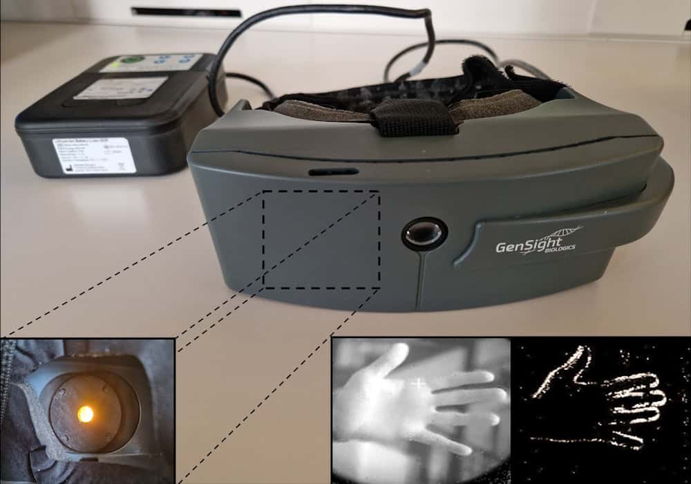
Visual stimulation system The specialized goggles (centre) send visual information to a processor (upper left) that converts the information into patterns of light. A projector within the glasses (lower left) sends these patterns back to the eye. Lower right: the system converts the image of a hand into a simplified light pattern that’s projected onto the retina. (Courtesy: Chloé Pagot, Streetlab, Paris)

Using the light-stimulating goggles, they then assessed the visual performance of treated eyes, before and after injection. The goggles contain a camera that detects changes in light intensities pixel-by-pixel and converts this visual data into pulses of amber light. This light is projected onto the eye to activate ChrimsonR in the modified retinal ganglion cells. Vision tests revealed that the treatment increased light sensitivity in seven of the 10 participants, by a factor of 2.0 to 62.3, with six patients showing a clinically meaningful improvement.

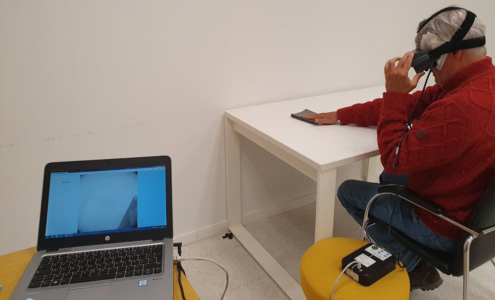
The patients also performed a series of tasks to detect, localize and touch a notebook and a staple-box. In the notebook task, five of eight participants had higher accuracy when wearing the goggles than when not wearing them, while four showed improved accuracy in the staple-box task. In a task determining the orientation of a single bar, accuracy was higher with the goggles in five of eight participants.

To provide an objective measurement of brain activity in response to light, the researchers obtained electroencephalographic (EEG) recordings in five patients performing two visual tests: detection of a tumbler placed in front of them (with the eyes open or closed); and passive viewing of periodic visual stimuli.

In the tumbler test, EEG decoding accuracy was higher with goggles than without when the eyes were open, but not when they were closed – implying a genuine improvement in visual processing rather than an artefact. The second test also showed increased decoding accuracy, which correlated with mean improvements in the notebook and staple-box tasks.

Visual behavioural test
Visual behavioural test After receiving optogenetic therapy, participants used a visual-stimulation system to complete tasks. Here, a participant reaches for a notebook. (Courtesy: Chloé Pagot, Streetlab, Paris)

A bright future

The changes in visual performance were consistent across multiple tasks and testing sessions, with responses observed in all three dose groups. Participants who spent longer being trained to use the goggles performed better in the visual behavioural tests. The researchers point out, however, that functional changes were modest: improving object detection and discrimination capabilities but not restoring normal sight. The small sample size also limited their ability to characterize treatment response.

Several projects are currently underway to further enhance the technology. This includes digital holographic goggles with an eye tracker being developed by the team of Valentina Emiliani at the Institut de la Vision in Paris. The system will project 20 µm-wide images that stimulate individual cells in the retina.

“With the eye tracker, we can make sure we are projecting exactly onto the area of the retina that we want. We’ve already developed a prototype, and it is moving forward,” says Sahel.

Elsewhere, University of Pittsburgh scientists are working with Roska’s group to develop cell-specific gene therapy vectors. “There are different types of cells: some respond to the onset of light, some to the offset of light, some to movement and direction,” Sahel explains. “We are trying to selectively stimulate the ‘on’ and the ‘off’, and discriminate the signal we are sending.”

“We are optimistic about advancing this approach toward future clinical trials,” he tells Physics World.

The post Optogenetic therapy shows promise for vision restoration appeared first on Physics World.

  •  

Inventors of optogenetics win Nobel Prize in Physiology or Medicine

The 2026 Nobel Prize in Physiology or Medicine has been awarded to Karl Deisseroth, Peter Hegemann and Georg Nagel for “their discoveries concerning light-gated ion channels and optogenetics”.

Optogenetics is a method that uses light to characterize and manipulate the activity of neurons in a living brain. Hegemann and Nagel discovered the light-sensitive protein channelrhodopsin, while Deisseroth transformed the protein into a light-controlled switch for nerve cells.

“Optogenetics provides opportunities for mapping the brain in a way that we could once only dream of,” says Per Svenningsson, chair of the Nobel Committee for Physiology or Medicine.

From the pond to the brain

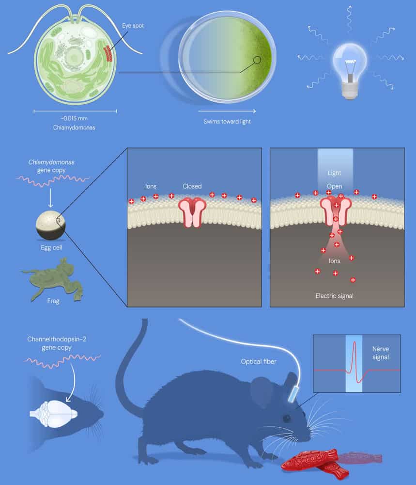
Speaking in the press conference following the announcement, neuroscientist Abdel El Manira from the Karolinska Institute described how the breakthrough began with the study of a single-cel­led green alga, Chlamydomonas, known for its ability to swim towards light using a tiny “eyespot” on its surface as a sensor. Hegemann was intrigued as to how this simple organism could transform light into action.

Prize-winning optogenetics research
Top to bottom: The algae Chlamydomonas senses light using its eyespot; Chlamydomonas genes in frog eggs, showing the ion channel proteins on the surface; activation of nerve cells in the brains of living mice. (Courtesy: © The Nobel Committee for Physiology or Medicine. Ill. Mattias Karlén)

Using electrical measurements to solve this mystery, Hegemann discovered that the eyespot contains a light-sensitive protein that almost instantly converts light into an electrical signal, enabling the alga to move. Hegemann next teamed up with Nagel, who used the gene for Chlamydomona to produce the protein in frog egg cells. They discovered that the protein itself was a light-gated ion channel that opens when exposed to light, allowing positively charged ions to flow into the cell and rapidly generate an electrical signal.

“Nagel and Hegemann realised exactly what they had found. They had just discovered the switch neuroscientists had long dreamed of – a light-gated ion channel they named channelrhodopsin,” says El Manira.

Appreciating the potential of this discovery, Deisseroth – at the time studying ways to establish causal links between specific brain circuits and behaviour – decided to test whether channelrhodopsin could be used as a switch to control the activity of neurons with light. By introducing the DNA encoding the protein (supplied by Nagel) into mammalian neurons and illuminating them with light, he observed a remarkable response: the neurons reacted on command, instantaneously and with millisecond precision.

The real test was whether channelrhodopsin could control neurons in living animals. Deisseroth and colleagues demonstrated that this was possible. By introducing the gene for channelrhodopsin into specific nerve cells in the brains of living mice and illuminating these cells via a thin optic fibre, they activated selected group of neurons and elicited a distinct behaviour.

“For the first time, causal links between specific brain circuits and behaviour had been achieved. The technology, which soon became known as optogenetics, transformed neuroscience,” notes El Manira. “Optogenetics was rapidly and widely adopted all over the world.”

Future medical potential

The field of optogenetics has now enabled researchers to establish causal links between neural activity and behaviour, identifying, for instance, neural circuits associated with pain, social behaviours, thirst, food consumption, reward and attention. Beyond the healthy brain, the technique also sheds light on how specific brain circuits can be disrupted and how this impacts psychiatric and neurological conditions.

And while optogenetics is primarily a tool for basic research and understanding of disease, optogenetic therapy is also showing early promise as a medical treatment. In particular, a landmark study has demonstrated partial recovery of visual function in a blind patient with retinitis pigmentosa, by insertion of a channelrhodopsin-like protein in their retina.

“In this disorder, retinitis pigmentosa, patients lose their photoreceptors, the rods and cones in the retina,” explains Svenningsson. “But there are still healthy cells in the retina and researchers have used optogenetics to stimulate these healthy cells and thereby activate the optic nerve and generate vision perception in the brain. This has been done and there are several clinical trials ongoing with this approach.”

Elsewhere, optogenetics is also under investigation for applications including control of spinal cord activity and bladder activity in small animals, inhibition of nerve pain and guidance of morphogenesis in a living embryo.

Friends together

Karl Deisseroth was born in 1971 and received his PhD and MD from Stanford University in the US, where he performed the work for which he was awarded the prize. He is currently an investigator at the Howard Hughes Medical Institute and professor at Stanford University.

Peter Hegemann was born in 1954 and received his PhD at the Max-Planck-Institute for Biochemistry in Martinsried, Germany, where he made his prize-awarded discoveries. He is now a senior professor of neuroscience at Humboldt University of Berlin, Germany.

Georg Nagel was born in 1953 and received his PhD at the University of Frankfurt, Germany. His prize awarded discoveries were made at Max Planck Institute for Biophysics in Frankfurt. He is currently professor of molecular plant physiology at the University of Würzburg, Germany.

Thomas Perlmann, secretary-general of the Nobel Assembly, described his call to the laureates earlier this morning: “All three were very surprised and all three said the same thing, that it was absolutely wonderful to receive the prize with the other two, called them ‘my friends’, and that it was such an honour to receive them together”.

  • Read about the use of organic dyes in optogenetics, in a book chapter co-authored by Peter Hegemann, now free to read on IOP Science.

SmarAct Group logo

SmarAct proudly supports Physics World‘s Nobel Prize coverage, advancing breakthroughs in science and technology through high-precision positioning, metrology and automation. Discover how SmarAct shapes the future of innovation at smaract.com.

The post Inventors of optogenetics win Nobel Prize in Physiology or Medicine appeared first on Physics World.

  •  

Radiotherapy trials shape future treatments of brain metastases

At this week’s ASTRO 2026 meeting in Boston, MA, researchers shared the results of clinical trials addressing important and longstanding questions in cancer care. In a dedicated news briefing highlighting noteworthy research studies, two speakers presented new evidence that could help inform how we deliver care to patients with brain metastases.

Brain metastases – cancerous cells that spread to the brain from another site in the body – are typically removed surgically or treated with radiotherapy. For most types of cancer, the preferred approach is stereotactic radiosurgery (SRS), in which high-dose radiation is precisely directed to individual lesions while limiting dose to surrounding healthy brain tissue.

Randomized trials have shown that SRS results in superior cognitive preservation and quality-of-life compared with whole-brain radiotherapy (WBRT), where the entire brain is irradiated to ensure treatment of both visible tumours and microscopic disease. There’s an exception, however. Historically, for brain metastases arising from small cell lung cancer (SCLC), a rare, fast-growing cancer, WBRT remains the standard-of-care. Results of a new trial challenge this approach.

Chad Rusthoven from the University of Colorado Anschutz School of Medicine shared the findings of the first phase III randomized trial to compare SRS versus WBRT for brain metastases from SCLC. The trial found that after adjusting for patient and disease characteristics, SRS was associated with a 40% lower risk of death than WBRT.

Rusthoven noted that recent advances have improved outcomes for patients receiving WBRT. “This includes addition of memantine [a drug that helps protect cognitive function], as well as hippocampal avoidance techniques,” he explained. “As a result, HA-WBRT plus memantine is now the preferred whole-brain treatment strategy for cognitive preservation.”

As such, the NRG-CC009 trial – which included 151 patients in the US and Canada – randomly assigned participants to receive either focused SRS to visible metastases or HA-WBRT plus memantine. All subjects had a median of two SCLC metastases, although some had more than 20.

For the primary endpoint – the time before a patient experienced cognitive decline – the study found no significant difference between the two treatment groups. However, patients treated with SRS had a median overall survival of 17.4 months, compared with 8.6 months for those in the HA-WBRT group. Rates of serious treatment-related side effects or death from neurologic causes were similar for the two cohorts.

Although no differences in neurocognitive failure were observed, the superior overall survival with SRS addresses a fundamental historical objection to SRS alone for small cell lung cancer brain metastases. That is, the concern that omission of WBRT would lead to worse overall survival in this population. Overall these findings support SRS as a treatment option for patients with brain metastases from small cell lung cancer,” Rusthoven concluded.

The power of fractionation

Speaking at the same briefing, Ayal Aizer from Mass General Brigham/Dana-Farber Cancer Institute explained how fractionated radiosurgery can reduce the risk of cancer recurrence after surgery for large brain metastases, without significantly increasing side effects. Fractionation – in which the radiation dose is divided into several treatments – gives healthy tissue time to recover between doses while still effectively treating the surgical cavity.

Large metastases, which can cause pressure on the brain leading to symptoms such as seizures, headaches or difficulties with speech or movement, can be removed via surgery. This is usually followed by radiotherapy to destroy any microscopic cancer cells left behind in the surgical cavity. Aizer explained that current guidelines recommend treating the cavity with SRS, following two pivotal trials demonstrating favourable outcomes for SRS compared with postoperative WBRT and observation.

Ayal Aizer
Ayal Aizer: “Postoperative fractionated SRS reduced the likelihood of one-year surgical bed recurrence.” (Courtesy: © ASTRO/Luke Franke 2026)

For larger surgical cavities, however, the challenge, is how to deliver enough dose to eliminate any remaining cancer cells without exposing too much surrounding healthy brain to radiation. “In both of these trials where single-fraction SRS was utilized, the one-year surgical bed recurrence rates were high: 40% in one trial, 28% in the other,” he explained. “Meanwhile, alternative approaches such as fractionated SRS to the surgical bed had been emerging.”

Aizer described Alliance A071801, the first randomized phase III trial to directly compare single-fraction versus fractionated SRS for resected brain metastases. The multi-centre trial enrolled 242 patients with one to four brain metastases, one of which was larger than 2 cm and surgically removed. The metastases arose from different primary tumour types, the most common being lung cancer.

The team randomly assigned patients to receive either a single session of SRS, with dose based on target size, or fractionated SRS: 27 Gy in three fractions for smaller targets or 30 Gy in five fractions (9%) for larger ones.

One year after treatment, 87% of patients who received fractionated SRS were free from recurrence at the surgical site, compared with 81% of those who received single-session radiosurgery. Side effects such as radiation necrosis and cerebral oedema were low in both treatment groups, with no significant differences between the two.

The overall survival was also improved after fractionated SRS, with patients surviving a median of 29 months, compared with 20 months for those treated in a single session. The researchers note that this survival difference was unexpected and it is not yet clear why this occurred.

“In conclusion, we can say that fractionated SRS improved surgical bed control compared with single-fraction SRS after resection of a brain metastasis,” said Aizer. “The improvement in surgical control was achieved without an apparent increase in toxicity. For patients undergoing resection of a larger brain metastasis, postoperative fractionated SRS should be considered the standard-of-care.”

The post Radiotherapy trials shape future treatments of brain metastases appeared first on Physics World.

  •  

Siemens Healthineers unveils ultrafast Accela radiotherapy system

The annual meeting of the American Society for Radiation Oncology is the place to be to discover the latest radiotherapy technology developments and new product launches. And this year was no exception. The ASTRO 2026 meeting, held this week in Boston, MA, saw Siemens Healthineers unveil its newest radiotherapy system – Accela – which combines precision delivery with speeds fast enough to enable stereotactic treatments in a single breath-hold.

Accela achieves this by implementing a new delivery technique known as NeoArc, or dynamic modulated arc therapy (DMAT). DMAT is a type of arc therapy, in which the treatment beam rotates continuously around the patient during treatment, with the beam shape and intensity controlled to deliver the prescribed treatment plan.

But while conventional arc-based approaches such as volumetric-modulated arc therapy (VMAT) primarily optimize treatment plans for the dose distribution, DMAT evaluates dosimetric objectives and delivery efficiency simultaneously. This enables clinicians to tailor treatments for both speed and precision, to optimize the balance between them and, ultimately, to reduce the patient time on the treatment table.

Sasa Mutic, head of radiation oncology solutions at Siemens Healthineers, tells Physics World that the product launch at ASTRO was a great success. “The response to Accela from customers and the medical community has been overwhelming and overwhelmingly positive,” he says. “It was extremely gratifying to see that the ASTRO audience instantly grasped the significance of what we’ve accomplished and the impact it will have on clinicians and patients.”

Done in 60 seconds

Accela integrates high-speed gantry rotation of up to 2.5 rpm, dose rates of up to 40 Gy/min and a dual-layer multileaf collimator (MLC) with leaf speeds of up to 6.5 cm/s. This combination means that, as well as single breath-hold stereotactic treatments, Accela can deliver multi-site stereotactic body radiotherapy (SBRT) in as little as 60 s and go from imaging to beam-off in as low as 90 s – including on-couch imaging with a large HyperSight cone-beam CT panel, couch shifts and the treatment delivery.

An additional benefit of ultrafast radiotherapy is the potential to actually alter the body’s biological response.

“Because the treatment times are potentially as quick as a single breath hold, there is much less blood irradiated,” explains Dee Khuntia, chief medical officer at Varian, the Siemens Healthineers cancer-care business. “This allows for preservation of lymphocytes, which are necessary for helping the body heal, fight infection and fight cancer. It is also well documented that patients undergoing radiation or chemo-radiation who have lymphopenia [not enough lymphocytes in the blood] have lower survival rates.”

The first patient treatments with Accela are scheduled for December in Europe. By this time, the system will have the CE mark, with FDA clearance expected early next year. One of the first centres to use Accela will be Maastro in the Netherlands, which plans to initially treat small lung tumours, lung metastases, brain tumours and metastases in the vertebrae – sites that require a high radiation dose administered extremely precisely, while sparing healthy tissue and nearby vulnerable organs.

  • Updated on 6 October 2026 to amend the imaging to beam-off time and add a clarification from Siemens Healthineers: Accela and NeoArc are 510(k) pending. Not available for sale or quotation. No guarantee of commercialization or feature availability.

The post Siemens Healthineers unveils ultrafast Accela radiotherapy system appeared first on Physics World.
  •  

Pathways to commercial and academic success in medical physics

This episode of the Physics World Weekly podcast features the medical physicist Nick Zacharopoulos, who is CEO of US-based Aktina Medical.

In conversation with Physics World’s Tami Freeman, Zacharopoulos reflects on the twists and turns in his career developing new technologies for radiation oncology. His journey began when he was a child helping his father build radiotherapy components in the family’s garage and led to academic and commercial success.

Zacharopoulos talks about his role in the development of a pivotable product for Aktina and how his persistence – and a chance lunch with a colleague – led to his enrolment in a PhD programme at McGill University.

The post Pathways to commercial and academic success in medical physics appeared first on Physics World.

  •  

Tiny biodegradable stars enhance drug delivery to the skin

Delivering drugs directly to the skin provides a targeted means of treating dermatological conditions while minimizing side effects. The approach is limited, however, by the stratum corneum – the skin’s outer layer – which acts as a barrier that can restrict the effectiveness of most topically applied drugs. To overcome this, a team at Georgia Tech has developed biodegradable “STAR particles” that use microscopic needles to painlessly puncture the skin and increase its absorption of medication.

“Very few drugs can be absorbed effectively into the skin, which means that many drugs in dermatology are given by mouth or injection,” explains team leader Mark Prausnitz. “This exposes the whole body to the drug, often causing side effects and reducing drug efficacy. STAR particles painlessly make micropores in the skin that allow drug that is rubbed on the skin to be absorbed. This targets drug delivery exactly to the site where it is needed.”

Previously, the researchers fabricated the STAR particles from a ceramic material (titania), which is safe to use on the skin, but not biodegradable, raising possible environmental concerns. In their latest work, reported in Advanced Healthcare Materials, they designed polymer STAR particles that dissolve or biodegrade after use, reducing their potential environmental impact.

STARs effectively create micropores

Prausnitz and colleagues created STAR particles from three polymers: water-soluble poly(vinyl alcohol) (PVA), enzyme-degradable cellulose acetate (CA) and hydrolysable polylactic acid (PLA). All three materials have previously been used to create microneedle patches, which enhance drug delivery but can generally only be used on small areas of skin.

“For dermatological conditions like eczema and psoriasis, patients need to treat skin with variable and sometimes large areas,” says Prausnitz. “STAR particles provide the power of a microneedle patch to increase skin permeability with the flexibility to apply them over large and variable areas by simply rubbing a gel or cream containing STAR particles on the skin.”

The researchers fabricated the polymer STAR particles using femtosecond laser micromachining to create star-shaped structures with sharp, well-defined microneedle tips and a tapered profile. They tested the ability of the various particles to puncture pig skin samples.

While PVA STAR particles cannot be applied using water-based vehicles, when suspended in non-aqueous formulations such as isopropyl palmitate (a non-toxic ingredient widely used in dermatology), they successfully punctured the skin. The STAR particles remained intact after use and the formulations worked equally well after storage for one week.

Similarly, CA and PLA STAR particles in water formulations demonstrated consistent skin-puncturing ability, even after a week’s storage, with no visible damage to the particles after application. The researchers note that titania STAR particles in water (examined as a control) generated more pores than the polymer particles, due to their higher hardness.

Drug delivery demonstration

The team next investigated how the STAR particles could enhance the delivery of three drugs – tacrolimus, methotrexate and copper tripeptide-1 – into pig skin samples.

Tacrolimus is used to treat inflammatory skin conditions and is soluble in non-aqueous solvents. After topical drug delivery, rubbing the skin with PVA STAR particles in isopropyl palmitate for 10 s or 30 s increased drug levels within the skin 1.7- or 3.2-fold, respectively, compared with control samples.

For CA STAR particles, the researchers examined methotrexate, which is commonly used for psoriasis treatment but cannot be delivered topically due to its very low skin permeability. Treatment with CA STAR particles for 10 or 30 s increased intradermal delivery of methotrexate 5.4- or 25.2-fold, respectively.

Finally, they used PLA STAR particles to deliver copper tripeptide-1, a skincare ingredient employed for anti-aging, wound healing and skin regeneration applications. The particles enhanced intradermal drug delivery 12.1- or 37-fold, after 10 or 30 s application, respectively.

Safety considerations

As the STAR particles are designed to create micropores in skin, it’s possible that they could unintentionally end up in other parts of the body, where the microneedles could cause damage. While this risk is likely small (as the STAR particles require forceful application to be effective), the water-soluble PVA STAR particles eliminate any potential risk as they rapidly dissolve upon contact with wet tissues.

The CA and PLA STAR particles will likely retain their structure immediately after use, but will eventually become blunt and weak upon enzymatic degradation or hydrolysis. Likewise, any potential environmental consequences of non-degradable STAR particles are reduced or eliminated with the water-soluble and biodegradable materials.

The team concludes that the biodegradable STAR particles address environmental and safety concerns while enhancing drug delivery to the skin. The approach paves the way for improved patient outcomes and broader applications, enabling delivery of hydrophilic drugs and larger molecules that are usually blocked by the stratum corneum barrier. Prausnitz tells Physics World that the research is primarily targeted at treatment of dermatological diseases that can spread over large areas of skin – such as psoriasis, eczema, vitiligo and allergic rashes – as well as cosmetic applications.

“We have licensed the STAR particle technology to a company that is preparing for a clinical trial next year using STAR particles to deliver siRNA as a novel eczema treatment,” he says. “At Georgia Tech, we are focused on advanced materials, manufacturing and formulations for the next generation of STAR particles.”

The post Tiny biodegradable stars enhance drug delivery to the skin appeared first on Physics World.

  •  
❌