❌

Normal view

AI Reads Tea Leaves’ Light Signatures to Measure Frost Damage

11 October 2026 at 09:12

Frost is one of the most punishing adversaries of tea cultivation. A single cold snap can scorch tender leaves, derail an entire season’s harvest, and degrade the delicate chemistry that gives tea its value. For growers and breeders, however, judging how badly a field has been damaged has long been a stubbornly subjective exercise: visual ratings lump continuous injury into crude categories, and laboratory assays destroy the very tissue they measure. A new study published in Plant Methods by Zhengtong He of Shihezi University, Zhiwei Chen of the Tea Research Institute of Shandong Academy of Agricultural Sciences, and colleagues offers a way out, pairing hyperspectral imaging with a purpose-built deep learning model to read frost damage directly from the way leaves reflect light.

The core problem the team confronted is that frost injury is not a binary event but a continuum. Leaves exposed to sub-zero temperatures suffer a cascade of overlapping stresses: photosynthetic pigments degrade, the machinery of photosynthesis falters, and cell membranes and walls break down. Any single measurement captures only a sliver of that cascade. To capture the whole picture, the researchers collected hyperspectral data together with eight key physicochemical indicators from tea plants growing across natural frost damage gradients in the field, rather than in the artificial conditions of a growth chamber. From these multidimensional physiological responses they constructed a composite measure, the tea cold stress response index, or TCSRI, which expresses frost damage severity as a continuous value instead of a coarse visual class.

Building TCSRI is a meaningful advance in itself. Traditional frost assessment often relies on ordinal scales, where an observer assigns a score of mild, moderate, or severe. Such scales throw away information and make it difficult to track recovery or compare cultivars with fine precision. By integrating several physiological dimensions into one index, the team created a target variable that reflects the plant’s actual internal state. The challenge then became predicting that index, and the individual chemical indicators behind it, from spectral data alone, without touching or destroying a single leaf.

Hyperspectral sensing is well suited to this task in principle. Across hundreds of narrow wavelength bands, plant leaves encode a wealth of information: chlorophyll absorption in the visible range, water and structural features in the near infrared. But the data are high-dimensional, with far more spectral channels than samples, a regime that invites overfitting, especially in field studies where collecting ground-truth measurements is laborious. Conventional chemometric approaches, from partial least squares regression to standard machine learning pipelines, often struggle to separate genuine physiological signals from noise, sensor drift, and the confounding effects of leaf surface properties and illumination variation.

The researchers’ answer is a model they call STAMPformer, short for spectral-prior-guided multi-view attention pooling. Its architecture reflects a deliberate design philosophy: rather than letting a neural network rediscover everything from scratch, the model is steered by what spectroscopists already know about how plants respond to stress. At its front end, a parameter-free attention convolution extraction module jointly models both the raw reflectance spectra and their first-order derivatives. Derivative spectra are a classic spectral prior: they accentuate subtle local features such as absorption peaks and shoulders that are easily drowned out in raw reflectance, while suppressing broad baseline drift and noise. By processing both views together, the module sharpens the local spectral signatures that carry physiological meaning.

On top of this extraction stage sits the second innovation, a multi-view attention pooling module. Deep networks processing spectral sequences produce long streams of features, but not all wavelengths matter equally for a given prediction. The pooling module adaptively aggregates sequence features drawn from different feature subspaces, learning to amplify the responses that are most diagnostic of cold stress and to down-weight the rest. This attention mechanism acts like a learned spotlight, and because it operates across multiple subspaces, it can capture complementary patterns that a single attention head might miss. The result is a compact representation tailored to the prediction task at hand, whether that is estimating a specific chemical indicator or the composite TCSRI.

The performance numbers are striking. Across all nine prediction tasks, spanning the eight physicochemical indicators and the TCSRI itself, STAMPformer achieved coefficients of determination above 0.87 and ratios of performance to deviation above 2.80, thresholds generally regarded as indicating reliable quantitative prediction. In other words, from a leaf’s reflectance fingerprint alone, the model could reconstruct the plant’s internal frost-stress state with an accuracy that approaches laboratory measurement, while remaining entirely non-destructive. The team did not stop at headline metrics. Ablation experiments systematically removed each proposed module to confirm that both the attention convolution extraction and the multi-view pooling contributed measurably to the gains, and paired statistical tests across multiple random seeds ensured the improvements were not artifacts of a lucky initialization.

Interpretability was a further priority, and here the study delivers something rarer than raw accuracy. Applying SHAP analysis, a technique that attributes each prediction to the contributions of individual input features, the researchers identified the wavelengths the model relied on most heavily. These key spectral regions correspond to physiologically meaningful processes: pigment degradation, photosynthetic impairment, and the disruption of cellular structure. That alignment between machine attention and plant biology matters on two fronts. It gives spectroscopists confidence that the model has learned genuine stress signatures rather than spurious correlations, and it hands plant physiologists a data-driven map of which parts of the spectrum track which facets of cold injury, potentially guiding future sensor design and breeding assays.

Crucially, the model also proved itself on data it had never seen. Independent external validation, using samples not involved in training, demonstrated that STAMPformer’s predictive ability generalizes beyond its original dataset, addressing one of the most common failure modes of machine learning in agricultural sensing. For a field where models are often tuned and tested on the same plots, this external check is an important credibility marker, suggesting the framework could be deployed across different farms, seasons, and frost events rather than remaining a laboratory curiosity.

The practical implications reach well beyond tea. An accurate, non-destructive, continuous measure of cold damage opens the door to rapid post-frost field surveys, letting growers quantify losses within hours instead of waiting for symptoms to develop or sending samples to a lab. For breeders screening for cold-tolerant germplasm, a spectral readout of TCSRI could accelerate the evaluation of hundreds of candidate lines without destructive sampling. And in the emerging vision of the smart tea garden, where drones and handheld spectrometers roam the rows, a model like STAMPformer could feed real-time frost maps into irrigation, shading, and recovery-management decisions. The authors frame their work as supporting frost monitoring, cold-tolerant germplasm evaluation, and intelligent garden management, and the architecture’s reliance on general spectral priors rather than tea-specific tricks suggests the approach could be adapted to other crops facing abiotic stress. As climate variability makes late-season frosts increasingly erratic, tools that turn light reflected from a leaf into a precise physiological diagnosis may become as essential to the tea garden as the thermometer.

Subject of Research: Hyperspectral deep learning prediction of continuous frost damage severity in tea plants

Article Title: STAMPformer: spectral-prior-guided multi-view attention learning for hyperspectral prediction of continuous frost damage severity in field-grown tea plants

Article References: He, Z., Chen, Z., Ding, Z., Wang, Z., Zhang, F., Zhao, Y., & Dong, C. (2026). STAMPformer: spectral-prior-guided multi-view attention learning for hyperspectral prediction of continuous frost damage severity in field-grown tea plants. Plant Methods. https://doi.org/10.1186/s13007-026-01597-4

Image Credits: AI Generated

DOI: 10.1186/s13007-026-01597-4

Keywords: tea plant, frost damage, hyperspectral imaging, deep learning, attention mechanism, cold stress index, non-destructive prediction, SHAP analysis, precision agriculture, spectral priors, plant physiology, smart farming

Why Metal Ions Make Fluorescent Probes Glow or Go Dark: New Design Rules Emerge

11 October 2026 at 09:08

Metal ions are everywhere in biology and the environment, and telling them apart quickly and cheaply has long been a challenge for chemists. A new open-access review published in Discover Chemistry by M. S. Ajay Kumar, Pradeep Bhaskar, and B. Anindita of Presidency University in Bengaluru takes a fresh look at one of the most versatile tools in the detection toolbox: organic fluorescent probes. Rather than simply cataloguing probes and their detection limits, the review argues that the real key to building better sensors lies in understanding why different metal ions produce fundamentally different fluorescence responses, from brilliant turn-on emission to complete quenching.

The stakes are high. Essential ions such as copper, zinc, and iron participate in enzymatic catalysis, cellular signaling, redox regulation, and gene expression, but when their balance is disrupted they can contribute to oxidative stress, metabolic disorders, and neurodegenerative diseases. Toxic heavy metals such as mercury and lead offer no biological benefit at all and are hazardous even at trace levels because of their persistence, bioaccumulation, and long biological half-lives. Conventional analytical techniques like atomic absorption spectroscopy and inductively coupled plasma mass spectrometry deliver excellent accuracy, but they demand expensive instruments, trained personnel, and complex sample preparation, making them poorly suited for real-time or on-site detection. Fluorescent probes, which convert metal-ligand interactions into measurable optical signals, promise something different: fast, simple, and biologically compatible sensing.

The central insight of the review is that probe performance depends not only on the fluorophore but on the compatibility between the target ion and the sensing mechanism. Paramagnetic ions such as Cu2+ and Fe3+ usually promote fluorescence quenching through non-radiative decay, intersystem crossing, and charge-transfer pathways. Diamagnetic Zn2+, by contrast, more readily supports turn-on emission through suppression of photoinduced electron transfer, known as PET, and chelation-induced rigidification of the molecular framework. Soft heavy metals such as Hg2+ and Pb2+ occupy a middle ground: they bind strongly to carefully selected donor atoms, yet their heavy-atom character can trigger spin-orbit coupling that quenches emission, so designers must balance strong binding against signal reliability.

The authors walk through the major photophysical mechanisms one by one. PET-based probes contain an electron-rich donor unit that quenches the excited fluorophore in the free state; when a metal ion coordinates to the donor, the electron-transfer pathway is blocked and fluorescence returns. This OFF-ON switching works beautifully for closed-shell d10 ions like Zn2+ but often fails for paramagnetic targets, where suppressing PET merely opens the door to stronger quenching routes such as ligand-to-metal charge transfer. Intramolecular charge transfer, or ICT, systems integrate the binding site directly into the conjugated framework, producing wavelength shifts that enable ratiometric sensing, a more reliable readout than simple intensity changes because it reduces dependence on probe concentration and excitation conditions. Twisted intramolecular charge transfer adds a conformational dimension: metal coordination can restrict torsional motion, suppressing non-emissive twisted states and boosting emission.

Chelation-enhanced fluorescence and its counterpart, chelation-enhanced quenching, are presented not as separate mechanisms but as opposite outcomes of the same coordination event. When metal binding locks a flexible ligand into a rigid, planar geometry, non-radiative decay is suppressed and brightness increases; when the bound ion instead introduces intersystem crossing or electron transfer, the same binding event extinguishes the signal. Excited-state intramolecular proton transfer, or ESIPT, offers another elegant route: photoexcitation drives a proton from a donor group to a nearby acceptor, generating a keto tautomer with a much larger Stokes shift. Metal ions can either disrupt the hydrogen bond required for this process or stabilize it, giving designers a tunable switch between enol and keto emission bands.

The review also highlights through-bond energy transfer, an emerging mechanism in which excitation energy moves along a covalently linked donor-acceptor backbone rather than through space as in FRET. Because TBET does not require strict spectral overlap or precise donor-acceptor distances, it can generate large pseudo-Stokes shifts and support ratiometric, multi-channel detection. A representative Hg2+-responsive cassette shows weak emission in the OFF state but strong acceptor fluorescence once metal binding enhances electronic communication between the chromophores. The authors caution, however, that TBET systems demand precise molecular architecture and often struggle with aggregation-induced quenching in water.

Computational chemistry receives a notably critical treatment. Density functional theory and time-dependent DFT are now standard in probe studies, but the authors argue their value is frequently squandered on decorative HOMO-LUMO diagrams. Used properly, ground-state DFT can confirm binding sites, coordination geometry, and structural rearrangements such as rigidification or ring opening, while TD-DFT can assign whether an observed transition is locally excited, charge-transfer, or ligand-to-metal in character. The review stresses that a smaller HOMO-LUMO gap alone proves nothing; orbital localization, charge-transfer direction, and experimental fluorescence output must all be correlated. Method choice matters too: hybrid functionals like B3LYP suit organic fluorophores, while effective core potential basis sets such as LANL2DZ are needed for heavy metals, and long-range corrected functionals like CAM-B3LYP or omega-B97XD better describe charge-transfer states.

Metal-by-metal, the review distills design lessons from dozens of representative probes. For Cu2+, quenching dominates because of its paramagnetic d9 configuration, with peptide-based probes reaching detection limits as low as 1.5 nanomolar, while rhodamine ring-opening and ratiometric platforms offer turn-on and quantitative alternatives. Iron sensing is even harder because Fe3+ is a hard, highly charged, paramagnetic d5 ion that promotes ligand-to-metal charge transfer, so most probes are turn-off or colorimetric. Zn2+ is the friendliest target: ratiometric and turn-on probes routinely achieve low-nanomolar detection in seconds, though discriminating Zn2+ from its fellow d10 ion Cd2+ remains the field’s stubborn challenge. For Hg2+ and Pb2+, the standout performers combine aggregation-induced emission, near-infrared output, and ratiometric readout, with some systems reporting detection limits down to the picomolar range in water, food, and living cells.

The application landscape is shifting rapidly from bench-top demonstrations to real-world deployment. Recent probes have been validated in tap water, groundwater, river water, milk, soil, fruit juice, and plant tissues, with recovery values typically between 90 and 102 percent. Smartphone-assisted detection, 3D-printed sensing devices, and paper test strips are turning molecular recognition into field-ready tools. In bioimaging, ratiometric probes have tracked intracellular Cu2+ in HeLa cells, an AIE probe with a 130-nanometer Stokes shift visualized Hg2+ in A549 cells and in the roots of rice plants, and a peptide-based self-assembling system detected Pb2+ in groundwater and living cells below 10 nanomolar. Nitrogen and sulfur co-doped carbon dots have even combined metal sensing with logic-gate behavior in a single platform.

Significant hurdles remain before fluorescent probes fulfill their promise. Absolute selectivity in complex matrices is still elusive, because borderline and soft ions such as Pb2+, Hg2+, and Cd2+ exhibit overlapping coordination behavior. Quantitative accuracy inside cells is compromised by probe distribution, intracellular heterogeneity, and interference from endogenous biomolecules like glutathione. Many probes lack reversibility, subcellular targeting, or the long-term stability needed for continuous monitoring. The review’s closing message is clear and likely to shape the next generation of research: successful probes must be mechanism-guided rather than structure-driven, matching the electronic personality of each metal ion with the right signal-transduction pathway, and validated not under ideal laboratory conditions but in the messy, competitive environments where metal ions actually matter.

Subject of Research: Organic fluorescent probes for detecting metal ions through mechanism-guided photophysical design and bioimaging

Article Title: Organic fluorescent metal ion probes for understanding design rules photophysical mechanisms and bioimaging applications

Article References: Ajay Kumar, M. S., Bhaskar, P., & Anindita, B. (2026). Organic fluorescent metal ion probes for understanding design rules photophysical mechanisms and bioimaging applications. Discover Chemistry, 3(1), Article 472. https://doi.org/10.1007/s44371-026-00857-5

Image Credits: AI Generated

DOI: 10.1007/s44371-026-00857-5

Keywords: fluorescent probes, metal ion sensing, photoinduced electron transfer, intramolecular charge transfer, ESIPT, chelation-enhanced fluorescence, aggregation-induced emission, DFT calculations, zinc detection, mercury detection, lead detection, bioimaging

Scientists have identified a 'protein signature' in blood that could predict healthy aging

9 October 2026 at 16:00

Two people born on the exact same day can age in totally different ways β€ͺ—‬ one in relative health and the other with chronic disease. Now, scientists have found a key difference in the immune systems of people who are more prone to age-related conditions, and it could help explain why some people remain healthier longer.

In young people, the immune system contains many naive T cells, a type of white blood cell. Over time, exposure to pathogens and vaccinations transforms some of these T cells into memory cells, which can respond quickly to defend the body against germs.

These memory cells make different types of characteristic proteins, called granzymes, which kill infected cells. They come in different "flavors," including granzyme K and granzyme B.

In a new study, published Friday (Oct. 9) in the journal Immunity, scientists found that individuals with more memory cells that make granzyme K than cells that make granzyme B tend to have healthier aging.

In the future, the balance of cells making granzyme K and granzyme B in a person's blood could be used as a test to determine "whether you're on the right trajectory of aging or not," study co-author Maxim Artyomov, an immunologist at Washington University in St. Louis, told Live Science.

Hunting for a signature of healthy aging

In earlier research, the same team found that, in old lab mice, granzyme K-making cells accumulated in several tissues and were linked to inflammation. They also observed the same increase in granzyme K cells in blood samples from older adults.

In the new study, they uncovered another piece of the puzzle: a dynamic with granzyme B-producing cells.

They began by analyzing immune cells sampled from over 2,600 people ages 18 to 97, looking at the balance of granzyme K- and granzyme B-making cells. They found that people with a lower ratio of granzyme K to granzyme B cell populations were more likely to have autoimmune diseases or other chronic immune conditions.

In a second analysis, they looked at both immune cells and blood-borne proteins sampled from 329 people, using the data to pinpoint a "protein signature" linked to an abundance of granzyme B-making cells.

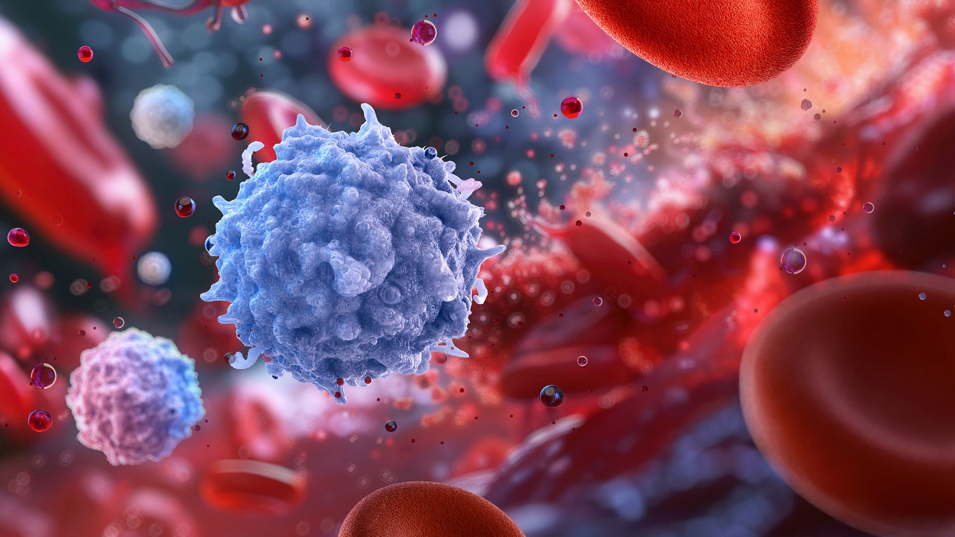
An illustration of a spiky purple sphere in the midst of blood cells

The balance of cells that make different granzymes may be important to how the immune system functions with age. (Image credit: RUSLANAS BARANAUSKAS/SCIENCE PHOTO LIBRARY via Getty Images)

They went on to hunt for this protein signature in around 48,000 participants who'd provided data to the UK Biobank, a large database of health data from U.K.-based adults. People who were predicted to have higher levels of granzyme B-making cells based on their blood proteins had worse health outcomes in the following 17 years, with a higher risk of death and of conditions like diabetes, high blood pressure, and liver and kidney disease.

The findings might point to an aspect of immune aging that wasn't previously understood.

"I think that's really exciting," Claire Gustafson, an immunologist at the Allen Institute, said of the results. "It highlights there's potentially some novel biology that we haven't really observed before," Gustafson, who was not involved in the research, told Live Science.

However, while there's a strong association between the balance of these cell populations and disease risks, Artyomov emphasized that the study does not prove that this ratio directly causes the poor health outcomes. Further studies would be needed to understand why having more granzyme B cells is linked with poorer health.

"This observation might be just a consequence, not a cause of the disease," Artyomov noted.

It's also important to note that granzyme K-making cells in mice are not the same as granzyme K-making cells in humans. In mice, they represent a more mature, specialized stage of immune cell development, whereas in humans, granzyme B cells better fit that description, he told Live Science.

That points to a key way that immunity in mice and humans is different, which was "underappreciated" before, Gustafson said. So when we're comparing these cells in mice and humans, "we're not looking at apples to apples," she said. Scientists now specifically "need to look more into humans, as it's not reflected in animal models."

In people, immune cells' features can also vary across diverse human populations, Gustafson added. We need more data across different populations to get a "true understanding of universal immune health," she said. The K-to-B ratio may be a "novel" clue that can help scientists understand healthy aging, but more data is needed to unpack why and how that ratio is linked to healthy or unhealthy aging.

"This is kind of like the first step to be able to get there," she told Live Science.

This article is for informational purposes only and is not meant to offer medical advice.

Would you trust AI to calculate your true 'biological age'?

8 October 2026 at 17:09

Scientists have recently harnessed artificial intelligence (AI) to build "aging clocks" that aim to estimate a person's true biological age.

While a person's chronological age reflects the number of years they've been alive, their biological age is intended to capture how well their bodily systems are functioning. The idea is that people with older biological ages have a higher risk of age-related diseases β€ͺ—‬ such as cancer, heart disease or dementia β€ͺ—‬ while younger biological ages come with less risk. Some aging clocks even aim to predict people's impending risk of death.

Many aging clocks look at epigenetic markers, meaning chemical tags that bind DNA and change how genes activate, while others measure features such as the pace and pitch of people's voices. But some scientists say we don't know how to use these aging clocks yet, arguing that the tools are useful for studying aging at a population level but not for guiding individual health decisions.

Others posit that the data the clocks are based on is too noisy, and thus, there's potential for drawing faulty conclusions about what they're measuring. They say, for example, that we don't know if the clocks are measuring the downstream effects of aging or its underlying causes. And in any case, we don't currently have any treatments to "wind back" the clocks.

What do you think? Would you use these AI tools to measure your biological age? Let us know in the poll below, and explain your reasoning and additional thoughts about the technology in the comments.

'Zombie cells' may drive aging, and scientists are devising new ways to get rid of them

7 October 2026 at 11:00

Emerging treatments aim to reverse or stall signs of aging in the body and thus cut the risk of age-related diseases. One of those signs is an accumulation of "zombie cells" β€” damaged cells that spew harmful substances rather than dying off and getting cleared away by the immune system.

Some scientists are designing treatments aimed at killing those zombies directly. But what if we instead helped the body's cleanup crew get rid of them?

"I think rejuvenating the immune system could rectify a lot of problems," said Roel de Maeyer, an immunologist at the University of Oxford. "We know hospitals are full of older people suffering from infections that younger people don't need to go to [the] hospital for."

The aim of these rejuvenating therapies would be to help people live healthier for longer, he told Live Science.

How do cells turn into zombies?

The immune system constantly gets rid of old, dysfunctional and dead cells to keep tissues healthy, but that cleanup gets less effective with age.

When cells die, they display proteins on their surfaces that act as "eat me" signals for immune cells, called macrophages, to engulf and destroy them. However, some cells refuse to die and instead become senescent. These zombified cells can lurk in tissues for years, releasing chemicals that drive inflammation and damage.

Senescent cells can form due to DNA damage from oxidative stress, radiation, or even normal cell division. Cells lose bits of DNA each time they make copies of themselves, and they have intricate mechanisms to decide when it becomes a "bit too dangerous to keep dividing," de Maeyer said. If a cell can no longer divide, it usually undergoes a process called programmed cell death or becomes senescent, he said.

Senescence can be beneficial in some contexts; it helps heal wounds, for example. But an accumulation of senescent cells is linked to damaging inflammation and impaired organ function.

"If there are many senescent cells in a tissue, that's usually a bad sign," de Maeyer said. The number of senescent cells goes up with age and is linked to many age-related conditions, such as heart disease and dementia.

Slaying zombies

Studies in mice have shown that using drugs to kill senescent cells can reverse inflammation and some signs of cellular aging. These drugs, called senolytics, force senescent cells to enter programmed cell death, thus reducing their numbers, said Jure Povsin, a biochemist at the Max Planck Institute of Biochemistry in Germany. This ultimately reduces inflammation and improves tissue function, he told Live Science.

But while senolytics can help rid the body of zombie cells, it's not clear why the immune system fails to eliminate senescent cells in the first place. To find answers, scientists began looking at macrophages, the cells tasked with devouring dying cells.

Research pointed to a receptor on macrophages called EP2. One study, published in July in the journal Science, found that old macrophages have a greater number of these receptors and cannot eat senescent cells effectively.

Genetically deleting the EP2 receptor in lab mice restored the macrophages' function and reduced the number of senescent cells. Compared with normal mice of the same age, the genetically tweaked mice showed less cognitive decline, muscle loss, cardiac dysfunction and systemic inflammation.

On some of these metrics, such as the ability to remember familiar objects, the modified mice were comparable to younger mice.

Gobbling up cells requires a ton of energy.

Dr. Katrin Andreasson, a neurologist at Stanford University

A problem with energy

Why is the EP2 receptor at the root of this problem? Scientists found a clue in a huge study of older adults.

A molecule called PGE2 plugs into the EP2 receptor. It's a type of prostaglandin β€” a molecule that helps cells communicate during stress and injury and contributes to fever, pain and swelling. Drugs such as ibuprofen reduce inflammation by blocking prostaglandin production.

In 2001, a large study uncovered a connection between these prostaglandin-blocking drugs and Alzheimer's disease: People ages 55 and older who routinely took the drugs for at least two years had a lower risk of developing Alzheimer's than nonusers did. The study could not prove that the drugs directly prevented the disease, but it added to growing evidence that inflammation related to EP2 activity plays a role in Alzheimer's.

This caught the attention of Dr. Katrin Andreasson, a neurologist at Stanford University and co-author of the recent Science study.

"I thought, 'Wow, this is interesting,'" she told Live Science. "I wondered if there's a connection."

Andreasson and colleagues had an important breakthrough in 2021: They found that aging macrophages had higher levels of the EP2 receptor and that increased EP2 activity disrupted how the cells used their energy. Instead of burning glucose to produce energy like young macrophages do, these cells stashed it away.

As a result, the cells could not do their job properly, as "gobbling up cells requires a ton of energy," Andreasson said. Blocking the EP2 receptor restored normal energy use in the macrophages. In old mice, it also led to reduced inflammation and better scores on cognitive tests, compared with mice in which EP2 activity remained high.

Making senescent cells vulnerable

In their recent study, Andreasson's team uncovered another consequence of aging: Old mice accumulated many senescent neutrophils, a type of short-lived immune cell that acts as the body's first line of defense against germs. Neutrophils normally die within days and are then cleared away, but with age, they become more prone to senescence and accumulate.

"I think that's kind of an odd concept because they're [usually] very short-lived cells," said de Maeyer, who was not involved in the Science study. "Neutrophils are inherently quite inflammatory, so wanting to clear them from tissues is very valuable."

Old mice have too much EP2 activity in their macrophages, and that means they can't get rid of these neutrophils, Andreasson's team found. That may be why deleting EP2 seems to have anti-aging effects in multiple organs, they concluded.

But increased EP2 activity is just one side of the story; some studies suggest that zombie cells actively suppress macrophages' ability to eat them. They do this by using a tricky protein called CD47.

CD47 is a "don't eat me" signal that stops macrophages from destroying cells, Povsin said. "It signals that the cell is healthy, alive and doesn't need to be eaten."

However, that signal gets amplified in senescent cells, Max Planck researchers showed in a 2023 study. That means that, when macrophages interact with senescent cells, macrophages' function becomes impaired. Afterward, they even lose the ability to engulf dying, nonsenescent cells. This allows a ton of cellular debris to accumulate.

Microscope images show red and blue stained macrophages engulf senescent cells (fluorescent green)

Top row: Macrophages (stained blue and red) incubated with young, proliferating cells will engulf dead cells and debris (green) when they encounter it. By contrast, macrophages grown in culture with senescent cells will not engulf dead cells, allowing cellular trash to accumulate (bottom row). This is because the aging cells release a strong "don't eat me" signal. (Image credit: Adapted from Schloesser et al, J. Cell Biology, 2022.)

So, rather than just blocking EP2, it could also be beneficial to reduce that "don't eat me" signal in senescent cells, this research hints. But CD47 is found on all living cells, so scientists can't just deplete its numbers across the body.

A specific enzyme modifies CD47 on senescent cells, and in the 2023 study, blocking the enzyme helped spare macrophages from the effects of CD47. Therefore, blocking the enzyme could be a more selective approach to weaken the "don't eat me" signal on senescent cells, Povsin said.

A long road to treatments for people

Translating these ideas into anti-aging treatments for humans would be a major success in the science of aging, but experts say there are significant problems to overcome.

Although blocking EP2 had consistent results in lab mice, the rodents are genetically identical and bred in controlled environments. Humans are inherently more variable in their biology and the environmental factors they're exposed to, introducing confounding factors that would need to be addressed, de Maeyer said.

Additionally, in clinical trials, EP2-blocking drugs have so far been explored as cancer treatments, but they are "underexplored" in the context of aging, he added.

Drugs targeting CD47 are in an even earlier stage of development. In the 2023 research from Max Planck, the team used both human and mouse macrophages in their experiments, but the approach has not been tested as an anti-aging treatment in living mice or humans yet.

In 2020, de Maeyer's team used a different strategy to rejuvenate macrophages in older adults. They blocked a protein called p38, whose activity was high in the macrophages of older adults. Blocking the protein helped macrophages better recognize and eat dying cells in specific tests.

In that study, the team gave adults ages 65 and up an experimental drug called losmapimod to block p38. Although it helped restore their immune cell function, it was not suitable for long-term use because it caused liver problems in later trials, de Maeyer said.

In their mouse studies, Andreasson's team was able to block EP2 without causing any negative side effects. But she cautioned that we "really need to drill down on the research and confirm all this stuff in humans."

"The next logical step," she said, "would be to figure out how to inhibit this receptor in a safe way."

This article is for informational purposes only and is not meant to offer medical advice.

Help us improve Live Science Pro: We're always trying to make our content better. Leave us feedback about Pro here.

❌