❌

Normal view

Tiny Genetic Fragments Crucial for Signaling Brain Rest Identified

19 June 2026 at 20:51

In a groundbreaking study shedding light on the molecular intricacies of arousal regulation, researchers from Pompeu Fabra University (UPF) and the Centre for Genomic Regulation (CRG) have unveiled the profound influence of neuronal microexons on behavioral states in zebrafish. This study elucidates how subtle alterations in these tiny genetic fragments can trigger hyperarousal—a state marked by heightened neural excitability and pronounced insomnia-like symptoms—which echoes the pathophysiological features observed in various neurodevelopmental disorders. The implications extend far beyond aquatic models, offering a window into the conserved mechanisms of arousal that potentially inform conditions such as autism spectrum disorder and schizophrenia in humans.

Arousal represents a fundamental neurophysiological process essential for survival, enabling organisms to respond to external and internal stimuli with appropriate behavioral and neural adaptations. This highly conserved mechanism across species ensures a meticulously balanced state, modulating responsiveness between lethargy and sensory hypersensitivity. Dysregulation of this balance manifests clinically as either diminished responsiveness or excessive wakefulness and sensory overload, typical hallmarks of stress and various neurodevelopmental pathologies.

Fundamental to this regulatory system is the diversity of proteins synthesized during development and adulthood through alternative splicing—a sophisticated post-transcriptional gene editing process. Alternative splicing enables the generation of multiple functionally distinct protein isoforms from a single gene, often mediated by the inclusion or exclusion of microexons. Microexons are exceptionally short exonic sequences within neuronal genes that profoundly influence protein function and neuronal circuit dynamics despite their minuscule size.

The investigative team employed zebrafish larvae, leveraging their optical transparency and genetic tractability to scrutinize the behavioral consequences of neural microexon misregulation. Larvae exhibiting abnormal microexon patterns demonstrated conspicuous hyperarousal behaviors including disrupted swim patterns and shortened sleep duration. “These larvae not only sleep less frequently but also take considerably longer to initiate sleep,” remarks first author Tahnee Mackensen. This behavioral hyperactivity parallels neural hyperexcitability, suggesting microexon regulation as a pivotal determinant of neurobehavioral states.

At the cellular signaling level, the researchers identified dysregulated cyclic adenosine monophosphate (cAMP) cascades as a key mediator of the observed hyperactive state. cAMP is a ubiquitous second messenger involved in modulating neuronal excitability and synaptic plasticity. The altered splicing of microexons modulates cAMP synthesis and degradation pathways, leading to an aberrant excitation of forebrain neurons in hyperaroused larvae. Notably, this altered cAMP signaling manifests as heightened cAMP-dependent protein kinase A (PKA) activity and subsequent phosphorylation of the transcription factor CREB, implicating the canonical cAMP-PKA-CREB pathway in the regulation of arousal.

The study’s pharmacological interventions underscore the centrality of cAMP regulation in arousal control. Application of cAMP inhibitors normalized the elevated neural activity and behavioral hyperarousal in mutant fish, whereas artificially elevating cAMP in wild-type fish recapitulated the hyperactive phenotype. This bidirectional modulation fortifies the concept that neuronal cAMP levels function as a ‘thermostat’ for arousal states, fine-tuning neuronal excitability and behavioral responsiveness.

Beyond the immediate findings in zebrafish, this research builds upon prior observations in drosophila models demonstrating that microexon disruption similarly impairs sleep and elevates arousal. “The parallel between species indicates an evolutionarily conserved arousal mechanism,” explains Manuel Irimia, senior author. This conservation implies that microexon misregulation, while mechanistically nuanced, may contribute to the neuropsychiatric symptomatology observed in mammals, including humans.

Human neurological disorders such as autism and schizophrenia are often accompanied by sleep disruption and sensory processing anomalies attributed, in part, to aberrant microexon splicing. While microexon alterations are unlikely to be sole causative factors, they may exacerbate or modulate disease phenotypes by disturbing neural excitability homeostasis. These insights prompt a reevaluation of therapeutic strategies aimed at restoring microexon splicing fidelity or modulating cAMP signaling pathways to alleviate neurodevelopmental symptomatology.

Moreover, the link between this microexon-cAMP pathway and mood disorders such as anxiety and depression opens compelling avenues for future research. The cAMP-PKA-CREB axis has well-documented roles in synaptic plasticity and mood regulation, suggesting that microexon-associated dysregulation could contribute to broader neuropsychiatric conditions. “This discovery might just scratch the surface of a complex regulatory network influencing brain function,” notes Mackensen.

The transparency and genetic accessibility of the zebrafish model provided unparalleled opportunities to visualize and quantify internal states through behavioral readouts. Advanced imaging of larval swimming patterns and direct measurement of neurochemical parameters furnished robust correlative evidence linking genetic alterations to functional outcomes. These technical advancements highlight the integrative power of model organisms in neuroscience.

Importantly, this research received support from an array of prestigious funding bodies, including the Horizon 2020 Framework Programme, the Marie Sklodowska-Curie Actions, and the Wellcome Trust, emphasizing the global significance and collaborative nature of this work. The study’s publication in Science Advances confirms its high impact and relevance to the scientific community.

As the team pursues translational studies, the prospect of correcting arousal imbalances by manipulating cAMP pathways or restoring microexon expression presents a promising frontier. This is especially critical given that aberrant arousal and sleep disturbances profoundly impair quality of life in neurodevelopmental disorders. The findings pave the way for multidisciplinary approaches integrating molecular genetics, neurobiology, and pharmacology to develop targeted interventions.

In summation, the identification of neuronal microexons as key modulators of arousal states via the cAMP-PKA-CREB pathway in zebrafish represents a seminal advance in our understanding of the molecular substrates governing complex behavioral phenotypes. This research not only deciphers fundamental biological mechanisms but also holds translational potential to inform therapeutic avenues for neuropsychiatric conditions marked by disrupted arousal and sleep.


Subject of Research: Animals

Article Title: Neuronal microexons modulate arousal via the cAMP-PKA-CREB pathway in zebrafish

News Publication Date: 19-Jun-2026

Web References: 10.1126/sciadv.ady8291

Image Credits: UPF – CRG

Keywords: Exons, Gene splicing, Developmental neuroscience, Anxiety, Sleep disorders, cAMP signaling, Zebrafish

How H5N1 Bird Flu Went Undetected for Weeks in Dairy Cattle

19 June 2026 at 20:45

In early 2024, a baffling outbreak of H5N1 avian influenza emerged among dairy cattle in the Texas Panhandle, presenting a new and unexpected clinical manifestation of the virus. Unlike the typical respiratory illness caused by H5N1 in other mammals, affected cows exhibited severe necrotizing mastitis, an inflammatory condition predominantly targeting the mammary glands. This atypical tissue tropism challenged existing paradigms and delayed the accurate diagnosis, as initial veterinary investigations focused mainly on bacterial pathogens common to mastitis. The revelation that H5N1 was the etiological agent marked a critical moment in understanding how influenza viruses can adapt to novel hosts and tissues.

The study, recently published in Science Advances by researchers at the University of Pittsburgh School of Public Health, provides pioneering mechanistic insights into this unusual manifestation. The research team, led by Dr. Suresh Kuchipudi, elucidated the molecular basis for the H5N1 virus’s propensity to infect bovine mammary tissue rather than respiratory organs. This phenomenon, now observed in over 100 avian and mammalian species globally, underscores the remarkable plasticity of the H5N1 virus and its ability to exploit unique receptor architectures in different host species.

Traditionally, H5N1 and related influenza viruses target sialic acid glycan receptors present in the respiratory tract, facilitating viral entry primarily via respiratory epithelial cells. However, in bovines, the absence of clinical respiratory disease despite the presence of these glycans suggested a more nuanced receptor interaction paradigm. Kuchipudi’s group hypothesized that not just the presence but the precise molecular subtype and distribution of influenza receptors could dictate tissue tropism. Their investigation focused on delineating the receptor landscape across bovine tissues using cutting-edge glycomic methods combined with binding assays and ultra-high-resolution microscopy.

The team employed glycomics—a comprehensive approach to cataloging glycan structures—collaborating with expert Dr. Lauren E. Pepi from Harvard Medical School. They identified that among various glycan receptors, only a particular subclass known as N-linked sialic acid receptors demonstrated a high affinity for the H5N1 virus. Crucially, these receptors were found to be prevalent and densely expressed in the mammary epithelial cells of the udder but conspicuously scarce in bovine airway tissues. This spatial receptor distribution offers a molecular explanation for the virus’s unusual targeting of the mammary gland, enabling productive viral replication and resulting clinical mastitis.

The discovery sheds light on the critical role of receptor architecture in species-specific influenza virus infection. Prior reports indicated the presence of flu-related glycans in bovine respiratory tissues, but those receptors did not support significant viral binding or replication. The differential receptor expression patterns in respiratory versus mammary tissues effectively reprogrammed the virus’s tissue tropism. This specialization allows H5N1 to hijack the bovine mammary environment as a niche, explaining why infected cattle shed the virus in milk rather than displaying respiratory symptoms.

Beyond animal health, this finding carries important public health implications. The intense viral shedding into milk poses a risk to farm workers exposed to raw milk and raises concerns regarding zoonotic transmission routes. Dr. Kuchipudi points out that while pasteurization effectively neutralizes the virus, the common practice of feeding raw milk to domestic pets, notably cats, can facilitate cross-species transmission. Indeed, previous studies documented feline fatalities linked to H5N1 infection. This highlights the hidden dangers of viral reservoirs in dairy cattle and underscores the need for enhanced surveillance and biosecurity protocols within agricultural settings.

Methodologically, the study stands out for its innovative multimodal approach. The integration of high-precision receptor mapping with functional binding experiments allowed researchers to move beyond conventional histochemical staining. This enabled a granular understanding of the receptor types mediating viral entry, paving the way for predictive models of viral host and tissue susceptibility. By decoding the receptor landscape at this molecular scale, the research team demonstrated an unprecedented ability to identify viral tropism determinants, which could serve as early warning indicators for emergent influenza strains.

Looking forward, these insights form the foundation for a new paradigm in influenza research and surveillance. Scientists can now preemptively screen various animal species—not just for the presence of virus-compatible receptors but for specific tissue vulnerabilities within those hosts. This capability is transformative for anticipating how H5N1 and related viruses might manifest clinically, whether through respiratory symptoms, mastitis-like conditions, or neurological involvement, as previously seen in felines. Such proactive screening could revolutionize public health responses, affording precious time to deploy targeted interventions and prevent widespread outbreaks.

The implications of receptor-based viral tropism extend beyond livestock to potential future zoonoses affecting humans. Understanding the molecular basis of host adaptation provides critical clues into viral evolution and spillover risk. This knowledge also informs vaccine development and therapeutic strategies by highlighting tissue-specific pathways of viral entry and replication. As zoonotic influenza viruses continue to adapt, harnessing receptor glycomics will be vital for staying ahead of viral evolution and protecting both animal and human health.

The study was a collaborative effort involving multiple institutions and experts, including contributors from Pennsylvania State University, Harvard University, and North Dakota State University. Their combined expertise spanned infectious diseases, glycomics, virology, and veterinary medicine, enabling a comprehensive and multidisciplinary approach. Supported by the U.S. Department of Agriculture’s National Institute of Food and Agriculture and Pitt Public Health, this research exemplifies how fundamental science can directly inform and improve agricultural biosecurity and zoonotic disease preparedness.

Ultimately, this investigation marks a significant milestone in understanding influenza virus host adaptation. By revealing the receptor basis for the bovine-specific tissue tropism of H5N1, the study not only clarifies an enigmatic veterinary health issue but also establishes a versatile scientific framework for future viral threat assessment. As influenza viruses continue to evolve in complex ecological contexts, such mechanistic insights will be indispensable tools to foresee their next unexpected moves and mitigate their impact on global health.


Subject of Research: Animals
Article Title: Receptor Basis of Unusual Tissue Tropism of Avian Influenza H5N1 Clade 2.3.4.4b Virus in Cattle
News Publication Date: 19-Jun-2026
Web References: https://doi.org/10.1126/sciadv.aea2068
Image Credits: Department of Infectious Diseases and Microbiology, School of Public Health, University of Pittsburgh
Keywords: Avian influenza, Glycomics, Public health, Viruses

'A completely different story': 300 million-year-old fossils reveal the first vertebrate land dwellers weren't what we thought, researchers claim

19 June 2026 at 11:26

Never-before-seen fossils of newly hatched crocodile-like creatures are shining new light on how our aquatic ancestors conquered land.

Early four-limbed vertebrates (tetrapods), which would eventually give rise to humans, took their first steps on land in the Devonian period, some 419 million to 359 million years ago, marking one of the most important periods in the evolutionary history of animals.

Now, a new study published Thursday (June 18) in the journal Science has revealed that these early tetrapods were less like amphibians and more like us. Rather than having a tadpole phase in their development, as many amphibians do today, new evidence suggests that they were direct developers — growing from smaller to bigger versions of themselves, like their ancestors, humans and many other animals.

The study is an important contribution to our understanding of early tetrapods' reproductive developmental biology, said Tim Smithson, a visiting academic at the University of Cambridge who specializes in early tetrapods but was not involved in the study.

It suggests that the "earliest tetrapods that took those first steps on to land were able to rely on the successful reproductive and developmental strategies of their forebears," Smithson told Live Science in an email. "Direct development made life easier — one less thing to worry about!"

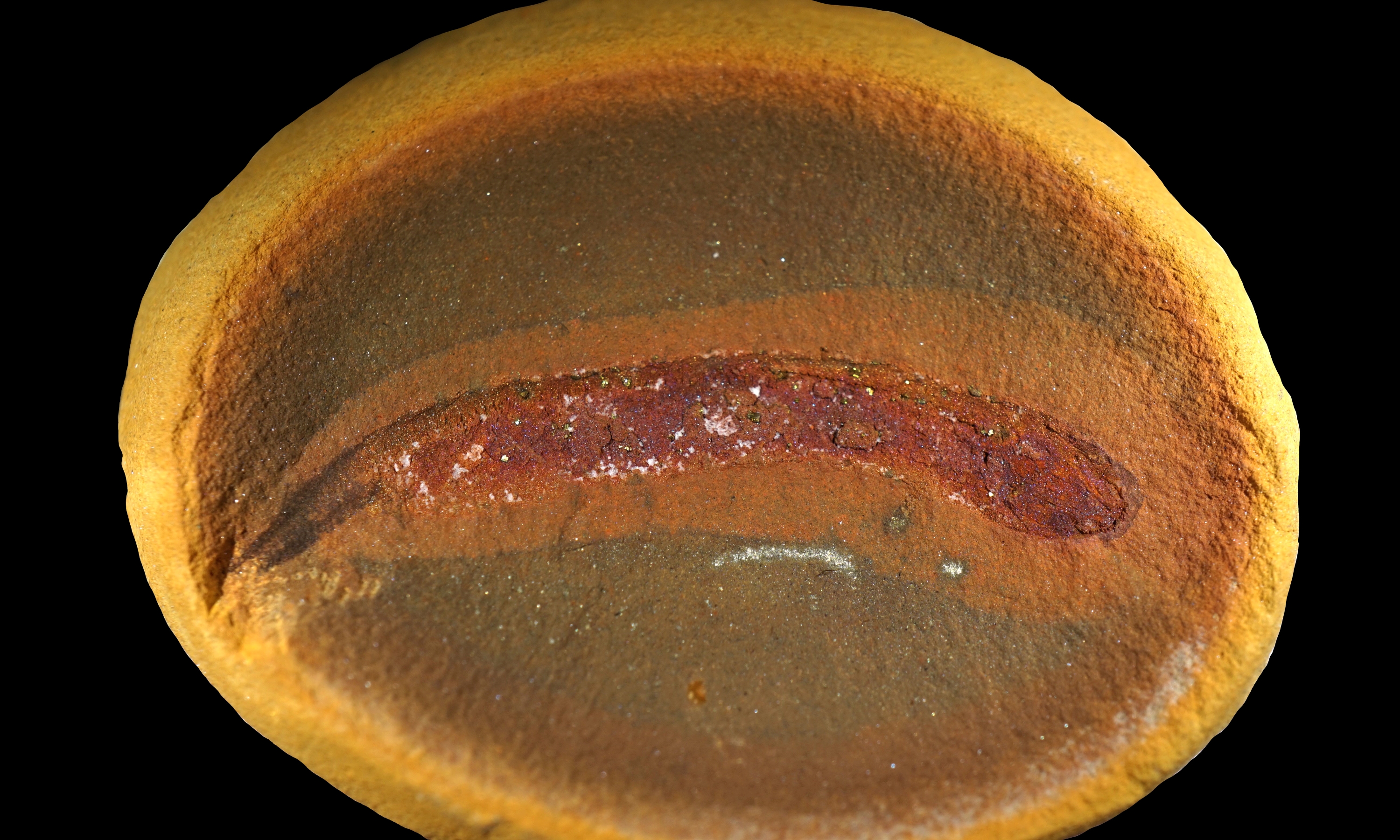
The new research was based partly on fossils from early land-dwelling predators called embolomeres. These animals looked like a cross between a crocodile and an eel and ruled river, lake and swamp habitats 350 million to 280 million years ago, during the Carboniferous and Permian periods. While these creatures could grow to more than 10 feet (3 meters) long as adults, the study unveils rare fossils from Mazon Creek, near Chicago, that preserved embolomeres as hatchlings that were days to a couple of weeks old.

"These are intimate details of the first moments of these animals' lives, and we've never seen that before for this entire part of the evolutionary tree," study co-author Jason Pardo, a postdoctoral fellow of evolutionary biology at Vilnius University in Lithuania and a research associate at the Field Museum in Chicago, told Live Science.

A photo of an embolomere fossil.

Researchers studied exceptionally well-preserved fossils from Mazon Creek, Illinois. (Image credit: Arjan Mann)

These fossils didn't show evidence of external gills and other tadpole-like features that the researchers would have expected from an early land dweller. The researchers then checked other fossils from before and during the "fin-to-limb transition" but found no evidence of an amphibian-like life cycle in those, either.

"For as long as we've understood evolution, we've assumed this story of how we made that transition from water to land," Pardo said. "We in fact have a completely different story."

Science upended?

A statement released by the Field Museum claimed that the study upends scientists' understanding of how animals conquered the land. However, the experts Live Science spoke to disagreed with this assertion.

"The Mazon Creek material is wonderful, the study is interesting and the interpretation of the fossils is sound, but I don't think the results are terribly surprising," Per Ahlberg, a professor of evolutionary organismal biology at Uppsala University in Sweden, told Live Science in an email.

Ahlberg, who was not involved in the new study, specializes in the early evolution of tetrapods. He noted that scientists knew some early tetrapods had a larval stage similar to that of modern salamanders ‪—‬ namely, those belonging to the group Temnospondyli, which he described as the ancestral stock of modern amphibians. However, he contends that this didn't mean scientists assumed every early tetrapod was the same.

"Nobody has been arguing in recent years that ALL early tetrapods had such a larval stage or that this was essential for enabling the transition to land," Ahlberg said. "I mean, I have been working right at the core of this research field for 40 years and I have never given it any thought."

In response, Pardo agreed that specialists in the field recognized that the data didn't support that early tetrapods had an amphibian-like development. However, he argued that even among specialists, assumptions were still made about metamorphosis — a major developmental transition, like a tadpole transforming into a frog — and amphibian-like bodies.

Young embolomeres would grow up to become apex predators in rivers, lakes and swamps.

Young embolomeres, illustrated here, suggest that early tetrapods didn't undergo an amphibian-like metamorphosis. (Image credit: Berit Godring)

"Onto something big"

Study co-author Arjan Mann, an assistant curator of early tetrapods at the Field Museum, first saw the study's first baby embolomere fossil during a 2016 trip to the Field Museum while working on his doctorate. At the time, the fossil was a mystery.

Mann and Pardo mused over the fossil's identity for years before high-resolution scans with scanning electron microscopy at the Canadian Museum of Nature confirmed that the ancient creature was an embolomere, according to the museum's statement.

"I think Jason and I both knew we were onto something big, since fossils of this kind of animal and from this phase, and developmental state in early tetrapod evolution have never been found or studied before," Mann told Live Science in an email.

Along with the embolomeres, the researchers looked at megalichthyid fish from before the land transition and limbless, snake-like creatures known as aistopods from during the land transition. All showed signs of direct development, they said.

"I think the take home message of this study is that we should always challenge conventional wisdom in science, especially when these older ideas do not have substantial backing," Mann said.

❌