Many people are familiar with melatonin, a hormone that helps ready the body for sleep. But there's another, lesser-known chemical in the brain, called orexin, that tips the scale in the other direction: waking the body up.
In 1999, two scientists independently uncovered the function of orexin and published papers about it. Dr. Masashi Yanagisawa, a molecular biologist and physician now at the University of Tsukuba in Japan, came across orexin while studying the interaction of receptors and chemicals in the brain. Dr. Emmanuel Mignot, now the director of the Stanford Center for Narcolepsy, homed in on orexin while studying narcoleptic dogs. (His Chihuahua, named Watson, has narcolepsy, and he previously had another dog with the condition, named Bear.)
This year, the pair of researchers won the coveted Albert Lasker Basic Medical Research Award for this fundamental finding that helped reshape our understanding of sleep. Their work has since led to the development of drugs for insomnia and narcolepsy.
Yanagisawa told Live Science that, at the start of his research career, he didn't expect to end up studying sleep. Mignot, on the other hand, had an early interest in understanding narcolepsy so that better treatments for the neurological disorder could be developed. Live Science spoke with Mignot about the discovery of orexin and how the field might progress in the future.
Nicoletta Lanese: What initially drew you to studying narcolepsy?
Dr. Emmanuel Mignot: There were three things that attracted me to narcolepsy: Number one, it was a human problem. It was already known [at the time] that it was not super rare. I'm not saying it's super common, but 1 person per 3,000, 0.03% — it's reasonably common. And nobody cared about it. I would talk to neurologists and they'd say, "Oh, I've never seen one case in my entire life." So I would say, "Yes, of course; you miss them all." That was the state of the play at the time.
Number two, was what this was a quintessential sleep disorder [in which sleep itself is disrupted]. I thought that if we found the cause of this disease, we could potentially discover something totally new about sleep — a fantastic entry point to a mystery.
The third thing that made me ready to go there was that it was tractable. A lot of problems are not tractable.
NL: Why did you start working with the dogs with narcolepsy?
EM: I studied the pharmacological basis because I was trained in pharmacology, and as a psychiatrist, and I discovered how this drug modafinil [a stimulant-based narcolepsy treatment] was working. But quickly, I realized I'm not going to find the cause by doing pharmacology. I'm only going to explore what's known.
That's when I started to fall in love with genetics, even though I was not trained as a geneticist. I said, "We have to find this gene in the dogs." It looks like they have all the symptoms. It was a huge bet, and it took me 10 years because I was definitely … overoptimistic. But it was worth the price at the end.
After narcolepsy was described — the dog gene — suddenly narcolepsy became much more famous.
NL: And scientists already knew about narcolepsy in dogs at that time?
EM: The guy at Stanford that started the sleep program was called Dr. [William] Dement. He was a graduate student in Chicago in the 1950s, when [Eugene] Aserinsky and [Nathaniel] Kleitman discovered REM sleep. He had the foresight to understand that this was an important discovery.
[Later], he discovered that narcolepsy was really a disorder where people go into REM sleep and have this very abnormal dreaming. He came in 1970 to Stanford, and the first thing he did after his medical school was to start a clinic to see patients with narcolepsy. Around then, he spoke at an American Medical Association conference in San Francisco, and he discussed different sleep disorders, including narcolepsy. And then there was someone in the audience that said, "Oh my god; I have a dog with your disease. Every time it gets excited — boom — it collapses. And it sleeps all the time."
That dog had already been euthanized, but it still gave him the idea that these dogs have narcolepsy — maybe we should try to get some. So he went to a lot of veterinarians to talk to them about it, and they identified a whole series of dogs, including the first poodle, Monique, who had narcolepsy. And they started this little colony of dogs. Initially, they tried to breed them, but most cases of narcolepsy are actually not genetic. Then, in 1977, they got a whole litter of Dobermans, and then Labradors, that had some form that was genetic.
So when I arrived, it was in 1987. They already had the dog colony. I came to study the pharmacology, to try to find a better treatment for narcolepsy. But I did stand on the shoulders of giants.
NL: At that time, it's not like you had a complete dog genome. Did that make studying their genetics challenging?
EM: It was crazy. A couple of human genes had been isolated, and mouse genes, but it was a handful. We didn't even have the full genome map in humans. And meanwhile, dogs — no pun intended — were in no man's land. Very little was known.
It took me 10 years. I had times where some people didn't believe that the dogs had narcolepsy. There were a few depressing moments, but I was convinced that was the right thing to do.
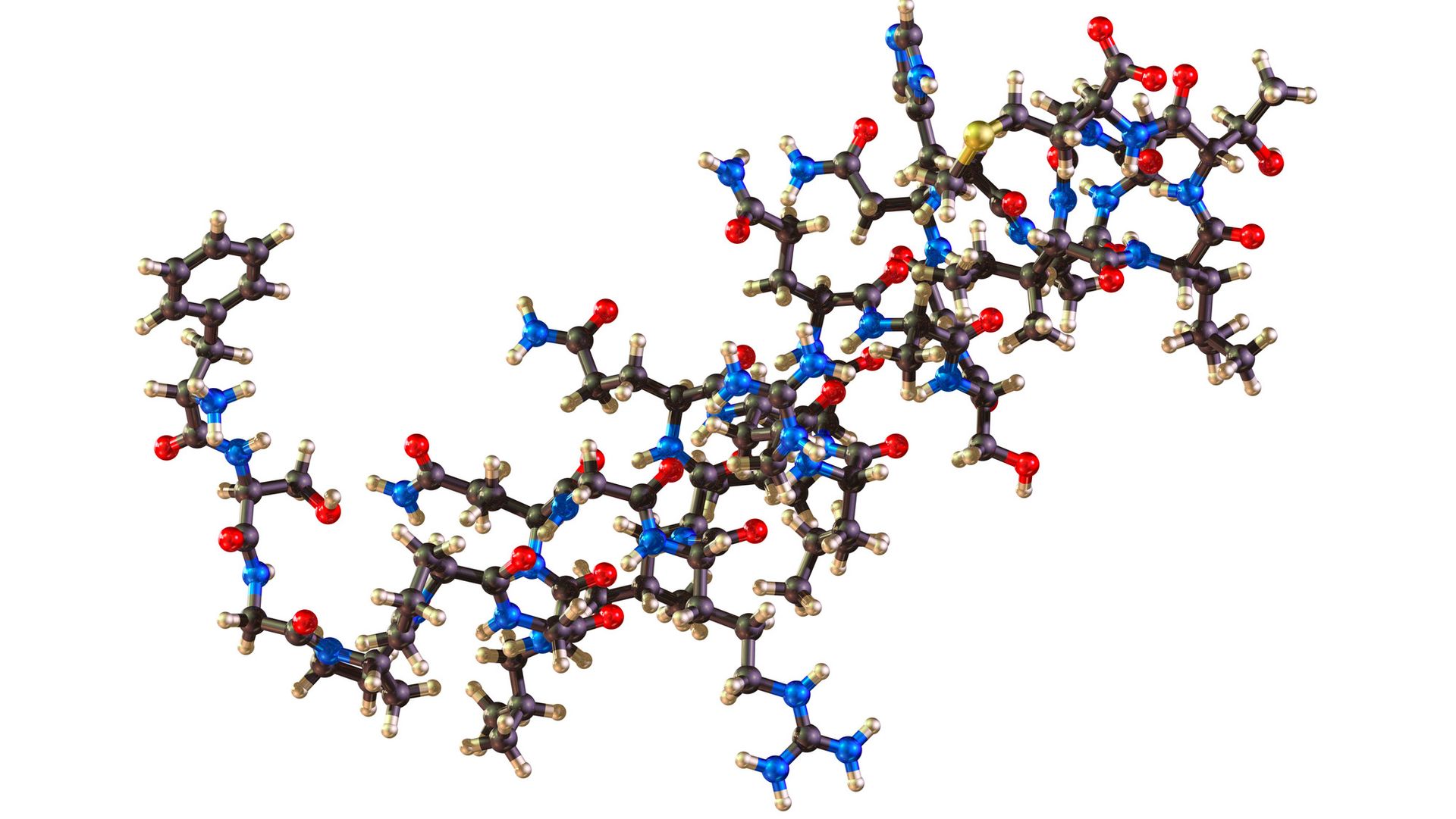
Orexin (pictured) is a peptide in the brain that promotes wakefulness. (Image credit: theasis via Getty Images)
NL: When you ultimately found the receptor for orexin, did it surprise you? It strikes me as surprising that there would be a discrete "switch" in the brain that controls wakefulness.
EM: I was lucky. Honestly, I had no idea. I got enamored with genetics because what I love [about it] is you can find something without any hypothesis. You just search for it, and what you find tells you what it is. You have no prerequisite for knowing what you would find.
I could have found something much less significant. That was a dream, to find a GPCR [G-protein coupled receptor, a protein on the outside of cells]. Can you imagine? A drug target.
Then, it turned out to be immediately applicable to humans. That, too, was a lot of luck; let's be honest. I was just trying to find the cause of one thing and one step at a time; it could have been something very complicated. It turned out to be simple.
Was I surprised? No, because I was not expecting one or another.
NL: Do you recall what the reception was like when the initial finding was published?
EM: It had an immediate effect, for several reasons. For example, I was seeing a lot of patients with narcolepsy at the time, but they were all old folks that had had narcolepsy forever. A lot of people would tell me, "I think I had it from when I was born, because I don't remember." We were never seeing children.
But after narcolepsy was described — the dog gene — suddenly narcolepsy became much more famous. And what happened is that people started to pay attention to narcolepsy, and we started to see kids. In kids, the picture is so much different. They gain an enormous amount of weight; it's very abrupt. It's a little bit different than what you see in adults, where they have adapted to it.
That was, I think, a practical consequence of the discovery. It really put narcolepsy on the map.
The second thing that happened is, all the drug companies started to develop hypnotics [sleeping pills], trying to block the orexin. They developed hypnotics relatively quickly, and they are very effective. They are probably safer than benzos [benzodiazepines] and so forth.
NL: Is it considered safer because the orexin blockers better replicate natural sleep?
EM: We know that the orexin definitely drops during sleep, so blocking it is definitely a way to recapitulate that drop; that's true. It's an awake-promoting system, so if you remove something that makes you more awake, it's safer than inducing sleep by shutting down the brain.
NL: As you moved to studying narcolepsy in humans, you found evidence that there might be an autoimmune response involved; it's been suggested that such a response may be harming the cells that make orexin in the brain. Was there suspicion that the condition had an autoimmune element previously?
EM: It's not something I 100% discovered. In 1983, before I came to study narcolepsy, there was a guy in Japan who I met, called Yutaka Honda. [Editor's note: Honda's research linked narcolepsy to the human leukocyte antigen (HLA) region of the genome, which helps regulate immune responses.]
The HLA is the way the immune system sees the world. That had just been discovered as very important for transplantation, because it's very polymorphic [takes on different forms] from one person to the next. They [Honda's group] found that all narcoleptics had a particular genetic variant, called HLA-DR2. This was a total surprise.
When I came, that was already known. But then people really looked and tried to see if the disease was autoimmune, but they couldn't find anything — no autoantibody, nothing. It was all negative.
I did a study with African Americans, and in African Americans, I found that this DR2 marker was not the best marker for narcolepsy. It was a gene just next to it that was called DQB1*06:02. [Editor's note: That HLA variant is now recognized as a strong risk factor for narcolepsy, particularly for narcolepsy type 1 (which involves a symptom called cataplexy).]
I'm very proud of that study because I think it's the first time that people used human diversity to actually map down a genetic factor more precisely, which now is used commonly. It's called transethnic mapping.
NL: New treatments that target orexin have just been approved. Do you see those therapies as an extension of your earlier findings?
EM: Of course. I was trained as a pharmacologist originally, so obviously, I do a lot of clinical trials. I see my patients totally transformed by this medication. It's just amazing. I'm so fortunate. How could you get a better reward than seeing your patients totally transformed by this medication?
It's going to have a lot of other applications [beyond narcolepsy]. Some people think it's going to be very helpful for ADHD; some people think it could be very helpful for depressed people with excessive sleepiness. We don't know, really; there's a lot of possibilities.
Another thing I'm excited about is that I have a few patients with [both] narcolepsy and schizophrenia. You cannot give them stimulants. If you give them modafinil or another dopamine stimulant [for narcolepsy], they have even more hallucinations. Often, they are sedated by their medication, and we are very poor at making people more motivated and active. These [orexin-activating] drugs seem to have effects beyond just waking people up. They seem to increase motivation.
You don't know until you try, but I'm sure it's going to have other applications.
This interview has been condensed and edited lightly for clarity. It is for informational purposes only and is not meant to offer medical advice.
This year's Lasker Awards — coveted prizes for biomedical research that are sometimes called "America's Nobel Prizes" — have gone to scientists behind fundamental discoveries in sleep science and to researchers who developed a groundbreaking treatment for hemophilia.
Additionally, a Lasker Award for public service went to actor Michael J. Fox for his advocacy for patients with Parkinson's disease that has helped advance research into the neurodegenerative condition.
The prizes, each of which comes with an honorarium of $250,000, will be presented in person Sept. 17 in New York City. The awardees were announced Wednesday (Sept. 9) by the Lasker Foundation, which was established in 1942 by the philanthropists and health advocates Albert and Mary Lasker.
A deeper understanding of sleep
The first of the three awards, called the Albert Lasker Basic Medical Research Award, went to two scientists who independently discovered a key substance in the brain that helps control the sleep-wake cycle and is missing in people with some forms of narcolepsy — the neurological disorder marked by excessive daytime sleepiness and, sometimes, cataplexy (sudden episodes of muscle weakness).
The researchers — Dr. Emmanuel Mignot of the Stanford University School of Medicine and Dr. Masashi Yanagisawa of the University of Tsukuba in Japan — arrived at the discovery through different approaches. Mignot wanted to understand the underlying causes of narcolepsy, while Yanagisawa was investigating a curious class of receptors that were known as "orphans" because the chemicals that activated those receptors were unknown.
"We never thought we would end up in sleep," Yanagisawa told Live Science. "This was a kind of biochemical fishing expedition."
In particular, Yanagisawa was interested in proteins at the surface of cells called G protein-coupled receptors (GPCRs). When the right chemicals are plugged into these receptors, they trigger a chain of events within cells. Various compounds that activate GPCRs had already been found to be useful as drugs, but there were many receptors with unknown triggers.
Yanagisawa and colleagues went fishing for those triggering chemicals in rat brains, isolating substances called peptides from the tissue and then exposing GPCRs to those peptides. "We got very lucky, initially," Yanagisawa said. The fifth or sixth receptor they tested had a hit. Through further experiments, they landed on two closely related peptides as the trigger, and they found a second related receptor to match.
They discovered that the peptides came from a part of the brain called the hypothalamus, which was known to be involved in eating behaviors. So they named the peptides "orexins" after the Greek word for appetite, "orexis."
It turned out, though, that orexins' main job wasn't to control appetite; rather, they help promote wakefulness, the opposite of sleepiness. Mice without functional orexin genes would suddenly fall over and remain immobile for a minute or two, the researchers found. Peering into those mice's brains, they found that the animals were suddenly entering REM sleep, a phenomenon also seen in people with narcolepsy with cataplexy.
One of this year's Lasker Awards went to scientists who helped uncover a fundamental driver of sleep across many animal species, including humans. (Image credit: FreshSplash via Getty Images)
At the same time, Mignot, the other winner of the Lasker Award, was learning that orexin deficiency is also behind a form of narcolepsy seen in dogs. He then went on to study the mechanism in humans with narcolepsy.
"It became, suddenly, a very, very convincing story," Yanagisawa said of the researchers' converging findings. Previously, the study of sleep involved damaging parts of the brain and then seeing how a lab animal's sleep changed in response.
"There was no molecular level, genetic level, description of sleep regulation mechanisms at all," he said. "So our papers really shed new light on sleep regulation."
Together, the researchers' work paved the way to a new class of insomnia treatments, called orexin receptor antagonists, which promote sleepiness by turning down wakefulness. They have also led to new narcolepsy treatments called orexin receptor agonists, which boost wakefulness by increasing the receptors' activity. The first in the latter class of drugs was just approved in August.
A life-changing hemophilia treatment
The second award, called the Lasker~DeBakey Clinical Medical Research Award, went to three scientists affiliated with the Japanese drug manufacturer Chugai Pharmaceutical. These researchers — Kunihiro Hattori, Takehisa Kitazawa and Tomoyuki Igawa — created an antibody that interacts with key proteins in the blood that produce clots and thus stop bleeding.
This antibody treats hemophilia A, a severe bleeding disorder caused by a genetic mutation. That mutation causes the body to not make enough factor VIII, one of the key blood-clotting proteins. Normally, factor VIII grabs hold of two other clotting factors, and this enables a chain reaction that results in normal blood clotting. Without enough factor VIII, there is abnormal bleeding.
The severity of hemophilia A varies, but people with the disorder frequently experience blood pooling in their joints, which causes chronic pain and impairs mobility; they can also bleed uncontrollably even in the absence of trauma.
The antibody treatment works by binding two other clotting proteins — factors IX and X — to create a structure that fulfills the function of the missing factor VIII. Hattori, who is no longer at Chugai Pharmaceutical, initially launched the effort in 2000 after hatching the idea to use an antibody to bind several clotting factors together. Kitazawa and Igawa then brought the idea to fruition using pharmacology and chemical engineering, respectively.
The antibody, called emicizumab (marketed as Hemlibra), was approved by the U.S. Food and Drug Administration in 2017, and over 100 additional countries have approved the drug since. Over 30,000 people have received the antibody treatment to date, according to the award announcement.
The third and final award — the Lasker~Bloomberg Public Service Award — went to Fox, actor and founder of The Michael J. Fox Foundation for Parkinson's Research.
Fox began experiencing symptoms of early-onset Parkinson's in 1990 at age 29. He publicly announced the diagnosis in 1998, sparking new discussions around the often-stigmatized disease. Through interactions with existing Parkinson's advocacy groups, Fox realized the field faced a lack of funding that likely hindered the pursuit of better treatments and a cure.
Since its inception in 2000, the foundation has funded more than $3 billion in Parkinson's research, according to the award announcement.
"When we launched the Foundation, our goal was simple, yet ambitious — cure Parkinson's," Fox said in a statement. "I am humbled to be recognized by the Lasker Foundation and jury. I accept this honor on behalf of everyone living with Parkinson's and everyone with us in our shared mission to end the disease."
This article is for informational purposes only and is not meant to offer medical advice.
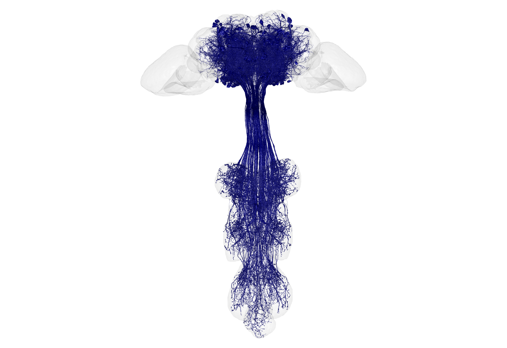
A fruit fly's brain is roughly the size of a poppy seed — and yet that tiny package contains over 100,000 neurons. A new map charts every single neuron in the male fruit fly brain, as well as the insect's equivalent of a spinal cord, totaling more than 166,000 neurons.
This new map joins a map of a female fruit fly brain that was unveiled in 2024 and covers about 140,000 neurons. These two wiring diagrams, also called "connectomes," can now be compared to see if there are differences between the sexes' brains that help to explain behavioral differences reflected during mating or in aggressive actions, including sex-specific fighting moves.
"It is the first time we can compare both sexes of an animal with complex social behavior," study co-author Gerry Rubin, head of biology and a senior group leader of the Howard Hughes Medical Institute's Janelia Research Campus, said in a statement. "Male and female flies have a lot of differences in their behavior, and neuroscientists want to understand how the brain controls those behaviors. This now allows us to easily home in on the neurons that are causing those differences."
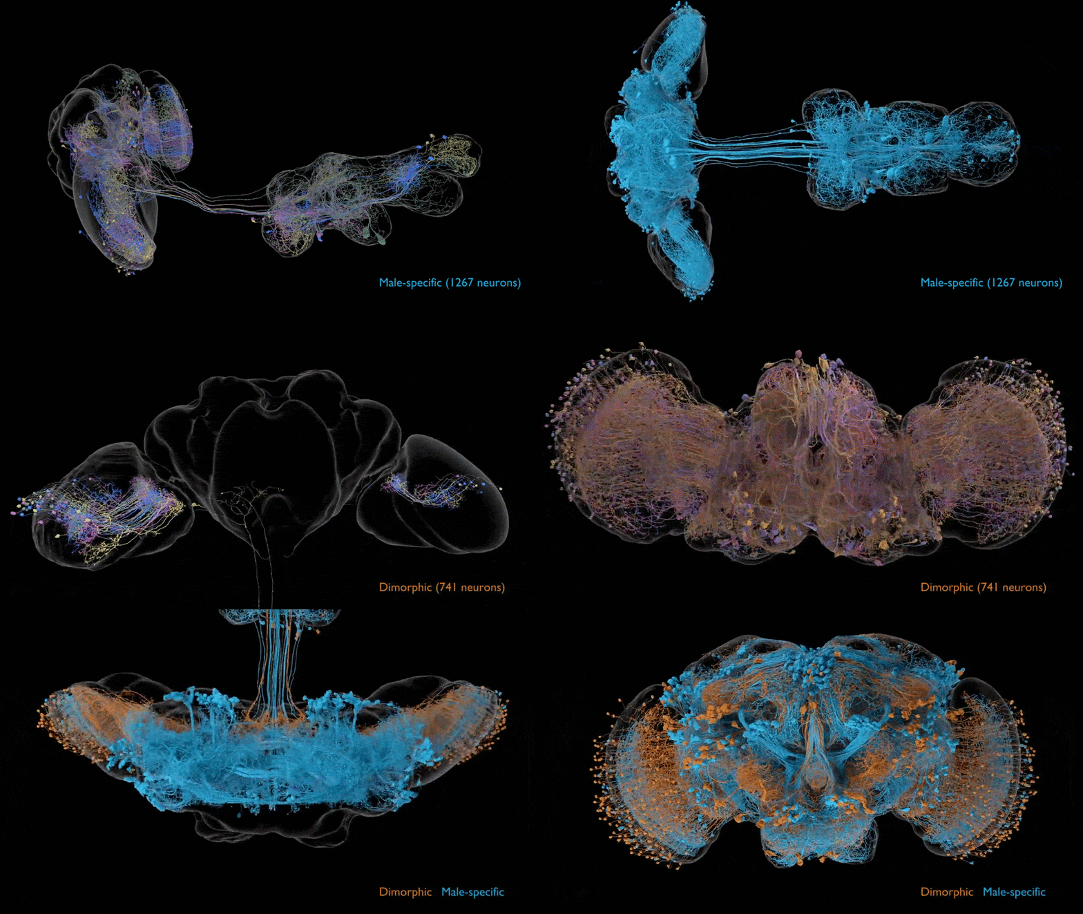
This animation shows neurons that are specific to the male connectome, as well as neurons that are present in both males and females but differ between them. The male connectome will allow researchers to compare male and female fly brains and understand complex social behaviors — like mating and aggression — that can vary by sex. (Image credit: Data acquired and analyzed by the FlyEM Project Team at HHMI’s Janelia Research Campus, the Cambridge Connectomics Group, and Google Research. Video by Philip Hubbard/HHMI Janelia Research Campus)
Initially released as a preprint, the new fly brain map was published in the journal Cell and Current Biology Thursday (Sept. 3). The study describing the map was published alongside three other papers, each of which uses the new data to explore a specific aspect of fruit fly neurobiology.
"The fly nervous system performs remarkably sophisticated computations with relatively few neurons and little energy, and its architecture could suggest principles for designing more efficient artificial systems," said Carlos Ribeiro, a principal investigator at the Champalimaud Foundation in Lisbon, Portugal, whose team contributed to the brain map and led one of the related studies.
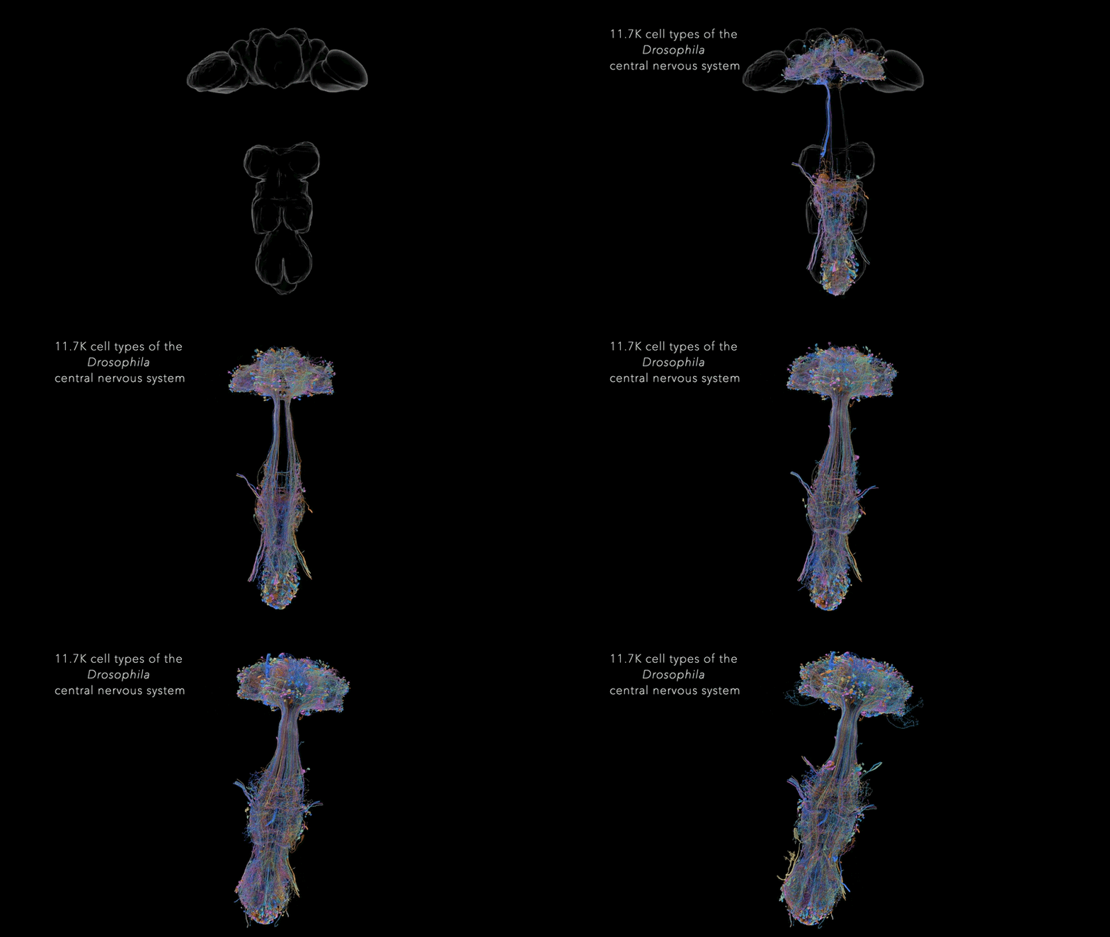
This animation shows all the types of neurons in the central nervous system of the male fruit fly. The newly completed connectome is the first map of an entire male fruit fly central nervous system, comprising all the neurons in the brain, both optic lobes and the ventral nerve cord. (Image credit: Data acquired and analyzed by the FlyEM Project Team at HHMI’s Janelia Research Campus, the Cambridge Connectomics Group, and Google Research. Video by Philip Hubbard/HHMI Janelia Research Campus)
"This work also provides a technical roadmap for more ambitious connectomics projects in the future, such as those for mice and humans," Ribeiro said in a separate statement from the Champalimaud Foundation.
In the near term, the scientists aim to map the brains of larval zebrafish (Danio rerio) and adult danionin fish (Danionella). In the long run, the goal of this research is to understand how vertebrates' brains enable complex behaviors, and then use that knowledge to help unravel the basis of neurological and psychiatric disorders in humans.
These neurons are responsible for allowing flies to process and react to tastes. (Image credit: Inês de Haan Vicente)
The study led by Ribeiro and other Champalimaud Foundation scientists aimed to map the circuits in the fly brain dedicated to the insect's sense of taste. Fruit flies have taste receptors in many body parts, including their legs, wings, mouthparts and inside of the throat. The researchers identified these taste receptors and traced their connections back to the fly brain. From there, they examined how those circuits interacted with those that govern behaviors such as swallowing or walking. When it comes to eating behaviors, this circuitry helps the fly determine whether a given morsel is safe or harmful and ultimately choose whether to eat it, the researchers concluded.
This diagram of taste processing is a "hypothesis-generation tool," study co-author Inês de Haan Vicente, a research technician in Ribeiro's lab, said in the Champalimaud statement. "Suppose you are interested in how taste controls locomotion. Now you can go to the map and ask: which sensory neurons are connected to the neurons controlling locomotion? Which intermediate neurons should I manipulate? It gives you a place to start."
The other two papers published alongside the map respectively explore the circuits behind vision processing in the fly brain and sex-specific differences in male and female fly brains. The first showed that visual processing extends deep into the brain, involving more than half of the 11,000 or so types of neurons identified in the map. The latter study found a network of cells that's specific to the male brain and appears to coordinate male-specific behaviors, such as specific aspects of fly courtship and physical aggression. (For example, female flies tend to headbutt while males lunge at their targets.)
While there were some networks specific to each sex, the circuits for sensation and movement are largely shared between males and females, the research found. At times, specific switches within those circuits reroute signals to different destinations in the male and female brain; the consequences of that rerouting will be a focus of future investigations.
See how much you know about the most complex organ in the human body with our brain quiz!
Of the former National Football League (NFL) players who died in recent years, at least 25% had brain changes related to chronic traumatic encephalopathy (CTE), a new study suggests.
The study, published Aug. 25 in the journal The BMJ, examined donated brain tissue from 338 former NFL players who died between 2008 and 2021. A total of 1,712 former players died in that time frame, so the analysis included only about 20% of the players' brains that could theoretically have been examined. Then, the researchers used that subset of brains to estimate the likely prevalence of CTE in the overall group.
Among the donated brains, over 93% showed physical signs of CTE, meaning an accumulation of abnormal proteins in specific places within the tissue. About 30% had stage IV CTE, the most severe form that comes with the most extensive brain changes.
These physical signs of CTE aren't always associated with overt symptoms, such as cognitive decline, mood and behavioral changes, or dementia. Only about 60% of the individuals with CTE-related brain changes were also diagnosed with dementia in this study. That said, those with stage IV CTE were more likely to have dementia, with 90% having a diagnosis.
Across the full study time frame, the researchers estimated that between 18.5% and 98.7% of the deceased players likely had CTE. Zooming in on the period with the highest number of brain donations — 2016 to 2021 — they estimated the rate to be between 24.5% and 97.7%. That latter time frame was likely the more representative sample, the researchers determined.
What can this study tell us about the CTE risk that current football players face, from youth sports to the professional level? To put the research into context, Live Science spoke with Thayne Munce, an associate professor and director of the Jayhawk Athletic Performance Laboratory at the University of Kansas, who was not involved in the BMJ study. Munce researches brain health and concussion in contact sports, primarily among young athletes. He cautioned that the BMJ study's findings shouldn't be extrapolated to all levels and ages of play, or even to today's NFL players.
Nicoletta Lanese: This study estimated the rate of CTE among this era of NFL players. Was the prevalence surprising to you, or in line with your expectations?
Thayne Munce: It's not surprising to me. This group and their colleagues have published previous work from donors — so these are former players who have donated their brains for research. They have reported even higher rates than 25% among just their donor sample [in this study]. This really high rate — over 90% of CTE cases in the donor group — is in line with what previous research has shown for these brain banks. So that's not terribly surprising.
This is a different era of football. So we should not be making any estimations about what current CTE rates are based on this study, because a lot has changed in 50 years.
What's different in this study is they compared the donors over a certain period of time versus all of the deaths among NFL players during that period of time and estimated the range based on how representative that donor sample was to the overall group of NFL players.
For example, if players just randomly selected to donate their brain to the brain bank and then they found that 98% of all of their samples had CTE — if that's representative of all the players from that era, then that's where you get the higher estimate. But if the majority of players that actually had CTE were the only ones who donated their brains because maybe they were showing outward signs or symptoms of dementia or cognitive decline or behavioral changes while they were living, then it becomes a very highly biased sample.
So that's where you get this big range. [The true prevalence] really depends on how representative that donor sample was to the peer group of players from that era.
NL: Does that broad range of estimates make it difficult to pin down the risk for current players?
TM: I think even more problematic than that big range is to understand that this is a historical study.
So these were players who died between 2008 and 2021, and the average age, I believe, was a little over 70 years of age. So if you extrapolate backwards, they probably played in the NFL starting around age 21 or 22 or 23 — that puts their playing days back in the '60s and '70s. So these were players who played in the NFL predominantly in the '60s and '70s, and would have played college and high school football in the '60s and '50s.
This is a different era of football. So we should not be making any estimations about what current CTE rates are based on this study, because a lot has changed in 50 years. It [the CTE rate] could be the same. It could be greater. It could be less. We really don't know.
I suspect, based on improvements in protective equipment and rules and advances in sports medicine, that the risk is likely lower. I don't have evidence that that's the case, but nobody does. We don't really know how the rates today compare to in the past, since CTE can't be diagnosed in the living. We can't take a sample of current players and look to see how many of them have CTE.
But that is the challenge of this study. It's interesting [in that] it gives us a range of what CTE may be in that group of former players. But it doesn't give us a lot of information about what rates are in current players in the NFL. And even more so, whatever the CTE rate is in professional NFL players does not translate to former collegiate, high school or youth football players, because CTE is an exposure-related disease. For players who have less exposure to these repetitive head impacts, it should follow that they have a considerably lower risk of CTE.
We don't want to confuse a range of CTE prevalence from players who played 50 years ago with contemporary NFL players and definitely don't want to conflate that with what CTE rates may be for current high school and youth players, who have far less levels of exposure.
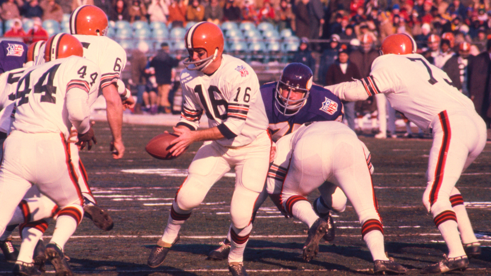
Cleveland Browns quarterback Bill Nelson hands off to running back Leroy Kelly during the 1969 NFL Championship Game against the Minnesota Vikings on January 4, 1970. Many safety precautions that are applied to today's NFL players did not exist at that time. (Image credit: James Drake via Getty Images)
NL: You mentioned that football has become safer over time. Could you spell out how the game has changed?
TM: We can look at different categories: With the rules back in that era, spearing — hitting with the crown of the head — was allowed, and oftentimes encouraged, as a style of play. That's now penalized and not taught, or shouldn't be taught.
There are changes in equipment, for sure. Modern football helmets — the material, the padding, the way in which these manufacturers are using head impact data to construct position-specific helmets and materials that are designed now to not just prevent catastrophic injuries, like skull fracture, but also to minimize the force from having repetitive head impacts — that's come a long way.
There's the Guardian Caps; you may have seen the NFL has adapted those. So they wear these soft-shell hats on their helmets during the preseason and during practices. That's now required for many of the position groups.
Virginia Tech has a helmet rating system where they test football helmets, and every year, the helmets score better on these tests. They now have star ratings for youth helmets, so manufacturers of youth football helmets are now being held to account, as well.
Maybe the most important thing is just the education around and recognition of concussion and brain injury in the sport. Now, when an athlete has a concussion, they are more likely to be recognized for having a concussion and be pulled from play and have an opportunity to recover before they return to the game. You hear anecdotal stories of former NFL players from that era, like the '60s and '70s, that suffered multiple concussions, oftentimes in the same game, and were not removed from play and continued to practice under those conditions. That medical recognition and treatment management of concussion and brain injury has moved ahead.
You look at the way in which practice was conducted, from full contact, two-a-day practices, which were common even in high school 15, 20 years ago. Now it's really not allowed at any level of play. So the amount of hitting that they do, the amount of contact that players are exposed to in practice and in the preseason is far less.
NL: When players weren't able to recover from concussions, did that likely increase their brain-injury risk?
TM: Theoretically, yes. There's evidence that if the brain is injured and then it's exposed to a second insult or injury before it's properly recovered, that makes the condition even worse.
There's actually a term called "second-impact syndrome," and it's very rare, so I don't want to scare people. But there have been instances where players — it's often, unfortunately, young players, high school players — who have had a concussion which, in retrospect, went undiagnosed and then there was a second concussive blow that led to brain swelling and death. That's the extreme, but there are cases of that [repetitive damage] happening even on a lower scale.
Even if you don't get another concussion, just those nonconcussive repetitive head impacts on top of an injured brain would not be good. It certainly is only going to delay recovery, likely is going to compound your inflammation and all of the downstream consequences of that initial injury.
I suspect that that was fairly common in years past, mainly because concussions generally weren't diagnosed. No one was looking for them, and players certainly weren't encouraged to report if they had the symptoms. So it's very likely that many of these players, including the players that were in the [brain donor] sample, had concussions and played through them and continued to receive additional trauma on top of a brain injury.
Now whether that leads to CTE, we can't really say. But I think it's reasonable to suspect that it probably would increase that risk.
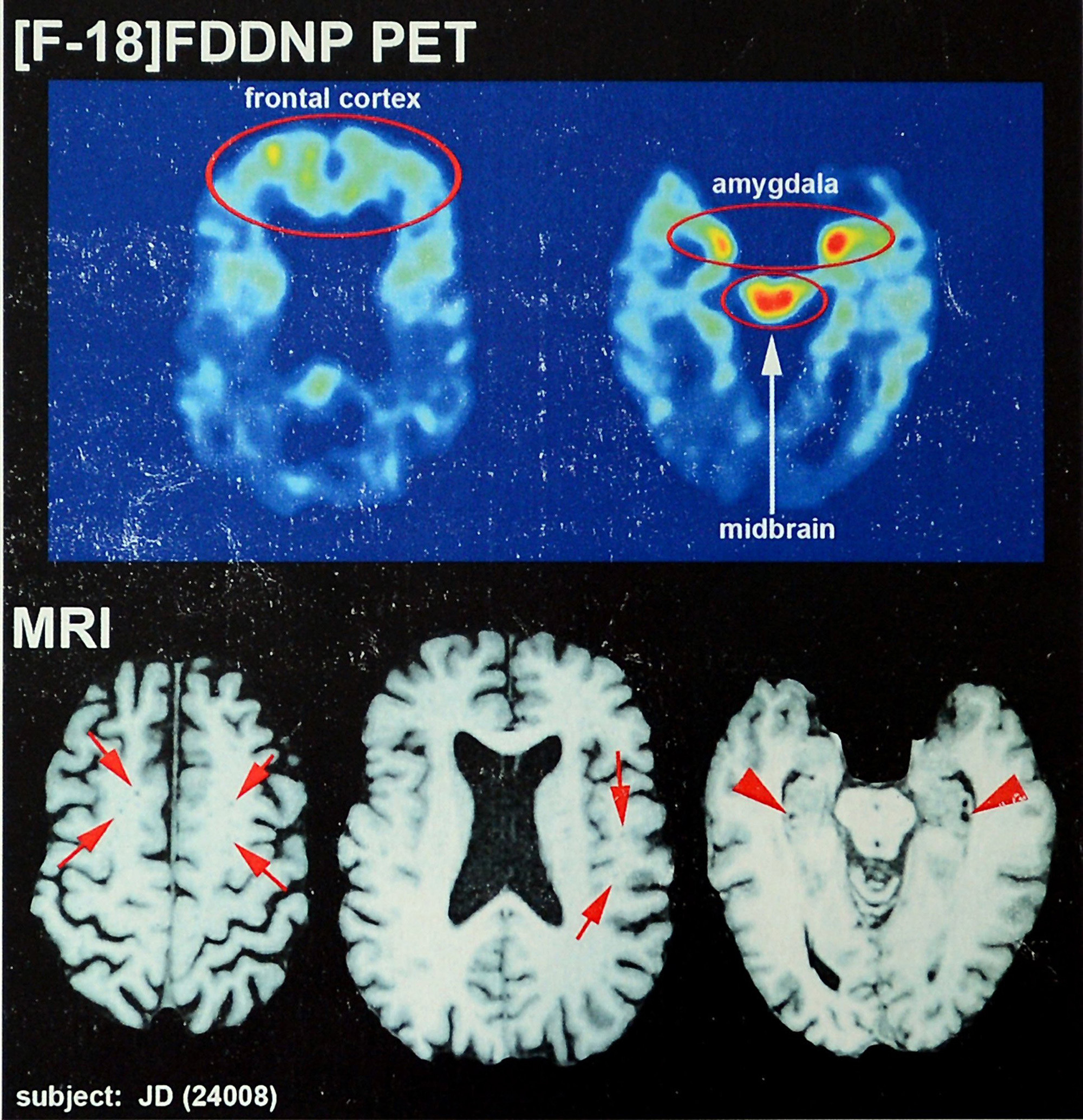
Recent brain scans of NFL hall-of-fame football player Joe DeLamielleure, who was diagnosed with CTE. (These are not from the new study.) (Image credit: Charlotte Observer via Getty Images)
NL: Could you explain whether the physical signs of CTE in the brain tend to come with symptoms, like cognitive decline? Or is the link more nuanced?
TM: So CTE is a neurodegenerative disease that can only be diagnosed by neuropathologists — people that look at slices of the brain and look for particular proteins that are present in certain regions of the brain. [This examination is done after death.] It's a pathological marker. It means that there are changes in the structure of the brain.
The clinical side of that is changes in mood, cognition, behavior, things like dementia or cognitive decline.
Even in the study, they showed that they [the CTE brain changes and symptoms] weren't necessarily linked. Now I believe they showed that for those who had more severe CTE — like the stage IV — that there was a pretty high rate of those individuals who also had subsequently diagnosed dementia. They had these clinicians that looked at the records and diagnosed them. That's a little bit of a limitation of the study: that a lot of the dementia cases were diagnosed after the person had passed away, based on medical records.
To your more immediate point, the presence of CTE: So the presence of these markers in your brain does not necessarily translate into a clinical disease or a clinical diagnosis. There can be signs or symptoms that somebody may have — let's say memory loss or changes in behavior — that may lead someone, like a former football player, to suspect that, "Oh, maybe I have CTE because I played football and I've heard that that's linked with dementia."
That may be true, but it may also be that it's due to old age, or it could be due to genetics or some other condition.
Many of these neurological diseases and conditions can be managed clinically, and there's treatment. There's therapy; there's pharmacological treatments. There's a whole toolbox available for people to seek help. That's, I think, an important message for former athletes or individuals who may be concerned about developing CTE, is to recognize that there is a disconnect between clinical disease and the pathological disease. Just because you have some outward signs or symptoms that doesn't mean that you have CTE.
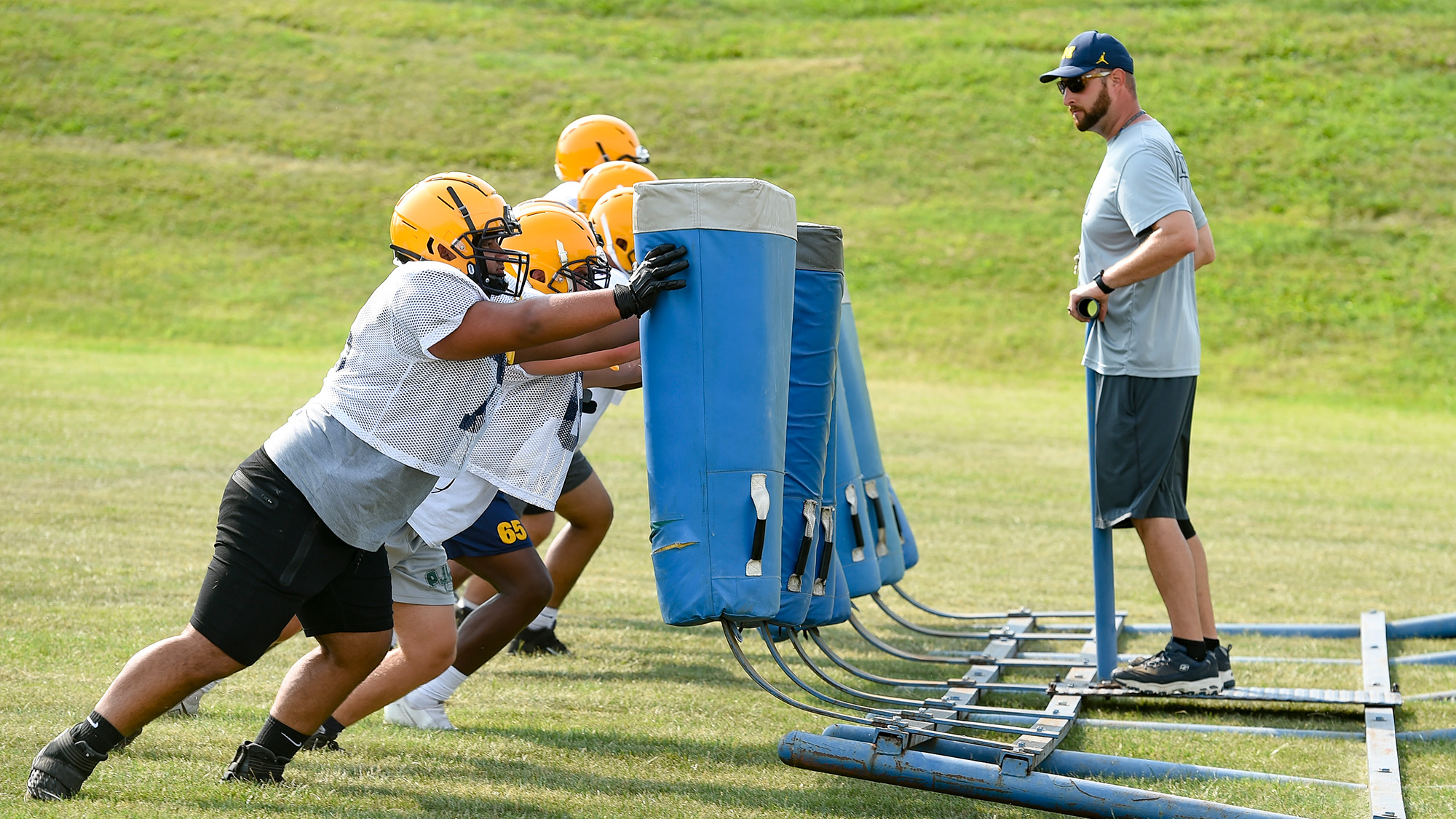
Football players at Muhlenberg High School in Pennsylvania do drills during practice. These young players likely don't have the same degree of risk as professional players when it comes to CTE. (Image credit: MediaNews Group/Reading Eagle via Getty Images via Getty Images)
NL: Since there was a higher likelihood of dementia in the stage IV cases, it seems like there's a stronger link between the pathological and clinical diagnoses at advanced stages. Is that a fair read?
TM: Again, from this study and other studies, just the presence of CTE does not necessarily mean that somebody's mood, behavior, cognition is altered. It seems to be more likely — it seems to be a tighter relationship at the more severe stages, which would kind of speak to what you're talking about. A certain threshold of disease progression seems to be more likely to manifest itself in clinical disease [with overt symptoms].
NL: What should current NFL players take from this study?
TM: I would go back to what we discussed earlier: just understanding the time frame in which the study was performed and when those players were in the NFL. I think we need to be cautious about projecting those rates; it's a pretty wide range of estimates, and it has to do with how representative the sample is. We need to use caution if we are trying to interpret those rates for the current players.
If anything, I think it probably validates all of these efforts that we discussed to try to improve brain health and minimize risk of exposure.
Even if the prevalence is on the low end — that 25% of players who played in the '60s and '70s developed CTE — that's pretty alarming. That in and of itself should get the NFL's attention. Players shouldn't have a 1-in-4 chance or greater of developing this disease.
So let's take steps to reduce that risk — and I think it validates all of the changes that have taken place over the last 50 years. I mean, a lot of them have taken place over the last 10 to 15 years. That's probably the biggest message: to say we were right to do these things and we need to continue to see if there are further opportunities to make the game safer.
Hopefully we've made a lot of progress and current-day rates are much lower than they were in the past. We don't know that, but let's not be idle. Let's continue to push forward to reduce as much risk as possible in a game where you can't completely eliminate the risk. That's something that I think everyone acknowledges; every player has to acknowledge that there is some risk.
NL: Looking forward, what further improvements could be made?
TM: There's a couple of areas that are really in the infancy stages right now. If you think about brain health as a continuum where you would have a disease like CTE on one end and then optimal health on the other end — let's say repetitive head impacts take away from your brain health and tip the scale in favor of developing disease, what are things we can do to tip the scale back in favor of improving brain health?
We know that sleep is really powerful and could learn more about that in and of itself. And what about nutrition? What about pharmacological interventions? Wouldn't it be great if we could develop a medicine, a drug at some point that could be taken preventatively to reduce athletes' inflammation? Really looking into ways in which we can optimally improve brain health as a countermeasure to the damage or the trauma that is occurring.
It's not just football where these treatments could be helpful; it's also with soccer, hockey, other sports and occupations where there is exposure to [head] trauma. I think that's a promising area.
And then, certainly, diagnostic techniques. We talked about CTE only being able to be diagnosed at death. It would certainly be a game changer if we could diagnose it convincingly in the living so it could be recognized early. People are going to have different risks, and some of that is probably genetically determined; some people are going to have the same level of exposure and develop CTE while some aren't. If we could identify individuals who are at higher risk or in the earliest stages of the development of the disease, you can then change lifestyle, behavior, exposure — that would be huge as well.
When I do talk to players and coaches and parents, I often like to remind them that, generally, the benefits of playing sports, including tackle football, outweigh the risk.
NL: To shift gears slightly, could you describe your own research in youth sports?
TM: Most of my work over the last 15 years has been focused on youth football players. That's been an interest in measuring repetitive head impacts and neurologic function in youth football players and some work in high-school-age players, as well. So a lot of that is focused around subconcussive injury [head trauma that does not trigger overt symptoms of concussion, like nausea, light sensitivity or thinking issues]. We're looking at the effects of these nonconcussive head impacts on brain health.
That ties into the CTE question in the sense that CTE is thought to be due to cumulative exposure to brain trauma — repetitive head impacts — and certainly most of the work, like in this study, has been done in NFL players. The question for a lot of the general population is, "What about youth players, high school players, that make up 90% of all football players?" So from a public health perspective, it's those younger players that I think we really need to be studying and concerned about.
That's not to say this study isn't really important. It certainly is, but my focus has been on that younger population. And then I do have some experience working more in a hospital background [on] clinical concussion management and [developing] objective pictures of concussion.
NL: With recent research, do we better understand how those nonconcussive impacts affect brain health?
TM: Going back maybe 15 to 20 years, people started to recognize that there were some cumulative effects of concussion. Those were some of the earlier studies showing that former NFL players who had multiple diagnosed concussions had worse outcomes later in life — that kind of put the spotlight on concussion. Concussion went to the forefront, in terms of how we thought about brain injury in football.
And then, over time, this recognition of what has been termed subconcussive or nonconcussive brain injury has emerged, as there's been some imaging studies in collegiate athletes, and even high school athletes, that have found that players who don't get concussed sometimes have changes in their brain, even over the course of the season.
That dovetails with some of the CTE research which has shown that history of concussions isn't necessarily predictive of who developed CTE. It's more of a cumulative effect of repetitive brain trauma. So the CTE researchers have essentially concluded that it's history of repetitive brain trauma, rather than history of concussion, per se, that's a leading risk factor for CTE.
With CTE, it's always looking backwards, because it's something that's diagnosed at death. For researchers like myself who study concussion, or study active football players, that has also then been a reaction of saying, "Well, if it is repetitive trauma, if it's repetitive head impacts, that is a risk factor for neurodegenerative disease such as CTE, then that makes it even more important to study it in active players." What does that head impact exposure look like at different ages, and how can we reduce that exposure? How can we reduce the number and severity of head impacts that players are experiencing?
So there's both the CTE angle, which is, again, retrospective — I'm looking at former players. And the contemporary issues of players who are currently playing and what's the risk for concussion, but also what's their exposure to repetitive head impacts? So it's really tied together.
That's why there's so much concern about repetitive head impacts and why there have been rule changes at the collegiate and high school level about the amount of contact and limiting those types of activities, with the idea of reducing that overall head-impact burden.
NL: When you talk about repetitive head impacts broadly, I assume that covers both concussive and nonconcussive impacts?
TM: Yep — so every hit would be considered a head impact, and then certain impacts are strong enough, or hit you in the right spot, to cause a concussion. The vast majority of impacts are nonconcussive.
NL: How do you speak to these athletes' families about the risks of concussion and CTE?
TM: When I do talk to players and coaches and parents, I often like to remind them that, generally, the benefits of playing sports, including tackle football, outweigh the risk. Oftentimes we get so concerned about the risk, which is real, that we forget about the benefits and about the alternatives of not playing. Almost always, the benefits — from the physical, the psychological and social components, the mental health components — are going to outweigh the risks. That's one thing to keep in mind.
Second, the risk of injury — in this case, CTE — is likely going to be far less in those younger players than older players, NFL players because their exposure, the number of impacts, the severity of impacts is far less. With my research, I've measured those head impacts and compared them to high school players and collegiate players, and extrapolated to NFL players because that data isn't available. And you see this big stepwise increase in exposure from the youth, the high school, the collegiate, to the NFL players.
[With parents], I'm reassuring them that the risk is far less for the younger players than what they hear about in this type of study. We're talking about an apples-to-oranges comparison.
I also like to remind them that, in my opinion, the game of football has never been safer. I've got a bit of a personal perspective on this as well, because I played football myself through college.
TM: I played for a Division II school in South Dakota called — it's Augustana University now; it was Augustana College when I played there.
So I played 12 years of tackle football, and I can compare what tackle football was like when I was in fifth grade versus now — with the training that coaches have and the type of equipment they have and the way the practices are structured. It's completely night and day.
Even at the collegiate level, the way in which collegiate practices are conducted and the safety measures that are in place for current collegiate players is nothing like what we had when I was playing. So I like to reassure players that this is probably the safest time to be playing the sport of football.
At the same time, I acknowledge that this is an individual decision that parents need to make with their kids and evaluate that risk. One of my goals as a researcher is that I want to provide more empirical evidence so that they can make an informed decision, so that a parent would be able to say, "My son's risk of getting a concussion while playing youth football is 1 in whatever" — you know, 1 in 10, 1 in 5, or 1 in 100, whatever that case may be. And his risk of getting CTE by playing four years of youth football or four years of high school football would be this. Then you make an informed decision. Is that risk worth the benefit of participation?
Unfortunately, we don't know what that risk is [currently]. We can't tell parents, "Here's objectively what your risk of injury is going to be." It makes it tough for parents. So I like to reassure them that a lot of the concerns that are expressed for NFL players are greater than they should have as parents of youth players.
This interview has been condensed and edited lightly for clarity. It is for informational purposes only and is not meant to offer medical advice.
See how much you know about the most complex organ in the human body with our brain quiz!

 Login