This episode of the Physics World Weekly podcast features Katharine Hunt, Head of Radiosurgery Physics at Sheffield Teaching Hospitals NHS Foundation Trust in the UK. She explains how Gamma Knife radiosurgery is being used to treat arteriovenous malformations (AVMs) and other benign brain conditions.
Sheffield is home to one of the world’s longest-running Gamma Knife centres, and Hunt shares the facility’s insights into treatment planning, dose precision, paediatric care, and long-term outcomes.
This podcast is sponsored by Elekta, which produces the Gamma Knife radiosurgery system.
A radiation shielding vest tested during the uncrewed Artemis I flight in 2022 has been shown to significantly reduce the radiation dose that future astronauts or deep-space tourists might receive.
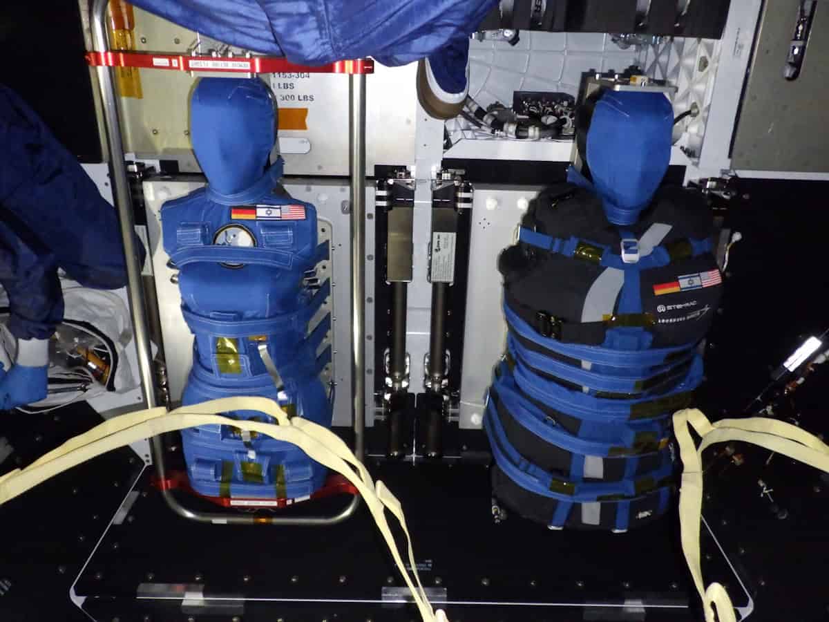
The data for this analysis were obtained via seating two human-tissue-equivalent female torso phantoms in the Orion crew capsule. While the “Zohar” phantom wore the new AstroRad vest, its travel companion “Helga” did not. Numerous radiation dosimeters placed in and on the phantoms allowed the received radiation dose to be compared.
“Exposure to radiation in space is unavoidable, and a single major solar particle event (SPE) can deliver more than one-third of NASA’s 600 millisievert career effective-dose limit. A three-year round trip to Mars would equal or exceed the career limit, which corresponds to a 3% mortality risk from radiation-induced cancer. So we developed the AstroRad vest to significantly reduce this hazard,” explains immunologist Oren Milstein, CEO of StemRad Radiation Protection, which began developing the vest with Lockheed Martin in 2015.
Dosimetry phantoms The Helga and Zohar (wearing the AstroRad vest) phantoms inside the Orion spacecraft at the NASA Vehicle Assembly Building prior to the launch of Artemis I. (Courtesy: NASA/Lockheed Martin/DLR)
The shielding thickness varies throughout the garment according to the radiation sensitivity of the underlying organs, with breasts, colon, lungs, ovaries, stomach and red bone marrow having concentrated shielding in the test vest. Milstein was inspired to develop this targeted architecture based on the experiences of his PhD supervisor, who treated the first responders to the 1986 Chernobyl nuclear power plant disaster.
With damage to bone marrow the accepted cause of death for all those emergency workers who lost their lives within months of the accident, “that heritage instilled in me a desire to protect people from radiation by selective shielding of the bone marrow,” Milstein explains, adding that the female focus of this experiment is because women have a higher predicted risk of radiation-induced cancer than men.
Astronauts currently shield from SPEs in “storm shelters” consisting of barricades of supplies, or by moving into areas containing more hardware against the walls. AstroRad provides a similar level of protection to the most robust shelter configuration while also “allowing the astronaut wearing it to move around the cabin and continue mission-critical tasks,” radiation physicist Jordan Houri, lead scientist for space exploration at StemRad, tells Physics World.
To maintain flexibility, the vest is constructed from thousands of hexagonal tessellated rods of high-density polyethylene (HDPE) sandwiched between two layers of elastic fabric. The shielding components can also slide past one another and stretch. HDPE was chosen for its high proportion of hydrogen – which is extremely effective at shielding against charged particle space radiation.
While Artemis I did not encounter a solar storm, Orion traversed the inner Van Allen belt, whose protons cover a similar energy range to those in SPEs. This enabled the team to construct a Monte Carlo model of the experiment for testing different radiation environments.
“We created digital twins of Helga and Zohar, the AstroRad vest, radiation detectors and spacecraft shielding,” explains Houri. “Using this model, we generated hundreds of billions of virtual protons and electrons representing the radiation environment of the inner Van Allen belt, before comparing the predicted detector responses directly with the measurements recorded during Artemis I.”
With close agreement found, the researchers then simulated measured SPEs from August 1972 and October 1989. As detailed in their recent Science Advancespaper, this revealed that wearing AstroRad would reduce effective dose by around 60% in an August 1972-type scenario, and by almost 40% for a 1989-type SPE – sparing astronauts the dose equivalent from up to 193 and 131 days of deep space travel, respectively.
Combining these results with those from previous experiments, which tested the vests’ ergonomics on the International Space Station, is allowing the researchers to improve comfort and ease of motion, and reduce mass. They are also exploring whether lighter, empty vests could be filled with recycled polyethylene after launch.
Alongside the health benefits, reducing radiation exposure makes economic sense as it extends astronauts’ careers, concludes Milstein, who hopes that the AstroRad vests can help “make lunar habitation and Mars travel truly sustainable and not just a dream”.
Join us for an in‑depth conversation with Simon Cherry, former editor‑in‑chief of Physics in Medicine & Biology (PMB), as he shares his expert perspective on the evolving landscape of medical physics research, molecular imaging, and non-invasive imaging technologies. Drawing on decades of leadership in biomedical imaging and academic publishing, Simon will reflect on major advances shaping the field, define the characteristics of high‑impact imaging research, and highlight how PMB continues to support rigorous, influential scientific work through its editorial standards and peer‑review practices.
This session will additionally spotlight the research of Xinchen (Alison) Deng, a finalist of the PMB 2024 Early Career Award. Attendees will gain insights into her contributions to the advanced medical imaging community, such as photon-counting CT for clinical applications and how her work supports broader advances in medical physics and X-ray/CT imaging research.
Simon will also discuss what makes early‑career research stand out and share strategies for researchers aiming to increase the visibility, reach, and long‑term impact of their publications within competitive scientific journals.
Left to right: Carol Clark, Simon Cherry, Xinchen (Alison) Deng
Moderator
Carol Clark, Publisher,Physics in Medicine & Biology
Speakers
Simon R Cherry, PhD received his BSc (Hons) in physics with astronomy from University College London in 1986 and a PhD in medical physics from the Institute of Cancer Research, University of London in 1989. Simon is currently Distinguished Research Professor in the Departments of Biomedical Engineering and Radiology at UC Davis and Professor of Total-Body Imaging at King’s College London. Simon’s research interests centre around biomedical imaging. His major contributions have been in developing systems for positron emission tomography (PET), including designing the microPET scanner for small-animal imaging, the earliest examples of simultaneous PET and MRI studies and co-leading the EXPLORER consortium that developed the world’s first total-body PET scanner. Simon is a Fellow of the Royal Society and Member of the U.S. National Academy of Engineering and National Academy of Inventors. He has received several international awards including the Society of Nuclear Medicine and Molecular Imaging Cassen Prize in 2022. He served as editor-in-chief of the journal Physics in Medicine & Biology from 2011–2020. Simon is the author of more than 350 peer-reviewed journal articles, review articles and book chapters in the field of biomedical imaging. He is also lead author of the widely-used textbook Physics in Nuclear Medicine.
Xinchen (Alison) Deng, PhD, is a diagnostic imaging physics resident at The Ohio State University. Her research focuses on advancing photon-counting CT for clinical applications, including multi-contrast imaging, early disease detection, detector optimization, and AI-based improvements in image quality. Before beginning the residency, Xinchen (Alison) was a postdoctoral fellow in medical physics at the University of Victoria, where she worked with Magdalena Bazalova-Carter and collaborated with Redlen Technologies on the optimization of photon-counting detectors and the clinical applications of spectral CT. She received her PhD in medical physics from the University of British Columbia, where she applied Raman spectroscopy, machine learning, and explainable AI to study radiation treatment response in cancer cells and tissues. Her broader research background spans photon-counting CT, spectral X-ray imaging, Raman spectroscopy, radiotherapy response assessment, and AI-driven image and data analysis. Her photon-counting CT research has been featured in the Physics in Medicine & Biology Early Career Researcher Focus Collection. She is a finalist for the PMB Early Career Researcher Award.
About this journal
Physics in Medicine & Biology. The international journal of biomedical physics and engineering, published by IOP Publishing on behalf of the Institute of Physics and Engineering in Medicine (IPEM)
Safety first: Internal Dosimetry is the practice of estimating intakes and risk from occupational exposures to radioactive materials (Credit: iStock/magicmine)
Radioactive materials have become a vital tool in our modern society, enabling the delivery of powerful medical treatments, the detection of toxic substances, and the generation of energy on a massive scale. But they also pose significant risks to human health, which means that organizations handling radioactive isotopes must have robust procedures in place to manage and monitor the dose received by their workforce and the wider public.
Dosimetry badges provide a visible and straightforward way to measure external exposure to radiation, but they are unable to determine the amount of radioactive particulates that have entered the body. Quantifying these intakes requires internal dosimetry, which typically uses measurements of urine or faecal samples – or less commonly in vivo radiological counts – to estimate the intake of specific radioisotopes. While the internal dose cannot be measured directly, monitoring data from these bioassays can be combined with biokinetic models to assess the likely impact of an individual inhaling, ingesting or being injected with a radioactive substance.
But internal dosimetry is not an exact science, and organizations with limited experience of radiation safety can struggle to translate regulatory requirements and the associated guidance into monitoring programmes that provide appropriate protection for their employees. Luckily, help is at hand. Dade Moeller & Associates, a wholly owned subsidiary of NV5, is a consultancy company specializing in radiation safety and dosimetry monitoring. Dade Moeller’s team of senior and certified health physicists can help organizations to balance worker protection with regulatory compliance.
Fit for purpose
“We encourage people to think about what they are monitoring for,” says Dr Brett Rosenberg, a certified health physicist at Dade Moeller with expertise in internal dosimetry. “The dosimetry programme must be tailored to the activity and half-life of the materials being handled, how those particular radionuclides interact with the body, and the maximum dose that an individual is likely to receive.” All of these considerations are important for prescribing a monitoring programme that does not become too onerous in costs and implementation while still providing an accurate assessment of occupational exposures.
In low-dose environments, for example, Rosenberg would advise organizations to run a confirmatory programme, in which samples from a subset of the workforce are used to check for any unexpected intake that would warrant further investigation. In contrast, routine internal dosimetry becomes a necessary requirement when there is a greater risk of workers receiving doses that exceed monitoring criteria – or even limits – set by the regulations.
Such situations are likely to arise, for example, in facilities that develop and manufacture medical radioisotopes, such as highly volatile iodine-131 for treating thyroid conditions. Radionuclides with the potential to generate higher doses are also produced throughout the fuel cycle for nuclear power generation, from extracting and refining the uranium – which creates radioactive dust that is easily inhaled – through to the management of nuclear waste.
In the US, such commercial operations are overseen by the Nuclear Regulatory Commission (NRC), which sets maximum limits both for the total radiation dose and the intake that a worker can receive. Companies must demonstrate compliance with these limits, but the NRC does not dictate the methodologies that are used for internal monitoring, or for estimating the intake from lab-based measurements. “Programme development and design is not based on regulation, it’s based on guidance,” says Rosenberg.
Flexibility leads to variability
That leaves organizations with significant flexibility in how they design their monitoring strategies, which allows programmes to be tailored to operational realities rather than forced into a rigid template. However, it also leads to variability in monitoring approaches and in the interpretation of results. Annual audits required by the NRC offer an opportunity for experts like Rosenberg not just to verify compliance, but also to identify opportunities for improvement. “I look for technical shortfalls in internal dosimetry, and check that the measures in place are effective and appropriate,” he says.
Many companies value the input they receive through these regular evaluations. Dan Sowers, for example, is the Global Radiation Protection Manager for Westinghouse, a nuclear services company that, among other things, fabricates uranium fuel and maintains the inner workings of nuclear reactors when they are not being operated. “My priority is the health of our workers,” he says. “Ultimately, we want our internal dosimetry programme to demonstrate that workplace controls are effective.”
Regular input from external advisors helps Sowers to ensure that robust procedures are in place for contamination control, and for assigning a dose to an employee when there is an intake of radioactivity. “The experts at Dade Moeller have enough real-world experience to know what’s right, which can go beyond the guidance we get from the regulators,” says Sowers. “They bring a fresh set of eyes to our operation and share best practice from other sites.”
The team at Dade Moeller also provides expert input to the US Department of Energy (DOE), which runs the national labs as well as facilities for nuclear security. Unlike the NRC, the DOE prescribes structured programmes for internal dosimetry that are tailored to the radioactive materials being handled at each site. It mandates lower dose thresholds than specified by the NRC for workers to undergo routine internal dosimetry checks, and uses a newer set of recommendations for assessing intake and estimating the dose received by different parts of the body. (However, notes Rosenberg, neither the NRC nor DOE use the latest set of recommendations, which are being adopted internationally.)
Creating consistency
Many of those recommendations come from the International Commission on Radiological Protection (ICRP), an independent body that provides guidance on dose limits and monitoring methodologies. The ICRP also develops and publishes the biokinetic models that are used by dosimetrists to translate the radiation counts from bioassays into estimates of intake and effective dose. Specific models have been developed to assess the impact of inhaling or ingesting different radioactive species, but the results are open to interpretation and depend on the version of the model being used.
To avoid these ambiguities, national regulators in Europe have combined forces to create a unified framework for internal dosimetry. Based on the outcomes from a co-ordinated research project in the early 2000s, this framework defines a standard set of methods and assumptions to ensure that different organizations calculate similar dose estimates from the same monitoring data. However, no such consistency exists in the US, where the NRC does not specify the details of the monitoring programme or the modelling methods a company should use, or between regulators in other parts of the world.
That lack of consistency can create difficulties for international companies like Westinghouse, which operates across 21 countries in different geographic regions. “Our internal dosimetry programmes are slightly different in each location,” says Sowers. “Those differences don’t affect the fundamentals of how we protect the health and safety of the worker, but the fine print of the regulations often requires us to do things slightly differently to make sure we are compliant.”
Dealing with uncertainty
To improve consistency, Rosenberg is involved in both national and international standards initiatives that aim to define best practice. However, he says, even the most carefully designed monitoring programme cannot eliminate the inherent uncertainty in estimating the effective dose from lab-based measurements of excreted samples.
“Even the most up-to-date models may not be very accurate,” says Rosenberg. “While the bioassay results are taken from a real individual, the model assigns the dose to a reference person who weighs 70 kg, excretes 135 g of faeces per day, and has both ovaries and a prostate.” As a result, the effective dose calculations based on excreted samples can carry errors of 300% when considering the anatomy and physiology of an “average worker”, and the practical limitations around counting statistics and sampling. “For practitioners, the challenge is not to remove uncertainty, but to understand and manage it,” says Rosenberg.
Given these intrinsic uncertainties, Rosenberg’s mission is to help regulators and commercial organizations to deploy the most effective methodologies for improving the standard of care. As part of these efforts, he has developed a week-long training course that aims to demystify the process of internal dosimetry for managers and regulators who need to understand the guidance from regulatory and international bodies like the ICRP.
“We do see uncertainty associated with inappropriate dosimetry practices, and to advance the field both the regulators and the practitioners need to stay current with the latest recommendations and models,” he says. “It’s easy to follow NRC regulations, but putting structured monitoring programmes in place and using the most appropriate models will help companies to produce more accurate evaluations and improve the safety of their workers.”
Radiation therapy is an essential component of cancer care and is used in the treatment of roughly half of all cancer patients. To ensure that these treatments are both safe and effective, it’s critical that the linear accelerators, or linacs, that deliver the radiation are as reliable, efficient and accurate as possible.
The US-based medical physics consultancy One Physics aims to help hospitals and cancer centres meet these goals by bringing together a team of highly-trained expert medical physicists to provide a wide range of radiotherapy and diagnostic imaging physics services.
This episode of the Physics World Weekly podcast features James Giltz, director of commissioning services at One Physics. In a wide-ranging conversation with Physics World’s Tami Freeman, Giltz explains how the company provides comprehensive medical-physics support for radiotherapy – from planning new centres through to ongoing clinical operations.
For Keamogetswe Ramonaheng, physics was never just about equations – it was about clarity. “From a young age, I was attracted to mathematics and science as a way of understanding complex phenomena through a structured approach,” she says. “Physics was the area that spoke to me the most because it is the foundation for the fundamental principles that govern the natural world.”
Ramonaheng’s academic journey began at the University of the Free State (UFS), where she completed her undergraduate and honours studies before starting an internship at Universitas Academic Hospital in Bloemfontein. There she saw how a rigorous physics training can lead to tangible, clinical benefits. “The ability to comprehend and harness the interaction between radiation and matter in the human body demonstrated the power and relevance of scientific inquiry,” she recalls.
In many ways, nuclear medicine found me
Keamogetswe Ramonaheng
Thanks to a fellowship from the International Atomic Energy Agency (IAEA), Ramonaheng completed a clinical placement at Royal North Shore Hospital in Sydney, Australia. She later continued her postgraduate studies at UFS, becoming the first Black South African woman to earn a PhD in medical physics for nuclear medicine. “In many ways, nuclear medicine found me,” says Ramonaheng, who is grateful to the encouragement of various senior staff members who saw her potential and guided her into the field.
Multifaceted role
Following a spell as an independent medical physicist and manager at Universitas Academic Hospital and lecturer at UFS, Ramonaheng joined NuMeRI in 2024 and the University of Pretoria. Along with the team of scientists she leads, Ramonaheng oversees the safe and effective use of ionizing radiation at NuMeRI used to treat and diagnose disease in a safe and effective manner.
It’s a varied role, which stretches from providing patient-focused clinical services to carrying out applied research. “We integrate research with operations,” says Ramonaheng. “That requires careful planning and rigorous quality assurance, ensuring that innovation does not compromise safety.”
Among her duties, Ramonaheng carries out dosimetry calculations for innovative radiopharmaceuticals, works on new forms of quantitative imaging, and helps to develop novel radionuclide therapies, including using alpha particles to treat cancer. She also uses gamma-ray cameras equipped with highly sensitive cadmium-zinc-telluride detectors, which allow radiopharmaceuticals to be quantified and imaged more precisely.
Ramonaheng is particularly interested in “theranostics” – a form of “precision medicine” that combines therapy with diagnostics. It involves giving a patient a tumour-targeting molecule labelled with a radionuclide. This allows the tumour to be visualized using techniques such as positron emission tomography (PET) or single-photon emission computerized tomography (SPECT). The same molecule – or one similar to it – is then used to deliver a therapeutic radionuclide directly to the tumour.
Daily challenges
For Ramonaheng, a typical day is fast-paced. Mornings often begin with her overseeing radiation-safety protocols and ensuring that radiation imaging and counting equipment are working as well as possible, such that they meet quality assurance standards. Through the day, Ramonaheng also oversees all operational medical-physics activities and carries out her duties as chair of NuMeRI’s radiation protection committee.
As the day progresses, she might find herself reviewing clinical theranostics dosimetry workflows to carrying out patient-specific dose calculations or evaluating quantitative imaging metrics from SPECT/CT and PET/CT systems. Other tasks include reviewing research protocols for cancer theranostics, mentoring postgraduate students at the University of Pretoria, and examining clinical trials.
Innovation accelerates when silos are dismantled
Keamogetswe Ramonaheng
Ramonaheng works in a highly interdisciplinary environment, collaborating with radiographers, nurses, radiochemists, radiopharmacists, medical physicists and clinicians to address live issues in real time. “Innovation accelerates when silos are dismantled,” she says.
The work is not without its challenges. Funding for postgraduate training is a persistent concern. Clinical physics is also a highly specialized field, which means it can be hard to recruit people with the right skills, who might be drawn to better-paid industry jobs. In addition, NuMeRI is an operationally complex mix of advanced imaging systems, radiopharmaceuticals and clinical regulations, which requires good project-management and planning skills.
But Ramonaheng, who recently won two awards at the 8th Theranostics World Congress in Cape Town, feels the benefits outweigh the challenges. “It is very fulfilling to see the translation of research into clinical application,” she says. Just as gratifying, she adds, is watching her students move from their studies to publications and clinical applications. “You see the entire process of scientific advancement.”
A more promising future
Looking ahead, Ramonaheng envisages a growing use of artificial intelligence (AI) in her work. She also collaborates with national and international partners to automate workflows and enhance efficiency, precision and patient-centred care. Another ambition for Ramonaheng is to further strengthen NuMeRI as an Africa-wide hub for research, clinical service and training – a vision reinforced by the IAEA recently naming NuMeRI as one of 18 global “anchor centres” for its work in radiotherapy and medical imaging.
Ramonaheng believes medical physics will grow rapidly in Africa over the next 10 years, fuelled by an expansion of theranostics and precision medicine. Her hope is to guide this growth through mentorship and leadership, ensuring that Africa develops its own talent pool of medical physicists who can address the continent’s unique healthcare needs.
Africa suffers, for example, from limited access to advanced imaging and targeted therapies. Ramonaheng’s aim is to optimize personalized and precision medicine for cancer patients, ultimately improving treatment outcomes and quality of life. Eventually, she hopes, medical physics will be recognized as a profession across the continent. “We are building not only research outputs but human capital.”
Leadership is not only about the creation of paths, but the creation of paths where there were no paths previously
Keamogetswe Ramonaheng
Being a pioneer in the field has required resilience on her part. “Competence must be coupled with confidence,” says Ramonaheng, who has had to learn the unwritten rules of a world dominated by men. As a mentor, her guiding principle is the African concept of motho ke motho ka batho babang – a person is a person only through others. “Leadership is not only about the creation of paths,” she says, “but the creation of paths where there were no paths previously.”
Her message to young physicists – particularly women and those from other underrepresented groups – is clear. “Medical physics is a dynamic and impactful field at the intersection of physics, medicine and technology,” she says. “ It allows you to see the direct translation of science to patients.” Medical physics requires resilience, curiosity and commitment, but for Ramonaheng its beauty is that equations don’t stay on paper – they become a tool for healing.
Two years ago, the ESTRO 2024 meeting in Glasgow dedicated a conference session to the discussion of upright radiotherapy. In particular, the speakers pondered whether this emerging technique – in which patients are treated sitting up rather than lying down – offers hope of increasing access to advanced radiotherapy, or whether it’s merely hype.
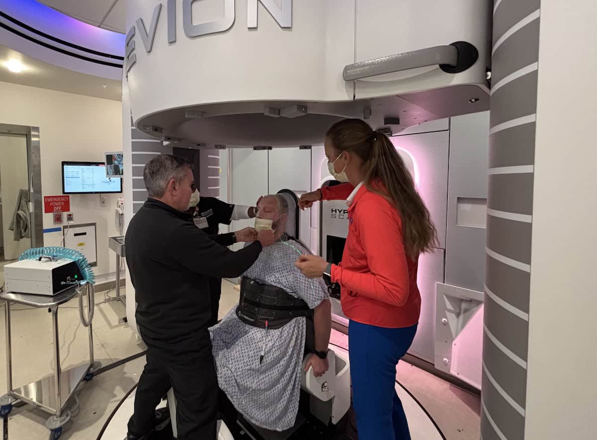
Things have moved on since then. Leo Cancer Care introduced its upright photon therapy system, Grace, and received commercial approval in the US and (just last week) Europe for its Marie upright positioning and CT system. Stanford Medicine recently unveiled the world’s first ultracompact proton therapy facility, pairing Mevion Medical Systems’ compact S250-FIT proton therapy system with the Marie platform. Meanwhile, the body of published research on the feasibility and patient experience of upright treatments continues to grow.
At this year’s ESTRO 2026 meeting in Stockholm, the theme was revisited by four experts in the field, who debated the motion that “Upright radiotherapy will be a mainstream and standard radiotherapy delivery option in 2035”.
The customary pre-debate vote revealed that just one quarter of the audience thought that photon-based upright radiotherapy would become mainstream, with the remainder believing that it would remain a niche technique. When it came to upright proton therapy, however, the vote was split roughly 50:50. So could the speakers persuade the attendees to change their minds?
Patient-centred care
The debate began with Tomas Kron from the Peter MacCallum Cancer Centre in Australia arguing the case for upright X-ray radiotherapy. He pointed out that upright positioning is not a new idea. “Historically, photons and upright have been around for a very long time. It has been, if not standard practice, widely used. But what role will it play in 2035?”
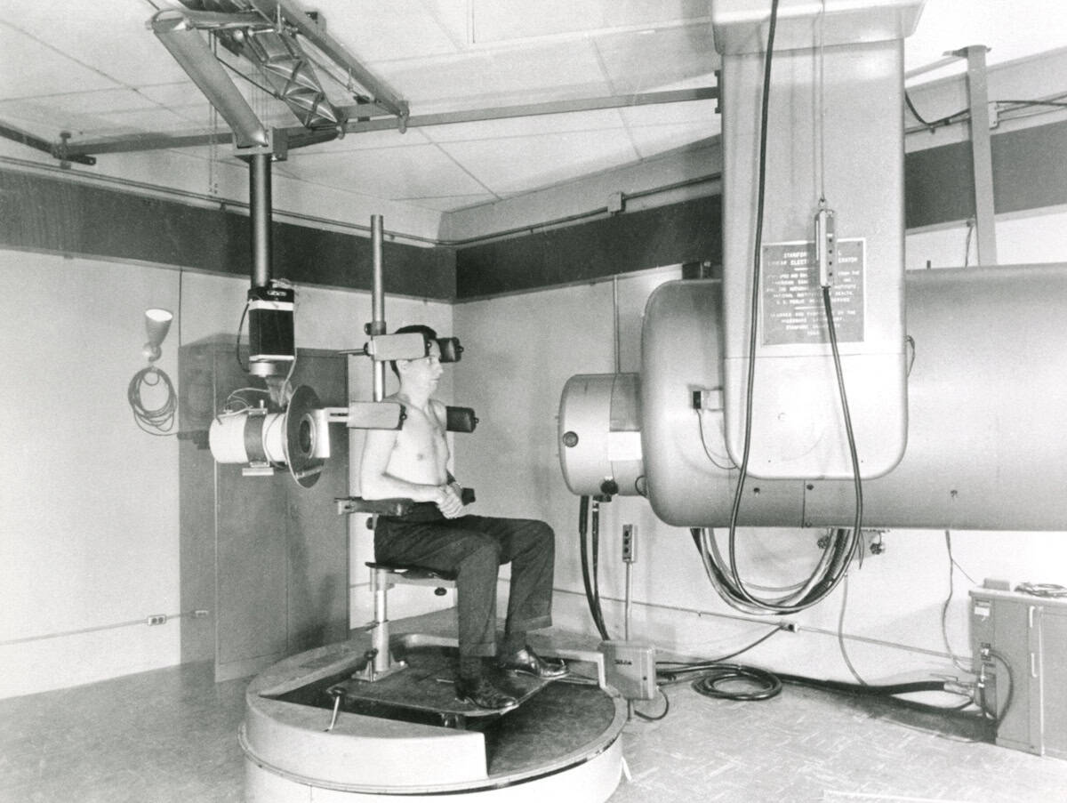
Not a new idea The first high-energy linear accelerator for medical use, known as LA-1, was developed at Stanford in the 1950s. Patients with head-and-neck or lung cancer could sit on an isocentrically mounted chair for treatment. The machine was decommissioned in 1972 and now resides in the Smithsonian Institution. (Courtesy: Stanford Medicine)
Kron described a clinical imaging trial underway at Peter Mac investigating upright cone-beam CT for planning lung cancer radiotherapy. The study showed that image quality was good enough for adaptive treatment planning, and that the lung was expanded and moved less in the upright position. Kron noted that patient setup and imaging was “really, really easy”, taking just a few minutes.
But what’s more important, he emphasized, is the patient experience. Patients treated while sitting up can maintain eye contact with the doctors throughout, they feel more involved and empowered, with one patient commenting: “My breathing was strong, I felt comfortable, the band around my chest was giving me a bear hug.”
“It’s really all about patient-centred care. Physical comfort and emotional wellbeing are top priorities,” Kron said. “Clearly, in an upright scenario this is much more likely to be the case.”
Upright radiotherapy offers many other unique features, including anatomical advantages and the ability to customize the chair, for example, for bariatric or paediatric patients. An upright treatment system is also more compact than a couch-based machine, requiring a smaller bunker. It could also be used as a mobile radiotherapy unit, said Kron – reducing the need for patient travel.
Kron’s team found that 80–90% of their patients could be treated just as well with upright radiotherapy as supine (lying down). “There are anatomical advantages with upright, there are patient preferences, there are economic benefits. What’s not to like,” he concluded.
The myth of mainstream
“Upright radiotherapy will not be mainstream and standard,” declared the second speaker, Livia Marrazzo from the University of Florence in Italy.
“Mainstream means widely adopted, used across the majority of radiotherapy centres, the default in clinical practice … and standard is even stronger, backed by clinical evidence, guideline-endorsed, reproducible and validated,” Marrazzo told the delegates. “It’s not ‘it works in some centres, is technically feasible, has early adopters, may have advantages for some patients’. But that is where we are with upright radiotherapy.”
The art of persuasion Livia Marrazzo argued that upright radiotherapy will not be mainstream by 2035. (Courtesy: Icro Meattini)
From a practical standpoint, most of the roughly 16,000 radiotherapy systems worldwide are linac-based recumbent machines with a typical lifecycle of 10 to15 years. Many were recently replaced with supine systems optimized for intensity-modulated and image-guided radiotherapy. “The installed base is locked into supine geometry for another full cycle,” Marrazzo explained.
She refuted many of the advantages proposed by Kron. “We have limited clinical evidence supporting comfort advantages,” she said. “It may benefit specific patient groups and conditions, but this doesn’t mean mainstream.” Overall, clinical experience is limited, with no comprehensive evaluations of plan quality and no comparative clinical studies.
She highlighted the particular challenges of breast cancer treatments, which account for 25-30% of cases in her radiotherapy department. “When we place a breast cancer patient upright, we lose the natural breast separation, so have much more difficulty in hitting the target and avoiding the contralateral breast,” she explained. “This exemplifies how upright is not a plug-and-play replacement for a conventional supine workflow.”
“Are we sure we would like to have upright as the standard radiotherapy delivery option by 2035 or do we want to push our efforts somewhere else?” Marrazzo concluded. She would prefer a focus on introducing technologies such as AI-driven planning and contouring, fully adaptive workflows, ultra-hypofractionation or biology-guided treatment adaptation. “These are all solutions that can be software-driven, scalable and compatible with existing supine infrastructure.”
The motion for protons
With half of the audience already agreeing that upright proton therapy will become mainstream, Petra Trnkova from Czech Technical University had perhaps a slightly easier task as she presented the case for upright protons. Nevertheless, she began by suggesting that her opponents are simply “scared of progress and won’t accept that, even without evidence, we can move forward in radiotherapy”.
Trnkova reiterated the benefits of upright radiotherapy cited by Kron: favourable patient anatomy, lower installation cost, improved sustainability, and patient-centric management. “For proton therapy, these improvements are much more significant,” she noted.
For starters, upright systems could help address the massive disparity in access to proton therapy around the globe. Sharing a map showing how proton therapy facilities are mostly distributed in wealthy countries, Trnkova noted: “My opponents may tell you that it’s not possible to do this by 2035, but when you look at this map, I ask you, can we wait any longer?”
Saving space The compact S250-FIT proton therapy system is small enough to fit in a standard linac vault. (Courtesy: Stanford Medicine)
Increasing access to proton facilities is enabled by the extreme size reduction when eliminating the need for a large rotating gantry, enabling proton therapy systems small enough to fit in a standard linac vault. Upright proton therapy can also reduce machine complexity, increase rotation speed and lower energy consumption – reducing costs, improving system upgradeability and increasing environmental sustainability.
“Another consequence of smaller facilities is we can really have patient-centred care,” Trnkova added. Recalling the 10 to 15 year linac lifetime mentioned by Marrazzo, she suggested another option: “You can replace your linac with proton therapy. Then you can have the full set of treatments available for each patient”.
Upright proton therapy could also ease the introduction of new treatment techniques, such as proton arc therapy, which offers dosimetric benefits over intensity-modulated proton therapy, but it is difficult to deliver with a gantry. It could also enable in vivo dosimetry, using shoot-through protons for range verification, or mixed-beam delivery of protons and photons.
“Upright positioning offers many opportunities, it’s the only way towards the democratization of proton therapy,” Trnkova concluded. “Stop asking what opportunities upright radiotherapy brings, start asking what you can do to bring it faster to clinical practice.”
The reality check
The final speaker, Carles Gomà from Clinic Barcelona in Spain, reflected upon what makes a good radiotherapy system. “In my view, it’s a three-legged stool: beam delivery, imaging and immobilization,” he said. “And progress comes with a combination of the three.”
For example, focusing too heavily on beam delivery and imaging can lead to immobilization being forgotten. “Immobilization means comfort, and if we are comfortable, we are still,” Gomà explained. “I cannot care less how many papers say patients are more comfortable in an upright position,” he added, pointing out that people will pay five times more to fly in business class where they can lie down.
The other reason cited for moving to upright proton therapy is its lower cost. “But is proton therapy expensive?” Gomà asked. He described the situation in Catalonia, which has a population of eight million and in 2018 spent Euro 42.2M on external-beam radiotherapy. “This is exactly the same cost as one immunotherapy drug for the same population,” he pointed out. “Proton therapy is not expensive; photon therapy is ridiculously cheap.”
Gomà also considered whether “suboptimal protons” are better than photons. “I’m going to answer no,” he said, describing two recent phase III, randomized trials comparing photons with protons for oropharyngeal cancer. The US trial concluded that proton therapy provides a new standard-of-care option, but the UK trial reported no difference between the two modalities.
“Let’s learn from history and not repeat the same mistakes,” he concluded. “True progress is improvement without compromise. If we want to make the stool higher, we have to work on all three legs at the same time.”
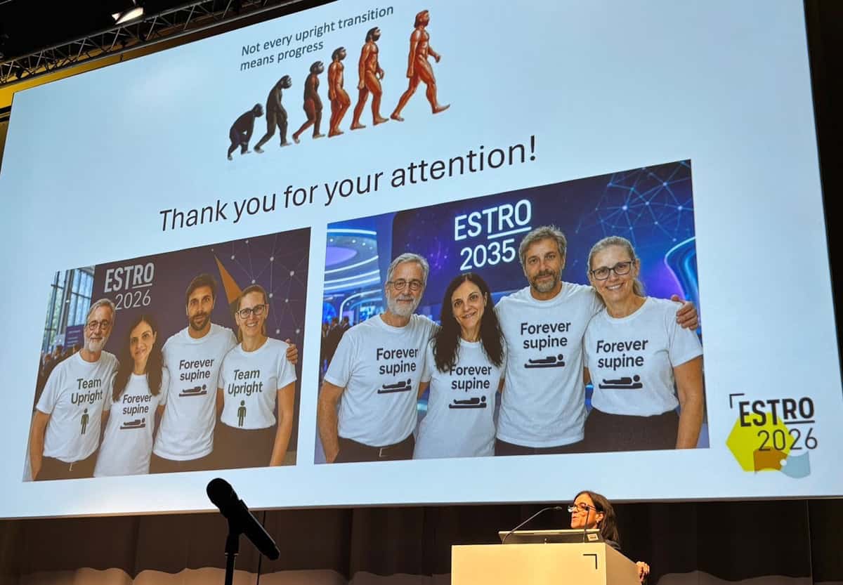
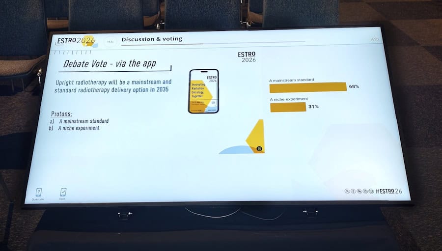
The final vote At the end of the debate, the ESTRO delegates strongly supported upright proton therapy. (Courtesy: Ye Zhang)
The debate concluded with decisive a final vote: while support for upright photon therapy reduced a little, over two-thirds of the audience believed that upright proton therapy will indeed become mainstream and standard by 2035.
Writing on LinkedIn, session co-chair Ye Zhang from the Paul Scherrer Institut noted: “The debate sparked an inspiring shift in perspective, with final voting showing slightly increased scepticism toward mainstream upright photon therapy (dropping from 23% to 18% support), but a dramatic surge in favour of upright proton therapy, which jumped from 47% to a 69% majority.”

 Login