❌

Normal view

Decades of Financial Hardship Linked to Faster Brain Aging

28 July 2026 at 20:13
Long-term financial stress accelerates age-related cognitive decline. Image credit: Thomas Breher.

A large British study spanning more than 70 years has found that people who experience chronic money struggles over their working lives show measurable declines in memory and processing speed by their 50s, along with signs of accelerated brain shrinkage decades later.

The post Decades of Financial Hardship Linked to Faster Brain Aging appeared first on Sci.News: Breaking Science News.

Short-Chain Fatty Acids: New Insights Into Health Effects

29 July 2026 at 14:15

Short-chain fatty acids (SCFAs)—especially acetate, propionate, and butyrate—are produced when gut microbes ferment carbohydrates that the human body cannot digest. Over the past decade, these small molecules have moved beyond being viewed as simple fuel. Instead, they are now recognized as signalling messengers that help tune metabolism, shape immune responses, and support physiological balance across organs. A new review in Nature Metabolism brings together scattered findings to clarify how SCFAs are made, how they are processed in the body, and why they matter for health.

The article emphasizes that SCFA production is not uniform. It depends on which microbial communities inhabit the gut, which dietary substrates reach the colon, and how microbes share resources through cross-feeding. When multiple bacterial species coordinate, fermentation pathways can shift, increasing the output of particular SCFAs. In that way, the “hardware” of the microbiome and the “inputs” provided by diet jointly determine the chemical signals generated in the intestine.

After their formation, SCFAs are handled by host tissues through absorption and further metabolic conversion. The review highlights that acetate, propionate, and butyrate can influence local and systemic physiology, helping link intestinal chemistry to whole-body regulation. Their effects are therefore both immediate—through signalling pathways—and longer-lasting—through changes in gene regulation.

Mechanistically, the authors focus on receptor-mediated communication. SCFAs activate specific receptors on immune and metabolic cells, altering inflammatory tone and metabolic signalling. This receptor layer helps explain how intestinal events can reverberate in distant tissues that rely on tightly controlled energy and immune states.

Beyond receptors, SCFAs also affect epigenetics. By influencing chromatin-modifying enzymes and related regulatory networks, they can change how genes are expressed without altering DNA sequence. Such effects provide a plausible bridge between diet–microbiome interactions and sustained shifts in health trajectories.

The review further surveys tissue-specific outcomes, from effects on gut barrier function to immune regulation and metabolic homeostasis. Importantly, this heterogeneity helps reconcile why SCFAs sometimes show protective associations while other contexts yield mixed results—differences in dose, timing, microbial ecology, and host physiology can all matter.

When the evidence is assembled, the paper argues that SCFAs are strongly implicated in metabolic health and disease risk. Conditions such as obesity, insulin dysregulation, and inflammatory disorders have been linked to altered SCFA profiles, though causality remains an active area of investigation. The authors also discuss the limitations of current studies, including differences in measurement methods and confounding dietary factors.

Finally, the review evaluates translational opportunities. Rather than treating SCFAs as isolated supplements, it frames dietary and therapeutic strategies around pathways that enhance beneficial SCFA production or mimic their downstream effects. With low fibre intake becoming a public health concern worldwide, the work positions SCFAs as a key biological rationale for improving diet quality.

Overall, this Nature Metabolism review synthesizes what is known—and what is still uncertain—about SCFAs as central mediators between gut microbes and host biology. It suggests that targeting microbial fermentation and SCFA signalling could become a practical lever for metabolic disease prevention.

Subject of Research: Gut microbiota–derived short-chain fatty acids and their physiological roles
Article Title: Short-chain fatty acids
Article References: Dagbasi, A., Cai, M., Hirdaramani, A. et al. Short-chain fatty acids. Nat Metab (2026). https://doi.org/10.1038/s42255-026-01579-9
DOI: https://doi.org/10.1038/s42255-026-01579-9
Image Credits: AI Generated

Rannasangpei crocin-1 improves valproate-induced autism-like behaviors by reducing oxidative stress

19 July 2026 at 14:49

A new study reporting in Translational Psychiatry suggests that a traditional medicinal formulation called Rannasangpei—and particularly its constituent crocin-1—may help blunt autism-like behaviors triggered by prenatal exposure to valproic acid (VPA). The work frames autism-related impairments not only as behavioral phenomena, but also as downstream consequences of disrupted redox balance and chronic neuroinflammatory signaling in the brain.

The researchers used a VPA-induced model to mimic key aspects of autism-like neurodevelopmental disruption. Within this framework, they assessed whether Rannasangpei components could restore biological stability and translate into measurable improvements in behavior. The experimental logic is straightforward: if oxidative stress and neuroinflammation are causal amplifiers, then reducing them should lead to behavioral rescue.

A central finding is that treatment with Rannasangpei correlated with reduced oxidative stress markers. Oxidative stress is increasingly viewed as a bridge between genetic/environmental risk and neuronal dysfunction, because reactive oxygen species can disturb synaptic integrity, neuronal maturation, and signaling cascades essential for social and cognitive behaviors.

Equally important, the study reports a suppression of neuroinflammation. Neuroinflammation can reshape neural circuits through glial activation and altered cytokine profiles, potentially worsening developmental trajectories. By dampening inflammatory responses, crocin-1–linked effects appear to protect the brain environment during a sensitive developmental window.

The authors also emphasize that crocin-1’s contribution is not merely supportive but functionally significant, consistent with the bioactive chemistry of crocins that have been studied for antioxidant and anti-inflammatory activity. In this sense, the paper positions crocin-1 as a mechanistic candidate within a multi-component formulation.

Importantly for translational enthusiasm, the results connect molecular readouts to behavior, strengthening the argument that the observed changes are not cosmetic. Instead, they suggest an integrated pathway: oxidative imbalance and inflammatory tone shift in parallel with autism-like phenotype severity.

Overall, the study adds to a growing viral science-news narrative in neurodevelopment: natural compounds may modulate the biological “stress–inflammation” axis that shapes risk models like VPA. While animal data cannot be directly generalized to humans, the mechanistic coherence makes crocin-1 and Rannasangpei an attention-worthy direction for future preclinical and clinical exploration.

In the meantime, the headline is clear: Rannasangpei and crocin-1 show promise in reducing VPA-induced autism-like behaviors by calming oxidative stress and neuroinflammation, bringing a traditional medicine ingredient into modern neurobiological spotlight.

Subject of Research: Autism-like behaviors induced by valproic acid; oxidative stress and neuroinflammation
Article Title: Rannasangpei and its constituent crocin-1 ameliorate valproic acid–induced autism-like behaviors accompanied by reduced oxidative stress and neuroinflammation.
Article References: Qiu, R., Li, L., Yao, T. et al. Transl Psychiatry (2026). https://doi.org/10.1038/s41398-026-04283-0
Image Credits: AI Generated
DOI: https://doi.org/10.1038/s41398-026-04283-0

Microglia Shape Developing Cortical Blood Vessels via PD-1 Signaling

16 July 2026 at 14:03

New blood vessels flood the developing brain, then must be sculpted into a functional network. In the cortex, this pruning depends on selective elimination of redundant connections, but the cellular command center behind vascular refinement has remained only partly understood. A new study now points to microglia—the brain’s immune sentinels—as active architects of cerebrovascular pruning during postnatal development.

Using live visualization in the frontal cortex of postnatal day 11 mice, researchers tracked how microglia behave around forming blood vessels. They observed that microglia do not merely associate with vasculature; instead, they progressively migrate toward vessels as development proceeds. As vessel remodeling advances, microglia become enriched at sites where pruning occurs, suggesting a causal role rather than passive surveillance.

To test function, the team manipulated microglia–vessel interactions and assessed vascular outcomes. Intervening in microglial activity altered the extent of vessel elimination, consistent with the idea that microglia mediate pruning to refine the emerging vascular map. The results connect microglial behavior directly to the efficiency and organization of the developing cortical circulation.

Central to the mechanism is an immune signaling pathway previously linked to neuronal synaptic refinement: PD-L1 and its receptor PD-1. The study identifies PD-L1–PD-1 signaling as a regulator of microglia-driven vascular pruning. When this axis was disrupted, pruning was impaired, indicating that immune checkpoint-like communication can shape vascular architecture in the developing brain.

The authors further implicate a specific molecular source from microglia. They report that microglial CD5L secretion promotes the PD-1–dependent pruning program. In other words, microglia release CD5L, engage PD-1 signaling through PD-L1 interactions, and thereby influence which vessels persist and which are eliminated.

Collectively, the work reframes cerebrovascular pruning as a neuroimmune-guided developmental process. By showing that microglia actively target redundant vasculature and by defining a PD-L1–PD-1/CD5L pathway, the study provides mechanistic evidence for how immune cells can tune vascular networks to match developing brain circuitry.

Beyond development, the findings raise broader questions about whether similar immune checkpoint signaling contributes to abnormal vascular remodeling in disease, where pruning can be either excessive or insufficient. For now, the study delivers a clear developmental role: microglia help determine what the brain keeps growing and what it discards.

DOI: https://doi.org/10.1038/s41593-026-02378-x

Article Title: Microglia prune developing cortical blood vessels through PD-1 signaling.

Article References: Zhang, M., Wang, Y., Yang, R. et al. Microglia prune developing cortical blood vessels through PD-1 signaling. Nat Neurosci (2026). https://doi.org/10.1038/s41593-026-02378-x

Image Credits: AI Generated

Gaia Data Show That the Initial Mass Function Is Not Universal

8 July 2026 at 22:48

star-forming cloud OMC-2

Do all star-forming regions birth stars with the same distribution of masses? Theory suggests that they do not, but researchers often assume they do for simplicity. Now, ultra-precise observations from Gaia have demonstrated that the initial mass function varies across star clusters in the Milky Way.

A Universal Initial Mass Function?

JWST image of galaxies in the iconic Hubble Ultra Deep Field

The light from distant galaxies is dominated by high-mass stars, requiring the use of the IMF to estimate the stellar mass of a galaxy. [ESA/Webb, NASA & CSA, G. Östlin, P. G. Perez-Gonzalez, J. Melinder, the JADES Collaboration, the MIDIS collaboration, M. Zamani (ESA/Webb); CC BY 4.0]

When a molecular cloud fragments, collapses, and births a star cluster, how many stars form, and what are their masses? The answer to that question is what astronomers term the initial mass function, or IMF: the distribution of masses of stars born in a cluster.

The IMF is important in many areas of astronomy, and it’s relied on especially heavily in studies of distant galaxies. The light from these galaxies is dominated by high-mass stars, with the emission from lower-mass stars lost in the glare, so astronomers must use the IMF to reconstruct the stellar populations of these far-off locales.

Though theory suggests that the IMF should vary with factors like temperature and metallicity, demonstrating these variations has been difficult, even within our own galaxy. Because of this, researchers often adopt a universal IMF drawn from observations of star clusters in the Milky Way. But is the IMF truly universal?

The More the Slope Changes, the More the Break Mass Stays the Same

Determining whether the IMF is universal is more complicated than simply comparing the stars in one cluster to those in another. That’s because what astronomers measure in present-day star clusters is not the initial mass function, but a version of the IMF that has been skewed by millions to billions of years of evolution; over time, high-mass stars die and low-mass stars get ejected, warping the shape of the mass function.

Plots of the stellar mass function

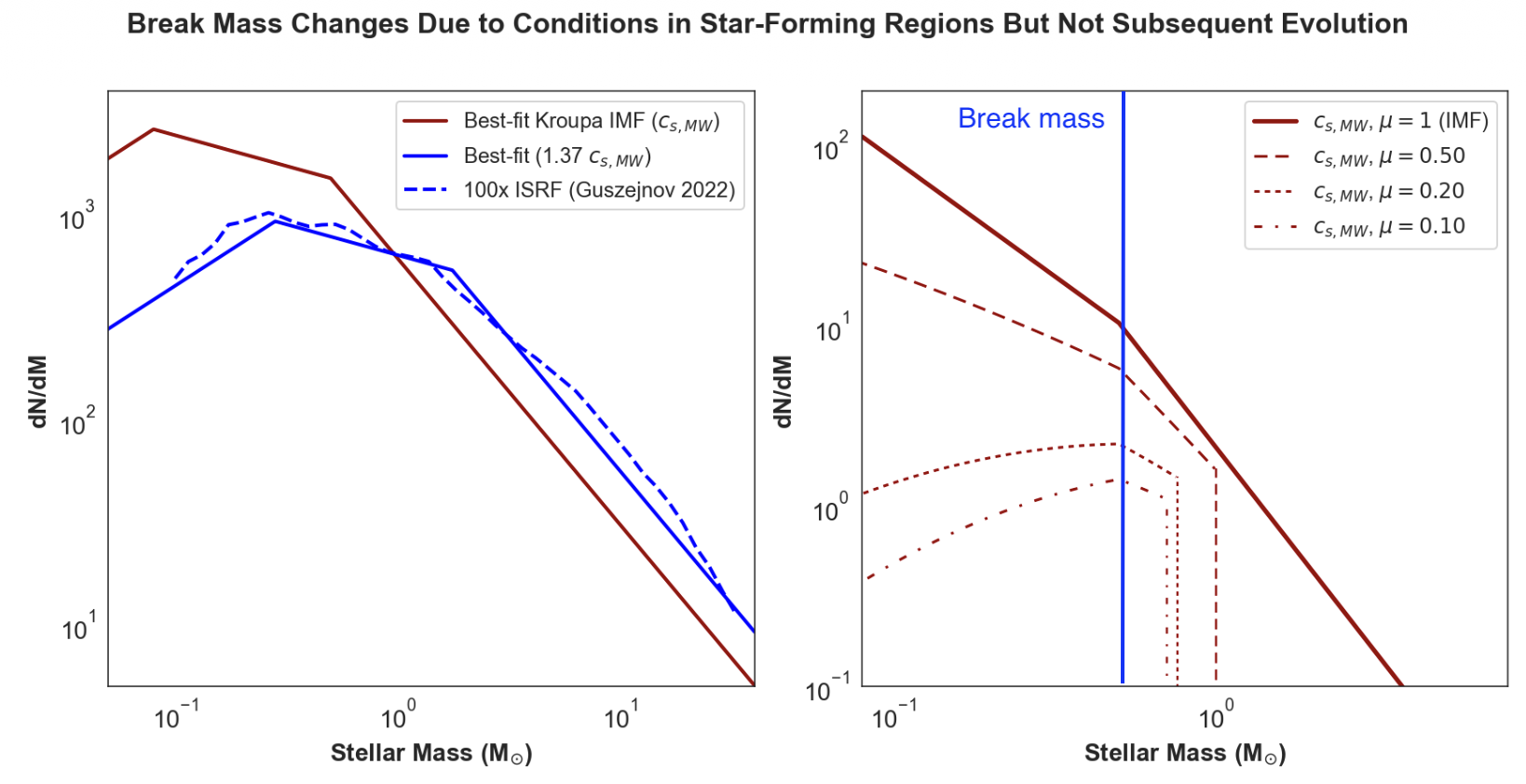
Left: Different break masses arise for different properties (here, the sound speed) of a given star-forming cloud. Right: Evolution of the mass function over time for a single star cluster. Over time, high-mass stars die and low-mass stars are ejected from the cluster, resulting in a decrease in the remaining mass fraction, μ, from 1 to 0.1. The slopes change, but the break mass does not. Click to enlarge. [Adapted from Steinhardt et al. 2026]

However, as demonstrated in a recent research article led by Charles L. Steinhardt (University of Missouri), certain aspects of the IMF may remain unchanged as clusters evolve. When the mass function of a star cluster is described using a broken power law (commonly called a “Kroupa-like” IMF in reference to work by Pavel Kroupa), the slopes of the power-law segments are affected by stellar evolution and dynamical interactions. Crucially, these effects appear not to alter the break mass — the mass at which the IMF transitions from one slope to another. Therefore, differences in the break mass from cluster to cluster reflect differences in the underlying IMF rather than the stellar and dynamical evolution of the clusters.

Insights from Gaia

Thanks to the Gaia spacecraft, which made precise observations of more than 2 billion stars in the Milky Way and beyond, it’s now possible to measure and compare the break masses of individual star clusters. Out of an initial catalog of 7,167 Milky Way clusters observed with Gaia, Steinhardt’s team selected just 110 that were of high enough quality and contained a sufficient number of stars to measure cluster break masses accurately.

Plot of stellar mass function for four Milky Way clusters

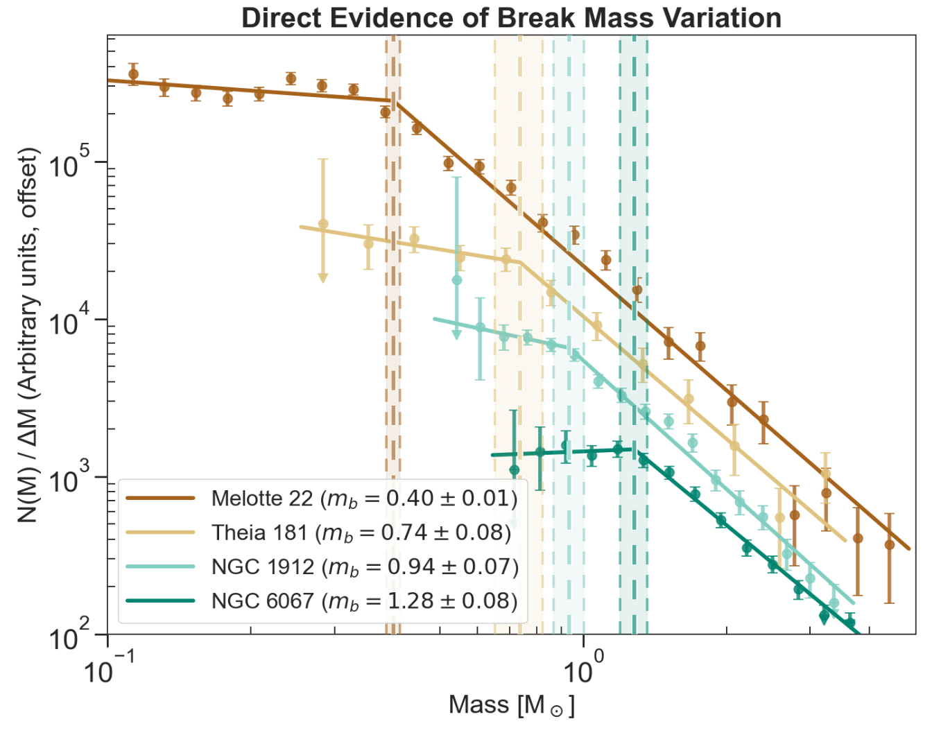
Observed mass functions of four Milky Way star clusters. The break masses are clearly different, showing that the IMF must vary from cluster to cluster. Click to enlarge. [Steinhardt et al. 2026]

The team found that the break mass differs considerably between the clusters in their sample, showing that the IMF is not universal. The observed variety of break masses appears to be linked to the age of the cluster, but instead of reflecting the impact of stellar and dynamical evolution — which have no effect on the break mass — these differences reflect the properties of the clouds in which the clusters were born. This implies that the properties of star-forming clouds in the Milky Way have, on average, changed over cosmic time. Thus, a star cluster that formed billions of years ago would have had a different IMF than one forming today.

This finding has implications for studies of high-redshift galaxies, where the properties of star-forming clouds are markedly different from those in the Milky Way. Going forward, Steinhardt and coauthors recommend developing a flexible parameterization of the IMF to capture how this critical function behaves in environments vastly different from our own.

Citation

“Direct Evidence for Stellar Initial Mass Function Variation in the Milky Way,” Charles L. Steinhardt et al 2026 ApJL 1005 L40. doi:10.3847/2041-8213/ae7444

The post Gaia Data Show That the Initial Mass Function Is Not Universal appeared first on AAS Nova.

New Technology Uncovers How the Heart Produces Cells with Regenerative Potential

24 June 2026 at 12:28

In a groundbreaking advancement for cardiovascular science, researchers at the Centro Nacional de Investigaciones Cardiovasculares Carlos III (CNIC) in Spain, in collaboration with the Karolinska Institute in Sweden, have devised a cutting-edge technique to analyze the proteome of individual cardiomyocytes. These cells, essential for heart contraction, have long eluded comprehensive single-cell proteomic scrutiny due to technological limitations. This novel approach now allows for a detailed protein-level characterization of single heart muscle cells, opening unprecedented avenues for understanding heart biology and regeneration.

The heart has traditionally been viewed as an organ comprised of relatively homogeneous cardiomyocytes. However, recent findings highlight remarkable heterogeneity even within this single cell type. Variability in protein expression among cardiomyocytes may underpin differences in their functional capacity and regenerative potential. This study, published in Genome Biology, leverages an innovative integration of optimized cell isolation methods, advanced mass spectrometry, and sophisticated bioinformatic algorithms to dissect this proteomic diversity with extraordinary precision.

Central to their investigation was Myc, a transcription factor already noted for its role in regenerative strategies for heart repair. Despite its promise, until now the specific mechanisms by which Myc modulates protein expression in individual cardiomyocytes remained elusive. Applying their novel single-cell proteomics technology, the researchers uncovered that Myc expression differentially alters metabolic enzyme levels across single cardiomyocytes. This heterogeneity results in a subset of cells exhibiting distinct immature states, which in turn possess enhanced regenerative potential—a finding that transforms our comprehension of how cardiac regeneration might be selectively targeted.

The technical feat achieved by these scientists hinges on the fusion of multiple cutting-edge methodologies. First, the meticulous isolation of adult mouse cardiomyocytes ensures viable, intact cells reflecting true physiological states. These cells then undergo mass spectrometry procedures tailored for minute sample volumes, capable of identifying thousands of proteins with high sensitivity and quantitative accuracy at the single-cell level. The ensuing data are processed with bespoke computational pipelines designed to handle the intrinsic variability and noise characteristic of single-cell omics measurements.

By revealing the proteomic fingerprints that distinguish a pro-regenerative subpopulation of cardiomyocytes, this research elucidates a previously uncharted layer of cardiac cellular complexity. Understanding how Myc drives metabolic remodeling at the protein level provides a molecular explanation for its regenerative effects and establishes a foundation for engineering therapeutic approaches that harness or mimic these cellular states.

Regeneration in the adult mammalian heart remains a formidable challenge, owing largely to the low intrinsic capacity of cardiomyocytes to proliferate or replace damaged tissue after injury such as myocardial infarction. Previous models relying largely on bulk tissue analyses have masked the nuanced cellular dynamics at play. This single-cell approach offers resolution sufficient to detect rare but critical cell subsets that might hold the key to effective myocardial repair.

Moreover, this study underscores the transformative impact of integrating proteomics with single-cell biology, moving beyond transcriptomic analyses that often fail to predict functional protein abundance accurately. Proteins, as the primary effectors of cellular function, are the crucial link between gene regulation and phenotype. Hence, this proteomic lens provides a direct readout of the biochemical state of cardiomyocytes under regenerative cues.

The implications of this work extend beyond cardiac biology. It exemplifies the broader potential of single-cell proteomics to deepen our grasp of cellular heterogeneity in health and disease, from cancer biology to neurodegeneration. The refinement and dissemination of such methodologies will empower researchers across biomedical fields to uncover subtle but impactful cellular states previously hidden in ensemble measurements.

Miguel Torres and Jesús Vázquez, who co-led the CNIC teams responsible for this study, emphasize the critical novel insights gained. According to them, dissecting Myc’s action at the single-cell level illuminates pathways and protein networks that govern cellular immaturity and regeneration, which are masked in population-level studies. This knowledge fundamentally shifts how regenerative medicine strategies might be designed, focusing on targeted modulation of key proteins identified within pro-regenerative cardiomyocyte subgroups.

Consuelo Marín-Vicente, the study’s first author, highlights the heterogeneous response to Myc induction in adult cardiomyocytes. Instead of a uniform effect, Myc induces diverse proteomic profiles, driving some cells toward a state primed for regeneration. This discovery points to the existence of intrinsic regulatory mechanisms that govern cardiomyocyte plasticity, with possible manipulation routes to expand these favorable subpopulations therapeutically.

This advancement also signifies a leap toward personalized regenerative therapies. By characterizing the protein signatures specific to regenerative-competent cardiomyocytes, future interventions could selectively enhance these signatures in patient hearts, enabling more effective recovery post-injury. It also offers biomarkers to monitor therapeutic efficacy or to stratify patients likely to benefit from Myc-based or related regenerative treatments.

The CNIC, recognized as a Severo Ochoa Center of Excellence and funded by a unique public-private partnership, remains at the forefront of cardiovascular research. Under the leadership of Dr. Valentín Fuster, CNIC’s mission to translate scientific discoveries into clinical innovations is exemplified by this study’s promise. Collaboration with leading international centers like the Karolinska Institute further underscores the global effort to unravel heart regeneration’s complexity.

In sum, by pioneering a technique for single-cell proteomic analysis of adult cardiomyocytes, this research opens transformative possibilities for cardiovascular medicine. It elucidates how transcription factors like Myc can orchestrate heterogeneous proteomic landscapes within single cells, generating subpopulations equipped with regenerative capabilities. These insights herald a new era in designing precision therapies that could one day restore heart function after injury, dramatically improving outcomes for millions suffering from cardiovascular diseases worldwide.


Subject of Research: Cells

Article Title: Pro-regenerative fingerprints identified in a sub-population of adult mouse cardiomyocytes by integrative single-cell proteomics

News Publication Date: 22-May-2026

Web References:
10.1186/s13059-026-04110-1

Image Credits: CNIC

Keywords

Cardiomyocytes, Single-cell proteomics, Myc transcription factor, Heart regeneration, Mass spectrometry, Cellular heterogeneity, Cardiovascular disease, Protein expression, Regenerative medicine, Proteome, Metabolic enzymes, Cardiovascular research

Exercise and Liraglutide Boost Vascular Health, Reduce Inflammation

24 June 2026 at 12:12

In the ongoing battle against obesity and its associated cardiovascular risks, new scientific insights are shedding light on the complex interplay between weight loss strategies and vascular health. Recent research from the S-LiTE trial, presented in a landmark study by Sandsdal et al., unfolds groundbreaking findings on how exercise and the GLP-1 receptor agonist liraglutide profoundly influence vascular function and systemic inflammation during weight loss maintenance. This investigation not only advances our understanding of therapeutic approaches but also charts a promising path for improving cardiovascular outcomes in individuals struggling with obesity.

Obesity, recognized as a critical driver of cardiovascular disease, involves pathological alterations in the vascular system and chronic low-grade inflammation. These pathological changes elevate the risk of atherosclerosis, hypertension, and ultimately, heart failure. Consequently, effective weight loss maintenance after initial reductions is paramount for sustaining vascular benefits and mitigating long-term health risks. Despite existing weight loss regimens, relapse rates are high, and how these interventions impact vascular integrity during the maintenance phase remains incompletely understood. The S-LiTE trial rigorously targeted this knowledge gap, merging pharmacological and lifestyle components for a nuanced examination.

Exercise has been universally endorsed as a cornerstone intervention in weight management and cardiovascular disease prevention. Its benefits extend beyond simple calorie expenditure, encompassing modulation of endothelial function, arterial stiffness, and inflammatory profiles. Liraglutide, a glucagon-like peptide-1 (GLP-1) receptor agonist, has garnered attention for its potent weight-lowering effects through appetite regulation and metabolic improvements. However, the independent and combined vascular effects of exercise and liraglutide during the maintenance phase after weight loss had yet to be elucidated prior to this study.

The S-LiTE trial meticulously randomized participants who had achieved significant weight reduction to either an exercise program, liraglutide treatment, a combination of both, or placebo, allowing for a comprehensive comparison across groups. Over the course of the trial, vascular health parameters such as carotid artery compliance, endothelial function assessed via flow-mediated dilation, and systemic inflammatory markers including C-reactive protein (CRP) and interleukins were closely monitored. The multidimensional assessment unveiled striking differences attributable to intervention modalities.

Exercise alone was associated with notable improvements in arterial compliance and a favorable reduction in markers of vascular inflammation. The mechanical stimuli generated during physical activity promote shear stress on vessel walls, which in turn enhance nitric oxide bioavailability and endothelial repair mechanisms. These physiological adaptations are critical for maintaining vascular elasticity and preventing atherogenic processes. This study underscores the indispensable role of exercise not just in weight regulation but as a direct modulator of vascular integrity.

Liraglutide, beyond its appetite-suppressant and glycemic control effects, demonstrated intrinsic vascular benefits. The study highlighted liraglutide’s capacity to enhance endothelial function independently of exercise, potentially through GLP-1 receptor-mediated pathways that reduce oxidative stress and inhibit pro-inflammatory signaling cascades. This offers compelling evidence supporting the use of GLP-1 receptor agonists as adjunct therapies in cardiovascular risk reduction within the obese population.

Intriguingly, the combination of exercise and liraglutide yielded additive benefits, with participants exhibiting the most pronounced improvements in vascular compliance and the greatest suppression of inflammatory cytokines. This synergistic effect suggests that the pharmacological and mechanical influences converge on complementary pathways, amplifying the overall vascular protective effect. Such findings advocate for integrative treatment frameworks that leverage both lifestyle and pharmacotherapy in obesity management.

Delving deeper, the study’s prespecified secondary analysis elucidated how these interventions influenced various immune cell populations that are pivotal contributors to vascular inflammation. It was evident that the combined approach modulated monocyte and macrophage activation states, steering them towards anti-inflammatory phenotypes. This immune modulation is instrumental in curbing chronic inflammation and subsequent endothelial dysfunction, which are foundational in the pathogenesis of vascular disease.

Beyond the vascular sphere, the inflammatory milieu impacts insulin sensitivity and adipose tissue dynamics, central to metabolic health. The study posits that improvements in vascular health might interplay with metabolic regulation, facilitating a virtuous cycle of weight loss maintenance and cardiovascular risk attenuation. This holistic perspective on obesity interventions highlights the interconnected nature of metabolism, immunity, and vascular function.

Crucial to real-world applicability, the study design mirrored long-term maintenance conditions, providing practical insights for clinicians. The sustainability of vascular improvements with continued exercise and pharmacological support affirms the need for ongoing adherence and monitoring. Moreover, the tolerability profile and patient-centered outcomes reported provide a realistic framework for incorporating such regimens in routine care.

This research not only emphasizes the potent vascular benefits tied to weight loss maintenance but also brings to light the nuanced biological effects of different modalities. The implications extend beyond cardiovascular disease prevention, touching on broader aspects of healthy aging and quality of life, as vascular health is integral to multiple organ systems. Hence, the S-LiTE trial’s findings hold substantial promise for shaping future clinical guidelines.

In the broader context of obesity treatment paradigms, these insights challenge the notion that weight loss alone is sufficient. Instead, they advocate for multidimensional interventions addressing vascular function and inflammation, which are crucial mediators of obesity-related morbidity. Such transformations in therapeutic strategies could pivot the field towards more personalized and mechanistically informed approaches.

While the study is robust, the authors acknowledge limitations including the homogeneity of the cohort and potential confounders inherent to lifestyle interventions. Future investigations are warranted to explore molecular signaling pathways in greater detail, diverse populations, and longer-term cardiovascular outcomes. Additionally, integrating imaging modalities and biomarker profiling could refine our understanding of treatment impacts at the microvascular level.

On a scientific and societal level, the S-LiTE trial represents a milestone in conceptualizing weight loss maintenance not simply as weight stabilization but as an active process with profound systemic effects. Public health strategies might evolve to promote combined regimens, potentially alleviating the healthcare burden posed by obesity-related vascular diseases. This research, therefore, stands at the confluence of basic science, clinical innovation, and health policy.

The findings from Sandsdal and colleagues’ work resonate with an emerging narrative in metabolic medicine: holistic, multimodal approaches yield superior benefits than isolated interventions. With obesity prevalence continuing to climb globally, translating these findings into accessible, scalable solutions remains both a challenge and an imperative. The S-LiTE trial ignites hope for more effective, durable solutions to one of the most pressing health crises of our time.

In summary, the S-LiTE trial reveals that exercise and liraglutide individually and synergistically improve vascular health and reduce inflammation during weight loss maintenance. This study offers compelling evidence for the inclusion of GLP-1 receptor agonists alongside structured exercise programs to sustain cardiovascular health in individuals maintaining weight loss. These innovative insights redefine obesity treatment and open new avenues for reducing the global burden of cardiovascular disease through integrated, targeted therapeutic strategies.


Subject of Research: Effects of exercise and GLP-1 receptor agonist liraglutide on vascular health and inflammation during weight loss maintenance.

Article Title: Effects of exercise and liraglutide on vascular health and inflammation during weight loss maintenance: a prespecified secondary analysis of the S-LiTE trial.

Article References:
Sandsdal, R.M., Holt, J., Alkhefagie, H.G.A. et al. Effects of exercise and liraglutide on vascular health and inflammation during weight loss maintenance: a prespecified secondary analysis of the S-LiTE trial. Nat Metab (2026). https://doi.org/10.1038/s42255-026-01554-4

Image Credits: AI Generated

DOI: https://doi.org/10.1038/s42255-026-01554-4

Inflammation and Metabolism Trigger Early Puberty in Obese Kids

21 June 2026 at 09:32

In a groundbreaking study published in Pediatric Research on June 21, 2026, researchers led by Wang, Yan, and Liu have unveiled critical insights into how metabolic, hormonal, and inflammatory pathways intertwine to accelerate precocious puberty in children suffering from obesity. This pioneering research uncovers the complex biological dialogue that propels early pubertal onset among obese youth, a phenomenon that has raised alarm bells worldwide due to its profound health implications.

Puberty, the process marking the transition from childhood to sexual maturity, is classically regulated by a symphony of endocrine signals, with the hypothalamic-pituitary-gonadal (HPG) axis as the conductor. However, the timing of this process is increasingly disrupted in obese children, leading to precocious puberty—defined as puberty occurring before age 8 in girls and 9 in boys. The new study delves deeply into the serum profiles of obese children to unravel the mechanistic underpinnings of this hastened developmental timeline.

Central to the pathophysiology elucidated by the researchers is the role of systemic inflammation, a hallmark of obesity. The team identified a distinctive inflammatory signature characterized by elevated levels of cytokines and acute phase reactants that converge on the HPG axis. This pro-inflammatory milieu is postulated to trigger neuroendocrine alterations that prematurely activate gonadotropin-releasing hormone (GnRH) neurons, thereby advancing pubertal initiation.

Complementing the inflammatory dimension are metabolic dysregulations that obesity engenders. The study meticulously profiles alterations in insulin and leptin dynamics—two pivotal hormones intimately linked to energy homeostasis and reproductive function. Elevated insulin levels, common in obese states due to insulin resistance, were found to potentiate GnRH neuronal activity. Meanwhile, hyperleptinemia, reflective of adipose tissue abundance, appears to modulate hypothalamic circuits governing puberty, providing a metabolic ‘green light’ for sexual maturation.

The research team employed high-throughput serum profiling techniques alongside advanced statistical modeling to choreograph the intricate interplay of metabolic and inflammatory mediators. This comprehensive approach allowed them to construct a predictive biochemical tableau correlating specific serum markers with rapid pubertal progression. Crucially, these findings bridge the gap between clinical observations of precocious puberty and the molecular pathways that orchestrate it in obesity.

Beyond identifying biomarkers, the study explores potential mechanistic pathways. It proposes that chronic low-grade inflammation potentiates neuronal sensitization within the hypothalamus, thereby lowering the threshold for GnRH secretion. Concurrently, metabolic hormones such as leptin may amplify neuroendocrine responsiveness, creating a synergistic environment favoring early puberty onset. This dual-hit model of inflammation and metabolic disturbance advances our understanding of the biological complexity underlying pubertal acceleration.

Furthermore, the research highlights the nuanced hormonal crosstalk influencing gonadal maturation. Upregulation of adrenal and gonadal steroidogenesis appears to be downstream of the altered serum milieu, fostering a hormonal cascade that further catalyzes secondary sexual characteristic development. This insight underscores the multifactorial nature of obesity-driven puberty alterations, extending beyond central neuroendocrine changes to peripheral endocrine feedback loops.

One of the notable technical innovations in this study is the longitudinal analysis of serum samples from a diverse pediatric cohort. This enabled the researchers to pinpoint temporal shifts in inflammatory and metabolic markers relative to pubertal milestones. The temporal dynamics revealed suggest potential windows for therapeutic intervention that could recalibrate aberrant biological signals before irreversible pubertal advancement occurs.

The implications of these findings are profound, particularly as precocious puberty is linked to increased risks of psychosocial stress, metabolic syndrome, and certain cancers later in life. By elucidating the biological signatures that accelerate puberty in obese children, the study opens avenues for early diagnostic screening and targeted therapeutics aimed at mitigating long-term health impacts.

Medical professionals and endocrinologists stand to gain new tools for assessing pubertal risk by integrating serum inflammatory and metabolic profiles into routine evaluations of obese pediatric patients. This could transform clinical practice by enabling proactive management strategies tailored to individual biochemical risk landscapes, rather than relying solely on phenotypic assessments.

From a public health perspective, the study amplifies the urgency of addressing childhood obesity not only for its well-known metabolic complications but also for its disruptive influence on normal developmental timing. Interventions that reduce systemic inflammation and restore metabolic balance might delay unwanted precocious puberty, improving both physical and mental health outcomes.

Looking forward, the researchers advocate for expanded studies to explore therapeutic modulation of the identified pathways. Immunomodulatory agents, metabolic enhancers, or lifestyle interventions targeting systemic inflammation and hormone regulation could serve as novel strategies to realign pubertal timing in susceptible children. Such approaches hold promise for personalized medicine frameworks in pediatric endocrinology.

This investigation also raises important questions about environmental and genetic factors that may interact with inflammatory and metabolic signals to influence puberty. Understanding how external exposures or inherited susceptibilities modulate these biochemical networks could refine predictive models and prevention efforts in diverse populations.

In conclusion, Wang, Yan, Liu, and colleagues have charted an unprecedented map of the intersecting biological forces accelerating precocious puberty in obese children. Their integrative analysis of inflammatory and metabolic serum markers fundamentally shifts our comprehension of childhood development, offering hope for interventions that could recalibrate the endocrine clock disrupted by obesity. As the obesity epidemic continues to burden global health, studies like this provide crucial scientific foundations to mitigate its far-reaching consequences on human growth and maturity.


Subject of Research: The interplay of metabolic, hormonal, and inflammatory factors in precocious puberty and obesity in children.

Article Title: Serum inflammatory and metabolic signatures accelerate precocious puberty in obese children.

Article References:

Wang, L., Yan, H., Liu, J. et al. Serum inflammatory and metabolic signatures accelerate precocious puberty in obese children.
Pediatr Res (2026). https://doi.org/10.1038/s41390-026-05214-x

Image Credits: AI Generated

DOI: 21 June 2026

Hydrogen Sulfide Eases Hypertension-Linked Brain Deficits

20 June 2026 at 09:30

In a groundbreaking study that promises new horizons in the treatment of hypertension-associated cognitive decline, researchers have uncovered the potential of hydrogen sulfide (H₂S) as a therapeutic agent to alleviate spatial learning deficits. This novel work, spearheaded by Kumar, Mishra, and Goswami, delves into the intricate molecular pathways by which H₂S mediates neuroprotection in the context of angiotensin II-induced hypertension, revealing the suppression of endothelial NOX2 and subsequent neuroinflammatory cascades as a key mechanism. These findings not only expand our understanding of the pathophysiological link between hypertension and cognitive impairment but also highlight the therapeutic promise of gaseous signaling molecules in neurovascular health.

Hypertension, a pervasive cardiovascular disorder, has long been implicated in cognitive decline and an increased risk of neurodegenerative diseases. The relationship between elevated blood pressure and impaired spatial learning is complex, involving multifactorial processes such as oxidative stress, endothelial dysfunction, and neuroinflammation. This study addresses the critical gap in understanding how these pathological mechanisms interplay and how specifically targeting them might prevent or reverse cognitive deficits commonly observed in hypertensive patients.

A central player in this pathology is the NADPH oxidase 2 (NOX2), an enzyme prominently expressed in the endothelium and a major producer of reactive oxygen species (ROS). Overactivation of NOX2 leads to excessive oxidative stress, which in turn triggers inflammatory pathways damaging neuronal networks essential for spatial learning and memory. The research identifies NOX2 not merely as a bystander but as a pivotal driver of the neuroinflammatory processes exacerbated by angiotensin II, a peptide hormone central to blood pressure regulation.

The role of hydrogen sulfide in cellular signaling has emerged as a vibrant area of research, particularly its cytoprotective effects in the cardiovascular system. This gasotransmitter is synthesized endogenously and exerts diverse biological functions, including vasodilation, anti-apoptotic actions, and antioxidant effects. Intriguingly, H₂S is shown to modulate the activity of endothelial cells, suggesting a potential intersection between vascular health and cognitive function. The study meticulously explores how administrating exogenous H₂S can modulate endothelial responses in hypertensive models.

Utilizing advanced in vivo models of angiotensin II-induced hypertension, the researchers employed comprehensive behavioral assays to evaluate spatial learning and memory deficits. These studies revealed pronounced impairments consistent with neuroanatomical and functional alterations within the hippocampus, a brain region integral to spatial navigation. The administration of hydrogen sulfide donors elicited a marked reduction in cognitive deficits, underscoring the functional significance of H₂S in this setting.

At the molecular level, the investigation demonstrated that hydrogen sulfide suppresses NOX2 expression and activity in endothelial cells exposed to hypertensive stimuli. This effect culminates in reduced oxidative stress and diminished activation of downstream pro-inflammatory mediators such as cytokines and microglial markers. The attenuation of these pathways underscores the multifaceted influence of H₂S in preserving the integrity of the neurovascular unit under conditions of elevated blood pressure.

Moreover, this research sheds light on the intricate interaction between endothelial dysfunction and neuroinflammation, which has emerged as a critical nexus in the pathogenesis of cognitive decline. The findings suggest that endothelial-derived oxidative stress and inflammation contribute to microglial activation and neuronal damage, processes that H₂S effectively counters. This adds a new layer to our conceptual framework of how vascular health dictates neural outcomes.

From a translational perspective, the study offers compelling evidence supporting hydrogen sulfide donors as viable therapeutic agents. The modulation of NOX2 and inflammatory pathways by H₂S paves the way for pharmacological strategies aimed at mitigating hypertension-induced neural deficits without directly targeting blood pressure itself. Such approaches could complement existing antihypertensive therapies, broadening the arsenal against cognitive impairment in hypertensive populations.

Importantly, the safety profile and physiological relevance of hydrogen sulfide administration were addressed, with the researchers noting minimal adverse effects in the animal models. This aspect is crucial given the dual nature of H₂S, which at elevated concentrations can be toxic. The study’s careful titration of dose and delivery methods serves as a valuable blueprint for future clinical investigations.

Another notable contribution of the study is its use of molecular imaging and histological techniques that corroborate the behavioral findings. These methodologies revealed decreased markers of oxidative damage and neuroinflammation in treated samples, providing robust evidence that H₂S exerts its effects through tangible cellular and tissue-level changes. Such multimodal validation strengthens the case for H₂S’s integrative role in neurovascular protection.

The work also prompts intriguing questions about the broader implications of gasotransmitters in neurodegeneration. Given the overlapping mechanisms of vascular and neurodegenerative diseases, the therapeutic modulation of gas signaling pathways may represent a paradigm shift in managing multifactorial brain disorders. This article can stimulate further research into the cross-talk between vascular function and cognitive health, championing hydrogen sulfide and related molecules as key modulators.

Furthermore, the research adds to the growing body of literature underscoring the critical importance of the endothelial interface in neural function. By targeting endothelial NOX2, hydrogen sulfide indirectly protects neural cells from oxidative and inflammatory insults, delineating an elegant mechanism wherein vascular and neural health are inextricably linked. This insight encourages a more integrated approach to cardiovascular and cognitive care.

As hypertension continues to affect millions worldwide and remains a predominant risk factor for dementia and stroke, therapies that confer cognitive resilience hold immense value. Hydrogen sulfide’s ability to mitigate vascular and neural insults in this context may redefine therapeutic goals, prioritizing the preservation of cognitive faculties alongside blood pressure control. Such dual-focused treatment paradigms could dramatically improve patient outcomes.

In conclusion, the pioneering study by Kumar and colleagues stands out as a beacon in the endeavor to unravel and counteract hypertension-induced cognitive dysfunction. By elucidating the molecular substrates by which hydrogen sulfide ameliorates endothelial oxidative stress and neuroinflammation, the research opens a new therapeutic frontier. As the science community advances these findings, we may soon witness the integration of gasotransmitter-based interventions in clinical practice, offering hope for millions at risk of cognitive decline associated with hypertension.

Subject of Research:
Article Title:
Article References: Kumar, G., Mishra, S., Goswami, S. et al. Hydrogen sulfide mitigates spatial learning deficits in angiotensin II-Induced hypertension via inhibition of endothelial NOX2 and neuroinflammation. Transl Psychiatry (2026). https://doi.org/10.1038/s41398-026-04178-0
Image Credits: AI Generated
DOI: https://doi.org/10.1038/s41398-026-04178-0

Tiny Genetic Fragments Crucial for Signaling Brain Rest Identified

19 June 2026 at 20:51

In a groundbreaking study shedding light on the molecular intricacies of arousal regulation, researchers from Pompeu Fabra University (UPF) and the Centre for Genomic Regulation (CRG) have unveiled the profound influence of neuronal microexons on behavioral states in zebrafish. This study elucidates how subtle alterations in these tiny genetic fragments can trigger hyperarousal—a state marked by heightened neural excitability and pronounced insomnia-like symptoms—which echoes the pathophysiological features observed in various neurodevelopmental disorders. The implications extend far beyond aquatic models, offering a window into the conserved mechanisms of arousal that potentially inform conditions such as autism spectrum disorder and schizophrenia in humans.

Arousal represents a fundamental neurophysiological process essential for survival, enabling organisms to respond to external and internal stimuli with appropriate behavioral and neural adaptations. This highly conserved mechanism across species ensures a meticulously balanced state, modulating responsiveness between lethargy and sensory hypersensitivity. Dysregulation of this balance manifests clinically as either diminished responsiveness or excessive wakefulness and sensory overload, typical hallmarks of stress and various neurodevelopmental pathologies.

Fundamental to this regulatory system is the diversity of proteins synthesized during development and adulthood through alternative splicing—a sophisticated post-transcriptional gene editing process. Alternative splicing enables the generation of multiple functionally distinct protein isoforms from a single gene, often mediated by the inclusion or exclusion of microexons. Microexons are exceptionally short exonic sequences within neuronal genes that profoundly influence protein function and neuronal circuit dynamics despite their minuscule size.

The investigative team employed zebrafish larvae, leveraging their optical transparency and genetic tractability to scrutinize the behavioral consequences of neural microexon misregulation. Larvae exhibiting abnormal microexon patterns demonstrated conspicuous hyperarousal behaviors including disrupted swim patterns and shortened sleep duration. “These larvae not only sleep less frequently but also take considerably longer to initiate sleep,” remarks first author Tahnee Mackensen. This behavioral hyperactivity parallels neural hyperexcitability, suggesting microexon regulation as a pivotal determinant of neurobehavioral states.

At the cellular signaling level, the researchers identified dysregulated cyclic adenosine monophosphate (cAMP) cascades as a key mediator of the observed hyperactive state. cAMP is a ubiquitous second messenger involved in modulating neuronal excitability and synaptic plasticity. The altered splicing of microexons modulates cAMP synthesis and degradation pathways, leading to an aberrant excitation of forebrain neurons in hyperaroused larvae. Notably, this altered cAMP signaling manifests as heightened cAMP-dependent protein kinase A (PKA) activity and subsequent phosphorylation of the transcription factor CREB, implicating the canonical cAMP-PKA-CREB pathway in the regulation of arousal.

The study’s pharmacological interventions underscore the centrality of cAMP regulation in arousal control. Application of cAMP inhibitors normalized the elevated neural activity and behavioral hyperarousal in mutant fish, whereas artificially elevating cAMP in wild-type fish recapitulated the hyperactive phenotype. This bidirectional modulation fortifies the concept that neuronal cAMP levels function as a ‘thermostat’ for arousal states, fine-tuning neuronal excitability and behavioral responsiveness.

Beyond the immediate findings in zebrafish, this research builds upon prior observations in drosophila models demonstrating that microexon disruption similarly impairs sleep and elevates arousal. “The parallel between species indicates an evolutionarily conserved arousal mechanism,” explains Manuel Irimia, senior author. This conservation implies that microexon misregulation, while mechanistically nuanced, may contribute to the neuropsychiatric symptomatology observed in mammals, including humans.

Human neurological disorders such as autism and schizophrenia are often accompanied by sleep disruption and sensory processing anomalies attributed, in part, to aberrant microexon splicing. While microexon alterations are unlikely to be sole causative factors, they may exacerbate or modulate disease phenotypes by disturbing neural excitability homeostasis. These insights prompt a reevaluation of therapeutic strategies aimed at restoring microexon splicing fidelity or modulating cAMP signaling pathways to alleviate neurodevelopmental symptomatology.

Moreover, the link between this microexon-cAMP pathway and mood disorders such as anxiety and depression opens compelling avenues for future research. The cAMP-PKA-CREB axis has well-documented roles in synaptic plasticity and mood regulation, suggesting that microexon-associated dysregulation could contribute to broader neuropsychiatric conditions. “This discovery might just scratch the surface of a complex regulatory network influencing brain function,” notes Mackensen.

The transparency and genetic accessibility of the zebrafish model provided unparalleled opportunities to visualize and quantify internal states through behavioral readouts. Advanced imaging of larval swimming patterns and direct measurement of neurochemical parameters furnished robust correlative evidence linking genetic alterations to functional outcomes. These technical advancements highlight the integrative power of model organisms in neuroscience.

Importantly, this research received support from an array of prestigious funding bodies, including the Horizon 2020 Framework Programme, the Marie Sklodowska-Curie Actions, and the Wellcome Trust, emphasizing the global significance and collaborative nature of this work. The study’s publication in Science Advances confirms its high impact and relevance to the scientific community.

As the team pursues translational studies, the prospect of correcting arousal imbalances by manipulating cAMP pathways or restoring microexon expression presents a promising frontier. This is especially critical given that aberrant arousal and sleep disturbances profoundly impair quality of life in neurodevelopmental disorders. The findings pave the way for multidisciplinary approaches integrating molecular genetics, neurobiology, and pharmacology to develop targeted interventions.

In summation, the identification of neuronal microexons as key modulators of arousal states via the cAMP-PKA-CREB pathway in zebrafish represents a seminal advance in our understanding of the molecular substrates governing complex behavioral phenotypes. This research not only deciphers fundamental biological mechanisms but also holds translational potential to inform therapeutic avenues for neuropsychiatric conditions marked by disrupted arousal and sleep.


Subject of Research: Animals

Article Title: Neuronal microexons modulate arousal via the cAMP-PKA-CREB pathway in zebrafish

News Publication Date: 19-Jun-2026

Web References: 10.1126/sciadv.ady8291

Image Credits: UPF – CRG

Keywords: Exons, Gene splicing, Developmental neuroscience, Anxiety, Sleep disorders, cAMP signaling, Zebrafish

New Study Seeks to Prolong Immune System Longevity

18 June 2026 at 00:45

In a groundbreaking advance poised to transform the landscape of age-related immune decline, University College London (UCL) researchers are spearheading a pioneering clinical trial aimed at rejuvenating worn-out T cells through an innovative immune restoration therapy. This Phase 1 first-in-human study will explore the therapeutic potential of metabolically resetting exhausted or senescent T cells, a subset of immune cells that accumulate with age and chronic disease and lose their robustness in coordinating immune responses. The initiative promises to reopen avenues for combating a spectrum of diseases, including cancer, HIV, and neurodegenerative disorders like dementia, by profoundly modifying the immune system’s intrinsic capacity to defend and repair.

Central to the research is the recognition that immune ageing, characterized by the progressive dysfunction and decline in immune cell performance, is a key factor leading to increased vulnerability in the elderly and those with chronic infections. T cells, particularly the CD4+ subtype—often regarded as the conductors of the immune orchestra—become less effective with time, diminishing the immune system’s ability to coordinate defenses accurately. These cells, rendered senescent or exhausted, exhibit impaired proliferation, cytokine production, and cytotoxic activity, undermining responses to pathogens, malignancies, and cellular damage.

The novel therapy, developed by SenTcell, a biotech startup founded by Dr. Alessio Lanna from UCL Medicine, employs a liquid formulation administered intramuscularly akin to conventional vaccines. This delivery method facilitates patient-friendly administration while targeting key metabolic pathways within dysfunctional T cells. By reprogramming the intracellular environment of these immune cells, the treatment aims to restore youthful functional properties, including enhanced proliferative capacity and efficient pathogen recognition, thereby revitalizing the immune system’s broad protective functions.

Dr. Lanna explains that although antiretroviral advancements have significantly improved lifespans for individuals living with HIV, many still experience premature immune ageing. Similar patterns of immune exhaustion are also detected in cancer patients and other chronic disease cohorts. The imminent clinical trial represents a critical stepping stone towards verifying whether exhausted immune cells can be safely and effectively rejuvenated in human subjects, with the potential to redefine therapeutic paradigms for diseases rooted in immune dysregulation.

At the molecular level, one of the intriguing aspects under investigation involves the role of telomeres—protective caps on chromosome ends whose shortening serves as a hallmark of biological ageing. Laboratory studies have suggested that rejuvenated CD4+ T cells may release telomere-containing extracellular structures, whimsically termed “telomere Rivers,” into the bloodstream. The presence and behavior of these structures might offer novel insights into how restored immune cells influence systemic health, extending their benefits beyond immediate immune niches to broader tissue health. Although this phenomenon has yet to be validated in human clinical contexts, it highlights the sophisticated interplay between immune rejuvenation and organismal aging.

The clinical trial enjoys regulatory support from the UK’s Medicines and Healthcare products Regulatory Agency (MHRA) through its Innovative Licensing and Access Pathway (ILAP), underscoring the therapy’s potential to meet the pressing unmet medical needs associated with immune senescence. This endorsement accelerates development timelines and affirms the significance of targeting immune metabolic dysfunction as a viable therapeutic strategy.

Participants in the upcoming Phase 1 trial will be meticulously selected adults exhibiting characteristics of immune dysfunction, including evidence of premature immune ageing or persistent viral infections. Comprehensive immune profiling will be conducted before and after therapeutic administration to discern immunological changes and safety outcomes. Although the initial focus is on evaluating biological activity and tolerability, successful results could pave the way for advanced trials assessing clinical efficacy.

Unlike conventional treatments that combat diseases by targeting individual pathogens or tumor cells directly, this therapy seeks to recalibrate the immune system itself—restoring its intrinsic resilience and regenerative functionality. Such a paradigm shift holds profound implications; by enhancing the immune system’s natural defensive repertoire, it may afford broad-spectrum protection and facilitate healthier ageing trajectories.

Beyond chronic infections like HIV and cancer, this approach could ultimately apply to autoimmune disorders characterized by immune exhaustion, wherein overly active yet dysfunctional immune cells drive pathology. Immune rejuvenation strategies offer a novel means of restoring balance without broadly suppressing immune activity, potentially mitigating disease progression while improving quality of life.

This research aligns with a growing global focus on understanding the cellular and molecular underpinnings of ageing and immune decline. By intervening at the level of T cell metabolism and function, scientists hope to extend not only lifespan but healthspan—preserving cognitive and physical function while reducing susceptibility to infections, cancer, and degenerative diseases.

As UCL celebrates two centuries of academic excellence and innovation, this project embodies the institution’s commitment to translating bold scientific ideas into therapies capable of reshaping medicine. If validated through clinical trials, this immune rejuvenation therapy could herald a transformative era in treating age-associated diseases, offering hope for millions affected by immune dysfunction worldwide.

Subject of Research: People
Article Title: Immune Rejuvenation: UCL’s Pioneering Trial Targets T Cell Exhaustion to Combat Age-Related Immune Decline
News Publication Date: Not provided
Web References: www.ucl.ac.uk
References: Not provided
Image Credits: Not provided
Keywords: Immune ageing, T cell exhaustion, CD4+ T cells, immune rejuvenation, senescent immune cells, telomeres, immune dysfunction, chronic disease, HIV, cancer, metabolic resetting, Phase 1 clinical trial

OPA3 Protein Maintains Heart Function via Calcium Regulation

17 June 2026 at 15:49

In a groundbreaking study published in Nature Communications, scientists have unveiled how a mitochondrial protein known as OPA3 plays a pivotal role in sustaining cardiac function by regulating calcium handling in male mice. This discovery sheds new light on the intricate cellular mechanisms that underpin heart performance and opens up promising avenues for treating cardiovascular diseases, which remain a leading cause of mortality worldwide. The findings emphasize the critical interplay between mitochondrial dynamics and calcium signaling within cardiac muscle cells, providing a fresh perspective on how cellular energy production influences heart health.

The heart’s ability to maintain rhythmic and efficient contractions is fundamentally dependent on the precise regulation of intracellular calcium levels. Calcium ions serve as essential second messengers in cardiac myocytes, triggering contraction through complex signaling cascades. However, the interplay between calcium dynamics and mitochondrial proteins, especially those involved in mitochondrial morphology and function, has remained elusive until now. The current study identifies the mitochondrial protein OPA3 as a key modulator in this regulatory network, maintaining both mitochondrial integrity and calcium homeostasis crucial for cardiac contractility.

OPA3 is traditionally recognized for its role in maintaining mitochondrial morphology, particularly in mitochondrial fission and fusion processes. These processes are vital for preserving the mitochondrial network’s dynamic nature, which is essential for cellular energy metabolism. Mitochondria not only produce ATP but also buffer intracellular calcium, thereby influencing calcium signaling within cardiac cells. By elucidating how OPA3 influences calcium handling, researchers have bridged an important gap between mitochondrial structural proteins and the functional output of cardiac cells.

Experimental models using male mice engineered for OPA3 depletion revealed significant impairments in cardiac function, demonstrating that loss of OPA3 disrupts calcium cycling in cardiac myocytes. Impaired calcium handling directly correlates with diminished contractile force and defective cardiac output. This suggests that OPA3’s influence extends beyond structural maintenance and into functional regulation of calcium channels or transporters. The dysregulation of such a critical ion pathway can lead to arrhythmias and cardiac failure, underscoring the potential clinical significance of these findings.

At the molecular level, the study highlights that OPA3 affects the expression and activity of key calcium regulators including the sarcoplasmic reticulum Ca2+ ATPase (SERCA) and ryanodine receptors (RyR). These proteins govern calcium reuptake and release in cardiac cells, orchestrating contraction and relaxation cycles. Through advanced imaging and electrophysiological techniques, the research team demonstrated that OPA3 deficiency impairs SERCA function and causes aberrant RyR-mediated calcium leak, leading to calcium overload or deficits. Such pathophysiological alterations are known contributors to cardiac hypertrophy and heart failure in humans.

The investigation also explores the bioenergetic consequences of OPA3 modulation. Mitochondrial bioenergetics in cardiac muscle cells are highly tuned to meet the energy demands of continuous contractions. OPA3 deficiency was found to compromise mitochondrial ATP production by disrupting inner membrane potential and electron transport chain efficiency. These mitochondrial defects culminate in inadequate energy supply for calcium pumps and ion exchangers, further exacerbating contractile dysfunction. This uncovers a feedback loop where structural mitochondrial proteins like OPA3 indirectly regulate calcium handling by maintaining energetic homeostasis.

A remarkable aspect of the study lies in its use of state-of-the-art imaging techniques such as live-cell confocal microscopy and super-resolution electron microscopy, which revealed morphological abnormalities in mitochondria lacking OPA3. Fragmented and swollen mitochondria with disrupted cristae architecture were prevalent in OPA3-deficient cardiomyocytes. These structural derangements are likely responsible for the observed bioenergetic failures and altered calcium buffering capacity, collectively impairing the cardiomyocyte’s functional integrity.

Furthermore, the team utilized optogenetics-based calcium sensors to monitor real-time calcium flux in isolated cardiomyocytes, providing unprecedented insight into how OPA3 influences calcium transients during excitation-contraction coupling. Data revealed that OPA3 loss leads to prolonged calcium decay time and increased diastolic calcium levels, conditions that predispose cardiac cells to arrhythmic events and contractile inefficiency. These calcium handling anomalies could explain the pathophysiology of certain forms of cardiomyopathy linked to mitochondrial dysfunction.

Importantly, the study addresses the potential sex-specific roles of OPA3, noting that experiments were conducted exclusively in male mice to control for hormonal influences on cardiac physiology. This raises intriguing questions about whether OPA3 functions similarly in female models and whether sex hormones modulate its expression or activity. Such considerations are critical as sex differences in cardiovascular disease outcomes are well-documented, emphasizing the need for further research in this domain.

The translational implications of these findings are profound. Therapeutic strategies aimed at restoring or enhancing OPA3 function could stabilize mitochondrial morphology and bioenergetics, thereby normalizing calcium handling and improving cardiac contractility. Such interventions might hold promise for treating heart failure syndromes characterized by mitochondrial and calcium dysregulation. Moreover, identifying small molecules or gene therapies targeting OPA3 pathways might complement existing treatments that primarily focus on calcium channels but neglect mitochondrial contributions.

Beyond the heart, the role of OPA3 in mitochondrial dynamics suggests potential relevance in other tissues with high energetic demand, such as skeletal muscle and the brain. Defects in mitochondrial morphology and calcium handling have been implicated in neurodegenerative diseases and metabolic disorders. Thus, understanding OPA3’s function could have broad implications in diverse biomedical fields, making it a protein of significant interest in physiology and pathophysiology.

This study also raises several compelling scientific questions for future research. How does OPA3 interact with other mitochondrial fission and fusion proteins, such as OPA1, MFN1/2, and DRP1, in regulating calcium signaling? What are the upstream regulators of OPA3 expression in cardiac tissue under physiological and pathological conditions? Additionally, can OPA3 modulation reverse established cardiac dysfunction or is it mainly preventive? Addressing these queries will deepen our understanding of cardiac mitochondrial biology and pave the way for novel therapeutic innovations.

Addressing cardiac diseases from a mitochondrial perspective represents a paradigm shift in cardiovascular medicine. Historically, calcium handling has been studied primarily through its direct regulators, but this research highlights the necessity of considering mitochondrial proteins like OPA3 as integral components of intracellular calcium homeostasis. This refined viewpoint may revolutionize how clinicians and scientists approach diagnosis, prognosis, and treatment of cardiac ailments.

In conclusion, the identification of mitochondrial protein OPA3 as a fundamental regulator of cardiac calcium handling marks a significant advance in cardiovascular biology. By elucidating the link between mitochondrial morphology, energy metabolism, and calcium signaling, this work provides critical insights into the molecular basis of cardiac function and dysfunction. It offers hope for innovative interventions that could alleviate the burden of heart disease, fostering improved patient outcomes and advancing the frontier of precision medicine in cardiology.

Subject of Research:
Mitochondrial protein OPA3 and its role in cardiac function via regulation of intracellular calcium handling mechanisms.

Article Title:
Mitochondrial protein OPA3 sustains cardiac function by regulating calcium handling in male mice.

Article References:
Geng, N., Chen, T., Li, H. et al. Mitochondrial protein OPA3 sustains cardiac function by regulating calcium handling in male mice. Nat Commun (2026). https://doi.org/10.1038/s41467-026-73991-4

Image Credits: AI Generated

Featured Image: Low-Mass Brown Dwarfs in a Class of Their Own?

14 July 2025 at 17:00

IC 348

The glowing green nebula in this JWST image surrounds the star cluster IC 348, which is the subject of a recent study by Kevin Luhman (Penn State University) and Catarina Alves de Oliveira (European Space Agency). Using JWST’s Near-Infrared Camera, Luhman and Alves de Oliveira searched the cluster’s young stellar population for free-floating brown dwarfs — objects that are less massive than stars but more massive than most planets — and discovered multiple candidates with masses down to just twice the mass of Jupiter. Follow-up JWST spectroscopy confirmed the masses of these objects, making them the lowest-mass brown dwarfs known to date. In addition to their mass, these newly discovered brown dwarfs are remarkable because their spectra show evidence of hydrocarbon molecules, the exact identities of which are not yet known. Luhman and Alves de Oliveira proposed that low-mass brown dwarfs bearing this chemical signature be inducted into a new spectral class called “H” for “hydrocarbon.” To add to the intrigue of these objects, the authors also discovered signs of circumstellar disks around two of them, suggesting that they may be capable of forming and harboring planets. To learn more about the low-mass brown dwarfs in IC 348, be sure to check out the full research article linked below.

Citation

“A New Spectral Class of Brown Dwarfs at the Bottom of the IMF in IC 348,” K. L. Luhman and C. Alves de Oliveira 2025 ApJL 986 L14. doi:10.3847/2041-8213/addc55

The post Featured Image: Low-Mass Brown Dwarfs in a Class of Their Own? appeared first on AAS Nova.

Event 201 Again | 2024 Desktop Exercise War-Gamed Bioterrorist Attack that Kills 280,000 Americans in a Single Day – on 4 July 2025

2 June 2025 at 22:48
In a podcast discussion, Dr. David E. Martin warns of a predicted bioterror attack on July 4, 2025, as outlined in the “National Blueprint for Biodefense” report, which anticipates 200,000 animal and 280,000 human casualties in one day. He compares it to the prior Event 201 exercise related to COVID-19, urging public alertness.

❌