Normal view
An Extraordinary White House Meeting

© Adriana Loureiro Fernandez for The New York Times
- Live Science
- 'Another dinosaur has entered the luxury collectibles market': Gus the T. rex just sold for $50 million. Here's what its loss means to science.
'Another dinosaur has entered the luxury collectibles market': Gus the T. rex just sold for $50 million. Here's what its loss means to science.
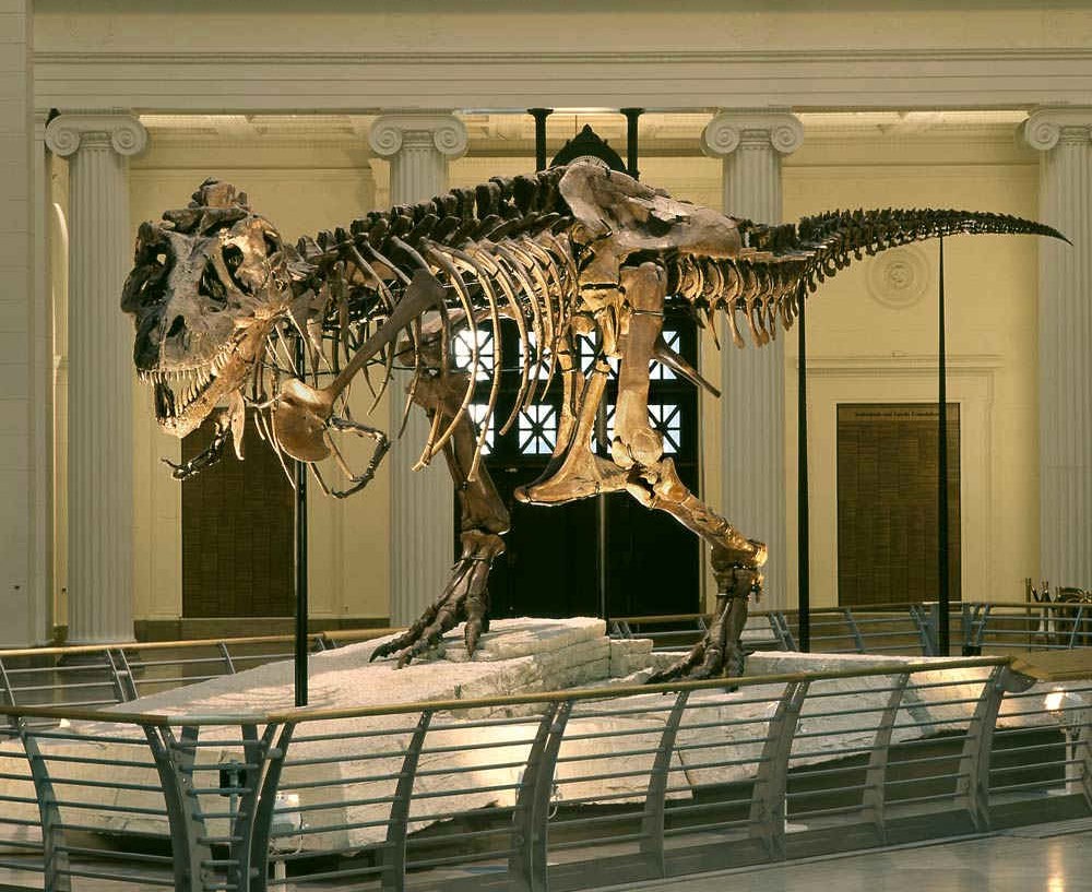
On July 14, 2026, "Gus," one of the most complete specimens of Tyrannosaurus rex, went to an as yet unidentified buyer for US$50.1 million. This auction at Sotheby's set a record for most valuable fossil ever sold. Another dinosaur has entered the luxury collectibles market, a reminder that even Earth's deepest history can be sold to the highest bidder.
To paleontologists like me, however, a fossil like "Gus" — excavated from the Hell Creek Formation in South Dakota over three years starting in 2021 by commercial collector Thomas Heitkamp and his team — is not a trophy or a work of art. It is an irreplaceable scientific archive. Fossils preserve evidence of evolution, extinction, growth, disease, injury and ancient ecosystems. They are finite, nonsubstitutable records of life’s history on Earth.
Science depends on independent verification of claims and healthy debate. Researchers must be able to revisit specimens, test earlier conclusions and ask new questions.
But once a scientifically important fossil enters a private collection, access for researchers is no longer guaranteed. Collectors typically sequester their fossils in their homes. Even when privately owned specimens are loaned to museums, the owners can change their minds, ending access at any time. This issue is especially of note when it comes to Tyrannosaurus rex; a 2025 study found that while there were 61 T. rex fossils in public trusts at that time, 71 were privately held.
That is why the Society of Vertebrate Paleontology, of which I'm a long-term member and president-elect, has long argued that scientifically significant vertebrate fossils belong in the public trust, curated in museums and universities that preserve them permanently, make them available for research and share them with the public.
Finding a fossil
Supporters of commercial fossil sales often argue that without sales to private collectors, specimens like "Gus" would remain buried or erode away. They’re right about one thing: Discovery matters. Many extraordinary fossils have been found by ranchers, hikers, amateur collectors and commercial excavators. Paleontology is accessible to everyone who has an eye for observing nature — you don't need to be an expert with academic credentials to make an important discovery.
But discovery is only the beginning. A fossil's scientific value depends on careful documentation of where it was found, the rocks surrounding it, and the plants and animals preserved alongside it. Those details allow scientists to reconstruct ancient ecosystems, understand how an animal lived and died, and interpret how its remains became fossilized. When that contextual information is incomplete or lost, much of the fossil's scientific value is lost as well.
Yet even discovery, excavation and publication barely scratch the surface of a fossil's scientific importance. The greatest scientific value of a specimen often comes decades later, when researchers ask new questions and apply new technologies that earlier generations never imagined. A specimen that seems fully studied today may yield surprising new information tomorrow, but only if it is still available for study.
Delayed discoveries
Consider the iconic dinosaurs, including T. rex, Triceratops, Diplodocus and Stegosaurus, first collected more than a century ago. Early paleontologists could describe their shapes but had no way to dig deeper by peering inside the bones. Because those specimens were preserved in museum collections, later generations could revisit them with technologies that didn't exist when they were discovered.
Paleontologist Larry Witmer and his collaborators at Ohio University started using CT imaging 20 years ago to reconstruct the internal anatomy of historic dinosaur fossils without damaging them, based on how X-rays travel through specimens. Brain cavities, inner ears, air spaces, nerves and blood vessels became visible for the first time, revealing how dinosaurs balanced, heard, smelled and perceived their world.
Henry Fricke, Thomas Cullen and other geochemists have used isotopic signatures preserved in fossil teeth and eggshells to reconstruct dinosaur diets, migration patterns and body temperatures. This research has revealed how dinosaurs lived: what they ate, how they moved through ancient landscapes, and even how warm their bodies were.
More recently, molecular paleontologist Jasmina Wiemann and her collaborators have identified chemical traces preserved in fossil bone, eggshell and skin that reveal aspects of dinosaur biology unimaginable even a generation ago. Until now, paleontologists had no way to know details about metabolic rates and reproduction or the colors of skin, feathers and eggs.
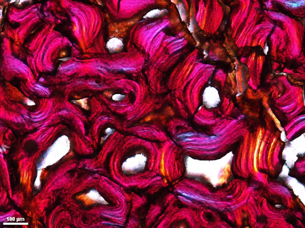
In my own research I use microscopes to uncover the hidden stories preserved inside dinosaur bones and teeth. Thin sections of fossil bones reveal that dinosaurs grew more like mammals and birds than like oversized reptiles. Microscopic modifications to bones capture traces of ancient scavenging, and tiny signatures deep inside baby dinosaur bones indicate the moment of hatching.
None of these discoveries would have been possible if the original fossils had vanished into inaccessible private collections.
Shared natural heritage, on the auction block
Fossils are not static objects whose scientific value is exhausted once they are described. Their value grows as science advances, but only if future researchers can continue to examine the original specimens.
Of course, sometimes dinosaur fossils are rescued from obscurity through purchase and immediate deposition or donation to natural history museums. Some of the world's most important dinosaur fossils are accessible today because individuals, companies or organizations with the means to acquire extraordinary specimens recognized that they belong where scientists can continue to study them and where future generations can learn from them.
Purchasing a fossil in order to place it permanently in the public trust is fundamentally different from acquiring it as a private collectible: One expands access, the other leaves access uncertain.
But as fossil prices rise into the millions, museums increasingly cannot compete. The most significant fossils are no longer reliably entering public collections. Instead, they are becoming luxury assets whose market value supersedes their scientific value.
Dinosaurs belong to our shared natural heritage. They inspire wonder because they connect all of us to a world unimaginably older than our own. For me, the question raised by auctions like the one on July 14 of "Gus" is not who can afford to own these relics of the past. It is whether future generations have the chance to study and learn from them.
This edited article is republished from The Conversation under a Creative Commons license. Read the original article.
How much do you know about the king of the dinosaurs? Test your knowledge with our T. rex quiz!
- NYT - Science
- Door-to-Door Search for Grapevines Sold at Costco Begins in California’s Santa Clara County
Door-to-Door Search for Grapevines Sold at Costco Begins in California’s Santa Clara County
Meet Earth’s Newest Monkey. (And Check Out Its Orange Lips.)
The Great Pacific Garbage Patch Is Becoming a Bizarre Floating Ecosystem
Trump Cuts Habitat Protections for Endangered Species

© Dave Sanders for The New York Times
- NYT - Energy & Environment
- I.U.C.N. Update Says Deep-Sea Mining Threatens Mollusks Around Hydrothermal Vents
I.U.C.N. Update Says Deep-Sea Mining Threatens Mollusks Around Hydrothermal Vents
The First Ancient Protein Analysis of Homo Naledi Has Revealed a New Burial Mystery
A team of researchers examined 23 teeth from at least 20 individuals from South Africa‘s Rising Star Cave system and found no evidence of the male-specific Amelogenin-Y protein.
A recent study, published in Cell, marks the first ancient protein analysis of Homo naledi and raises the possibility that the Dinaledi Chamber may contain predominantly, or possibly exclusively, female individuals. If confirmed, Homo naledi would become the first known non-human species to practice sex-specific burial.
A Decade-Old Puzzle
Since its discovery in 2013, Homo naledi has puzzled paleoanthropologists. This extinct human relative lived between 335,000 and 241,000 years ago and combined ape-like anatomy with distinct human features. So far, Homo naledi remains have only been found in the Rising Star Cave system in South Africa.
For over a decade, scientists noted that the adult fossils from the Dinaledi Chamber were nearly identical, lacking the skeletal variation usually seen between males and females. The reason for this similarity remained unclear.
Proteins Where DNA Fails
To investigate further, a team from the University of York, the University of Copenhagen, and additional institutions analyzed the remains at the molecular level. They used a minimally destructive acid-etching method to extract protein fragments, or peptides, from the enamel of the 23 teeth.
“Unlike those found in other remains like bone fragments, proteins in tooth enamel are preserved because dental enamel—the hardest tissue in the human body—shields proteins from environmental contamination even for millions of years. This makes them ideal carriers of genetic information from deep time. Our study helps explain the long-standing mystery of why Homo naledi lacked significant variation; it’s probably because they could have all belonged to one sex,” said Palesa Madupe, who conducted the research during her postdoctoral fellowship at the University of Copenhagen.
A Missing Male Marker
The researchers specifically searched for Amelogenin-Y, a protein produced only by the male Y chromosome, but detected none of it in the sampled teeth. To make sure the results were not due to modern contamination, a specialized chemistry team at the University of York independently confirmed that the amino acids were genuinely ancient. The findings remained consistent.
“The lack of male markers within the group is truly fascinating. It is incredibly exciting to gain a window not only into the biology of our ancestors, but also into how they lived. These findings offer rare insights into a culture that has, until now, been difficult to access directly. Advances in ancient protein analysis are opening the door to a far richer and more nuanced understanding of ancient hominins,” said Dr. Marc Dickinson from the university’s Department of Chemistry.
Ritual or Genetic Fluke?
The findings allow for two possible explanations. One is cultural: if the chamber were reserved for females, it could indicate that Homo naledi practiced sex-specific, symbolic mortuary behavior, a trait previously thought to be unique to modern humans.
The second explanation is biological. The researchers note that a small, isolated Homo naledi population could have lost or mutated the Amelogenin-Y gene, leaving males without the protein marker typically used to identify male remains.
Either explanation could reshape what scientists know about Homo naledi. If researchers confirm the site as an all-female burial chamber, it would suggest that symbolic behavior emerged earlier in the human lineage than previously believed. On the other hand, a genetic change across the population would show how isolation can shape a species’ biology in ways that fossils alone cannot reveal. This study shows how ancient protein analysis from teeth can open new windows into human evolution.
Austin Burgess is a writer and researcher with a background in sales, marketing, and data analytics. He holds an MBA, a Bachelor of Science in Business Administration, and a data analytics certification. His work focuses on breaking scientific developments, with an emphasis on emerging biology, cognitive neuroscience, and archaeological discoveries.
Studying Brain Mechanisms of Irritability Through Cross-Species Reward Frustration
In a groundbreaking study published in Translational Psychiatry, researchers have unveiled a novel cross-species approach to unraveling the neural circuitry underlying irritability—a pervasive emotional state with significant clinical implications. The investigation, led by WL Tseng, Z Li, and E Leibenluft, harnesses the concept of frustrative non-reward (FNR) as a potent experimental probe, enabling unprecedented insight into the brain mechanisms that drive irritability across humans and animal models.
Irritability, often characterized by heightened sensitivity to frustration and negative affect, is a hallmark symptom in various psychiatric disorders, including mood and anxiety conditions. However, the challenge in dissecting its neurobiological roots has historically stemmed from the lack of translational tools that can bridge findings from animal models to human pathology. By focusing on frustrative non-reward—the experience of being denied an expected reward—this study provides a quantifiable and comparable construct to systematically explore irritability’s brain basis.
Central to the research is the deployment of paradigms that elicit FNR responses in both humans and animals, enabling scientists to examine conserved neural circuits modulated by this emotional challenge. Functional neuroimaging data reveal that regions such as the anterior cingulate cortex, ventral striatum, and amygdala exhibit altered activity during frustrative episodes. These findings suggest a neurobiological signature of irritability that transcends species boundaries, emphasizing the role of reward processing and emotional regulation networks.
On a molecular level, the study points toward dysregulation in neurotransmitter systems implicated in reward anticipation and frustration tolerance, including dopaminergic and serotonergic pathways. These alterations may underlie the exaggerated irritability seen in clinical populations, offering targets for therapeutic intervention. Furthermore, the team’s use of advanced computational modeling to integrate behavioral, neuroimaging, and neurochemical data sets a new standard for mechanistic research in affective neuroscience.
Importantly, this cross-species strategy enhances the validity of animal models used in preclinical research on irritability, fostering improved translational potential. It opens avenues for testing pharmacological agents aimed at modulating FNR responses, thereby attenuating irritability symptoms with higher specificity. The quantitative nature of FNR as an experimental probe may also facilitate the development of biomarker-driven diagnostic tools in psychiatry.
The research underscores the significance of irritability not merely as a symptomatic complaint but as a fundamental affective process with distinct neurobiological underpinnings. By delineating the brain circuitry common to humans and animals during frustrative non-reward, the study propels the field toward more effective interventions for irritability-related psychopathologies.
As psychiatric research increasingly embraces an integrative cross-species framework, studies like this highlight the potential for converging evidence to unravel complex emotional states. The elucidation of neural mechanisms underlying irritability promises to revolutionize both our understanding and treatment of disorders marked by heightened frustration sensitivity.
This pioneering work marks a crucial step forward in affective neuroscience, establishing frustrative non-reward as a versatile and robust probe to decode irritability. Future investigations building on these findings are poised to translate laboratory insights into therapeutic breakthroughs that improve mental health outcomes worldwide.
Subject of Research: Neural mechanisms of irritability using frustrative non-reward as a cross-species probe
Article Title: Frustrative non-reward: A cross-species probe to study brain mechanisms of irritability
Article References:
Tseng, WL., Li, Z. & Leibenluft, E. Frustrative non-reward: A cross-species probe to study brain mechanisms of irritability. Transl Psychiatry (2026). https://doi.org/10.1038/s41398-026-04257-2
Image Credits: AI Generated
Global Warming Upends London Climate Week

© Pool photo by Yui Mok
- NYT - Energy & Environment
- Trump Administration Forms Partnership to Bank DNA From Imperiled Species
Trump Administration Forms Partnership to Bank DNA From Imperiled Species

© Lenny Ignelzi/Associated Press
- Live Science
- Antarctica's first dinosaur fossil belonged to a group of the largest land animals ever
Antarctica's first dinosaur fossil belonged to a group of the largest land animals ever
The first dinosaur found in Antarctica belonged to a group that included the largest animals ever to walk the planet, a new study finds.
A backbone from the 82 million-year-old giant was discovered more than 40 years ago, but at the time, researchers assumed it came from an ancient marine reptile. Now a new study, published June 29 in the journal Acta Palaeontologica Polonica, has revealed that it was actually a titanosaur — the group of long-necked sauropods that included the largest land animals on record.
"At first glance this appears to be an unremarkable fossil, but it holds an important place in the history of Antarctic exploration as the first dinosaur fossil found on the continent," study first author Paul Barrett, a paleontologist at the Natural History Museum in London, said in a statement.
While this fossil now holds the record for the first dinosaur fossil found in Antarctica, other dinosaur fossils were identified on the continent after its discovery, so it's not the only known Antarctic dinosaur. In fact, researchers have identified a variety of dinosaurs on Antarctica, with another sauropod fossil identified as a titanosaur in 2011.
The newly identified dinosaur was around 20 to 23 feet (6 to 7 meters) long. That's very small compared with the largest-known titanosaurs, which could grow up to 123 feet (37.5 m) long. However, as the fossil is just a fragment of a vertebra, researchers are unable to narrow down which species the Antarctic titanosaur belonged to, and it's possible that the individual was only a juvenile when it died.
Mike Thomson, a British Antarctic Survey geologist, found the fossil during an expedition to James Ross Island in 1985. The island, which has been the site of multiple dinosaur discoveries, is located off the northeastern Antarctic Peninsula, south of the 600-mile-wide (965 kilometers) Drake Passage that separates South America from Antarctica.
Antarctica is well known for its icy landscapes, but when titanosaurs roamed Earth, the continent was still attached to South America and full of temperate forests. Antarctica's dinosaurs were so far south that they would have lived in constant twilight during the winter months, according to a news article published by the Natural History Museum.
The authors of the new study identified the dinosaur using high-resolution CT scans, which enabled them to look inside the fossil. Dating back to the Cretaceous period (143 million to 66 million years ago), the titanosaur lived in the last age of the non-avian dinosaurs, before an enormous asteroid hit what is now Mexico's Yucatan Peninsula 66 million years ago and wiped them out.
- 'Last titan' of Thailand discovered, and it's the longest-necked dinosaur on record from Southeast Asia
- Giant 'cow of the Cretaceous' discovered almost 100 years ago identified as new duck-billed dinosaur
- 'He began to cry, and almost fell to the floor': The fluffy fossil that finally showed the world that birds are dinosaurs
The Antarctic titanosaur helps researchers better understand how dinosaurs spread across Earth's southern continents, which at the time were combined into a supercontinent called Gondwana. The presence of titanosaurs on Antarctica suggests that they may have used Antarctica to travel from what is now South America to New Zealand.
Other dinosaurs identified on Antarctica include small herbivores, armored ankylosaurs and bipedal predators like Imperobator, which would have shared the forests with the newly identified titanosaur. While researchers are beginning to piece together Antarctica's ancient ecosystems, they still have a lot to learn about the dinosaurs that lived there.
"There are likely many more dinosaurs to be discovered on the continent," Barrett said. "As climate change causes ice to retreat we may indeed find further evidence of this past biodiversity."
From Darkness to Light: How Blind Mexican Cavefish Reveal Brain Evolution
Deep within the labyrinthine caves of northeastern Mexico dwells a remarkable species of fish, the blind Mexican cavefish (Astyanax mexicanus), whose evolutionary journey over hundreds of thousands of years has led it to adapt to a life shrouded in perpetual darkness. This fish has lost its eyes and pigmentation, hallmark traits of cave-dwelling organisms, while simultaneously acquiring a suite of physiological and behavioral adaptations tailored to survive in nutrient-scarce subterranean environments. Its dual existence as both a sighted surface fish and numerous independently evolved cave forms presents a unique biological system for examining how extreme environments drive evolutionary changes in brain structure, sensory processing, and behavior.
Researchers from Florida Atlantic University, collaborating with experts from institutions across the United States, have leveraged cutting-edge genetic and imaging technologies to decipher the neural underpinnings that distinguish cavefish from their surface relatives. By employing genetically engineered lines of this species that express fluorescent markers in their neurons, the scientists conducted real-time, whole-brain imaging at cellular resolution, unveiling intricate patterns of neural activity in response to sensory stimuli. This approach allowed direct comparison of neural circuitry when fish were exposed to alternating light and dark conditions, providing profound insights into how evolution recalibrates brain function to orchestrate behavior appropriate for radically different environments.
The study, recently published in Science Advances, reveals an astonishing reversal in sensorimotor behavior between the two ecotypes. Whereas surface fish exhibit increased locomotor activity upon sudden darkness—presumably an adaptive mechanism to enhance exploration for light sources—the cavefish respond inversely, with heightened activity triggered by light exposure. This counterintuitive response is theorized to serve as an adaptive avoidance strategy, steering cavefish away from illuminated cave entrances where predation risk and environmental fluctuations are higher. Such behavioral rewiring exemplifies how neural circuits can be evolutionarily repurposed rather than completely reinvented.
Central to this behavioral dichotomy is the alteration of activity within the posterior tuberculum, a brain region long implicated in sensorimotor integration and motivational states. The researchers identified not only region-specific shifts in activity patterns but also characterized a previously undescribed neuronal cell type that appears to play a pivotal role in mediating light-evoked responses. Intriguingly, neurons that in surface fish were activated by darkness exhibited robust activation in cavefish under light stimulus, underscoring the concept of evolutionary neural circuit reutilization. This finding challenges conventional assumptions that new behaviors necessitate de novo neural architectures and instead supports a model where existing circuits are modified to generate novel functional outputs.
Further mechanistic insights emerged from the investigation of dopaminergic signaling pathways. Dopamine, a neurotransmitter extensively involved in motor control, reward processing, and sensory modulation across vertebrates, was found to be centrally implicated in mediating these light-driven behavioral shifts. The study demonstrates that evolutionary tuning of dopamine circuits underlies the behavioral adaptation in cavefish, representing a conserved neurochemical axis modified through natural selection. This work sheds light on the plasticity and evolutionary potential of neuromodulatory systems in shaping species-specific behaviors.
These findings have significant implications beyond the realm of evolutionary biology. Since dopaminergic circuits are highly conserved from fish to mammals, understanding how these systems adapt to environmental pressures provides a window into fundamental brain functions relevant to health and disease. For example, alterations in dopamine signaling underlie numerous neurodevelopmental and neurodegenerative disorders in humans, including Parkinson’s disease, schizophrenia, autism spectrum disorders, and attention deficit hyperactivity disorder (ADHD). By elucidating natural mechanisms of dopamine circuit modification, this research may inform therapeutic strategies targeting related pathologies characterized by dysregulated sensory processing and motor responses.
The genetically controlled inheritance of photokinetic behavior further underscores the robustness of these adaptations. Crossbreeding experiments between surface fish and cavefish yielded hybrid offspring exhibiting a spectrum of activity responses to light and dark, confirming that these sensorimotor traits are encoded within the genome. Dissecting the genetic loci and developmental pathways responsible for remodeling neural circuits remains a crucial next step. Such investigations promise to unravel molecular drivers of behavioral evolution and highlight how genetic variation translates into neural and behavioral diversity.
The blind Mexican cavefish stands as an exemplary model system to probe how sensory systems are sculpted by evolutionary forces and how brains dynamically reconfigure to interpret environmental signals. By disentangling the neural rewiring that facilitates survival in total darkness, researchers gain a clearer picture of how sensory inputs are transformed into adaptive motor outputs. This contributes to a broader understanding of brain evolution applicable across vertebrates and illustrates nature’s ingenuity in leveraging existing neural frameworks to solve complex ecological challenges.
Through advanced imaging and behavioral quantification, the Florida Atlantic University team has carved new ground in charting the neural circuit modifications that underpin a dramatic shift from dark-evoked to light-evoked photokinesis. This work provides a compelling paradigm illustrating that evolution does not invariably mandate the invention of novel structures but can preferentially repurpose and recalibrate pre-existing neural substrates to generate behavioral novelty.
The evolutionary narrative of the blind Mexican cavefish enriches our comprehension of neuroecology—the study of how nervous systems evolve in response to environmental variables. It reveals that sensory deprivation, far from being a mere loss, can instigate intricate neural and behavioral innovations that confer selective advantages. Such insights deepen our appreciation for the complexity and flexibility inherent in neural systems when confronted with extreme ecological niches.
Finally, this research exemplifies the power of integrative methodologies combining genetics, neurobiology, and behavioral science to unravel the enigma of brain evolution. It paves the way for future interdisciplinary studies to identify specific genes orchestrating neural circuit plasticity, decode developmental mechanisms driving neuronal subtype specification, and ultimately connect genotype to phenotype in a context of evolutionary adaptation.
Subject of Research: Animals
Article Title: Evolution of a central dopamine circuit underlies adaptation of a light-evoked sensorimotor response in the blind cavefish
News Publication Date: 22-May-2026
Web References:
DOI: 10.1126/sciadv.adv3770
Image Credits: Florida Atlantic University
Keywords: Evolutionary biology, Brain, Neurons, Sensory neurons, Photoreceptors, Neurological disorders, Neurodegenerative diseases, Parkinson’s disease, Schizophrenia, Fish, Brain activity maps, Behavioral ecology, Ecological adaptation, Neural pathways, Dopamine pathways
Scientists Rediscover Rare Island Fox Not Seen for More Than 20 Years
Eastern and Southern Asian Gymnosperms Face Extinction
Scientists Warn: Eastern and Southern Asian Gymnosperms Face Imminent Extinction Amid Accelerating Climate Change
The botanical world stands on the edge of a crisis as groundbreaking research exposes the severe vulnerability of gymnosperms inhabiting Eastern and Southern Asia. A study published recently in Communications Earth & Environment projects a grim future for these ancient plant species, contriving to predict their potential extinction driven by rapid, anthropogenic climate shifts. Gymnosperms, a critical group of seed-producing plants including pines, cycads, and ginkgos, have thrived on Earth for millions of years, yet they now face an unprecedented existential threat.
The research team, led by environmental scientists Tang C.Q., Matsui T., and Shitara T., delved deep into the combined effects of rising temperature, shifting precipitation patterns, and increased frequency of extreme weather events on gymnosperm populations. Their models integrate climate projections with species-distribution data, revealing that the traditionally temperate and subtropical niches occupied by these gymnosperms will dramatically shrink by the middle of the 21st century. The researchers underscored that this decline is not uniform; rather, certain species endemic to specific mountainous or coastal ecosystems display alarmingly higher risk profiles.
Gymnosperms perform indispensable ecological functions, including carbon sequestration, habitat provision, and soil stabilization. Their dwindling numbers could initiate cascading environmental consequences in Asia’s biodiverse forests. The botanical group’s deep-rooted historical presence has allowed ecosystems to flourish through millennia; however, current climate trends disrupt the intricate balance of these habitats at a speed too fast for evolutionary adaptation or migration to compensate.
The research methodology involved coupling high-resolution climate modeling with phylogenetic analysis of gymnosperms. By mapping physiological tolerances and reproductive traits against future climate scenarios simulated under various Representative Concentration Pathways (RCPs), the study offers one of the most comprehensive assessments to date. Crucially, it highlights that while some gymnosperm species maintain a degree of resilience due to wider ecological amplitude, many are specialists confined to narrow environmental parameters and thereby disproportionately susceptible.
Tang and colleagues emphasize the intricate relationship between temperature increases and drought incidence in the region, noting that these stressors synergistically impair gymnosperm vitality. Water stress impacts photosynthetic efficiency, disrupts nutrient uptake, and elevates susceptibility to pests and diseases. In marginalized microhabitats or high-altitude refugia, these effects intensify, leading to localized extirpations that can spiral into broader population collapses over time.
The implications extend beyond botanical loss; gymnosperms constitute foundational species in many forest ecosystems, affiliate with mycorrhizal fungi essential for nutrient cycling, and provide resources for myriad animal taxa. The potential disappearance of these plants risks triggering ethological and trophic-level shifts, reducing ecosystem resilience to further climate perturbations. Furthermore, economically important species used for timber, traditional medicine, and cultural practices face sharp declines, posing socioeconomic challenges.
This research also serves as a dire warning against complacency in regional conservation strategies. Protected areas alone may prove insufficient if climatic envelopes shift beyond the reach of current reserves. The scientists advocate for integrated approaches, including assisted migration, ex situ conservation, and the establishment of ecological corridors to facilitate natural dispersal. Prioritizing gymnosperms with limited dispersal ability and narrow ecological niches is urgent to formulate responsive management frameworks.
The study explores the genetic diversity patterns across gymnosperm populations to evaluate adaptive capacity. Low genetic variability combined with rapid environmental changes diminishes resilience, constraining evolutionary potential. Loss of phylogenetic diversity risks undermining ecosystem functions more severely than numerical species losses alone, as more distinct lineages harbor unique functional traits critical for ecosystem stability.
Researchers also discuss phenotype plasticity and reproductive timing alterations in response to shifting climate variables. Disruptions in phenology—such as mismatches in pollination timing or seed maturation cycles—further complicate species survival trajectories. These dynamics, observed empirically in several regional species, underscore the complex interplay of biotic responses to multifaceted climate stressors.
The comprehensive data synthesis provided by Tang et al. highlights key geographic hotspots where gymnosperm extinction risk concentrates. Mountain ranges and isolated forest patches in Eastern China, Vietnam, and parts of the Himalayas emerge as critical areas for intensified conservation actions. In contrast, some upland refuges might preserve pockets of gymnosperm diversity, warranting prioritized protection and monitoring.
The projected losses loom as one of the most significant biodiversity crises in plant history within this region. The consequences for carbon dynamics depend on how these gymnosperm populations, which often function as carbon reservoirs, respond to climate change. The decline of such reservoirs exacerbates atmospheric CO2 accumulation, amplifying global warming in a feedback loop.
This study further calls for robust interdisciplinary collaboration between climatologists, ecologists, foresters, and policymakers to devise adaptive strategies addressing these challenges. Integrating remote sensing, long-term ecological monitoring, and community-based conservation initiatives could bolster efforts to safeguard remaining gymnosperm populations.
In conclusion, this sobering new evidence underscores the urgent need for both mitigating climate change drivers and innovating species-specific conservation measures. By illuminating the precarious status of Eastern and Southern Asian gymnosperms, Tang and colleagues have provided an essential scientific foundation that demands immediate international attention and action. Failing to act risks erasing a vital part of Earth’s botanical heritage, with irreversible ramifications for ecosystems and human societies alike.
Subject of Research: Extinction risk of Eastern and Southern Asian gymnosperms under climate change.
Article Title: Eastern and southern Asian gymnosperms are doomed to extinction under climate change.
Article References:
Tang, C.Q., Matsui, T., Shitara, T. et al. Eastern and southern Asian gymnosperms are doomed to extinction under climate change. Commun Earth Environ (2026). https://doi.org/10.1038/s43247-026-03759-7
Image Credits: AI Generated

Tiny Genetic Fragments Crucial for Signaling Brain Rest Identified
In a groundbreaking study shedding light on the molecular intricacies of arousal regulation, researchers from Pompeu Fabra University (UPF) and the Centre for Genomic Regulation (CRG) have unveiled the profound influence of neuronal microexons on behavioral states in zebrafish. This study elucidates how subtle alterations in these tiny genetic fragments can trigger hyperarousal—a state marked by heightened neural excitability and pronounced insomnia-like symptoms—which echoes the pathophysiological features observed in various neurodevelopmental disorders. The implications extend far beyond aquatic models, offering a window into the conserved mechanisms of arousal that potentially inform conditions such as autism spectrum disorder and schizophrenia in humans.
Arousal represents a fundamental neurophysiological process essential for survival, enabling organisms to respond to external and internal stimuli with appropriate behavioral and neural adaptations. This highly conserved mechanism across species ensures a meticulously balanced state, modulating responsiveness between lethargy and sensory hypersensitivity. Dysregulation of this balance manifests clinically as either diminished responsiveness or excessive wakefulness and sensory overload, typical hallmarks of stress and various neurodevelopmental pathologies.
Fundamental to this regulatory system is the diversity of proteins synthesized during development and adulthood through alternative splicing—a sophisticated post-transcriptional gene editing process. Alternative splicing enables the generation of multiple functionally distinct protein isoforms from a single gene, often mediated by the inclusion or exclusion of microexons. Microexons are exceptionally short exonic sequences within neuronal genes that profoundly influence protein function and neuronal circuit dynamics despite their minuscule size.
The investigative team employed zebrafish larvae, leveraging their optical transparency and genetic tractability to scrutinize the behavioral consequences of neural microexon misregulation. Larvae exhibiting abnormal microexon patterns demonstrated conspicuous hyperarousal behaviors including disrupted swim patterns and shortened sleep duration. “These larvae not only sleep less frequently but also take considerably longer to initiate sleep,” remarks first author Tahnee Mackensen. This behavioral hyperactivity parallels neural hyperexcitability, suggesting microexon regulation as a pivotal determinant of neurobehavioral states.
At the cellular signaling level, the researchers identified dysregulated cyclic adenosine monophosphate (cAMP) cascades as a key mediator of the observed hyperactive state. cAMP is a ubiquitous second messenger involved in modulating neuronal excitability and synaptic plasticity. The altered splicing of microexons modulates cAMP synthesis and degradation pathways, leading to an aberrant excitation of forebrain neurons in hyperaroused larvae. Notably, this altered cAMP signaling manifests as heightened cAMP-dependent protein kinase A (PKA) activity and subsequent phosphorylation of the transcription factor CREB, implicating the canonical cAMP-PKA-CREB pathway in the regulation of arousal.
The study’s pharmacological interventions underscore the centrality of cAMP regulation in arousal control. Application of cAMP inhibitors normalized the elevated neural activity and behavioral hyperarousal in mutant fish, whereas artificially elevating cAMP in wild-type fish recapitulated the hyperactive phenotype. This bidirectional modulation fortifies the concept that neuronal cAMP levels function as a ‘thermostat’ for arousal states, fine-tuning neuronal excitability and behavioral responsiveness.
Beyond the immediate findings in zebrafish, this research builds upon prior observations in drosophila models demonstrating that microexon disruption similarly impairs sleep and elevates arousal. “The parallel between species indicates an evolutionarily conserved arousal mechanism,” explains Manuel Irimia, senior author. This conservation implies that microexon misregulation, while mechanistically nuanced, may contribute to the neuropsychiatric symptomatology observed in mammals, including humans.
Human neurological disorders such as autism and schizophrenia are often accompanied by sleep disruption and sensory processing anomalies attributed, in part, to aberrant microexon splicing. While microexon alterations are unlikely to be sole causative factors, they may exacerbate or modulate disease phenotypes by disturbing neural excitability homeostasis. These insights prompt a reevaluation of therapeutic strategies aimed at restoring microexon splicing fidelity or modulating cAMP signaling pathways to alleviate neurodevelopmental symptomatology.
Moreover, the link between this microexon-cAMP pathway and mood disorders such as anxiety and depression opens compelling avenues for future research. The cAMP-PKA-CREB axis has well-documented roles in synaptic plasticity and mood regulation, suggesting that microexon-associated dysregulation could contribute to broader neuropsychiatric conditions. “This discovery might just scratch the surface of a complex regulatory network influencing brain function,” notes Mackensen.
The transparency and genetic accessibility of the zebrafish model provided unparalleled opportunities to visualize and quantify internal states through behavioral readouts. Advanced imaging of larval swimming patterns and direct measurement of neurochemical parameters furnished robust correlative evidence linking genetic alterations to functional outcomes. These technical advancements highlight the integrative power of model organisms in neuroscience.
Importantly, this research received support from an array of prestigious funding bodies, including the Horizon 2020 Framework Programme, the Marie Sklodowska-Curie Actions, and the Wellcome Trust, emphasizing the global significance and collaborative nature of this work. The study’s publication in Science Advances confirms its high impact and relevance to the scientific community.
As the team pursues translational studies, the prospect of correcting arousal imbalances by manipulating cAMP pathways or restoring microexon expression presents a promising frontier. This is especially critical given that aberrant arousal and sleep disturbances profoundly impair quality of life in neurodevelopmental disorders. The findings pave the way for multidisciplinary approaches integrating molecular genetics, neurobiology, and pharmacology to develop targeted interventions.
In summation, the identification of neuronal microexons as key modulators of arousal states via the cAMP-PKA-CREB pathway in zebrafish represents a seminal advance in our understanding of the molecular substrates governing complex behavioral phenotypes. This research not only deciphers fundamental biological mechanisms but also holds translational potential to inform therapeutic avenues for neuropsychiatric conditions marked by disrupted arousal and sleep.
Subject of Research: Animals
Article Title: Neuronal microexons modulate arousal via the cAMP-PKA-CREB pathway in zebrafish
News Publication Date: 19-Jun-2026
Web References: 10.1126/sciadv.ady8291
Image Credits: UPF – CRG
Keywords: Exons, Gene splicing, Developmental neuroscience, Anxiety, Sleep disorders, cAMP signaling, Zebrafish
How H5N1 Bird Flu Went Undetected for Weeks in Dairy Cattle
In early 2024, a baffling outbreak of H5N1 avian influenza emerged among dairy cattle in the Texas Panhandle, presenting a new and unexpected clinical manifestation of the virus. Unlike the typical respiratory illness caused by H5N1 in other mammals, affected cows exhibited severe necrotizing mastitis, an inflammatory condition predominantly targeting the mammary glands. This atypical tissue tropism challenged existing paradigms and delayed the accurate diagnosis, as initial veterinary investigations focused mainly on bacterial pathogens common to mastitis. The revelation that H5N1 was the etiological agent marked a critical moment in understanding how influenza viruses can adapt to novel hosts and tissues.
The study, recently published in Science Advances by researchers at the University of Pittsburgh School of Public Health, provides pioneering mechanistic insights into this unusual manifestation. The research team, led by Dr. Suresh Kuchipudi, elucidated the molecular basis for the H5N1 virus’s propensity to infect bovine mammary tissue rather than respiratory organs. This phenomenon, now observed in over 100 avian and mammalian species globally, underscores the remarkable plasticity of the H5N1 virus and its ability to exploit unique receptor architectures in different host species.
Traditionally, H5N1 and related influenza viruses target sialic acid glycan receptors present in the respiratory tract, facilitating viral entry primarily via respiratory epithelial cells. However, in bovines, the absence of clinical respiratory disease despite the presence of these glycans suggested a more nuanced receptor interaction paradigm. Kuchipudi’s group hypothesized that not just the presence but the precise molecular subtype and distribution of influenza receptors could dictate tissue tropism. Their investigation focused on delineating the receptor landscape across bovine tissues using cutting-edge glycomic methods combined with binding assays and ultra-high-resolution microscopy.
The team employed glycomics—a comprehensive approach to cataloging glycan structures—collaborating with expert Dr. Lauren E. Pepi from Harvard Medical School. They identified that among various glycan receptors, only a particular subclass known as N-linked sialic acid receptors demonstrated a high affinity for the H5N1 virus. Crucially, these receptors were found to be prevalent and densely expressed in the mammary epithelial cells of the udder but conspicuously scarce in bovine airway tissues. This spatial receptor distribution offers a molecular explanation for the virus’s unusual targeting of the mammary gland, enabling productive viral replication and resulting clinical mastitis.
The discovery sheds light on the critical role of receptor architecture in species-specific influenza virus infection. Prior reports indicated the presence of flu-related glycans in bovine respiratory tissues, but those receptors did not support significant viral binding or replication. The differential receptor expression patterns in respiratory versus mammary tissues effectively reprogrammed the virus’s tissue tropism. This specialization allows H5N1 to hijack the bovine mammary environment as a niche, explaining why infected cattle shed the virus in milk rather than displaying respiratory symptoms.
Beyond animal health, this finding carries important public health implications. The intense viral shedding into milk poses a risk to farm workers exposed to raw milk and raises concerns regarding zoonotic transmission routes. Dr. Kuchipudi points out that while pasteurization effectively neutralizes the virus, the common practice of feeding raw milk to domestic pets, notably cats, can facilitate cross-species transmission. Indeed, previous studies documented feline fatalities linked to H5N1 infection. This highlights the hidden dangers of viral reservoirs in dairy cattle and underscores the need for enhanced surveillance and biosecurity protocols within agricultural settings.
Methodologically, the study stands out for its innovative multimodal approach. The integration of high-precision receptor mapping with functional binding experiments allowed researchers to move beyond conventional histochemical staining. This enabled a granular understanding of the receptor types mediating viral entry, paving the way for predictive models of viral host and tissue susceptibility. By decoding the receptor landscape at this molecular scale, the research team demonstrated an unprecedented ability to identify viral tropism determinants, which could serve as early warning indicators for emergent influenza strains.
Looking forward, these insights form the foundation for a new paradigm in influenza research and surveillance. Scientists can now preemptively screen various animal species—not just for the presence of virus-compatible receptors but for specific tissue vulnerabilities within those hosts. This capability is transformative for anticipating how H5N1 and related viruses might manifest clinically, whether through respiratory symptoms, mastitis-like conditions, or neurological involvement, as previously seen in felines. Such proactive screening could revolutionize public health responses, affording precious time to deploy targeted interventions and prevent widespread outbreaks.
The implications of receptor-based viral tropism extend beyond livestock to potential future zoonoses affecting humans. Understanding the molecular basis of host adaptation provides critical clues into viral evolution and spillover risk. This knowledge also informs vaccine development and therapeutic strategies by highlighting tissue-specific pathways of viral entry and replication. As zoonotic influenza viruses continue to adapt, harnessing receptor glycomics will be vital for staying ahead of viral evolution and protecting both animal and human health.
The study was a collaborative effort involving multiple institutions and experts, including contributors from Pennsylvania State University, Harvard University, and North Dakota State University. Their combined expertise spanned infectious diseases, glycomics, virology, and veterinary medicine, enabling a comprehensive and multidisciplinary approach. Supported by the U.S. Department of Agriculture’s National Institute of Food and Agriculture and Pitt Public Health, this research exemplifies how fundamental science can directly inform and improve agricultural biosecurity and zoonotic disease preparedness.
Ultimately, this investigation marks a significant milestone in understanding influenza virus host adaptation. By revealing the receptor basis for the bovine-specific tissue tropism of H5N1, the study not only clarifies an enigmatic veterinary health issue but also establishes a versatile scientific framework for future viral threat assessment. As influenza viruses continue to evolve in complex ecological contexts, such mechanistic insights will be indispensable tools to foresee their next unexpected moves and mitigate their impact on global health.
Subject of Research: Animals
Article Title: Receptor Basis of Unusual Tissue Tropism of Avian Influenza H5N1 Clade 2.3.4.4b Virus in Cattle
News Publication Date: 19-Jun-2026
Web References: https://doi.org/10.1126/sciadv.aea2068
Image Credits: Department of Infectious Diseases and Microbiology, School of Public Health, University of Pittsburgh
Keywords: Avian influenza, Glycomics, Public health, Viruses