 Login
Anthropic and Blackstone Launch Ode, a $1.5 Billion AI Implementation Company
16 July 2026 at 09:01
Anthropic Bets on Deployment AI models keep getting better. What enterprises actually do with them is still an



Many observations of solar flares suffer from saturation and blooming, causing crucial information to be lost. Researchers recently validated a technique for retrieving information from saturated flare images, unlocking a treasure trove of powerful solar flares for examination.
Since 2010, the Atmospheric Imaging Assembly (AIA) on the Solar Dynamics Observatory (SDO) has been capturing nearly continuous extreme-ultraviolet images of the Sun’s disk, revealing our star’s dramatic high-energy behavior. Though SDO/AIA’s observations have transformed our understanding of the Sun’s activity, they have limitations: powerful solar flares can saturate the detector, spilling charge into nearby pixels.
This saturation and overflow compromises our ability to study the most energetic solar flares — but the “lost” information may actually be recoverable. When light from an intense flare enters SDO’s instruments, not all of it pools in the saturated region of the detector: some of the light is diffracted by the optics, and by mathematically inverting this pattern of diffracted light, researchers can attempt to extract the encoded information about the saturated region. Researchers have used this technique for more than a decade, but options for validating the results were limited until now.
Launched almost 10 years to the day after SDO, the Solar Orbiter spacecraft provides an excellent opportunity to test inversion techniques for recovering information from saturated solar images. Solar Orbiter’s Extreme Ultraviolet Imager snaps brief photos of the Sun at a rapid cadence, and its images remain unsaturated even when faced with energetic solar flares.
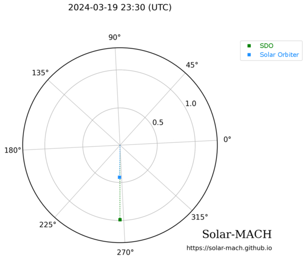
Positions of SDO (green) and Solar Orbiter (blue) during a solar flare on 19 March 2024. Click to enlarge. [Guastavino et al. 2026]
Guastavino’s team aligned and reprojected the data, accounting for the slightly different viewing angles of the two spacecraft, then applied an algorithm called Adaptive SE-DESAT to reconstruct the flux within the saturated portion of the SDO image. The team compared the desaturated SDO images to the unsaturated Solar Orbiter images over the course of the flare, finding the same flare morphology and similar time evolution between the two data sets.
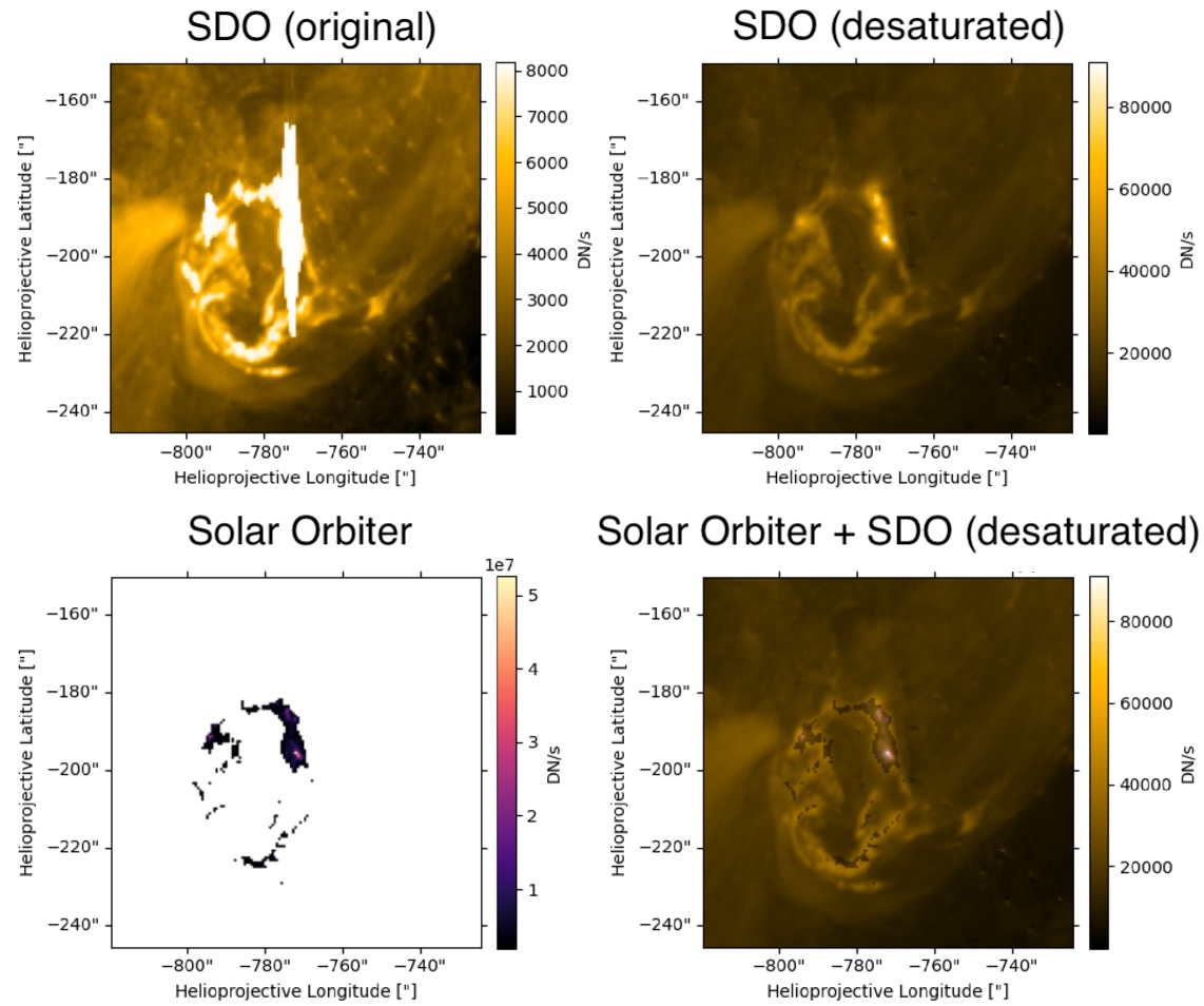
Example of unprocessed SDO data (top left), desaturated SDO data (top right), Solar Orbiter data (bottom left), and a combination of the two data sources (bottom right). Note that the Solar Orbiter observations have been clipped so that only the highest flare intensities were recorded. Click to enlarge. [Adapted from Guastavino et al. 2026]
Overall, these results demonstrate the effectiveness of the inversion technique, though Guastavino and coauthors noted that improvements could be made during the impulsive phase of the flare, when the emission is increasing. The team identified several reasons why the results may not match during this phase, including rapid evolution of the flare during individual exposures.
Despite these areas that require further attention, the validation of this technique unlocks 16 years of solar flare observations from SDO. This especially enhances our sample of powerful solar flares, increasing the available records of the most energetic solar flares by orders of magnitude.
“Validation of an Extreme-Ultraviolet Desaturation Technique for the Atmospheric Imaging Assembly on Board the Solar Dynamics Observatory Using Observations from the Extreme Ultraviolet Imager on Board Solar Orbiter,” Sabrina Guastavino et al 2026 ApJL 1005 L50. doi:10.3847/2041-8213/ae7d2e
The post When the Spacecraft Align: Rescuing Saturated Images of Solar Flares appeared first on AAS Nova.
Neuroscientists have long believed that language processing was only found in regions of the brain’s left hemisphere, including the well-known Broca’s area. However, new evidence suggests the brain’s language network is far more widespread than previously thought.
Researchers at the Massachusetts Institute of Technology (MIT) analyzed functional magnetic resonance imaging (fMRI) scans from 772 participants and identified 17 additional brain regions involved in language comprehension. These newly discovered areas are distributed across the brain, including the cerebellum, hippocampus, amygdala, and several regions of the cerebral cortex. In total, this accounts for five percent of the adult brain volume (roughly the size of a large strawberry).
“Even though there are all these distant components, it’s pretty restricted in terms of volume. You don’t need that much of the brain to do language,” says senior author Evelina Fedorenko, an MIT associate professor of brain and cognitive sciences and a member of MIT’s McGovern Institute for Brain Research, in a statement.
To find these hidden language-related regions in the brain, the researchers revisited years of brain imaging data collected since 2013. Additionally, participants completed a standard “language localizer” task, during which they read or listened to meaningful sentences and sequences of meaningless nonwords. By comparing brain activity between these two conditions, the research team identified regions that responded more strongly to language.
“When we initially started looking at language, in the first couple of papers, I tried to be comprehensive and include anything that seemed consistent across participants, and there was a huge amount of resistance,” said Fedorenko. “People would say things like, ‘Well, we know those are not language areas, so please focus on the language areas.’”
The team decided to adopt a more flexible statistical approach to detect weaker signals and identify previously overlooked language-sensitive regions beyond the classical language areas. They then compared these regions’ responses during a spatial working memory task to distinguish language-specific areas from those involved in what would be considered general cognitive functions.
Interestingly, five of the newly identified language regions are located in the cerebellum, a structure traditionally associated with movement and coordination. Some of these cerebellar regions were also active during non-language tasks, suggesting they may help integrate information across different brain systems rather than process language alone.
“It’s a very simple paradigm that lets you identify this core language system in individual brains,” says MIT postdoc Agata Wolna, the lead author of the paper.
Additional language-sensitive regions were identified in the hippocampus and amygdala, areas better known for their roles in memory and emotion. Their involvement suggests that understanding language may rely more on integrating memory, emotional context, and higher-order cognitive functions than previously understood in science.
“We always see this frontal temporal network, but there’s quite a lot of evidence that there are other regions that are also critical for language processing,” said Wolna. “By using a laxer threshold and zooming in on areas with weak MRI signal, we tried to maximize the chances of finding small and weakly responsive regions outside of this left frontal temporal system.”
The findings challenge long-held assumptions about the brain’s organization and can open new avenues for research into how language processing is understood in neuroscience.
The recent study was published in the Journal of Neuroscience.
Chrissy Newton is a PR professional and the founder of VOCAB Communications. She currently appears on The Discovery Channel and Max and hosts the Rebelliously Curious podcast, which can be found on YouTube and on all audio podcast streaming platforms. Follow her on X: @ChrissyNewton, Instagram: @BeingChrissyNewton, and chrissynewton.com. To contact Chrissy with a story, please email chrissy @ thedebrief.org.
Enzymes and other biomolecules are often anchored onto solid materials so they can be reused, purified more easily, and made more stable, a strategy biotechnology has relied on since industrial-scale immobilized enzymes first appeared in chemical manufacturing in the mid-twentieth century. Immobilized enzymes today drive processes from food and pharmaceutical production to biosensors and medical diagnostics. Fixing a biomolecule in place, however, is not as simple as gluing it down. The exact spot where it attaches, the direction it faces, and how much freedom it retains to move can all change how well it works. Testing every possible arrangement in the laboratory is slow and costly, which is why computer simulation has become such a valuable complement to experiments.
One of the main tools for this kind of study is molecular dynamics simulation, which calculates how atoms move over time by applying the basic laws of physics. Simulating every single atom of a protein, its surrounding water, and a supporting material gives rich detail, but is extremely demanding on computing power, especially when many attachment geometries need comparing. A widely used shortcut is coarse-grained modeling, in which small clusters of atoms are grouped into single, simplified particles. This keeps the essential physical behavior of the system while making the calculations far cheaper. The most established coarse-grained approach for biological molecules is the Martini force field, first developed in 2004 to describe fatty, membrane-forming molecules and later extended to proteins and, more recently, to a more accurate third generation of the model. Martini has since become a standard tool for simulating large biological assemblies that would otherwise be out of reach.
A team of researchers now introduces MartiniSurf 1, an open-source software package that automates the preparation of these coarse-grained simulations for biomolecules attached to surfaces. Building such a system by hand normally involves several separate programs and many manual steps, which makes it hard to reproduce results or compare different setups fairly. MartiniSurf instead combines the whole process into a single, streamlined workflow. Starting from the structure of a protein or a DNA strand, it converts the molecule into its coarse-grained form, builds a model of the supporting surface, positions the molecule in a chosen orientation, adds water and dissolved ions, and produces all the files needed to run the simulation with GROMACS, a molecular dynamics program that has been developed and refined for decades. Because every step is controlled by explicit settings rather than manual adjustments, two researchers using the same settings should obtain the same starting system, making comparisons across studies far more reliable.
A central feature of the software is its flexibility in representing how a biomolecule is attached. In one mode, selected regions of the molecule are held near the surface through simple restraining forces, without modeling the physical connector itself. In another mode, an explicit molecular linker is placed between the biomolecule and the surface, so that the linker’s own length and flexibility can influence the simulation. Because the properties of such linkers are known to affect how active and stable an attached enzyme remains, this second option offers a more realistic picture whenever the necessary coarse-grained description of the linker is available.
The supporting surfaces themselves can also be varied widely. The software can generate simple flat reference surfaces or draw on previously developed coarse-grained models of real materials such as graphene, graphite, carbon nanotubes, and agarose-like gels, the type of porous material commonly used to immobilize enzymes in the laboratory. Properties such as surface chemistry, electric charge, particle spacing, and added chemical groups can all be adjusted. Recent studies have shown that surfaces built without care can cause water molecules near the interface to line up in unrealistic, almost frozen patterns, an artifact rather than genuine physical behavior. The software includes options that let users introduce controlled irregularity into the surface to reduce this problem, while staying consistent with the wider Martini modeling framework.
To demonstrate how the approach works in practice, the study presents examples involving both proteins and DNA. For an enzyme attached to an agarose-like surface, the software reproduces several different attachment arrangements, including a complete system containing the enzyme together with its natural helper molecule and reaction substrate. These examples show how changing the attachment point or adding chemical groups to the surface can be explored systematically, without needing to reconstruct every model from scratch by hand.
The approach is also extended to DNA tethered to a graphene-like surface, comparing an electrically neutral surface with a positively charged one. The coarse-grained simulations reproduce the same general behavior previously reported in much more computationally demanding all-atom studies: the DNA strand remains mostly upright above the neutral surface but bends progressively toward the surface when it carries a positive charge, drawn in by electrostatic attraction. This comparison is not meant to reproduce exact experimental values, but rather to confirm that the automated setup captures the right physical trends, providing a check on the reliability of the workflow.
Every simulation generated by the software automatically records the settings, software versions, and modeling choices used to build it, so that another researcher can repeat or directly compare a given calculation later. Importantly, the software does not introduce new physical models or new force-field parameters of its own. Instead, it draws on already validated Martini tools and organizes them into a consistent, automated pipeline.
Overall, MartiniSurf addresses a practical bottleneck in computational chemistry rather than proposing a new simulation method. By automating the construction of surface-bound protein and DNA systems, it makes coarse-grained molecular dynamics studies faster to set up, easier to reproduce, and simpler to scale across many attachment geometries. This kind of tool should make it easier to systematically study how surface properties, attachment geometry, and molecular flexibility shape the behavior of immobilized biomolecules, supporting the design of better catalysts, biosensors, and other biotechnology applications.
Author: César Tomé López is a science writer and the editor of Mapping Ignorance
Disclaimer: Parts of this article may have been copied verbatim or almost verbatim from the referenced research paper/s.
The post Overcoming bottlenecks in bio-interface simulations: The MartiniSurf Approach appeared first on Mapping Ignorance.
In an era where environmental pollutants increasingly threaten public health, breakthrough scientific methodologies are emerging to decipher the complex puzzle of chemical exposures in everyday life. The U.S. Environmental Protection Agency (EPA), through its innovative Exposure Forecasting (ExpoCast) project, has unveiled a suite of high-throughput novel approaches that promise to revolutionize our understanding of human exposure to hazardous substances. These new approach methodologies integrate cutting-edge computational power, biomonitoring data, and machine learning to clarify how chemicals enter and navigate the human body via different exposure pathways.
At the core of this scientific leap is the urgent need to move beyond traditional, labor-intensive measures of exposure assessment. Conventional methods, reliant on direct environmental sampling or biomarker tracking alone, often provide fragmented or delayed insights. ExpoCast’s high-throughput frameworks address this by enabling the rapid processing of vast chemical datasets and synthesizing multifaceted exposure routes—dermal contact, inhalation, and ingestion—into cohesive predictive models. This integration forms the backbone of a new era in toxicological science, one that could empower preventive public health strategies with unprecedented accuracy and speed.
The advanced computational techniques deployed in this research pivot on two pivotal innovations. First, the ability to infer chemical intake directly from metabolite measurements represents a quantum leap in exposure science. Metabolites—the biochemical footprints left by toxicants within the body—offer a window into the internal chemical burden of an individual. This invertible analytic approach not only isolates the true extent of chemical accumulation but also contextualizes it within the dynamic processes of metabolism and excretion. Such resolution provides critical clues for both risk characterization and regulatory evaluation.
Complementing this metabolite-centric analysis are high-throughput models that simulate multi-pathway exposures. Unlike prior assessments that treated exposure routes in isolation, these models encapsulate the reality of simultaneous, overlapping chemical encounters encountered in everyday environments. By integrating data across air, dust, water, and dietary matrices, the models generate comprehensive exposure profiles that reflect the true cumulative burden experienced by populations. This holistic perspective marks a paradigm shift in exposure science, with far-reaching implications for epidemiological research and environmental policy.
Another cornerstone of the ExpoCast project’s success lies in its deployment of sophisticated machine learning (ML) algorithms to bridge pervasive data gaps. Chemical exposure data are notoriously incomplete, especially for emerging contaminants and complex mixtures. The machine learning methods exploit chemical structures and existing information repositories to predict missing values, refine exposure estimates, and identify likely high-exposure scenarios. Through these predictive analytics, the EPA’s approach dramatically enhances the resolution and reliability of exposure assessments without the untenable demands of large-scale physical sampling.
The strength of coupling high-throughput analytics with ML-based data gap filling is evident in a recently published case study application that specifically targets indoor environments. Indoor settings are recognized as critical exposure venues given the amount of time individuals spend inside homes, workplaces, and public buildings. In this case study, researchers synchronized biomarker findings from human subjects with media-specific chemical concentration measurements—dust, indoor air, and surface residues—to validate the accuracy and applicability of their models. The congruence observed underscores the potential of these next-generation methodologies to accurately capture exposure nuances in real-world scenarios.
Delving into the technicalities, the approach of inferring chemical intake from biomarker data involves mechanistic, pharmacokinetic models. These models simulate the absorption, distribution, metabolism, and elimination (ADME) of chemicals, allowing reverse calculation from measured metabolite concentrations back to estimated exposure doses. Integrating population variability and uncertainty quantification within these models further refines predictions, supporting more nuanced risk assessments that reflect real human diversity and exposure complexities.
High-throughput exposure models, meanwhile, employ automated workflows powered by large chemical libraries, exposure factor databases, and physiological parameters. These workflows rapidly generate exposure estimates across thousands of chemicals and scenarios, facilitating prioritization in regulatory contexts. The incorporation of multi-pathway assessments elevates these models beyond simple single-route frameworks, capturing interactive effects such as dermal absorption following inhaled volatilization, or ingestion arising from hand-to-mouth transfer of settled dust.
Machine learning strategies, spanning supervised and unsupervised techniques, utilize features derived from chemical structure descriptors, physicochemical properties, and known exposure pathways to fill data voids. These models can predict exposure likelihood, magnitude, and temporal patterns with quantifiable confidence. Importantly, the EPA team employed interpretability-focused ML algorithms to ensure that the models’ decision-making processes remain transparent to toxicologists and risk assessors, a critical factor for regulatory acceptance and scientific scrutiny.
Together, these novel methodologies embody a systems-level approach to understanding chemical exposures that integrates biological, environmental, and computational sciences. They represent a shift from descriptive monitoring to predictive and mechanistically informed exposure science, capable of anticipating emerging public health threats before widespread harm occurs. Such anticipatory capacity aligns with modern needs for proactive environmental health management amid ever-expanding chemical use.
The implications extend beyond academia or regulatory agencies. Public health practitioners, clinicians, and community advocates can leverage these insights to better understand exposure disparities, tailor interventions, and inform vulnerable populations. For instance, accurately predicting exposure levels in indoor environments helps prioritize remediation or behavior modification strategies for those disproportionately impacted by chemical pollutants, such as children or individuals in lower socioeconomic strata.
From an innovation standpoint, the ExpoCast project exemplifies the power of integrating novel in silico tools with empirical biomonitoring, fostering a virtuous cycle of model refinement and real-world validation. This iterative process ensures that computational predictions remain grounded in biological realities, enhancing confidence in exposure and risk evaluations. Furthermore, as chemical inventories expand and new compounds emerge, these scalable, automated methodologies will be indispensable for keeping pace with the evolving regulatory landscape.
The broader environmental science community stands to benefit from adopting similar high-throughput, machine-learning-enabled approaches. The generalizability of these methodologies means they can be adapted for global chemical monitoring programs, occupational health assessments, and ecosystem exposure analyses. Cross-disciplinary collaboration will be critical to extend these tools and harmonize data frameworks, advancing a unified, internationally coordinated approach to chemical safety.
In conclusion, the EPA’s ExpoCast-driven advancements represent a milestone in exposure science, knitting together innovative analytical methods to unravel the complexities of human-chemical interactions. By harnessing metabolite-informed intake reconstruction, multi-pathway exposure modeling, and machine learning-based data gap filling, the project paves the way for more accurate, timely, and insightful exposure assessments. This transformative toolkit holds promise to enhance chemical risk management, protect public health, and guide sustainable environmental stewardship in an increasingly chemical-dependent world.
The research outlined in the recent publication epitomizes the potential of high-throughput new approach methodologies to revolutionize exposure science across diverse settings—from homes and workplaces to broader communities. Continued development, validation, and deployment of such integrative frameworks will be paramount in confronting the escalating challenges posed by chemical exposures in the 21st century.
Subject of Research:
Development and application of high-throughput new approach methodologies for complex chemical exposure assessment, involving biomarker inference, multi-pathway exposure modeling, and machine learning-driven data gap filling.
Article Title:
Case study application of high-throughput new approach methodologies for exposure to the interpretation of matched biomarker and indoor media measurements.
Article References:
Jacketti, M.A., Biryol, D., Setzer, R.W. et al. Case study application of high-throughput new approach methodologies for exposure to the interpretation of matched biomarker and indoor media measurements. J Expo Sci Environ Epidemiol (2026). https://doi.org/10.1038/s41370-026-00926-y
Image Credits: AI Generated
DOI: 21 June 2026
In a groundbreaking study soon to redefine neuroscientific understanding of memory mechanisms, researchers have uncovered the pivotal role played by Neurensin-2 (NRSN2) in regulating hippocampal synaptic plasticity and memory functionality. This newly published research reveals how deficiency in NRSN2 leads to profound impairments in memory, traced back to disrupted excitatory synaptic transmission and weakened long-term potentiation (LTP) within the hippocampus, a brain region critically involved in memory formation and cognitive processing.
The hippocampus serves as the brain’s memory hub, integrating myriad neuronal signals to encode and retrieve information efficiently. Central to this integration is the function of excitatory synapses that employ glutamate receptors, especially the N-methyl-D-aspartate (NMDA) receptor, which plays an essential role in synaptic plasticity—the ability of synapses to strengthen or weaken over time. This plasticity underlies learning and memory. The study elucidates how NRSN2 exerts a regulatory influence on the expression of NMDA receptor subunits, with deficiency in NRSN2 tipping the balance unfavorably and compromising synaptic efficacy.
Utilizing advanced genetic models, the investigators engineered mice lacking NRSN2 to dissect its functional contributions. Behavioral assays showcased a marked decline in memory performance, particularly in spatial and recognition tasks reliant on hippocampal circuits. These deficits were not attributed to developmental anomalies but to disrupted synaptic physiology, signaling a direct role for NRSN2 in maintaining synaptic integrity and plasticity during adult cognition.
At the molecular scale, the absence of NRSN2 triggered a significant downregulation of key NMDA receptor subunits, notably NR1, NR2A, and NR2B. These subunits orchestrate receptor assembly and gating properties critical for calcium influx, a signal cascade necessary for potentiation of synaptic strength. The reduction in their expression correlated tightly with the inhibition of LTP induction, a process considered the cellular analog of memory encoding, confirming the hypothesis that NRSN2 deficiency impairs memory by hampering core synaptic mechanisms.
Electrophysiological recordings from hippocampal slices further substantiated the disrupted synaptic function. In control animals, robust LTP was readily induced by theta burst stimulation—a protocol mimicking physiological neuronal firing during learning. Conversely, hippocampi from NRSN2-deficient mice exhibited significantly weakened potentiation, underscoring the protein’s critical role in modulating synaptic responsiveness. This impairment was not a general defect of synaptic transmission but selectively impacted excitatory plasticity mediated through NMDA receptors.
Delving deeper, the team explored the intracellular cascades influenced by NRSN2, discovering that its absence perturbed the trafficking and surface expression of NMDA receptor complexes. NRSN2 appears to act as a chaperone or scaffold protein, ensuring proper receptor assembly and synaptic localization. Without it, receptor subunits fail to achieve adequate membrane insertion, resulting in diminished postsynaptic sensitivity to glutamate and compromised signal transduction.
This mechanistic insight offers a fresh perspective on how synaptic architecture and molecular composition govern cognition. The study’s integration of molecular biology, electrophysiology, and behavioral neuroscience draws a comprehensive picture of how a single protein’s deficit can destabilize the neural substrates of memory. Such findings not only enrich fundamental neuroscience but also hint at novel targets for cognitive-enhancing therapies.
Intriguingly, alterations in NRSN2 expression and NMDA receptor dysfunction have been implicated in various neuropsychiatric disorders, including schizophrenia and major depressive disorder. By illuminating this molecular nexus within the hippocampus, the research opens doors to understanding disease pathophysiology at a finer scale and may spur development of precision interventions aimed at restoring synaptic balance in affected individuals.
The investigators also examined compensatory mechanisms that might mitigate NRSN2 absence. While some plasticity-related proteins showed upregulation, these changes failed to rescue the deficits in NMDA receptor expression and synaptic potentiation. This highlights the non-redundant and indispensable nature of NRSN2 in maintaining synaptic homeostasis and cognitive function.
Beyond the laboratory findings, the study raises fascinating questions about how synaptic proteins coordinate to sustain the dynamic range of neural connectivity required for adaptive learning. The delicate equilibrium modulated by NRSN2 and NMDA receptors underscores the complexity of neuronal networks and their vulnerability when key molecular elements are compromised—insights that are pivotal as the field seeks to decode the neural basis of memory disorders.
Future research directions spawned by this work include investigating how NRSN2 interacts with other synaptic scaffold proteins and cytoskeletal elements, potentially shaping the three-dimensional organization of the postsynaptic density. Additionally, exploring the developmental timeline of NRSN2 expression could reveal critical periods during which intervention might prevent or reverse memory impairments.
From a translational perspective, the study’s revelations beckon the clinical neuroscience community to consider NRSN2 and its pathways as therapeutic candidates. Pharmacological strategies aimed at upregulating NRSN2 or mimicking its stabilizing effects on NMDA receptors could hold promise for enhancing cognition or slowing neurodegenerative progression in disorders marked by synaptic decline.
In sum, this landmark research defines NRSN2 as a vital regulator of excitatory synaptic plasticity and memory formation. Its deficiency causes a cascade of molecular disturbances culminating in impaired NMDA receptor expression and disrupted LTP, ultimately compromising hippocampal-dependent memory functions. This work not only advances our understanding of the molecular machinery underlying memory but also charts new territory in the quest to ameliorate cognitive dysfunction.
As neuroscience inches closer to mapping the intricate cellular and molecular landscape of cognition, findings such as these underscore the profound impact a single synaptic protein can have on brain function. The compelling evidence presented lays a robust foundation for future explorations into synaptic plasticity and memory, promising strides in both basic science and clinical applications that may soon transform lives burdened by memory loss.
Subject of Research: Impact of Neurensin-2 (NRSN2) deficiency on hippocampal memory functions and synaptic plasticity
Article Title: Impact of NRSN2 deficiency on memory: Altered excitatory synaptic plasticity associated with reduced expression of NMDA receptor subunits and impaired LTP in the hippocampus.
Article References:
Wei, J., Wu, Y., Yi, Y. et al. Impact of NRSN2 deficiency on memory: Altered excitatory synaptic plasticity associated with reduced expression of NMDA receptor subunits and impaired LTP in the hippocampus. Transl Psychiatry (2026). https://doi.org/10.1038/s41398-026-04163-7
Image Credits: AI Generated
In a groundbreaking departure from traditional approaches, researchers have unveiled a transformative perspective on EEG biomarkers for brain disorders, emphasizing a transdiagnostic dimensional framework that promises to revolutionize neuropsychiatric diagnostics and treatment. This pioneering study, conducted by Zebhauser, Heitmann, Henningsen, and colleagues, challenges decades-old conventions that pigeonhole brain disorders strictly according to categorical diagnostic criteria. Instead, it advocates for a more nuanced, continuum-based understanding of brain dysfunctions as reflected in electroencephalographic signals.
The conventional paradigm in neuropsychiatry has long relied upon discrete diagnostic labels—labels which often fall short in capturing the complex, overlapping symptomology exhibited across disorders such as schizophrenia, bipolar disorder, major depressive disorder, and anxiety disorders. Traditional EEG analyses have sought biomarkers confined within these diagnostic categories, leading to inconsistent results and limited clinical utility. The new study confronts these limitations by proposing that EEG biomarkers be analyzed through a dimensional lens that transcends categorical boundaries, enabling detection of underlying neural dysfunctions that span multiple disorders.
Central to this conceptual shift is the transdiagnostic approach, which posits that neurobiological aberrations are shared across different psychiatric conditions and manifest along spectrums of symptom severity and cognitive impairment. Through rigorous computational modeling and advanced signal processing applied to large EEG datasets, the research team delineated patterns of neural oscillatory activity and connectivity that correlate with continuous measures of cognitive and affective dysfunctions, irrespective of diagnostic categories. This approach underscores the shared neurophysiological substrates that traditional categorical classifications often obscure.
Methodologically, the study harnessed sophisticated machine learning algorithms to parse high-dimensional EEG data and extract signal features indicative of brain circuit dysregulation. By focusing on spectral power variations across frequency bands—delta, theta, alpha, beta, and gamma—as well as functional connectivity metrics between cortical networks, the researchers succeeded in identifying biomarkers reflective of symptom dimensions such as cognitive control deficits, emotional dysregulation, and sensory processing anomalies. These dimensions overlap across disorders, suggesting a common pathophysiological mechanism modulated along a continuum.
Importantly, the transdiagnostic dimensional framework enables more personalized and precise characterization of patients’ neural profiles. Instead of forcing diagnoses into rigid boxes, clinicians can now utilize EEG biomarkers to quantify an individual’s specific symptom constellation and severity in real-time. This precision facilitates tailored interventions that target dysfunctional brain networks directly, potentially enhancing treatment efficacy and reducing trial-and-error prescribing that often plagues psychiatric care.
The implications extend beyond diagnosis and treatment optimization. This novel EEG biomarker approach offers a powerful tool for preventative psychiatry by identifying at-risk individuals who may not yet meet full diagnostic criteria but exhibit measurable neural dysfunctions indicative of emerging disorders. By mapping these dimensional brain signatures early, clinicians can intervene preemptively, possibly altering disease trajectories before chronicity sets in.
Moreover, this paradigm fosters an integrative understanding of brain disorders as dynamic states rather than static labels. The EEG biomarkers track fluctuation in cognitive and affective states, capturing the temporal evolution of illness and response to therapy. Such temporally sensitive biomarkers pave the way for real-time monitoring of treatment response and disease progression via non-invasive methods, enhancing clinical decision-making and patient outcomes.
The study also confronts several technical challenges historically hindering EEG’s clinical translation. By developing robust preprocessing pipelines that mitigate artifacts and employing cross-validation techniques to ensure replicability of findings across diverse cohorts, the researchers set new standards for EEG research rigor. Their comprehensive methodology serves as a blueprint for future investigations aiming to unify neurophysiological data with psychiatric phenotypes in a clinically meaningful manner.
Furthermore, by adopting a transdiagnostic dimensional view, the research opens novel avenues for drug development. Pharmaceutical interventions can be engineered to target neural circuits and pathways implicated across multiple disorders, possibly leading to broader spectrum therapeutics with enhanced efficacy. This contrasts with the current trend of developing highly disorder-specific drugs, which may only benefit a subset of patients.
Underpinning this research is the recognition that brain disorders are multifactorial and multifaceted, involving intricate interactions between genetics, environment, and neural circuitry. The EEG biomarkers illuminated by this study reflect emergent properties of large-scale brain networks whose dysfunction cuts across traditional diagnostic divides. This holistic lens aligns with contemporary theories of brain function as distributed and dynamic, rather than localized and static.
Clinically, the transdiagnostic dimensional EEG biomarkers facilitate identification of subtypes within disorders, based on distinct neurophysiological profiles. Such stratification could improve prognostic accuracy and enable stratified clinical trials, ultimately accelerating discovery and application of targeted therapies. This personalized medicine approach marks a considerable leap in the field of psychiatry.
The study’s findings also resonate deeply with the Research Domain Criteria (RDoC) initiative advocated by the National Institute of Mental Health, which promotes research centered on fundamental dimensions of functioning rather than syndromic categories. The demonstrated EEG biomarkers provide tangible neurobiological correlates for RDoC constructs such as cognitive systems and arousal/regulatory systems, making a compelling case for their adoption in clinical and research frameworks.
Ethical and practical considerations emerge as these new EEG biomarkers are integrated into clinical practice. Ensuring equitable access to advanced neurodiagnostic technologies and protecting patient privacy with respect to neural data will be paramount. The authors acknowledge these issues and call for development of guidelines and safeguards alongside technological progress to maximize benefit and minimize harm.
This research heralds a new era in neuropsychiatry, where neural biomarkers derived from accessible EEG recordings become central to understanding, diagnosing, and treating brain disorders as dimensional phenomena. By transcending rigid diagnostic boundaries, this approach reflects the complex reality of brain function and dysfunction, offering hope for more effective, individualized care for millions worldwide impacted by mental illness.
In conclusion, the visionary framework proposed by Zebhauser and colleagues challenges entrenched conventions and illuminates a path forward marked by dimensional precision, cross-disorder integration, and clinically actionable EEG biomarkers. This work stands to reshape the landscape of psychiatric neuroscience and catalyze a shift toward truly personalized mental health care.
Subject of Research: EEG biomarkers and their application in a transdiagnostic dimensional framework for brain disorders
Article Title: Rethinking EEG biomarkers of brain disorders: a transdiagnostic dimensional view
Article References: Zebhauser, P.T., Heitmann, H., Henningsen, P. et al. Rethinking EEG biomarkers of brain disorders: a transdiagnostic dimensional view. Transl Psychiatry 16, 316 (2026). https://doi.org/10.1038/s41398-026-04187-z
Image Credits: AI Generated
DOI: 10 June 2026
Researchers built Sashimi-Bot to master one of robotics’ slipperiest challenges.In a groundbreaking study that redefines our understanding of how language is encoded in the human brain, researchers have mapped the diverse landscape of neurons distributed across frontal and temporal cortical regions, revealing a complex yet lateralized neural architecture for human language processing. By leveraging advanced single-neuron recording techniques alongside sophisticated language models, the team has identified not just the locations but the nuanced encoding properties of neurons implicated in various linguistic computations. This work illuminates how language, a uniquely human faculty, is instantiated at the neuronal level with precision and regional specificity.
The study’s comprehensive neuronal recordings spanned key brain areas historically associated with language, including the frontal cortex, anterior temporal cortex, and posterior temporal cortex. These regions were chosen given their established roles in speech production and linguistic comprehension, as well as their demonstrated selective responses in prior localized brain imaging studies. Importantly, the neurons examined were broadly distributed across both hemispheres and these cortical regions, showcasing the widespread neural substrate involved in language tasks.
One of the seminal findings was that roughly half of the neurons, 289 out of 579, exhibited selective responsiveness to one or more linguistic features. Intriguingly, this selectivity was evenly distributed across the three cortical sites — frontal, anterior temporal, and posterior temporal — and did not significantly differ between the left and right hemispheres. This suggests a foundational neural scaffold where language-related computations recruit a multitude of neurons across large-scale networks rather than isolated hotspots, emphasizing distributed processing over modular localization.
However, beyond this broad distribution, the study uncovered striking lateralization and regional differentiation in the strength and informational content of neural modulation. Neurons within the left hemisphere exhibited significantly stronger modulation—quantified by their z-scored activity changes—in response to linguistic features compared to their right hemisphere counterparts. This enhanced sensitivity encompassed a range of linguistic elements, including lower-order features like pitch, underscoring the superior role of the left hemisphere in detailed linguistic encoding.
Further dissecting these lateralized effects across cortical regions revealed that the posterior temporal cortex showed the most pronounced difference in left-right modulation strength, significantly outperforming other areas. This aligns well with the notion that posterior temporal areas are central to decoding complex linguistic dependencies and syntactic structures, functions critical for fluent language comprehension and production.
Conversely, the prefrontal cortex displayed the strongest overall neuronal modulation to these language features, both in the left and right hemispheres, compared to temporal regions. This finding suggests a hierarchical and possibly integrative role for the frontal cortex in orchestrating language processing, perhaps linked to higher-order syntactic planning, working memory, or executive control mechanisms required for natural speech.
The neural predictivity analysis, leveraging embeddings from cutting-edge contextual language models, further corroborated the lateralized and regionalized pattern. Neural activity in the left anterior temporal cortex was most accurately predicted by these language model features, highlighting this region’s role in abstract linguistic representation and semantic integration during language tasks.
Methodologically, the study capitalized on the precise temporal resolution of single-neuron recording combined with sophisticated statistical analyses, including permutation and rank-sum tests, to quantify neuronal response selectivity and modulation. This rigorous approach afforded a fine-grained characterization of how individual neurons adapt their firing patterns to various syntactic and prosodic elements of language in real-time.
Collectively, these data challenge conventional notions that neural language processing is confined to narrowly defined “language centers.” Instead, the findings advocate for a model of language representation as a widely distributed but regionally specialized system with marked hemispheric dominance. This has profound implications for understanding the neural basis of language disorders, suggesting that therapeutic interventions might need to consider both distributed network integrity and hemispheric targeting.
Moreover, the study’s integration of naturalistic language models with neurophysiological data marks a pioneering step in computational neuroscience, bridging theoretical frameworks of language with tangible neural mechanisms. By showing how models trained solely on linguistic input can approximate neuronal responses, this research points toward a future where artificial intelligence and neuroscience synergistically unravel human cognition.
Importantly, the results underscore the unique neural architecture supporting language that has evolved in humans, outstripping earlier simplistic models focused on gross anatomical mapping. These advances set the stage for future investigations that might explore how these neuronal populations interact dynamically during complex language tasks such as conversation, narrative comprehension, or bilingual processing.
In conclusion, this landmark research paints a detailed, high-resolution portrait of the neuronal building blocks underpinning human language. It reveals an exquisitely lateralized and regionally differentiated system whereby distributed neurons collectively encode the multifaceted features of speech. Such insights not only deepen fundamental neuroscience but also hold promise for novel clinical interventions and computational linguistic advancements.
Subject of Research: Neuronal encoding of human language features across frontal and temporal cortical regions; lateralization and regional differentiation of linguistic processing in the brain.
Article Title: Mapping the neuronal building blocks of human language with language models.
Article References:
Cai, J., Kfir, Y., Jamali, M. et al. Mapping the neuronal building blocks of human language with language models. Nature (2026). https://doi.org/10.1038/s41586-026-10691-5
Image Credits: AI Generated
A groundbreaking advancement in photonic technology promises to redefine the landscape of medical artificial intelligence (AI) diagnostics, as a research consortium led by Professor Han Zhang at Shenzhen University has unveiled a novel all-fiber photonic AI platform utilizing black phosphorus (BP)-based tunable modulators. This innovative approach, which hinges on photon-based computing, surmounts the intrinsic constraints posed by traditional electronic processors, delivering unprecedented energy efficiency and ultrafast performance crucial for real-time clinical decision-making.
The prevailing paradigm in medical AI, deeply reliant on electronic architectures such as GPUs, faces severe limitations related to energy consumption, heat dissipation, and processing latency. These factors collectively impede the integration of AI into time-sensitive diagnostic workflows and exacerbate systemic carbon footprints. Photonic computing, with its reliance on photons rather than electrons, offers dramatically accelerated data transmission speeds and parallelism through wavelength multiplexing, alongside near-zero heat generation. Until now, however, the practical implementation of photonic AI had been hampered by the bulkiness, inefficiency, and fabrication challenges synonymous with conventional optical modulators.
Professor Zhang’s team has ingeniously engineered an atomically thin van der Waals heterostructure by stacking black phosphorus and molybdenum disulfide (MoS2), leveraging the extraordinary optoelectronic properties of these 2D materials. This heterostructure is meticulously integrated onto a microfiber knot resonator (MKR), an ultra-fine looped optical fiber with a diameter narrower than a human hair. This configuration significantly intensifies the interaction between light and material, enabling modulation of the refractive index with minimal applied voltage, which shifts the light’s wavelength to encode computational information with remarkable precision and energy economy.
The fabrication process commences with mechanical exfoliation to obtain pristine BP/MoS2 bilayers, followed by a dry transfer technique to position these heterostructures onto the MKR. The MKR’s torsional geometry enhances evanescent field coupling, ensuring that the optical mode overlaps maximally with the active layered materials. To broaden the modulator’s linear response and suppress nonlinear distortions, the researchers incorporated a Ring-Assisted Mach–Zehnder Interferometer (RAMZI) design. This integration extends the operational voltage range, ensuring fidelity and robustness essential for medical-grade AI computations.
The team transcended single-device innovation by architecting a full all-fiber photonic neural network (PNN). By deploying two RAMZI modulators in tandem with a highly sensitive photoreceiver within a sophisticated time-division multiplexing schema, they realized a closed-loop, coherent computing unit capable of executing neural network operations entirely in the optical domain. This milestone substantiates the transition of photonic neural networks from theoretical constructs to viable, deployable platforms for clinical diagnostics.
Clinical performance validation was conducted in collaboration with esteemed hospitals. The PNN demonstrated expert-level diagnostic accuracy in two stringent medical tasks: detecting retinal detachment from B-scan ultrasound imagery and diagnosing hepatocellular carcinoma (HCC) using multiphase liver computed tomography (CT) scans. Diagnostic accuracy and specificity metrics paralleled those of seasoned radiologists, underscoring the system’s potential for integration into frontline healthcare.
Beyond accuracy, the photonic system showcased transformative efficiency advantages. While a conventional NVIDIA A100 GPU requires approximately 85 milliseconds to process a liver CT study, the photonic platform accomplished the same in a mere 0.8 milliseconds. Additionally, the energy expenditure per operation plummeted to an astonishing 0.608 femtojoules, a staggering 246-fold decrease compared to electronic counterparts consuming 150 femtojoules. This synergy of speed and efficiency paves the way for deploying AI diagnostics in resource-constrained or mobile medical environments like rural clinics and ambulances, dramatically expanding access to critical health services.
The system’s environmental implications cannot be overstated. Conventional AI’s sizable energy footprint contributes appreciably to healthcare’s carbon emissions. The BP/MoS2-based photonic platform fundamentally disrupts this model by combining ultrafast processing with near-negligible thermal dissipation, forging a sustainable pathway for green AI implementations in medicine. This breakthrough aligns with expanding global commitments to environmentally responsible technologies without sacrificing computational power.
Acknowledging the infancy of current implementations, the authors outline avenues for scaling. Presently limited to a single processing layer realized through two modulators, the system’s complexity and image resolution capacity necessitate expansion. A promising strategy involves wavelength-division multiplexing (WDM), exploiting the device’s ∼30 nm bandwidth resonances to channel up to 40 distinct wavelengths. This parallelism would amplify computational density approximately 40-fold while maintaining the existing clock rate, facilitating thousands of multiplications per network layer—an enhancement critical to tackling high-dimensional medical image data.
Device longevity and stability pose additional challenges, given the sensitivity of BP to environmental degradation. While the MoS2 layer affords interim encapsulation, the team plans industrial collaboration to pioneer large-scale protective coatings via atomic layer deposition of Al2O3 and scalable chemical vapor deposition processes. These advancements will bolster device durability, uniformity, and manufacturability, accelerating clinical adoption and commercial viability.
The study also signifies the maturation of fiber-based photonic neural networks into practical diagnostic platforms rather than experimental novelties. Optical fibers’ inherently low transmission loss (<0.2 dB/km), synergized with the modulator’s efficient 0.25 V·cm electro-optic response and expanded linear range, addresses critical bottlenecks impeding prior photonic computing endeavors. Consequently, the technology promises to empower future applications in drug discovery, genomics, and real-time clinical imaging with unmatched energy footprints.
The pioneering work led by Professor Han Zhang not only foregrounds interdisciplinary collaboration across physics, materials science, and biomedicine but also sets a precedent for industry-academia partnerships catalyzing the commercialization of ultrafast photonic devices poised to transform healthcare AI. As research progresses towards integrated multi-layer architectures and industrial encapsulation, the healthcare sector stands on the cusp of embracing a new generation of diagnostic tools that are faster, greener, and broadly accessible.
This heralds a paradigm shift where medical AI transcends the limitations inherent to electronic systems, embracing photon-driven computing as a sustainable, high-performance alternative that aligns with the twin imperatives of clinical excellence and environmental stewardship.
Subject of Research: People
Article Title: Tunable phosphorene modulators – accelerating medical diagnosis with ultra-efficient photonic platforms
News Publication Date: 28-May-2026
References:
Zhang, H., Liu, Y., Wang, H., Zhu, H., et al. (2026). Tunable phosphorene modulators – accelerating medical diagnosis with ultra-efficient photonic platforms. Opto-Electronic Advances. DOI: 10.29026/oea.2026.250332
Image Credits: Professor Han Zhang of Shenzhen University
Keywords: photonic computing, black phosphorus, molybdenum disulfide, van der Waals heterostructure, microfiber knot resonator, electro-optic modulator, photonic neural network, medical imaging AI, energy efficiency, wavelength-division multiplexing, ultrafast diagnostics, sustainable healthcare
