❌

Normal view

The bacteria behind gum disease may also harm your heart valves, early research suggests

20 July 2026 at 21:15

A common culprit behind severe gum disease may also stiffen and narrow the heart's aortic valve, preliminary research suggests. This may help explain links between gum problems and poor heart health.

Scientists have long linked severe gum disease to inflammation throughout the body and a higher risk of heart problems, including coronary artery disease, which can raise the risk of heart attack. Studies have also found that treating gum disease can improve blood-vessel health.

Now, researchers suggest that Porphyromonas gingivalis, one of the main bacteria involved in severe gum disease, may also contribute to calcific aortic valve disease (CAVD), a condition in which calcium builds up in the heart's aortic valve. The findings were presented in July at a meeting of the American Heart Association in Boston and have not been peer-reviewed yet.

Dr. Elena Aikawa, a professor of medicine at Harvard Medical School who was not involved in the study, said the findings "fit well with our current understanding of calcific aortic valve disease as an active, inflammation-driven disease rather than simply wear and tear of the valve."

Periodontal disease is likely just one contributor among many, rather than the sole cause of CAVD, she added. But the study does identify a biologically plausible pathway linking oral health to heart health that could open new opportunities for prevention and treatment.

How mouth bacteria harm the heart

CAVD affects millions of people worldwide, and there are no approved drugs that slow or stop its progression. As the disease worsens, people may develop fatigue, shortness of breath and chest pain, and in severe cases, they often require valve-replacement surgery.

Meanwhile, prior to the new research, it was known that bacteria and inflammatory signals from the mouth can sometimes enter the bloodstream and affect distant tissues, said Richard Lamont, chair of oral immunology and infectious diseases at the University of Louisville School of Dentistry, who was not involved in the study. Over time, these effects can accrue and gradually reshape how the immune system behaves, he said.

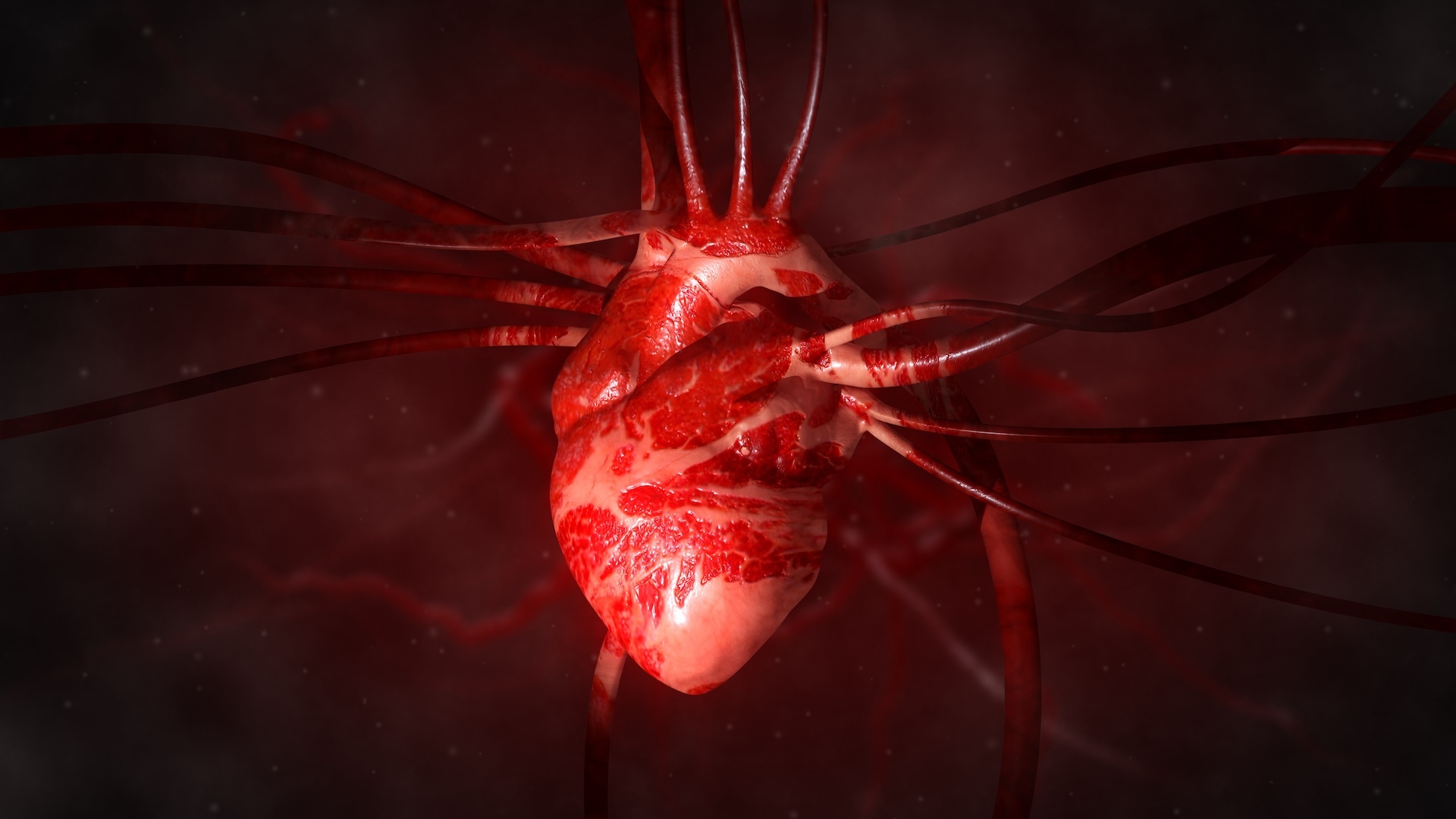
An illustration of a human heart

In CAVD, calcium builds up in the aortic valve, which connects the left side of the heart to the large artery that carries oxygen-rich blood to the body. (Image credit: Noctiluxx via Getty Images)

P. gingivalis is known to be particularly destructive to gum tissue, causing lots of inflammation, and it's been tied to cardiovascular disease in the past. To see if the bacterium might be directly impacting heart health, researchers at Fuwai Hospital in Beijing looked for P. gingivalis in aortic valve tissue removed during valve-replacement surgery. They compared this diseased tissue to valves that weren't calcified.

The calcified valves contained much higher levels of P. gingivalis DNA and proteins made by the bacterium. "P. gingivalis was about 30 times more abundant in calcified valves than in non-calcified valves," study first author Dr. Chenyang Li, a doctoral candidate at Fuwai Hospital, told Live Science in an email.

To test whether the bacteria might directly contribute to valve damage, Li and colleagues injected live P. gingivalis into the bloodstreams of healthy lab mice. The bacteria reached the animals' aortic valves, triggering inflammation, calcium buildup and narrowing of the valve.

"We were surprised to see valve calcification even in mice without high cholesterol," Li said. "This suggests P. gingivalis may directly contribute to valve disease, beyond traditional cardiovascular risk factors."

A comparison group of mice was given antibiotics before the injection and had less bacteria in their aortic valves and slower disease progression. Meanwhile, mice injected with dead P. gingivalis bacteria did not show any changes in their aortic valves.

The researchers traced the changes in the mice's hearts to interleukin-1 beta, a signalling molecule that normally helps the body fight infections. Exposure to P. gingivalis ramped up the production of this molecule, and in turn, the immune signal switched on genes that pushed healthy valve cells to behave more like bone-building cells, which is a hallmark of CAVD. This switch causes the cells to lay down calcium in the surrounding valve tissue.

When the scientists blocked interleukin-1 beta in some mice, the animals developed much less valve calcification even when the bacteria reached the valve. Lamont said that the mouse experiments strengthen the case that there's a biological mechanism directly linking gum and heart diseases. However, he cautioned that mice have very different oral microbiomes than humans, making it difficult to know whether the exact same process occurs in people.

That said, the team also saw the same pattern in human valve cells grown in the lab. Exposure to P. gingivalis increased inflammation and calcium buildup among the cells, while blocking interleukin-1 beta blunted those effects.

Both Aikawa and Lamont said more research is needed to confirm the relationship in humans. The findings give researchers a new pathway to explore for future treatments, raising questions of whether targeting inflammatory signals like interleukin-1 beta could help slow the progression of this widespread heart disease.

This article is for informational purposes only and is not meant to offer medical advice.

What do you know about the body's hardest-working muscle? Find out with our heart quiz!

New robotic heart mimics common, mysterious condition to help researchers study it

Scientists have invented a soft robotic heart that could give them a new way to study a mysterious condition that accounts for roughly half of all heart failure cases.

This form of heart failure, called heart failure with preserved ejection fraction (HFpEF), is a condition in which the heart pumps out a normal amount of blood but becomes too stiff to relax and refill properly between beats. Over 3 million Americans have the condition, but researchers still don't fully understand why it develops, making it difficult to design treatments that directly target the disease.

The new device, described June 1 in the journal Nature Communications, is the first soft robotic heart model that can actively adjust how it responds to changes in pressure. It tightens or relaxes its artificial muscles, enabling researchers to control how stiff the heart becomes. This allows it to better mimic a diseased human heart.

Current laboratory models of HFpEF have drawbacks. Traditional "mock circulation loops" use rigid pumps and tubing to recreate blood flow but don't physically resemble a beating heart. Lab animals capture more realistic biology but are expensive and don't perfectly mirror human disease, said study co-author Thanh Nho Do, a biomedical engineer and associate professor at the University of New South Wales in Australia.

"HFpEF has been notoriously difficult to study and treat," Nho Do told Live Science in an email.

Earlier versions of the robotic heart could realistically recreate cardiac movement, but they followed present commands that didn't adapt to changing conditions. The new system attempts to solve that limitation by allowing the artificial muscle fibers to "feel" the pressure created by the fluid they are pumping and dynamically contract or relax against it.

That feedback is critical for reproducing HFpEF, in which abnormal stiffness β€” rather than weakened pumping β€” lies at the core of the disease.

The researchers built a silicone replica of the left side of the human heart and wrapped it in artificial muscle fibers made from rubber tubes reinforced with spring coils. The model continuously senses the pressure generated by the fluid flowing through it and adjusts the force of its artificial muscles in response. By changing how much the muscles resist being stretched as the heart fills, researchers can dial in different levels of stiffness.

Instead of modeling a single snapshot of HFpEF, the researchers recreated several stages of the disease's progression. In early stages, patients' hearts start to show impaired relaxation between beats, while in more advanced forms, the heat is so stiff that it can't adequately fill with blood before the next contraction.

"If we can model its progression pathway, we might be able to use models like ours to develop medical devices that interrupt that trajectory, rather than just treating end-stage disease," Nho Do said.

Current treatment for HFpEF focuses largely on managing symptoms and associated conditions, such as high blood pressure, obesity and diabetes. In recent years, drugs known as SGLT2 inhibitors have been shown to reduce the risk of hospitalization in many patients by reducing excess fluid in the body. But there are still few therapies that directly target the stiffening of the heart muscle.

The researchers hope their new model might pave the way, but they emphasized that the work is an early proof of concept. Looking ahead, they hope to develop increasingly sophisticated robot heart models that can complement other data on HFpEF, including that from computer simulations, animal studies and clinical testing.

"We hope the biggest impact will be in understanding HFpEF mechanisms and improving the way we develop cardiovascular devices," Nho Do said.

Heart quiz: What do you know about the body's hardest-working muscle?

Heart issues tied to 'microdamage' in the brain might raise risk of memory loss, study hints

6 July 2026 at 22:20

Scientists uncovered a link between mild heart trouble and microscopic signs of brain damage that may raise the risk of memory problems down the line.

The new study, published Monday (July 6) in The Journal of Neuroscience, did not prove that the heart issues directly caused memory problems. But it "adds to the overall picture that preserving healthy brain-heart communication is key for healthy aging," said Dr. Jan Scheitz, a consultant stroke neurologist and head of the Brain-Heart Lab at the CharitΓ© University Hospital in Germany, who was not involved in the study.

"A healthy lifestyle that protects the cardiovascular system will not only benefit the heart but also the brain," he told Live Science in an email.

The work could one day help doctors identify patients at risk of memory problems early on through routine heart-function tests, said study co-author Dr. Xia Zhang, a doctoral researcher at the Max Planck Institute for Human Cognitive and Brain Sciences in Germany.

"The broader implication is that the brain may show subtle tissue-level changes related to cardiac dysfunction before we see obvious brain shrinkage or clinical dementia," Zhang told Live Science in an email.

Early warning signs

Researchers already knew that heart and brain function are closely interconnected. Heart diseases such as heart failure, atrial fibrillation and heart attack impair communication between the heart and the brain. This can contribute to thinking problems and dementia by restricting blood flow to the brain and causing chronic inflammation.

So far, though, scientists have only studied changes in the brain in people already diagnosed with heart conditions. They haven't necessarily assessed the changes that take place earlier on.

In the new study, researchers followed 73 patients who had sought care for heart-related symptoms at the Heart Center Leipzig in Germany, some of whom had confirmed coronary artery disease and/or heart failure. They also assessed 95 people without any heart-related symptoms. The researchers measured heart function at the start of the study. Then, about 3.5 years later, they performed cognitive testing and an MRI scan of the brain to take a snapshot of its structure.

Among all 168 study participants, those whose hearts pumped blood less efficiently at the start of the study showed greater signs of tissue damage in their gray matter years later. This was true regardless of whether or not they had diagnosed heart failure.

The cognitive tests were given only to the heart-disease patients, and they assessed attention, executive function, learning and memory. Only memory showed a link to weaker heart pumping. The participants with weaker heart pumping showed more microscopic damage in memory-related brain regions, and those brain changes correlated with worse memory scores.

The degree of brain damage also correlated with the levels of stress hormones released by the heart, but this link was only seen in patients who already had heart failure.

"What surprised us most" was that subtle reductions in the heart's ability to pump were tied to later brain changes, even in patients who did not meet clinical criteria for heart failure, Zhang said.

One limitation of the study is that the researchers didn't take MRI scans at the start. That means "we do not know whether some changes were already present at the beginning of the study period," Scheitz noted. This is something that Zhang and her colleagues plan to test in future studies.

"The next step for the field is replication in larger cohorts with multiple time points, so that cardiac function, brain microstructure, and cognition can be followed more precisely over time," she said.

The brain damage flagged in the 73 patients happened in parts of the brain that are important for memory and are vulnerable in Alzheimer's disease. "These regions helped identify a possible brain pathway through which poorer cardiac function may contribute to later memory problems," Zhang said.

However, until this idea is tested directly in future studies, it is too soon to say if the heart function-related brain changes seen on the MRI scans point to a higher risk of Alzheimer's, Zhang cautioned. The team did not measure proteins related to Alzheimer's, so they cannot conclude that the patients with measurable brain changes are developing Alzheimer's disease.

The study also did not directly test the effect of exercise on heart and brain health, but the findings may help explain why regular exercise is often linked to better brain health and cognitive aging, Zhang said.

"Regular exercise supports cardiovascular function, vascular health, and cerebral blood-flow regulation," she said, "all of which may help protect brain tissue over time."

This article is for informational purposes only and is not meant to offer medical advice.

What do you know about the body's hardest-working muscle? Find out with our heart quiz!

❌