Reading view

Next-Generation PET Tracer Revolutionizes Rapid, High-Precision Kidney Cancer Detection

A groundbreaking advancement in molecular imaging has emerged from recent clinical research, unveiling a novel PET tracer that targets carbonic anhydrase IX (CAIX) with remarkable precision. This innovative radiotracer, designated as ^68Ga-RCC78, has exhibited exceptional sensitivity in detecting clear cell renal cell carcinoma (ccRCC), a malignancy known for its aggressive nature and diagnostic challenges. The development of ^68Ga-RCC78 represents a pioneering step toward enhanced staging and personalized management of kidney cancer, as presented at the Society of Nuclear Medicine and Molecular Imaging (SNMMI) 2026 Annual Meeting.

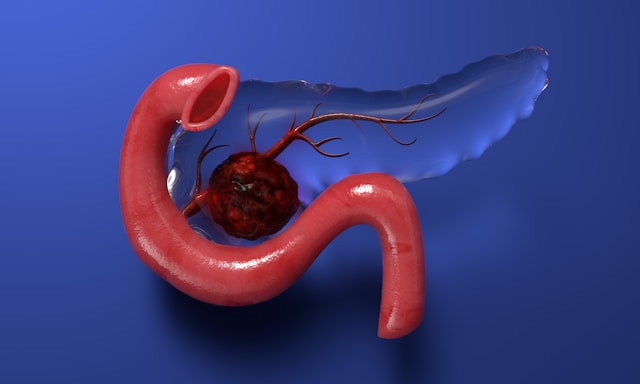
Clear cell renal cell carcinoma is characterized by the distinctive and constitutive overexpression of CAIX, a transmembrane protein involved in pH regulation within the tumor microenvironment. This pathological overexpression makes CAIX a highly attractive target for molecular imaging agents seeking to discern malignant lesions amidst the complex anatomical structures of the abdomen. Until now, molecular imaging probes targeting CAIX have been hampered by significant physiological expression in the gastrointestinal tract, resulting in high background signals that obscure tumor visualization and compromise diagnostic accuracy.

The novel ^68Ga-RCC78 tracer overcomes these limitations through the use of a uniquely engineered cyclic peptide that binds specifically to CAIX with high affinity. Unlike traditional antibody-based tracers requiring prolonged clearance times extending over days, ^68Ga-RCC78 achieves rapid accumulation in tumor tissues while simultaneously minimizing non-specific background uptake. This rapid pharmacokinetic profile not only accelerates imaging timelines but also markedly improves tumor-to-background contrast, a vital factor in identifying metastatic deposits.

The development process began with the synthesis of sixteen novel CAIX-specific cyclic peptides, each radiolabeled with the positron-emitting radionuclide gallium-68 (^68Ga). Cellular uptake assays systematically evaluated tracer affinity and specificity across cell lines with high and low CAIX expression, alongside blocking studies to confirm target-mediated binding. Subsequent in vivo evaluations entailed extensive PET/CT imaging and biodistribution analyses in ccRCC xenograft models and patient-derived xenografts, providing critical insights into tracer dynamics and tumor delineation.

Among the candidates, ^68Ga-RCC78 demonstrated superior performance, characterized by robust and sustained tumor uptake coupled with rapid clearance from non-target tissues. Intriguingly, this tracer enabled the detection of metastatic lesions in often elusive locations such as the mediastinum, pancreas, adrenal gland, and contralateral kidney, regions where conventional imaging modalities have traditionally shown limited sensitivity due to anatomical complexity and overlapping background activity.

A pivotal stage of the research involved a first-in-human clinical evaluation consisting of thirteen patients diagnosed with ccRCC. The study provided compelling evidence that ^68Ga-RCC78 could discern CAIX-positive tumors accurately, consistent with histopathological confirmation of CAIX expression via immunostaining. Furthermore, the intra-abdominal background activity was remarkably low, enabling clear visualization of both primary lesions and metastatic foci that eluded detection by standard ^18F-FDG PET imaging, which often suffers from non-specific uptake in renal and gastrointestinal tissues.

The clinical implications of these findings are profound. With enhanced tumor specificity and minimized background noise, ^68Ga-RCC78 not only offers potential improvements in initial staging accuracy but may also facilitate earlier detection of recurrent or metastatic disease. This capability is critical in the management of ccRCC, where timely therapeutic interventions significantly influence patient outcomes. By furnishing a more precise molecular map of the disease landscape, this tracer may inform personalized treatment strategies tailored to the unique tumor biology of each patient.

Moreover, the research team has highlighted the therapeutic potential of this molecular platform. Building on the diagnostic success of ^68Ga-RCC78, efforts are underway to conjugate the same cyclic peptide scaffold with therapeutic radioisotopes capable of delivering targeted radiation. This theranostic approach holds promise for simultaneously diagnosing and treating ccRCC, maximizing tumoricidal effects while sparing healthy tissues and minimizing systemic toxicity.

The development of ^68Ga-RCC78 addresses a critical unmet need in kidney cancer diagnostics by overcoming persistent challenges related to abdominal background interference that have historically limited CAIX-targeted imaging. The precise balance achieved between rapid tumor uptake and efficient background clearance is a testament to the sophisticated molecular engineering underlying this probe, paving the way for next-generation radiopharmaceuticals in oncology.

The current phase of clinical investigation remains early, necessitating expanded trials to validate safety, efficacy, and reproducibility across broader patient populations. However, the promising results from preclinical and first-in-human studies have set the foundation for larger multicenter trials anticipated within the next few years. Pending regulatory approvals, ^68Ga-RCC78 could transition into routine clinical practice, revolutionizing the diagnostic workflow for ccRCC and potentially other CAIX-expressing malignancies.

This advancement exemplifies the evolving paradigm of precision medicine within nuclear oncology, where highly specific molecular probes enable disease characterization at the cellular level. The integration of such targeted PET tracers reinforces the role of molecular imaging not only as a diagnostic tool but also as a critical component in the design of personalized therapeutic regimens, fostering improved prognosis and individualized patient care.

In summary, the introduction of ^68Ga-RCC78 marks a milestone in ccRCC imaging by delivering unparalleled tumor specificity combined with reduced physiological background interference. Its capability to visualize metastatic disease with high sensitivity promises to refine staging accuracy, guide therapeutic decisions, and propel the field toward an era of integrated diagnostics and therapeutics tailored to the molecular signature of each patient’s cancer.

Subject of Research: Development and clinical evaluation of a CAIX-targeted radiotracer for precision diagnosis of clear cell renal cell carcinoma.

Article Title: Development and Clinical Evaluation of a Novel CAIX-Targeted PET Radiotracer for Clear Cell Renal Cell Carcinoma.

News Publication Date: 2026

Web References:
– Society of Nuclear Medicine and Molecular Imaging 2026 Annual Meeting Abstracts: https://www.xcdsystem.com/snmmi/program/UtDKfSi/index.cfm?pgid=3058&sid=53902&mobileappid=5390200000
– SNMMI official website: http://www.snmmi.org/

References: Abstract 261784. “Development and clinical evaluation of a novel CAIX-targeted radiotracer for clear cell renal cell carcinoma precision diagnosis,” Sixuan Cheng et al., Union Hospital, Tongji Medical College, Huazhong University of Science and Technology.

Image Credits: Image courtesy of SNMMI.

Keywords: Clear cell renal cell carcinoma, CAIX, molecular imaging, PET tracer, ^68Ga-RCC78, precision medicine, radiotheranostics, cyclic peptide probe, tumor-to-background contrast, metastatic lesion detection.

  •  

Scientists Made Older Mice Biologically Younger Using Gut Microbes

Medical Hologram Human LiverScientists restored young gut bacteria in aging mice and saw signs of rejuvenation along with complete protection from liver cancer. Returning the gut microbiome to a more youthful state could help slow aging and lower the risk of liver cancer, according to research entitled “Restoration of a youthful gut microbiome reduces liver aging and suppresses [...]
  •  

This Drug Promises New Hope for Deadly Pancreatic Cancer

For years, pancreatic cancer has been one of the most feared cancer diagnoses. Unlike some cancers that can be detected early through screening or produce warning signs before spreading, pancreatic cancer often grows quietly. Many patients only learn they have the disease after it has already reached other organs. This late diagnosis is one reason […]

The post This Drug Promises New Hope for Deadly Pancreatic Cancer appeared first on Knowridge Science Report.

  •  

New Test May Help Breast Cancer Patients Avoid Chemotherapy

For many people diagnosed with breast cancer, one of the biggest fears is chemotherapy. While chemotherapy can save lives, it often comes with difficult side effects such as fatigue, nausea, hair loss, infections, and long-term health problems. Doctors have long known that some patients benefit greatly from chemotherapy, while others receive little extra benefit but […]

The post New Test May Help Breast Cancer Patients Avoid Chemotherapy appeared first on Knowridge Science Report.

  •  

China’s Rise in Drug Development Looms Over U.S.

Clinical trials in China are getting attention at an international oncology gathering in Chicago. China’s surging biotechnology industry is fueling alarm that U.S. dominance in the field is waning.
  •  

Innovative AI Technique Predicts Radiation Dosage Prior to Treatment in Advanced Prostate Cancer

A groundbreaking advancement in the realm of metastatic castration-resistant prostate cancer (mCRPC) therapy has emerged from a recent study involving machine learning and molecular imaging. Researchers have developed an innovative predictive model capable of estimating the radiation dose that tumors and critical organs might absorb during ^177Lu-PSMA radiopharmaceutical therapy, a leading treatment modality for mCRPC. This pioneering approach leverages data derived from pre-therapy ^18F-PSMA PET/CT scans, fundamentally transforming treatment planning by enabling more accurate, patient-specific predictions prior to the commencement of therapeutic intervention.

Dosimetry—the precise measurement of absorbed radiation dose—remains an indispensable component in refining and optimizing radionuclide therapies such as ^177Lu-PSMA. Traditionally, dosimetric evaluation relies heavily on imaging conducted post-treatment, which poses significant challenges due to its labor-intensive nature and the extensive resources required. The advent of a pre-therapy predictive tool utilizing widely available ^18F-PSMA PET/CT imaging represents a major leap forward by potentially circumventing these constraints. This shift not only promises to streamline clinical workflows but also extends the possibility of tailoring treatment intensity to individual patient profiles, thus maximizing therapeutic benefit while minimizing adverse effects.

The research, spearheaded by Dr. Amit Nautiyal and colleagues at the University Hospital Southampton and the University of Southampton, UK, employs a sophisticated machine learning framework combining mixed-effects modeling with multi-parametric data inputs. The model assimilates PET uptake metrics, radiomic features—which capture spatial and textural heterogeneity of lesions—and relevant clinical biomarkers. By integrating these multidimensional variables, the algorithm can accommodate inter-patient variability and predict absorbed dose distributions in tumors alongside vital organs such as salivary glands and kidneys with promising accuracy.

This proof-of-concept study analyzed data from nine mCRPC patients undergoing ^177Lu-PSMA therapy. Across these individuals, 57 tumors, 36 salivary glands, and 18 kidneys were evaluated, offering a robust dataset for model training and validation. The comparison of predicted absorbed doses with those calculated via conventional post-therapy imaging demonstrated the model’s potential in accurately forecasting dosimetric outcomes prior to treatment initiation. Such validation underscores how comprehensive image-derived quantitative features, when harnessed through machine learning techniques, can revolutionize personalized treatment planning in nuclear medicine.

One of the critical advantages of this approach lies in its capacity to inform patient selection. By predicting which patients are likely to receive optimal radiation doses in tumors while sparing normal tissue, clinicians can better stratify candidates for ^177Lu-PSMA therapy. This strategic selection inherently reduces the risk of treatment-associated toxicity and enhances the likelihood of favorable clinical responses. Furthermore, this predictive capacity may serve as an invaluable decision support tool during multidisciplinary team discussions, where tailored therapeutic regimens are formulated based on individual risk-benefit assessments.

The integration of radiomics—a burgeoning field that quantitatively analyzes medical images beyond conventional visual interpretation—marks a significant step forward in nuclear oncology. The nuanced information extracted from texture, shape, and intensity patterns within the ^18F-PSMA PET/CT images provides a rich dataset that machine learning algorithms can exploit to uncover complex relationships correlating with dosimetric parameters. When combined with patient-specific clinical biomarkers, this multifaceted modeling embodies the essence of precision medicine, ensuring treatment is dynamically adapted to each patient’s unique biological landscape.

Dr. Nautiyal emphasizes the transformative potential of this methodology, suggesting that, pending corroboration through larger cohort studies, it could redefine pre-treatment assessment strategies globally. Such validation would not only affirm the reproducibility and scalability of the model but also encourage its adoption into routine clinical practice. The ability to anticipate radiation dose distributions before therapy confers tangible benefits, including reduced need for extensive post-therapy imaging, diminished patient burden, and expedited initiation of treatment cycles.

The current research represents a foundational step in a comprehensive five-year initiative aimed at expanding the training dataset, refining the predictive accuracy of the model, and conducting rigorous external validation using multi-center patient cohorts. This longitudinal program aspires to establish a robust, clinically deployable tool capable of stratifying patients effectively and personalizing ^177Lu-PSMA radiopharmaceutical therapy. Importantly, the ongoing collaboration across institutions highlights the multidisciplinary nature of this endeavor, spanning nuclear medicine, radiology, oncology, and data science.

From a technical perspective, the employment of mixed-effects models within the machine learning framework allows for the accommodation of both fixed effects related to PET and clinical features and random effects capturing patient-specific variabilities. This statistical architecture enhances the model’s flexibility and adaptability across heterogeneous patient populations, which is paramount given the variability inherent in tumor biology and organ susceptibility. It also mitigates potential biases that might arise from limited sample sizes, fostering generalizability.

The implications of this work extend beyond prostate cancer and ^177Lu-PSMA therapy. The demonstrated feasibility of using pre-treatment imaging combined with advanced computational analytics to predict treatment dosimetry could inspire similar approaches across various theranostic applications. This positions imaging not merely as a diagnostic modality but as a dynamic, integral component of personalized therapy planning, bridging the gap between molecular visualization and actionable clinical insights.

In conclusion, this compelling study from the University of Southampton consortium delivers a visionary framework for enhancing the precision and efficacy of radionuclide therapy in advanced prostate cancer. By harnessing routinely acquired ^18F-PSMA PET/CT data through machine learning innovation, the research charts a path toward individualized treatment strategies that promise to improve patient outcomes significantly. As this technology progresses toward clinical translation, it heralds a paradigm shift in nuclear medicine, where therapy is foreseen and optimized well before a radioactive agent is administered.

Subject of Research: Machine learning for pre-therapy prediction of tumor and organ absorbed dose in ^177Lu-PSMA radiopharmaceutical therapy using ^18F-PSMA PET/CT radiomics and clinical biomarkers.

Article Title: Machine Learning-Based Pretherapy Prediction of Tumor and Organ Absorbed Dose in ^177Lu-PSMA Therapy Using ^18F-PSMA PET/CT Radiomics and Biomarkers

News Publication Date: 2026 (presented at SNMMI 2026 Annual Meeting)

Web References:

References:

  • Nautiyal A., Crabb S., Martinez Camacho R., Sundram F., Saad Z., Michopoulou S., Dewaraja Y., Dickson J. Machine Learning-Based Pretherapy Prediction of Tumour and Organ Absorbed Dose in ^177Lu-PSMA Therapy Using ^18F-PSMA PET/CT Radiomics and Biomarkers. SNMMI 2026 Annual Meeting, Abstract 262138.

Image Credits: Courtesy of SNMMI

Keywords: molecular imaging, positron emission tomography, radiopharmaceutical therapy, prostate cancer, ^177Lu-PSMA therapy, ^18F-PSMA PET/CT, dosimetry, machine learning, radiomics, personalized medicine, metastatic castration-resistant prostate cancer, nuclear medicine

  •  

Study Reveals Cancer Diagnostic Delays Linked to Population-Based Screening Using Cell-Free DNA Multicancer Early Detection Test

A groundbreaking investigation into the ramifications of population-based multicancer early detection (MCED) screening trials has shed new light on the nuanced interplay between enhanced diagnostic technologies and healthcare system demands. This study meticulously analyzed regional participation in a large-scale MCED screening trial, uncovering a subtle yet clinically relevant increase in diagnostic delays for patients evaluated for suspected cancers of the head and neck, lung, and upper gastrointestinal tract. While the rise in delay rates was modest, these findings are instrumental in understanding the secondary consequences that widescale screening initiatives may impart on healthcare delivery systems.

MCED screening, an innovative approach leveraging molecular genetic markers in circulating DNA, aspires to revolutionize early cancer detection across multiple tumor types simultaneously. This approach holds promise to identify malignancies at earlier, more treatable stages, fundamentally altering cancer morbidity and mortality trajectories. However, as MCED technology becomes integrated into population health strategies, it has become critical to scrutinize the broader systemic effects, particularly the potential for increased demand on diagnostic resources that could translate into delays in confirmatory diagnostic processes.

This comprehensive study deployed robust epidemiological methods to quantify diagnostic delay intervals within geographically stratified populations engaged in the MCED trial versus comparator regions. The researchers defined diagnostic delay as the time lag from initial clinical referral for suspected malignancy until definitive diagnosis. Intriguingly, regions participating in the MCED trial, despite the advanced molecular screening capabilities at their disposal, demonstrated a statistically significant yet clinically modest extension of diagnostic timelines for head and neck, lung, and upper gastrointestinal cancer referrals.

A key insight from the study is that the observed rise in diagnostic delays did not materially influence the interpretation of the primary MCED trial outcomes, suggesting that the benefits of early cancer detection via population-based molecular screening remain robust. However, the findings underscore a pivotal consideration for future large-scale screening interventions: the potential for system-level spillover effects that may inadvertently strain finite healthcare diagnostic infrastructures, thereby affecting timely patient management in complex oncologic pathways.

The implications of this research extend to the strategic planning and resource allocation necessary to optimize the clinical integration of MCED screening. Health systems must anticipate increased workload on diagnostic services, including imaging, endoscopic evaluations, and pathology, that follow positive molecular screening results. Without adequate capacity planning, these pressure points may culminate in unwarranted diagnostic bottlenecks, offsetting some advantages gained through early molecular detection.

A fascinating aspect of this study is its methodological emphasis on population-based real-world data, which enhances the external validity of its conclusions. By adopting a broad, regional perspective rather than isolated institutional analysis, the investigation captures the complex dynamics that define contemporary healthcare delivery, including referral patterns, diagnostic throughput, and multidisciplinary coordination inherent to cancer diagnosis.

The study also highlights the imperative for ongoing surveillance of diagnostic timelines as innovative screening technologies diffuse across health systems. Continuous monitoring can identify emerging gaps and enable adaptive resource adjustments. This is particularly critical in oncology, where diagnostic expediency directly influences therapeutic options and ultimately patient outcomes.

Technological advancements in molecular genetics underpin MCED screening, employing sophisticated assays that detect fragmented tumor-derived DNA circulating in the bloodstream. These approaches represent a paradigm shift from organ-specific screening towards a holistic, genome-informed assessment of oncogenic risk. Nonetheless, the downstream logistical consequences revealed by this investigation accentuate the need for harmonizing molecular innovation with pragmatic health services research.

Furthermore, the trial’s multi-cancer scope raises additional complexity in managing positive screening results, as heterogeneous cancer types often necessitate distinct and sometimes overlapping diagnostic workflows. This aspect may inherently contribute to the observed delay effect, reinforcing that translation of molecular screening into routine clinical care demands systemic agility and integrated pathways.

The investigators recommend that future research and clinical trial designs incorporate explicit metrics for system-level impacts, not solely patient-level outcomes. Understanding how innovations affect healthcare delivery dynamics is vital for achieving meaningful population health gains without inadvertently compromising service quality or accessibility.

By elucidating the delicate balance between pioneering molecular diagnostics and health system capacity, this study marks a seminal step towards precision public health. It encourages stakeholders—researchers, clinicians, policymakers—to engage collaboratively in anticipating, mitigating, and managing the ripple effects engendered by transformative screening technologies.

In summary, while population-based MCED screening heralds an era of unprecedented cancer detection capability, this study provides a clarion call for meticulous evaluation of the systemic implications inherent to large-scale deployment. The modest diagnostic delays identified serve as a harbinger of the complex, multifaceted challenges that lie ahead as molecular diagnostics increasingly permeate the oncology landscape.

Subject of Research:
Article Title:
News Publication Date:
Web References:
References:
Image Credits:

Keywords: cancer, multicancer early detection, MCED screening, diagnostic delay, head and neck cancer, lung cancer, gastrointestinal neoplasms, molecular genetics, circulating tumor DNA, health care delivery, oncology, clinical trials

  •  

Fish and Wildlife Service Clears a Weedkiller, Saying It Won’t Cause Extinction

The finding effectively paves the way for continued use of atrazine, a widely used herbicide that has been linked to birth defects and cancer in humans.

© Julie Ingwersen/Reuters

Test plots at a Syngenta research site in Junction City, Kan. Atrazine is made primarily by Syngenta.
  •  
❌