 Login

NIST-Developed Quantum Sensors Improve Nuclear Monitoring
To monitor the amount and type of nuclear material at power plants and weapons facilities, scientists look for

Scientists in Brazil have demonstrated that a combination of three non-destructive imaging techniques, visible-spectrum (RGB) photography, X-ray radiography, and three-dimensional magnetic resonance imaging (MRI), can detect and characterize hidden damage in soybean seeds that conventional quality tests frequently miss. The study, published in Smart Agricultural Technology, comes at a critical moment: Brazil is the world’s largest soybean producer and exporter, with an estimated harvest of 166 million tons for the 2024/2025 growing season, and seed quality is increasingly under threat from the very climate extremes that help drive the country’s agricultural productivity.
The research, led by Rafael Mateus Alves and colleagues at institutions including the Seed Technology Center of Embrapa Soybean and the University of São Paulo, was funded by the São Paulo Research Foundation (FAPESP). Its central premise is deceptively simple: because seed quality is largely determined in the field, before harvest even begins, researchers and seed companies need tools that can look inside a seed without killing it. Destructive assays such as the tetrazolium viability test, which stains metabolically active tissue red, are the industry standard, but they end the life of the sample. That is a particular problem for breeding programs, where each seed represents a unique genetic combination that must survive analysis if it is to be planted.
The seed itself is a small miracle of engineering. A protective coat, derived from the ovule integuments and marked by a scar called the hilum, regulates water uptake and shields the embryo within. The embryo contains the plumule, which gives rise to the plant’s first true leaves; the apical meristem, precursor of the aerial plant; and the hypocotyl–radicle axis, which breaks through the soil and forms the primary root. Flanking this axis are the cotyledons, storage organs packed with macromolecules that are metabolized and shipped to the growing axis during germination. Damage to any of these structures, from insects, fungi, drought, heat, or rough handling during harvest, can silently reduce a seed lot’s vigor, the capacity for rapid, uniform germination that underpins successful crop establishment.
To interrogate this architecture non-invasively, the team assembled ten representative seeds from production fields, commercial lots, and storage samples, each embodying a distinct damage category: stink bug injury, purple seed stain caused by fungi, green seeds from incomplete maturation, wrinkled seeds from heat and drought stress, weathering-related coat ruptures, severe structural failure, mechanical harvest damage, and internal galleries bored by the cigarette beetle (Lasioderma serricorne) during storage. Every seed was imaged sequentially with each of the three modalities and then subjected to the tetrazolium test as a qualitative reference standard.
The technical pipeline was meticulously standardized. For visible-light imaging, seeds were mounted on acetate sheets and photographed with a Leica DMLB microscope equipped with a DFC 310FX digital camera, oriented in four standardized positions with the hilum consistently to the right. X-ray radiographs were acquired on a Faxitron MX-20 DC-12 system operating at 34 kilovolts, with a nine-second exposure and an object-to-source distance of 11.4 centimeters; uniform contrast settings were applied to all images to make damaged tissues directly comparable. The MRI work used a horizontal 2 Tesla superconducting magnet coupled to a Bruker Avance III spectrometer operating at 85.24 megahertz for proton nuclei. Before scanning, seeds were conditioned to raise their moisture content from roughly 8–10 percent (wet basis) by incubation on moistened germination paper for three hours at 25 °C, a step that ensured sufficient hydrogen signal for imaging. A three-dimensional FLASH pulse sequence, with a 3-millisecond echo time and 75-millisecond repetition time, collected data on a 128×128×128 matrix over a 12.8-millimeter field of view, yielding isotropic voxels of 100 micrometers per side. Datasets were zero-padded to 256³ before Fourier transformation and rendered in three dimensions using ImageJ with the 3Dscript plugin.
The resulting image galleries are striking. On seeds punctured by stink bugs, the dominant pest of Brazilian soybean fields, RGB images revealed darkened patches on the cotyledons and near the plumule, the visible legacy of insect feeding that lacerates tissue and injects yeasts that degrade it from within. X-ray images flagged zones of reduced radiation absorption in the same darkened regions, while MRI cross-sections exposed the depth of tissue destruction with unusual clarity, precisely because degraded tissue no longer absorbs water and therefore produces no magnetic resonance signal. The tetrazolium test confirmed that these zones lacked respiratory activity, meaning they were metabolically dead.
Not every symptom tells the same story, however, and the multimodal approach proved especially powerful at distinguishing superficial from structural damage. Seeds with purple seed stain, caused by Cercospora fungi that secrete the toxin cercosporin, showed internal structures that appeared entirely normal in both X-ray and MRI. Yet the tetrazolium test produced an unusually intense red coloration at the stained coat, indicating that fungal activity had compromised the seed coat’s ability to regulate water uptake, allowing the staining solution to penetrate more deeply into the embryo. Similarly, green seeds, which retain chlorophyll because heat and drought interrupted maturation, showed no structural anomalies under X-ray or MRI, but their membranes proved unusually permeable to the tetrazolium solution, hinting at the physiological deterioration typical of immature tissue.
Where MRI truly distinguished itself was in detecting damage invisible to every other method. One seed that appeared flawless in visible light and radiographically normal in X-ray images revealed, in its three-dimensional MRI reconstruction, internal ruptures running through the central cotyledons, the likely consequence of rapid, uneven drying that forces differential cellular expansion and eventual tissue collapse. In mechanically damaged seeds, the X-ray images showed a crack in the hypocotyl–radicle axis that the camera could not see, but MRI went further, revealing the full extent of the fissure and the displacement of the axis relative to the cotyledons. The team also documented a dramatic structural failure nicknamed the “popcorn effect,” in which alternating wetting and rapid drying cycles tear the seed coat and force the cotyledons apart, a phenomenon increasingly reported in Brazilian seed lots as late-season weather grows more erratic.
To move beyond qualitative observation, the researchers extracted grayscale histograms from each X-ray image, normalizing pixel frequencies across the full 0–255 intensity range. Two statistical descriptors, mean gray level and Shannon entropy, were then compared across damage categories using one-way ANOVA followed by Tukey’s honest significance test. The analysis yielded clear separation: undamaged seeds showed the narrowest distributions with peaks at high grayscale values, while beetle-infested seeds and wrinkled seeds displayed the broadest distributions with heavy weight in the lower gray ranges, reflecting hollowed-out tissues and sparse, deformed structures. Mean gray level differed significantly across categories, peaking in undamaged seeds and bottoming out in wrinkled ones, while Shannon entropy, a measure of histogram complexity, was highest in beetle-damaged seeds and lowest in healthy ones. Because each damage category produced a characteristic radiographic fingerprint, the authors suggest these histogram metrics could support automated, quantitative screening in commercial seed laboratories.
Each modality, the authors stress, occupies a distinct niche. RGB imaging is fast, cheap, and ideal for surface screening but blind to anything beneath the coat. X-ray radiography, already a mainstay of seed testing labs, reveals internal voids, malformations, and cavities rapidly and non-destructively, though it cannot report on metabolic viability and demands careful standardization. MRI, with its superb soft-tissue contrast and genuine three-dimensional resolution, excels at detecting subtle physiological and structural alterations, latent mechanical injury, hydration-induced ruptures, and moisture redistribution, but its cost and acquisition time currently confine it to research settings. The tetrazolium test remains valuable as a reference but is destructive and subject to interpreter bias.
The authors argue the techniques should be viewed as complementary rather than competing, and they sketch a future in which RGB and X-ray screening handle high-throughput commercial quality control while MRI resolves ambiguous or high-stakes cases, such as breeding lines and seed physiology studies. Advances in imaging hardware, automation, and artificial intelligence-based analysis, they note, could eventually fuse these modalities into scalable phenotyping workflows. For now, the message is clear: in an era when a single season of heat, drought, and hungry insects can undermine an entire seed lot, the ability to see damage in all three dimensions, without sacrificing the seed itself, may prove as transformative for agriculture as it has been for medicine.
Article Title: When damage goes deep: a multi-technique approach including 3D for soybean seed damage assessment
Article References: Alves, R. M., Teixeira, J. M., Marassi, A. G., Henning, F. A., Umburanas, R. C., Gomes-Junior, F. G., & Tannús, A. (2026). When damage goes deep: a multi-technique approach including 3D for soybean seed damage assessment. Smart Agricultural Technology, 15, Article 102523. https://doi.org/10.1016/j.atech.2026.102523
Image Credits: AI Generated
DOI: 10.1016/j.atech.2026.102523
Keywords: soybean seeds, seed quality, MRI, X-ray imaging, RGB imaging, seed vigor, multimodal imaging, stink bug damage, tetrazolium test, non-destructive testing, seed coat rupture, cigarette beetle
Every spring, Europe’s forests are showered with far more than rain. Wind-pollinated trees release staggering quantities of pollen into the atmosphere, and much of it eventually settles on leaves, branches and the forest floor. New research now shows that this pollen is not merely biological debris — it is a measurable, taxon-specific driver of nutrient fluxes in the water that drips through forest canopies, a flow scientists call throughfall. The findings, published in Environmental Monitoring and Assessment, provide the first continental-scale evidence directly linking pollen deposition to forest water chemistry, and they suggest that decades of forest monitoring may have been quietly misattributing a slice of nutrient input to other sources.
The study was carried out by a large international team led by Elena Gottardini of the Fondazione Edmund Mach in Italy, working within the ICP Forests programme, a pan-European network that monitors air pollution effects on forests. The researchers analysed throughfall samples collected in 2018 from 60 intensive monitoring plots — known as Level II plots — across eight countries: Belgium, Finland, France, Germany, Italy, Norway, Switzerland and the United Kingdom. The plots ranged in altitude from 14 to 1,900 metres above sea level and were dominated by six tree genera: Abies, Fagus, Larix, Picea, Pinus and Quercus. In total, 196 throughfall samples were collected between February and July, covering the early vegetative season when most European trees flower.
The innovation of the study lies in its methodology. Rather than inferring pollen inputs from nearby aerobiological samplers, the team examined the very same water samples that were being chemically analysed. A known quantity of water from each sample was filtered through a 5-micrometre cellulose nitrate mesh, and a calibrated number of 10-micrometre polystyrene-divinylbenzene microspheres — roughly 155,000 per sample — were added as a counting standard. The filters were then dissolved in acetone, centrifuged, and the resulting pellet embedded in fuchsin jelly on microscope slides. Trained analysts identified and counted pollen grains under 400-times magnification, stopping when about 1 percent of the added microspheres had been counted. From these counts, the team calculated pollen deposition rates in grains per square metre per day, correcting for filtered volume, throughfall quantity and sampling duration.
The results were striking. Fifty-three pollen taxa were identified across the samples, with woody species accounting for 98 percent of all grains. Four genera — Pinus, Picea, Fagus and Quercus — together made up 91.4 percent of the total. Pine pollen alone represented more than half of everything collected, a dominance the researchers attribute to the buoyant air sacs on pine grains, which allow them to travel enormous distances from both local and distant sources. In beech, spruce and pine stands, the pollen of the dominant genus itself prevailed, making up 49.7, 59.1 and 84.3 percent of the local pollen spectrum respectively. In oak stands, oak pollen ranked second at 26.9 percent, behind pine. Data from fir and larch stands were excluded because sampling there did not align with those genera’s flowering periods.
By combining the measured pollen curves with published flowering phenology, the team defined main pollen seasons for each genus. Beech and oak flowering spanned April and May, with beech peaking in the last week of April and oak showing a double peak at weeks 19 and 23, likely reflecting multiple oak species flowering at slightly different times across the wide latitudinal and altitudinal spread of plots. Spruce pollen peaked around weeks 19 to 20, while pine peaked at week 19 with a secondary pulse at week 22. Overall, 57 percent of the throughfall samples fell within their stand’s flowering period for at least half of their collection window, providing a robust temporal overlap between pollen deposition and water chemistry measurements.
The chemical analysis, conducted at the Research Institute for Nature and Forest laboratory in Belgium, quantified major ions including potassium, calcium, magnesium, sodium, chloride, phosphate, sulphate, nitrate, nitrite and ammonium using ion chromatography, alongside total dissolved nitrogen and non-purgeable organic carbon measured on a TOC/TN analyser. Linear mixed-effects models then tested whether pollen deposition rates could predict the flux of these compounds, with plot identity included as a random effect and autocorrelation between repeated samples explicitly modelled.
The answer was a clear yes. Every statistically significant association between pollen and throughfall chemistry was positive, meaning more pollen corresponded to more nutrients reaching the forest floor. Potassium emerged as the star player: its flux was significantly explained by pollen from all four main genera, with pollen accounting for an average of 33 percent of the variance. This makes biochemical sense, because potassium is one of the predominant ions in pollen grains, and pollen washed from flowers and crowns by rain or deposited dry is a direct source of the ion during the flowering season — an idea first proposed decades ago but never confirmed at this scale. The strongest single relationships involved oak pollen, which explained 77.8 percent of the variance in ammonium fluxes and 55 percent in total dissolved nitrogen.
Broadleaved and coniferous pollen left different chemical fingerprints. Beech and oak pollen together significantly explained fluxes of non-purgeable organic carbon, dissolved organic nitrogen, total dissolved nitrogen, ammonium and magnesium, while the combined pollen of pine and spruce was linked with every measured compound except phosphate. Intriguingly, broadleaved pollen showed a weak negative association with nitrate fluxes. The authors urge caution in interpreting this pattern, since several non-exclusive mechanisms could be at play. Laboratory experiments have shown that pollen and its associated microbiota can transform extracellular nitrate into nitrite and nitrous oxide, an effect pronounced in beech and oak but weak or absent in pine and spruce. Canopy uptake of nitrogen and coincidentally lower atmospheric deposition during broadleaf flowering are also possible explanations that this single-year study cannot disentangle.
The dissolved organic carbon carried in throughfall also differed by forest type. Specific ultraviolet absorbance at 254 nanometres, a proxy for aromatic and recalcitrant carbon, was highest in spruce stands, whose needles release lignin derivatives and tannins. The humification index was significantly higher in oak throughfall, reflecting more decomposed, humic organic matter. Most revealing was the fluorescence index, which distinguishes microbial from plant-derived organic matter: beech and oak samples had significantly higher values, with medians near 1.9, than pine and spruce samples, which sat near 1.7. Values above 1.9 typically indicate microbially processed carbon, and the elevated readings under broadleaves suggest microbes play a larger role in transforming pollen-derived carbon in those forests — hinting that pollen inputs feed not just trees but entire soil microbial food webs.
For the authors, the implications reach well beyond academic curiosity. Routine throughfall monitoring has historically ignored pollen as a variable, meaning that chemical enrichment previously attributed solely to canopy leaching or atmospheric deposition may in fact be partly pollenic. As climate change pushes flowering earlier and increases airborne pollen amounts across arboreal taxa, these hidden inputs could grow. Masting behaviour in beech and oak — the episodic, synchronized production of enormous seed and pollen crops — introduces strong year-to-year variability that any nutrient budget must now reckon with.
The researchers acknowledge the limitations of a one-year, correlative dataset and call for multi-year studies incorporating canopy architecture, stand age, phenology and insect infestations, as well as stable isotope tracing or controlled exclusion experiments to nail down mechanisms. Still, the message is clear: pollen is not just about reproduction, or even allergies. It is a seasonal fertiliser delivered from the sky, and forests have been banking on it all along. Incorporating pollen surveys into long-term forest observation networks, the team argues, will be essential for accurate nutrient budgets in a changing climate.
Article Title: Deposited pollen from dominant tree genera drives nutrient composition of throughfall in European forests
Article References: Gottardini, E., Cristofolini, F., Cristofori, A., Vanguelova, E., Thimonier, A., Clarke, N., Wachtveitl, L., Ukonmaanaho, L., Lindroos, A.-J., Nicolas, M., Limić, I., Boeckx, P., Bodé, S., Bauters, M., & Verstraeten, A. (2026). Deposited pollen from dominant tree genera drives nutrient composition of throughfall in European forests. Environmental Monitoring and Assessment, 198(10), Article 1053. https://doi.org/10.1007/s10661-026-15883-0
Image Credits: AI Generated
DOI: 10.1007/s10661-026-15883-0
Keywords: Tree pollen deposition, Throughfall chemistry, Atmospheric deposition, Forest nutrient cycle, Potassium fluxes, Dissolved organic carbon, ICP Forests, Flowering phenology, Nitrogen cycling, European forests
 This paint could track the heart, muscles and brain during everyday movement.
Many volcano scientists spend their time monitoring volcanoes to predict if, or when, they’ll next erupt. The challenge for these scientists is to forecast the exact probability of an eruption and the moment it will occur.
To address this problem, a team of researchers developed an early-detection system to determine when to issue alerts about volcanic eruptions. Their motivation was that previous alert systems weren’t accurate enough because they couldn’t detect underground volcanic processes that occur right before an eruption.
From 2014 to 2023, the researchers conducted a series of tests to accurately detect temporary, low-frequency vibrations using an instrument called a seismometer. The researchers focused on these transient signals because they come from earthquakes and other ground phenomena, which scientists often rely on to predict volcanic eruptions. These phenomena include ground tilting caused by fracturing from the movement of magma and volcanic gases. The scientists named these signals the “Jerk” signals because they occur due to sudden movements.
The team set up their experiment at Piton de la Fournaise, a volcano at La Réunion Island off the east coast of Madagascar. They discovered the Jerk signals by detecting and analyzing mysterious short-term signals from existing and experimental seismic data. During real-time experiments, they detected signals 8 kilometers (about 5 miles) from the volcano at the Rivière de l’Est seismic station. These signals resulted from changes in ground elevation as magma moved, and they preceded eruptions by minutes and sometimes hours.
The researchers measured the Jerk signals in units of newton-meters per second, which conveys how fast physical changes occurred in or around the volcano. In addition to real-time experiments, the team checked the Jerk signals’ accuracy by analyzing the station’s past data to determine when these signals would have occurred before an eruption.
To avoid false alarms during their experiments, the researchers needed to distinguish between the signal amplitudes of ocean tides and the transient Jerk signals. So, they used computational software to predict the signals from ocean tides. This was necessary because the instruments that record ground phenomena are also sensitive to tidal movements.
The Jerk system sent its first automatic alarm in June 2014, 1 hour before the first volcanic tremor, signalling that magma was at the Earth’s surface. The team recorded the last signal in their study on July 2, 2023, 40 minutes before the eruption, at a slow 1.5 newton-meters per second. From 2014 to 2023, other Jerk detections occurred between a few minutes and up to 8 hours before an eruption.
To further assess the strength of the Jerk signals, the team examined previous eruption records at Piton de la Fournaise. They found that for the 24 eruptions between 1998 and 2010, a Jerk alarm would have gone off 83% of the time. Of the 48 eruptions that occurred between 1998 and 2023, including the eruptions during their real-time tests, about 42 Jerk alarms would have occurred. However, they found that the Jerk signals were more accurate during their real-time analysis than they would have been for the eruptions 20 years earlier.
Although the researchers successfully detected pre-eruption signals, they noted that the Jerk alarms occasionally arrived too late due to the data processing time, which takes at least 10 to 15 minutes. They reported 2 late alarms: one on June 11, 2019, and the other on February 10, 2020. Therefore, after 10 years of tracking the Jerk signals, the team evaluated their success rates at 22 successful alarms out of 24 eruptions, for a 92% positive rate.
Before using the Jerk signals, many volcanologists struggled to predict the exact moment a volcano erupts. The team concluded that, in addition to the usual methods for detecting pre-eruption earthquakes, observatory scientists could now use Jerk signals to confidently notify authorities in advance of volcanic eruptions, so they can alert people living near volcanoes worldwide.
The post A new method to detect volcanic eruptions appeared first on Sciworthy.
In a groundbreaking advancement for geriatric health assessment, researchers have unveiled a novel nonexercise approach to estimate cardiorespiratory fitness in older adults by utilizing seismocardiography (SCG). This innovative technology harnesses the subtle vibrations produced by cardiac activity, enabling precise monitoring of cardiovascular function without the need for physical exertion. As aging populations increase globally, the demand for accessible and reliable methods to evaluate heart and lung fitness has never been higher, making this development not only timely but also revolutionary.
Cardiorespiratory fitness traditionally requires exercise testing, involving treadmill or bicycle protocols that may pose risks or be impractical for elderly individuals with limited mobility or chronic conditions. The novel system employing SCG bypasses these challenges, offering a safe and feasible alternative that significantly minimizes patient burden. SCG captures micro-vibrations generated by the heart during each beat using sensitive accelerometers placed on the chest, translating mechanical cardiac events into quantitative data reflective of cardiovascular performance.
The study, led by Sundberg, Gates, and Boraxbekk among others, meticulously validated the use of SCG signals against established measures of cardiorespiratory fitness, such as VO2 max testing, across a diverse cohort of older adults. Their results demonstrated remarkable correlation, confirming that SCG can serve as a reliable proxy for exercise-based evaluations. This breakthrough is poised to transform clinical practice by facilitating widespread cardiovascular screening and monitoring in geriatric populations without exposing them to physical strain.
One of the pivotal technological advances underpinning this approach is the enhancement of signal processing algorithms that can accurately decipher the complex mechanical patterns of heart activity from SCG data. These algorithms filter noise and artifacts, isolating key features such as timing intervals between cardiac events that are indicative of cardiac output and fitness levels. By employing machine learning techniques, the research team optimized predictive models that link SCG metrics with cardiorespiratory capacity.
Beyond the impressive technical accomplishments, the application of SCG for fitness estimation aligns perfectly with the principles of personalized medicine. By providing continuous, noninvasive monitoring methods, clinicians can tailor interventions and lifestyle recommendations based on real-time fitness metrics, facilitating proactive management of cardiovascular health. This could substantially reduce the incidence of cardiovascular events, improve quality of life, and enhance longevity in aging adults.
Importantly, this methodology opens new avenues for remote health monitoring. The compact sensors utilized for SCG are lightweight, unobtrusive, and capable of wireless transmission, enabling integration with mobile health platforms. This may empower elderly individuals to measure their cardiorespiratory fitness regularly from the comfort of their homes. Such democratization of health data can lead to earlier detection of functional decline, prompt clinical interventions, and reduced healthcare costs.
From a physiological perspective, seismocardiography reveals intricate interactions governing heart mechanics, including myocardial contractility and valve function, which are directly tied to oxygen delivery and utilization — the cornerstone of cardiorespiratory fitness. This depth of insight surpasses conventional pulse or blood pressure monitoring, offering a richer dataset for comprehensive cardiovascular assessment. The capacity to obtain these insights passively without exercise expends minimal energy, crucial for frail patients.
The research also emphasizes the feasibility of SCG in monitoring dynamic changes in fitness over time. Whereas exercise-based testing reflects a snapshot requiring considerable effort and clinical resources, SCG’s accessibility permits serial measurements that track progression or regression in cardiorespiratory health. This longitudinal view enhances clinicians’ ability to evaluate treatment efficacy and adjust care plans appropriately.
Moreover, SCG technology exemplifies the convergence of biomechanics, biomedical engineering, and data science, epitomizing modern healthcare innovation. The fusion of robust sensors with advanced computational methods has produced a tool that transcends standard diagnostic modalities, offering a more comprehensive and patient-friendly option. This interdisciplinary synergy highlights the potential for future wearable technologies to expand their role in chronic disease management.
Addressing limitations, the study carefully acknowledges challenges such as variability in SCG signal quality due to anatomical differences and external factors. However, by employing adaptive algorithms and calibration protocols, the researchers have mitigated many of these issues, ensuring consistent performance across diverse patient profiles. Ongoing refinements aim to further enhance accuracy and user-friendliness, making broad clinical adoption realistic.
The implications of this research extend beyond geriatric cardiology, potentially influencing rehabilitation programs, sports science, and preventive medicine. With further validation, SCG could monitor athletes’ fitness without exhaustive testing or assist in evaluating pulmonary function in respiratory diseases. Its versatility underscores the broad applicability of biomechanical sensing in health monitoring paradigms.
In conclusion, the integration of seismocardiography for the nonexercise estimation of cardiorespiratory fitness in older adults marks a paradigm shift in cardiovascular health assessment. By circumventing the constraints of traditional exercise testing, this technology enables safe, efficient, and insightful evaluations that can be performed repeatedly and remotely. As the elderly demographic surges worldwide, the deployment of such innovative methods will be pivotal in optimizing healthcare delivery and promoting healthier aging.
The promise of SCG transcends mere technological novelty; it represents a compassionate approach to health evaluation that respects the physical limitations and safety of vulnerable populations. Empowering healthcare providers with precise, noninvasive tools enriches clinical decision-making and enhances patient outcomes. Looking ahead, continued research and integration with digital health ecosystems will likely establish seismocardiography as a cornerstone in the management of cardiovascular fitness.
Ultimately, the study spearheaded by Sundberg and colleagues not only broadens scientific understanding but also provides a tangible blueprint for implementing cutting-edge sensing technologies in everyday clinical practice. The synergy of engineering excellence, clinical insight, and patient-centric design encapsulates the future of medical innovation — one that prioritizes accessibility, safety, and actionable intelligence for all.
Subject of Research: Nonexercise estimation of cardiorespiratory fitness in older adults using seismocardiography.
Article Title: Seismocardiography for nonexercise estimation of cardiorespiratory fitness in older adults.
Article References:
Sundberg, M., Gates, A.T., Boraxbekk, C.J., et al. Seismocardiography for nonexercise estimation of cardiorespiratory fitness in older adults. BMC Geriatr (2026). https://doi.org/10.1186/s12877-026-07771-6
Image Credits: AI Generated
In a groundbreaking new study published in Pediatric Research, scientists have unveiled a novel biomarker within mothers’ milk that significantly advances our understanding of secretory activation in mothers of preterm infants. This discovery holds the potential to transform neonatal care by enabling earlier and more precise monitoring of lactation readiness, specifically through assessing sodium content without relying on the traditional sodium-to-potassium (Na:K) ratio. The implications for clinical practice and infant health are profound, especially for premature babies who depend on timely and adequate milk supply.
Secretory activation, the critical transition of the mammary gland to copious milk secretion, has long been a focal point for neonatal healthcare providers. Early and accurate detection of this physiological milestone can dramatically influence infant outcomes by ensuring that premature neonates receive the optimal nutrition needed for growth and immune support. Traditionally, the Na:K ratio in breast milk has served as the primary laboratory marker to signal secretory activation. However, this method presents challenges—samples must be analyzed carefully, and fluctuating electrolyte concentrations often complicate the interpretation, particularly in the dynamic physiology post-delivery.
The study spearheaded by Pei, Meier, Bendixen, and colleagues proposes a compelling alternative: monitoring the sodium (Na) concentration in breast milk independently, without referencing potassium (K) levels. By focusing solely on sodium levels, researchers discovered a more straightforward, faster, and potentially more sensitive biomarker to detect the onset of secretory activation. This shift could overhaul clinical protocols and streamline the path to identifying lactation progress in mothers of preterm infants.
Preterm birth, defined as birth before 37 weeks gestation, is a critical public health challenge affecting millions globally. Infants born prematurely are at elevated risk for complications stemming from immature organ systems, including underdeveloped gastrointestinal tracts that rely heavily on human milk to mitigate risks like necrotizing enterocolitis and infections. Timely secretory activation ensures that mothers can provide milk with adequate volume and composition tailored to meet these vulnerabilities, making the detection of this lactation stage an essential focus.
One of the key findings from the research reveals that sodium concentration alone, measured in mothers’ milk, reliably corresponds with the timing of secretory activation, offering enhanced accuracy over the Na:K ratio. The researchers harnessed cutting-edge biochemical assays and longitudinal sampling of milk from mothers of preterm infants, characterizing electrolyte fluctuations in detail. They demonstrated that sodium levels begin to decrease sharply after secretory activation due to the closure of tight junctions in mammary epithelium, making sodium a robust surrogate marker when potassium is disregarded.
This novel analytical approach holds significant clinical advantages. By simplifying the biomarker assessment to a single electrolyte—sodium—healthcare providers could deploy point-of-care testing devices that quickly inform about lactation status. This immediate feedback is not merely academic; it could empower bedside decision-making, maternal counseling, and targeted interventions such as lactation support or nutritional supplementation tailored to the mother-infant dyad’s needs.
Moreover, the researchers emphasized the particular relevance of this method for mothers of preterm infants, who face complex physiological challenges in initiating and maintaining milk production. The stress of prematurity, along with common maternal health complications, can delay secretory activation, placing these vulnerable infants at nutritional risk. Early detection of such delays via a straightforward sodium measurement could prompt timely remedial strategies, ultimately improving growth trajectories and developmental outcomes.
The molecular and cellular underpinnings lend biological credibility to these findings. Secretory activation involves hormonal cascades, primarily triggered by the precipitous drop in circulating progesterone after placental delivery, alongside rising prolactin levels. This hormonal milieu encourages the closure of mammary tight junctions, reducing sodium passage into milk. Hence, declining sodium concentrations mirror the biochemical shifts indicating functional milk secretion—transforming sodium into a powerful diagnostic proxy.
Beyond the immediate neonatal implications, this study’s methodology aligns with larger trends in personalized medicine and point-of-care diagnostics. By distilling complex physiological events into accessible, quantifiable milk component metrics, the research epitomizes the merging of biochemistry with clinical practicability. As lactation science evolves, such innovations hold promise to optimize maternal-infant health with greater precision and speed.
Nevertheless, the study authors acknowledge potential limitations and areas for further inquiry. The population was focused on mothers of preterm infants, and whether the sodium-only marker applies equally to term deliveries or diverse populations warrants exploration. Additionally, advancing from laboratory assays to user-friendly devices suitable for widespread clinical or even home use will require multidisciplinary collaboration in engineering, biochemistry, and healthcare delivery.
Intriguingly, the research opens doors to investigating how other milk components—such as immunoglobulins, hormones, or other electrolytes—could synergize with sodium assessment to map lactation physiology more comprehensively. Future studies might expand this framework to encompass not just secretory activation timing but also milk quality, volume forecasting, and infant growth correlation, painting a more holistic picture of lactation dynamics.
The implications extend beyond immediate clinical management. For neonatologists and lactation consultants, a reliable and rapid biomarker offers a potent tool for enhancing support programs, tailoring interventions, and potentially improving breastfeeding success rates and duration, especially in the fragile realm of neonatal intensive care. With breastfeeding recognized globally as a cornerstone of infant health, innovations like these resonate far across public health landscapes.
In sum, Pei and colleagues’ work represents a pivotal advancement in lactation science and neonatal nutrition. By elucidating the power of sodium measurements without the confounding Na:K ratio, they have charted a path toward more streamlined, accurate, and clinically actionable monitoring of secretory activation in mothers of preterm infants. This biomarker could become a vital asset in neonatal care, ensuring the earliest possible identification of lactation onset and facilitating optimal support for both mother and child during a crucial period.
As this research garners attention, it is poised to influence future guidelines and inspire technological innovation in lactation monitoring. The prospect of employing simple sodium assays in diverse care settings—from tertiary hospitals to community clinics—heralds a democratization of lactation science, empowering more mothers to achieve successful breastfeeding outcomes, regardless of prematurity challenges.
The cascade of benefits from this research is already rippling through the scientific and medical communities. Improved neonatal nutrition, reduced complications from feeding delays, and enhanced maternal confidence all emerge from a seemingly small shift in how we understand and measure breast milk composition. Such breakthroughs underscore the power of focused biochemical insight to generate transformative healthcare innovations.
Looking forward, collaborations between researchers, clinicians, and technologists will be essential in translating this discovery into effective clinical tools. As innovation speeds up, the integration of rapid sodium tests into neonatal intensive care workflows could soon become standard, ensuring that no preterm infant is left behind in the race toward optimal growth and development. This study not only advances science but also embodies a vital step toward better health outcomes worldwide.
Subject of Research: Secretory activation in mothers of preterm infants as detected through sodium concentration measurements in breast milk.
Article Title: Mothers’ milk Na without Na:K ratio detects secretory activation in mothers of preterm infants.
Article References:
Pei, Q., Meier, P.P., Bendixen, M.M. et al. Mothers’ milk Na without Na:K ratio detects secretory activation in mothers of preterm infants. Pediatr Res (2026). https://doi.org/10.1038/s41390-026-05206-x
Image Credits: AI Generated
DOI: 19 June 2026
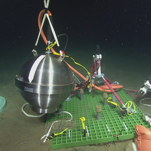
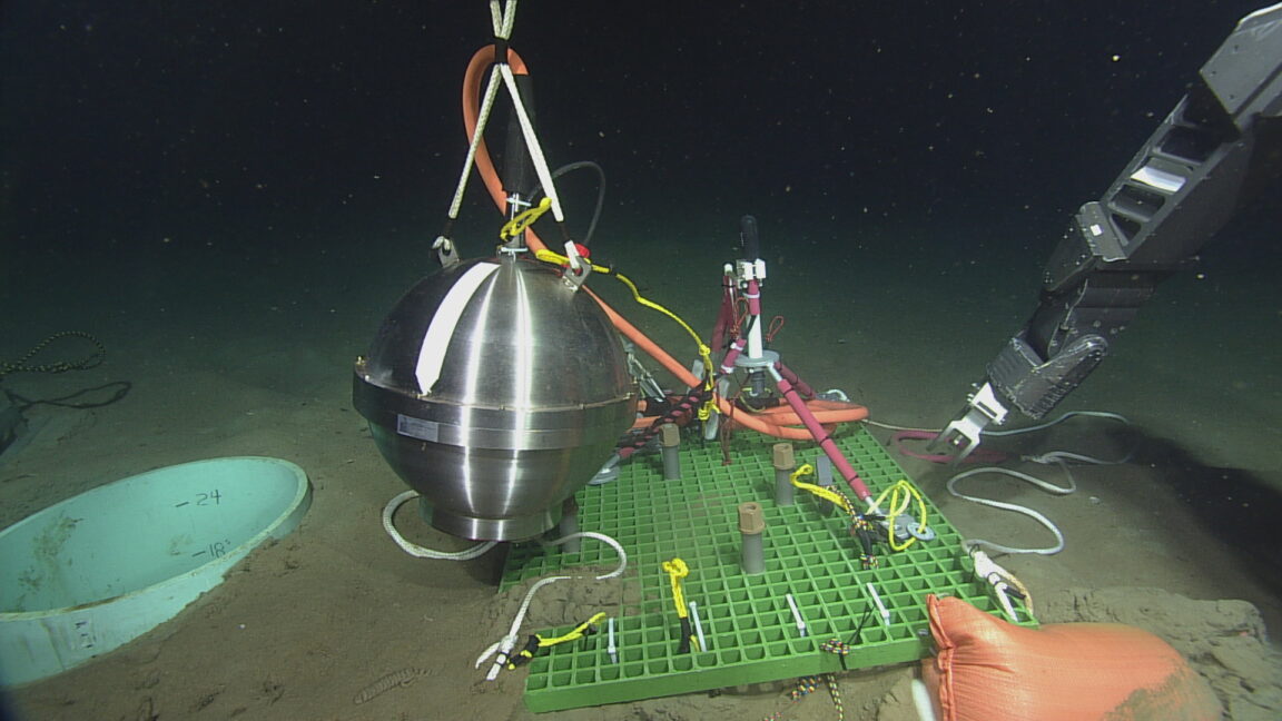
In May, the federal government announced without warning that it would take apart a network of ocean monitoring systems that it had spent over $350 million to build. No reason was given for the decision to shut down the Ocean Observatories Initiative (OOI), but suspicion immediately focused on the network's role in tracking climate change.
But the OOI also provides data that's useful for weather forecasting and fisheries management, leading to widespread opposition. Today, it appears that the opposition has won, as the government will announce that it's reversing the decision. The big remaining question is how much damage the OOI took during the intervening month.
As of now, there is no formal statement available from the federal government. However, The New York Times reports that the decision will be announced later today, and Ars received a statement from Zoe Lofgren, the ranking Democrat on the House Science Committee, indicating that the decision has been made.
© OOI
In May, the federal government announced without warning that it would take apart a network of ocean monitoring systems that it had spent over $350 million to build. No reason was given for the decision to shut down the Ocean Observatories Initiative (OOI), but suspicion immediately focused on the network's role in tracking climate change.
But the OOI also provides data that's useful for weather forecasting and fisheries management, leading to widespread opposition. Today, it appears that the opposition has won, as the government will announce that it's reversing the decision. The big remaining question is how much damage the OOI took during the intervening month.
As of now, there is no formal statement available from the federal government. However, The New York Times reports that the decision will be announced later today, and Ars received a statement from Zoe Lofgren, the ranking Democrat on the House Science Committee, indicating that the decision has been made.
© OOI
In a remarkable leap forward in food safety technology, researchers at the University of California, Berkeley, have developed a groundbreaking “electronic nose” capable of pinpointing spoiled food and detecting allergens with unprecedented accuracy. This innovative device combines a miniature array of gas sensors with sophisticated machine learning algorithms to outperform the human nose, which, while remarkably sensitive, often fails to detect early signs of food spoilage and allergen presence. The implications of this technology are vast, promising to revolutionize how consumers, retailers, and food producers monitor food freshness and safety.
The team’s new creation is composed of 16 ultra-sensitive gas sensors, each responding uniquely to specific volatile compounds released by different foods. These sensors act much like digital taste buds, capable of identifying complex chemical signatures through changes in electrical signals. By mapping these responses to known gas fingerprints, the electronic nose can distinguish between fresh and spoiled food items, along with categorizing various allergens including walnuts, peanuts, and other nuts known to trigger severe allergic reactions.
What makes this achievement especially notable is the integration of carbon nanotube technology in the sensors. Unlike traditional metal oxide sensors, carbon nanotubes offer extraordinary surface area relative to their size and maintain sensitivity at room temperature. This is crucial because many sensing materials degrade or require heating to function effectively, limiting their applications. The Berkeley team leveraged the thin, nanometer-scale layers of carbon nanotubes to create a sensor surface that is both highly selective and stable under everyday environmental conditions.
Central to the power of this electronic nose is machine learning. Lead author Carla Bassil developed models trained to interpret the nuanced electrical patterns emanating from the sensor array when exposed to gases released by seven different foods, including strawberry, blueberry, banana, walnuts, hazelnuts, cashews, and peanuts. The algorithm also discerns the freshness of common perishables such as raw chicken, milk, and eggs by detecting subtle changes in their gaseous emissions over time, reflecting degrees of spoilage after 24 and 48 hours at room temperature.
A key challenge in creating an effective gas sensor lies in the complexity of food odors, which consist of many overlapping volatile organic compounds. This electronic nose overcomes this hurdle by relying on the relative selectivity of each sensor and the pattern recognition capabilities of machine learning, enabling the system to sort through these chemical signatures and recognize distinct gas “fingerprints.” The result is a highly sensitive, objective, and scalable detection method that could transform food safety practices.
The development of multiplexed sensor arrays has historically been complex, with issues arising from manufacturing difficulties and material limitations. However, by utilizing a fabrication process known as drop casting, the team managed to deposit different sensing films onto a single chip in a single step. This scalability is pivotal for real-world adoption, allowing potential mass production of these chips at a reasonable cost and size suitable for use in household appliances or portable devices.
In terms of applications, the researchers envision integration within “smart” kitchen appliances such as refrigerators equipped with wireless connectivity and app-based interfaces. Imagine a refrigerator that could alert you via your smartphone, warning that your broccoli is nearing spoilage or that your chicken is at its last safe day for consumption. Such timely notifications could drastically reduce food waste and prevent foodborne illness.
The sensitivity of the device is particularly striking. The electronic nose demonstrated the ability to detect as little as 0.05 grams of walnut, which corresponds to approximately one hundredth of a shelled nut. This level of detection sensitivity is crucial not only for spoilage monitoring but also for allergen detection, where even trace amounts can have life-threatening consequences for sensitive individuals.
Although these findings are promising, challenges remain. For instance, the device’s performance amidst complex food environments—such as detecting walnuts hidden in salad or cakes, or isolating spoilage gases when multiple food items are stored together—is yet to be rigorously tested. Future developments will focus on enhancing sensitivity, reducing false positives, and ensuring reliability in real-world, cluttered atmospheres.
Furthermore, a portable version of the electronic nose, operable via an iPhone application, has been developed, offering exciting possibilities for on-the-go freshness and allergen detection. Such a compact and user-friendly device empowers consumers to make informed decisions at grocery stores, restaurants, and home kitchens, fostering safer, healthier food habits.
The senior author of the study, Ali Javey, emphasizes the importance of combining novel nanomaterials with advanced machine learning to solve complex sensing problems. This convergence of disciplines paves the way for a new era of digital olfaction technologies that are not only sensitive and selective but also accessible and affordable for everyday use.
Published in the journal Science Advances on June 17, 2026, this study marks a significant milestone in the fields of applied sciences and engineering, with a particular emphasis on food science and sensor technology. Supported by funding from the U.S. Department of Energy and the National Science Foundation, the research team comprises experts from UC Berkeley and collaborators from KAIST in South Korea.
As this technology continues to mature, it holds the promise of making kitchens safer and reducing the global burden of foodborne illnesses. By translating the chemical complexity of food odors into digital data streams, the electronic nose could soon become an indispensable tool for consumers worldwide, enhancing food quality and saving lives.
Subject of Research: Not applicable
Article Title: Scalable multiplexed machine learning gas sensor chips for food classification
News Publication Date: 17-Jun-2026
Web References: http://dx.doi.org/10.1126/sciadv.aec7965
References:
Bassil, C., Lee, K., Liao, X., Krishnan, D., Zhan, Y., Wijaya, T. J., Hester, E., Kim, M., Kim, I.-D., Park, I., & Javey, A. (2026). Scalable multiplexed machine learning gas sensor chips for food classification. Science Advances. https://doi.org/10.1126/sciadv.aec7965
Image Credits: Brandon Sánchez-Mejia/UC Berkeley
Food science, Sensors, Emission detectors, Electronic nose, Machine learning, Carbon nanotubes, Food safety, Food spoilage detection, Allergen detection, Smart appliances, Gas sensors, Nanotechnology
Rivers serve as the lifeblood of our planet, weaving through landscapes and ecosystems while sustaining human civilizations. Despite their critical importance, many rivers remain under-studied, often due to the difficulties presented by their accessibility and variability. Small streams can be intermittent and unpredictable, sometimes disappearing for months or even years. This presents a formidable challenge for hydrologists and environmental scientists tasked with understanding these dynamic systems and their impact on water resources. Now, cutting-edge satellite remote sensing technology is revolutionizing how researchers monitor and analyze river health and water quality on a global scale.
Professor Dongmei Feng of the University of Cincinnati is at the forefront of this emerging discipline. Specializing in hydrological modeling and remote sensing, Feng recognizes that satellite data offers an unprecedented opportunity to observe rivers wherever they flow, even those in remote or inaccessible regions. Through spectral analysis of satellite imagery, scientists can detect changes in water quality and identify key pollutants, nutrients, and biological stressors that are difficult to measure through traditional fieldwork alone. This technology enables continuous and comprehensive monitoring, which is essential in managing freshwater resources sustainably.
The article recently published in Nature Water by Feng and her international collaborators underscores the importance of a holistic yet detailed approach to river science. By combining systematic review techniques with satellite-based observations, the researchers advocate studying rivers both individually and collectively. Each river exhibits a unique combination of climatic conditions, geographical features, and human impacts, which influence its health and quality. Understanding these nuances at multiple scales is crucial for predicting the rivers’ response to environmental pressures and human activities.
Rivers are not isolated entities; they are integral to planetary processes, acting as conduits connecting terrestrial landscapes with oceans and the atmosphere. The sediment and nutrients transported from rivers to estuaries serve as essential nourishment for diverse marine ecosystems. Estuaries themselves act as critical breeding grounds and nurseries for countless marine species, making the health of upstream river systems vital to ocean biodiversity. Satellite remote sensing provides a powerful lens through which to monitor these interactions in near real-time with astonishing spatial coverage.
One of the most pressing global environmental concerns linked to river systems is nutrient pollution, which frequently leads to harmful algal blooms. These blooms, fueled by excess nitrogen and phosphorus from fertilizer runoff and sewage discharge, pose severe risks to public health and aquatic ecosystems. Professor Feng’s ongoing research, supported by a substantial grant from the National Science Foundation, aims to unravel nutrient dynamics over the past five decades across multiple rivers worldwide. This extensive historical analysis is anticipated to improve early warning systems, allowing water managers to preemptively respond to developing algal blooms before they reach toxic levels.
The biogeochemical cycles underlying these nutrient movements involve complex feedback mechanisms. Algal blooms block sunlight from penetrating the water column, suffocating submerged vegetation that supports aquatic life. Upon decomposition of the dead algae, oxygen levels in the water plummet, creating ‘dead zones’ where fish and other organisms cannot survive. This process, known as eutrophication, exemplifies how human-induced nutrient loading can trigger cascading ecological failures. Understanding and modeling this cycle is critical for devising targeted mitigation strategies.
Satellite technology, combined with sophisticated computer modeling, is revolutionizing the ability to track water quality parameters such as turbidity, chlorophyll concentration, and dissolved organic matter with unprecedented precision. Dr. Xiao Yang from Southern Methodist University highlights how spectral signatures obtained from satellites can differentiate among various pollutants and ecological indicators. This technological advancement enables continuous global surveillance and geographically detailed water quality assessments that were not feasible before.
The applications of these remote sensing tools extend beyond academic research; they have real-world implications for managing urban infrastructure and protecting public health. Cities that rely heavily on river water for drinking need accurate and timely data to prevent toxic algal contaminants from entering treatment plants. Given that around 90% of the world’s population lives within six miles of a river, technological breakthroughs in monitoring river systems have direct societal benefits, including safeguarding drinking water, supporting recreational activities, and preserving biodiversity.
Moreover, this satellite-enabled river science promises to illuminate previously hidden aspects of hydrological variability and change. By tracking fluctuations in river flow and quality across diverse climatic zones, researchers hope to better understand how climate change will impact freshwater availability and ecosystem services. The integration of historical datasets with contemporary remote sensing observations offers a pathway to predictive models that inform adaptation and conservation efforts worldwide.
The multidisciplinary collaboration evident in Feng’s study incorporates expertise from hydrology, environmental engineering, satellite physics, and data science. Such integrative research approaches are becoming increasingly necessary to tackle complex environmental challenges that span geographic and disciplinary boundaries. The comprehensive global perspective championed in this research paves the way for a new era of river science, where policy decisions and resource management are underpinned by data-driven insights and continuous observation.
As research progresses, the challenge will be to translate these tremendous amounts of data into actionable knowledge. This entails developing user-friendly platforms for water managers and policymakers to interpret satellite-derived information effectively. Ensuring accessibility while maintaining scientific rigor requires ongoing innovation in data analytics and communication. Nonetheless, the promise of these technological tools to safeguard freshwater resources against pollution and ecological degradation has galvanized the scientific community and stakeholders alike.
In conclusion, the integration of satellite remote sensing into river science marks a transformative advancement in understanding and managing one of Earth’s most essential natural resources. Pioneered by experts such as Professor Dongmei Feng, this approach enables scientists to overcome traditional barriers to studying rivers, track nutrient pollution trends, and predict ecological threats such as toxic algal blooms. As the global population continues to grow and climate change accelerates, technologies that provide real-time, global-scale insights into river ecosystems are indispensable for sustainable water management and environmental protection.
Subject of Research: Hydrology, water quality control, and nutrient dynamics in rivers using satellite remote sensing technology.
Article Title: Remote sensing and the new global river science
News Publication Date: 17-Jun-2026
Web References: DOI: 10.1038/s44221-026-00665-2
Image Credits: Andrew Higley
Keywords: Hydrology, Water quality control, Sewage treatment, Water delivery, Environmental engineering, Natural resources management, Pollution control
In a groundbreaking advancement in neuroscience technology, researchers from the Aerospace Information Research Institute at the Chinese Academy of Sciences have unveiled a state-of-the-art dual-mode wireless microsystem designed to simultaneously monitor electrophysiological activity and dopamine-related electrochemical signals within the brain. This innovative platform promises to illuminate the intricate neural mechanisms underlying pharmacological effects on cortical circuits by capturing real-time electrical and chemical brain dynamics, a feat that has long eluded scientists due to technical constraints.
Neuroscientific investigations often grapple with the challenge of correlating multidimensional neural signals that operate on different modalities. Electrophysiological recordings capture rapid neuronal spikes and local field potentials (LFPs), presenting a temporal resolution ideal for observing immediate neuronal events. However, these signals alone fail to convey the chemical milieu shaping neural behavior. Neurotransmitter dynamics, especially dopamine, play a crucial role in modulating brain function but require sensitive electrochemical detection methods for accurate monitoring. Historically, research platforms have predominantly focused on one modality—either electrophysiological or neurochemical—making it difficult to obtain a holistic understanding of brain states or drug effects.
The newly developed dual-mode microelectrode array tackles this limitation through a sophisticated electrode design. Using platinum nanoparticles (PtNPs) and poly(3,4-ethylenedioxythiophene) polystyrene sulfonate (PEDOT:PSS), the researchers engineered electrophysiological sites with enhanced conductivity and stability for precise spike and LFP recordings. Simultaneously, an electrochemical site tailored for dopamine sensing incorporates additional modifications with reduced graphene oxide (rGO) and Nafion, constructing a PtNPs/PEDOT:PSS/rGO/Nafion composite. This multi-layered structure significantly amplifies electrochemical sensitivity and ensures selectivity against common interferents such as uric acid, serotonin, ascorbic acid, and lactic acid.
On the hardware frontier, the team implemented a hybrid acquisition system that integrates a specialized electrophysiological chip alongside the AD5941 electrochemical front end. An intelligent dual-controller architecture comprising a field-programmable gate array (FPGA) paired with an ESP32 microcontroller orchestrates independent yet synchronized data capture and wireless transmission from both signal modalities. Critical to minimizing crosstalk and data corruption, electrophysiological and electrochemical data streams are transmitted simultaneously but via separate TCP ports. This architectural choice guarantees the temporal alignment and integrity of neural datasets, enabling researchers to dissect the interplay between electrical spikes, LFPs, and dopamine fluctuations with millisecond precision.
The comprehensive calibration and validation of this microsystem were first conducted in vitro. Dopamine calibration curves revealed a robust and linear electrochemical response, confirming the device’s sensitivity and dynamic range. Notably, selectivity tests demonstrated the system’s ability to discriminate dopamine oxidation currents amidst a cocktail of neurochemical interferents, ensuring fidelity in complex biological environments. Furthermore, wireless transmission integrity was rigorously assessed, demonstrating accurate data delivery across distances up to 25 meters with negligible latency or packet loss, underscoring the device’s suitability for freely moving animal experiments without tethers.
Translating these advances into a live animal model, the researchers implanted the dual-mode microsystem into the prelimbic (PrL) cortex of rats. This brain region is integral to executive functions and is sensitive to neuropharmacological modulation. By administering dexmedetomidine—a sedative with known but paradoxical and poorly understood effects on cortical activity—the team pursued a dynamic interrogation of neural electrical firing patterns alongside dopamine fluctuations. This sophisticated approach exposed nuanced dose-dependent changes: higher doses correlated with reduced spike firing rates and dampened peak-to-peak amplitudes, suppressed overall LFP power, increased delta-frequency activity (associated with decreased arousal), and reduced gamma oscillations, indicative of a reconfigured cortical state consistent with sedation.
Simultaneously, electrochemical monitoring unveiled an increase in amperometric signals related to dopamine concentrations as dexmedetomidine dosage escalated. This parallel observation of neurochemical enhancement accompanying electrophysiological suppression substantiates the microsystem’s capability to capture the complex, multifaceted neural correlates of sedation. Importantly, control saline conditions elicited minimal changes, affirming system specificity and the physiological relevance of the recorded responses. These findings reveal the microsystem’s potential as a powerful investigative tool for understanding the neurochemical underpinnings of anesthetic action and cortical state transitions.
The scientific implications of this dual-mode wireless microsystem are profound. Unlike prior single-modality or tethered platforms, this technology conserves 32 independent electrophysiological channels at high sampling rates while integrating dopamine-sensitive electrochemical pathways within the same device footprint. The dual-TCP wireless architecture enables real-time, synchronized acquisition and visualization of electrical and chemical signals, overcoming longstanding barriers in multimodal neural monitoring. Such comprehensive systems are invaluable for probing the mechanistic interactions between neural circuit activity and neurotransmitter dynamics under pharmacological challenges.
Looking forward, despite its remarkable performance, the current microsystem has limitations that present rich avenues for refinement. The modest in vivo sample size tempers broad generalization of the reported electrophysiological and dopamine-related observations. Amperometric readings in vivo remain susceptible to confounding factors such as electrode fouling, baseline drift, shifts in local pH or oxygenation, tissue inflammatory responses, and reference electrode stability. Thus, the data are best interpreted as dopamine-correlated signals rather than exact dopamine quantifications in extracellular space. Furthermore, the system currently relies on commercial electronic components, which constrain further miniaturization. It also lacks integrated stimulation or closed-loop modulation functionalities, which are critical for advanced neuromodulation studies.
Future developments aspire to embed dedicated, custom-fabricated integrated circuits that will shrink device size and power consumption. Incorporation of multimodal closed-loop control systems will enable responsive neurochemical-electrical feedback interventions, enhancing therapeutic and experimental utility. Expanding recording capacity to encompass multiple brain regions simultaneously will provide a more comprehensive spatial map of neurophysiological network dynamics. Cross-validation with orthogonal neurochemical detection modalities could further bolster confidence in dopamine measurements. Together, these advances would propel the technology from a sophisticated measurement tool to an instrumental platform for neurological disease research, neurorehabilitation, and cognitive neuroscience.
The research team responsible for this innovation includes Peiyao Jiao, Yilin Song, Jin Shan, Yu Liu, Qianli Jia, Qi Li, Ying Wang, Yan Luo, Pengfei Zhao, Juntao Liu, Zhenchang Wang, Mixia Wang, and Xinxia Cai. Their work represents a significant leap toward decrypting the intertwined electrical and chemical language of the brain in vivo, promising to inspire future explorations into neural circuit regulation and pharmacodynamics.
This pioneering study titled “A Dual-Mode Wireless Microsystem for Monitoring Dopamine and Spike Changes with Dexmedetomidine” was published on May 21, 2026, in the journal Cyborg and Bionic Systems. It was supported by funding from the National Natural Science Foundation of China, the Major Program of Scientific and Technical Innovation 2030, the Chinese Academy of Sciences Joint Foundation Program, and the Natural Science Foundation of Beijing.
Subject of Research: Dual-mode wireless monitoring of electrophysiological and dopamine-related electrochemical signals in vivo under pharmacological modulation.
Article Title: A Dual-Mode Wireless Microsystem for Monitoring Dopamine and Spike Changes with Dexmedetomidine
News Publication Date: May 21, 2026
Image Credits: Peiyao Jiao, Aerospace Information Research Institute, Chinese Academy of Sciences
Neural monitoring, dopamine sensing, electrophysiology, wireless microsystem, dexmedetomidine, neurochemistry, brain-computer interfaces, electrode modification, dual-mode recording, neuropharmacology, local field potentials, neural circuits
In a pioneering stride towards maintaining the integrity of scientific literature, MDPI has fully implemented its proprietary artificial intelligence-powered system called Ethicality. This sophisticated tool is now embedded within the publishing workflow to automatically screen all submitted manuscripts, revolutionizing the approach to safeguarding the scientific record. Ethicality is engineered to operate continuously throughout the editorial process, offering a proactive framework that enhances the evaluation and decision-making capabilities of editors globally. Presently, it processes around 2,000 submissions daily, handling a vast and diverse array of academic contributions from across the world.
The development of Ethicality reflects the pressing need for automated solutions in the face of surging scholarly output and increasing demands for transparency, speed, and quality in academic publishing. Traditional manual screening methods, once the norm, are no longer adequate to detect sophisticated manipulations or emerging threats posed by advanced generative AI techniques. Its design integrates an end-to-end integrity layer that scrutinizes manuscripts from the moment they enter the editorial system, ensuring continuous vigilance rather than episodic checks. This paradigm shift marks a new era in editorial practices, combining the precision of AI with human expertise to set robust guardrails around the publication pipeline.
Dr. Milos Cuculovic, MDPI’s head of technology innovation, emphasizes that the role of AI within this context is not to replace editors but to augment their capabilities. Ethicality functions as a vigilant assistant, flagging potential ethical issues and inconsistencies while leaving final judgment to experienced professionals. This human-in-the-loop approach enables a balance where automation streamlines routine tasks without compromising critical oversight. Such a model helps preserve transparency and trust at scale, combating increasingly sophisticated tactics that jeopardize scientific authenticity, such as synthetic content generation, fabricated submissions, and manipulated peer review reports.
Ethicality’s analytical engine examines each submission comprehensively by dissecting fundamental manuscript components, including the title, abstract, author metadata, main body, and reference lists. Beyond textual elements, it also reviews peer review reports sent with the manuscripts, employing natural language processing techniques and machine learning algorithms tailored for scholarly content. This holistic evaluation identifies a wide spectrum of potential integrity breaches, ranging from paper mill-generated manuscripts and spurious authorship claims to subtle citation manipulations and irregular referencing patterns that may indicate gaming of metrics or unethical publication practices.
One of the system’s remarkable features lies in its ability to detect AI-generated or altered text, a rapidly emerging threat in the digital publication ecosystem. Leveraging large language models fine-tuned to recognize nuances and inconsistencies characteristic of synthetic content, Ethicality can pinpoint sections likely created or tampered with by artificial intelligence. This capability is crucial amid widespread concerns that generative AI tools could be exploited to fabricate scientific results or obfuscate fraudulent data, undermining trust in scholarly communication. The AI’s detection mechanisms, however, are continuously refined against real-world data to minimize false positives and maintain editorial efficiency.
In addition to content analysis, Ethicality collaborates with external software suites to cover domains beyond textual scrutiny. For instance, image manipulation detection is managed via integration with specialized tools like Proofig, which applies forensic algorithms to uncover alterations in scientific figures and illustrations. For plagiarism and text duplication, the system incorporates iThenticate services to cross-reference submissions with an extensive repository of published works. This multi-layered approach strengthens MDPI’s ability to uphold ethical standards by combining internal AI capabilities with trusted third-party solutions to comprehensively monitor manuscript authenticity.
The advent of such an automated system answers growing concerns about the sustainability of peer review in an era characterized by exponential growth in submission volumes. By automating repetitive and technical screening tasks, Ethicality frees up valuable editorial resources, allowing human experts to concentrate on evaluating the scientific merit and originality of manuscripts rather than being overwhelmed by manual integrity checks. This redistribution of effort ensures that editorial workflows can keep pace with demand without compromising quality or publication timelines, addressing one of the major bottlenecks in modern scholarly publishing.
Dr. Enric Sayas, the product owner of Ethicality, highlights that the publishing sector is entering a technological race where adversaries leverage advanced AI to generate deceptive manuscripts and fake review reports. Traditional detection tools alone are insufficient to counteract such sophistication. Only by deploying equally advanced AI systems, capable of multi-modal analysis encompassing textual, image, and metadata anomalies, can publishers effectively defend the credibility of scholarly records. Ethicality embodies this next-generation defense mechanism, combining deep learning models with domain-specific heuristics to expose fraudulent submissions before they can impact the scientific corpus.
Ethicality’s role extends beyond mere detection; it facilitates informed decision-making by generating risk signals that prioritize editorial scrutiny. This nuanced output classifies flagged cases according to severity and type of ethical concern, enabling editors to apply their judgment where most critical while bypassing lower-risk items. Such targeted intervention reduces unnecessary delays in the publication process and improves the overall efficiency of manuscript handling. The system’s iterative design also permits continuous learning from editor feedback, enhancing accuracy and adaptability as new manipulation techniques emerge.
The timing of Ethicality’s deployment coincides with an inflection point in scholarly publishing, where generative AI tools have made it increasingly feasible to produce lifelike but fraudulent scientific content. This proliferation necessitates automated defenses that not only identify outright fraud but also detect subtler anomalies such as inconsistent author data, suspicious referencing behavior, and peer review irregularities. By integrating these multifaceted analyses, MDPI demonstrates a comprehensive and forward-looking strategy aimed at preserving the integrity and credibility of academic knowledge dissemination.
Crucially, Ethicality exemplifies how technology can be harnessed responsibly within complex human ecosystems. It respects the indispensable role of expert editors by operating as a sentinel that brings potential issues to attention while leaving interpretive authority intact. This model upholds core values of transparency, accountability, and collaborative oversight, providing a blueprint for other publishers grappling with integrity challenges in the AI era. Moreover, it underscores the broader imperative for publishers to innovate continuously in response to evolving threats and technological opportunities, ensuring that scientific publishing remains a trusted pillar of global knowledge advancement.
As MDPI’s journey with Ethicality moves forward, continuous enhancements and iterations are planned, driven by real-world data and editorial collaboration. This adaptive approach ensures the system evolves alongside the changing landscape of academic publishing and the advent of new manipulative tactics. Ultimately, Ethicality stands as a testament to the potential of AI-powered tools to reinforce scientific integrity, protect the scholarly record, and empower editorial professionals in an increasingly complex and dynamic environment.
Subject of Research: Artificial intelligence applications in scholarly publishing and research integrity
News Publication Date: 2024
Image Credits: MDPI
Academic publishing, research integrity, artificial intelligence, editorial workflow automation, AI-generated scientific content detection, paper mill detection, peer review screening, scholarly publishing technology, automated manuscript analysis, scientific record safeguarding
