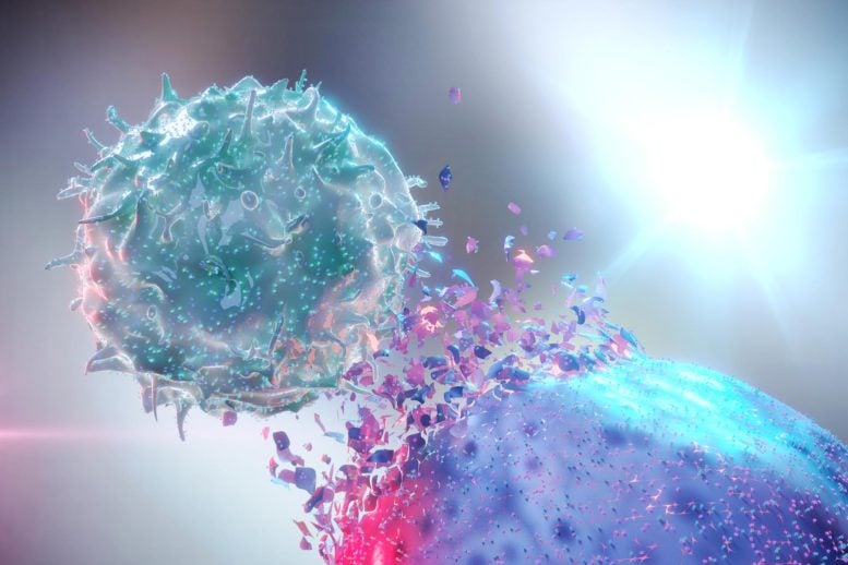
A protein called TRAILshort may act as a central immune brake that limits T-cell responses and CAR-T therapy. Cancer cells can weaken the immune system by producing a protein that prevents its most important defenders from attacking. Mayo Clinic researchers have identified how this protein works, revealing a possible target for treatments designed to improve [...]
Fuchs endothelial corneal dystrophy, a progressive disorder that can gradually cloud the cornea and impair vision, may be driven by a cellular process more complex than simple cell loss. A new study published in Cell Death Discovery reports that ultraviolet-A light can induce chronic hyperploidization in corneal endothelial cells, triggering changes associated with fibrosis. The findings connect long-term genome abnormalities in post-mitotic cells to the structural deterioration observed in this increasingly recognized eye disease.
The cornea depends on a thin inner layer of endothelial cells to maintain its transparency. These cells regulate the movement of fluid between the cornea and the anterior chamber of the eye, preventing the tissue from becoming waterlogged. Unlike many cell types, human corneal endothelial cells have limited ability to divide and replenish themselves. When they are damaged or lost, neighboring cells may enlarge and stretch to cover the affected area, but this compensation has limits. In Fuchs endothelial corneal dystrophy, the endothelial layer progressively becomes dysfunctional, allowing fluid accumulation and producing corneal swelling, haze and visual distortion.
The study focuses on hyperploidization, a condition in which cells acquire extra sets or copies of chromosomes. In a healthy cell cycle, DNA is replicated and then evenly distributed when a cell divides. Post-mitotic cells, however, are not expected to continue cycling through repeated rounds of DNA replication. If DNA replication occurs without successful cell division, the result can be a cell with an enlarged genome and multiple chromosome sets. This state, known as polyploidy or hyperploidization depending on its extent and context, can alter cell size, metabolism, gene activity and responses to stress.
The researchers investigated how ultraviolet-A, or UVA, light affects cells associated with Fuchs endothelial corneal dystrophy. UVA has a longer wavelength than ultraviolet-B and can penetrate biological tissues more deeply. Although the eye possesses protective mechanisms, persistent or excessive exposure to light-generated stress can damage cellular components, including DNA, proteins and mitochondria. The study indicates that UVA exposure does not merely cause an acute injury that cells either repair or fail to survive. Instead, it can establish a chronic hyperploid state in cells that no longer divide normally.
That distinction is important because a cell can remain alive while becoming increasingly dysfunctional. Hyperploid cells may continue to produce proteins, communicate with neighboring cells and modify their surrounding environment, even as their enlarged genomes disrupt normal regulation. In the corneal endothelium, such persistent abnormalities could interfere with the machinery responsible for maintaining fluid balance and tissue transparency. The research links this long-lasting cellular state to the activation of fibrotic processes, suggesting that damaged endothelial cells may help remodel the cornea rather than simply disappearing.
Fibrosis is the formation or accumulation of excessive connective-tissue components, particularly extracellular matrix proteins such as collagens. During wound healing, matrix production can provide temporary structural support. When the response persists, however, the tissue can stiffen, become disorganized and lose its normal optical properties. In the context of Fuchs endothelial corneal dystrophy, fibrosis may contribute to the loss of corneal clarity and the progression of visual impairment. The study’s central message is that chronic hyperploidization may act as an upstream signal that pushes post-mitotic cells toward this pathological remodeling.
The findings also help explain why a disease affecting a specialized, non-dividing cell population can progress over time. A conventional view of cellular damage often emphasizes apoptosis, necrosis or the gradual depletion of functional cells. The new work highlights another possibility: injured cells may survive in a permanently altered condition and actively influence disease progression. By retaining metabolic activity while carrying abnormal quantities of DNA, these cells could produce signals that encourage inflammation, matrix deposition or changes in the behavior of nearby cells.
The UVA connection raises questions about how environmental stress interacts with inherited susceptibility. Fuchs endothelial corneal dystrophy has genetic and age-related components, and the disease does not arise from a single universal cause. The study does not establish that UVA exposure alone causes the disorder, but it provides a mechanism through which light-associated stress could worsen cellular instability in vulnerable corneal tissue. This may help researchers examine whether cumulative exposure, cellular antioxidant capacity and pre-existing genetic changes combine to determine how rapidly the disease advances.
From a treatment perspective, the work points toward targets beyond replacing lost endothelial cells. Future strategies might seek to prevent abnormal DNA replication, stabilize genome integrity, suppress the fibrotic signals released by hyperploid cells or protect the corneal endothelium from chronic light-induced stress. Such approaches would require careful testing, because polyploidy can serve useful roles in some normal tissues, and broadly blocking cell-cycle or repair pathways could produce harmful effects. The challenge will be to distinguish pathological hyperploidization from adaptive genome changes that help cells survive.
The study ultimately presents Fuchs endothelial corneal dystrophy as a disease of cellular identity as well as cellular survival. UVA-induced chronic hyperploidization appears to place post-mitotic corneal endothelial cells in a persistent state of genomic imbalance, where they can contribute to fibrosis and tissue failure. By connecting light stress, abnormal DNA content and corneal scarring, the research offers a new framework for understanding why the disease can continue even after the initial injury has passed—and provides a potential roadmap for therapies designed to interrupt the process before vision is permanently compromised.
Subject of Research: Ultraviolet-A light-induced chronic hyperploidization and fibrosis in post-mitotic corneal endothelial cells affected by Fuchs endothelial corneal dystrophy
Article Title: Ultraviolet-A light-induced chronic hyperploidization causes fibrosis in post-mitotic cells affected by Fuchs endothelial corneal dystrophy
Article References: Adhikari, Y., Parekh, M., Deshpande, N. et al. Ultraviolet-A light-induced chronic hyperploidization causes fibrosis in post-mitotic cells affected by Fuchs endothelial corneal dystrophy. Cell Death Discov. (2026). https://doi.org/10.1038/s41420-026-03292-8
T cells may use a previously unknown CD64-dependent pathway to attack acute myeloid leukemia. T cells are generally thought to identify cancer through a well-established molecular recognition system. Yet when researchers removed a key part of that system from acute myeloid leukemia (AML) cells, the immune cells continued killing them. The unexpected result led scientists [...]
Macrobiological features have been cited as irreducibly complex, including animal joints and the trap mechanisms of carnivorous plants, even the cell itself.
A protein and signaling pathway may help overcome the developmental block that keeps some leukemia cells immature. Inside the bone marrow, immature blood cells normally pass through a tightly controlled sequence of development before taking on specialized roles. In aggressive forms of leukemia, some of these precursor cells become trapped partway through that process. Instead [...]
The Varta crisis has entered another severe phase after the German battery manufacturer lost the customer responsible for nearly all production at its specialized factory in Nördlingen.
Varta has not publicly identified that customer, but reports from German media and industry sources point to Apple. The iPhone maker is reportedly moving battery orders for a future AirPods generation to a lower-cost supplier in China.
The decision will lead Varta to stop producing rechargeable button cells in Nördlingen during fall 2026. Approximately 350 jobs will be eliminated across the factory and the company headquarters in nearby Ellwangen.
Describing the development as a complete corporate collapse would go too far. Varta continues operating other battery businesses and recently completed an extensive financial restructuring backed by Porsche. The loss of Apple, however, has destroyed the economic foundation of a production site created around one highly influential customer.
Varta Crisis Exposes Dependence on Apple
The Varta crisis illustrates how a large technology customer can transform a supplier and then leave it with capacity that cannot easily be reassigned.
Varta became an important producer of CoinPower lithium-ion cells, small rechargeable batteries designed for wireless earbuds and other compact devices. Teardowns of AirPods Pro models identified Varta cells inside the earbuds, although Apple does not publicly discuss most component suppliers by product.
Demand from premium wireless headphones helped Varta expand rapidly. In 2020, its Microbatteries and Solutions division generated approximately €508 million in revenue, representing growth of 49% from the previous year.
The company invested heavily in production as wireless earbuds became a mass-market category. Apple demand provided the volume needed to justify specialized lines, equipment and hiring in Germany.
That dependence became dangerous when AirPods production weakened and Apple began adding other battery suppliers. A factory developed for unusually large orders could not operate efficiently once those orders declined.
The customer now leaving Nördlingen had previously accounted for nearly the entire capacity of the site. Varta says it has gained other customers, but their combined demand is not sufficient to keep the specialized factory running.
The plant is therefore closing not because rechargeable button cells have disappeared, but because replacing one customer of Apple scale requires far more than several smaller contracts.
Price Reportedly Decided the AirPods Order
German reports indicate that price was the decisive factor behind the supplier change.
Varta battery quality was reportedly equal to or better than the competing product. The German manufacturer could not, however, match the cost offered for the next generation of cells from China.
Battery production in Germany faces higher labor, energy and operating expenses than many Asian manufacturing centers. Chinese suppliers also benefit from extensive domestic electronics supply chains, access to materials and production volumes spread across numerous customers.
Apple has enormous leverage when negotiating component contracts. AirPods ship in sufficient quantities for a small difference in the cost of each battery to produce substantial savings across an entire product cycle.
The company regularly qualifies multiple suppliers for components when technical requirements and available capacity permit. Competition can reduce prices, protect production against interruptions and prevent one manufacturer from gaining excessive influence over the supply chain.
For Varta, that process brought the opposite effect. The supplier expanded to serve Apple and then faced declining utilization as the customer divided orders among additional manufacturers.
The latest move appears more final. Varta expects the contract to expire at the end of October, removing the volume that kept Nördlingen economically viable.
Image Credit: Apple Inc.
Apple Supplier Strategy Is Not Simply About China
The reported decision does not mean Apple is moving its complete production network toward China.
Apple has spent several years expanding device assembly and component production in India, Vietnam and other countries. AirPods manufacturing has grown in India, while Macs, iPads and wearables are assembled across a wider group of Asian locations.
The Varta case concerns a particular component for which a Chinese supplier reportedly offered a more competitive price. It shows that diversification away from one country can coexist with cost-driven sourcing from that same country.
Apple may reduce its dependence on Chinese final assembly while continuing to purchase Chinese batteries, displays, enclosures or electronic components when suppliers provide the required scale and pricing.
That balance creates a difficult environment for European manufacturers. Political interest in domestic technology production does not guarantee that major customers will accept higher component prices.
Premium engineering can protect a contract when it produces a measurable advantage in battery capacity, safety, dimensions or lifespan. Once competing suppliers reach comparable performance, procurement decisions become increasingly sensitive to cost.
The Varta crisis suggests that manufacturing location and supplier history carried less weight than the economics of the next AirPods battery.
Varta Was Already Fighting Financial Failure
Apple did not create every problem affecting Varta.
The company entered the current dispute after years of debt, costly investments, volatile customer orders, supply-chain problems and weaker demand in parts of its energy-storage business. A cyberattack in early 2024 also disrupted production and delayed financial reporting.
Varta warned that its earlier restructuring plan might fail after Apple reduced AirPods production. By mid-2024, the company had reached a point where it could no longer service its debt without another intervention.
A pre-insolvency restructuring reduced financial liabilities from approximately €485 million to €200 million. Porsche and majority shareholder Michael Tojner agreed to provide new capital, while creditors supplied additional financing.
Existing shareholders lost their investment as Varta was removed from the stock market. The process preserved the operating company but demonstrated how little financial room remained for another major disruption.
Varta reported a loss of €64.5 million for 2024. Although that was an improvement from the previous year, the business remained dependent on successful restructuring and stronger performance from its surviving divisions.
The Apple contract loss therefore did not independently bring down a healthy battery group. It struck a manufacturer that had already narrowly avoided insolvency.
Image Credit: Wikipedia
Porsche Cannot Protect Every Varta Business
Porsche participation gives Varta a future in automotive batteries, but it does not rescue the Nördlingen button-cell operation.
The automaker invested in the restructuring and took control of the V4Drive business, which develops high-performance cylindrical lithium-ion cells for hybrid and performance vehicles. Porsche gains greater access to battery technology while helping retain part of that expertise in Germany.
Varta also continues producing household batteries and energy-storage systems. Those businesses serve different customers and do not depend on the machinery used for tiny CoinPower cells.
Employees affected by the Nördlingen closure may be offered positions at other locations, although Varta has not indicated how many can realistically be transferred. The company and its works council are negotiating measures for the workers whose roles will disappear.
The shutdown leaves a modern production unit without enough demand only a few years after wireless earbuds were presented as one of Varta most promising growth markets.
Its remaining automotive operation now has the security of a strategic customer with a direct ownership interest. The microbattery division learned the cost of building a factory around a customer that never made the same commitment.
Aging appears to alter how key cerebellar neurons fire, with measurable effects on movement and coordination. Aging often brings slower movement, weaker balance, and reduced agility, but the brain changes behind these losses have been difficult to identify. McGill University researchers have now directly linked declining activity in Purkinje cells, an important type of neuron [...]
Controlling ribosomal RNA could help researchers target diseases driven by too little or too much protein production. Cells do not simply make proteins at a fixed pace. They continually adjust production to match their needs, and those changes can help determine whether a cell keeps dividing, adopts a specialized identity, or retains the developmental flexibility [...]
Researchers have developed the first entirely artificial cell, made completely from non-living chemical components, which they say is even capable of replicating.
A team of researchers at the University of Minnesota, Twin Cities, College of Biological Sciences, has described their syntheticcell, which they call SpudCell, in a recent preprint. SpudCell marks the first time scientists have replicated elements of life, such as energy use, reproduction, growth, and development, in a synthetic biological organism.
The Secrets of Life
While many mysteries of life and death remain for science, the team’s achievement represents a significant breakthrough, moving a concept once relegated only to science fiction into science fact. The team is hopeful that new solutions to medical and engineering problems will also arise from their achievement.
“This is likely the most exciting project I’ve ever worked on,” said co-author Kate Adamala. “We’ve replicated in chemistry what only used to be possible in biology: the complete set of behaviors of a cell. It proves that the most fundamental functions of life, like growth and replication, do not need a mysterious magical spark.”
Fluorescent microscopy of SpudCell undergoing division (Image Credit: Kate Adamala, Adamala LabCredit: Kate Adamala, Adamala Lab).
The artificial SpudCell performs some of the most complex activities of its biological counterparts, including selection, genome replication, genetically encoded division, resource acquisition via feeding, and growth.
Like a Cell, Yet Different
Although the synthetic cell copies many biological functions, there are some differences. It lacks the internal scaffolding, called a cytoskeleton, which natural cells use to divide. Instead, SpudCell crowds proteins together on its membrane surface until enough mechanical stress builds up to cause the membrane to split.
In addition to being slightly different from a biological cell, the team refined SpudCell throughout their research, at one point altering the cell’s genetics to increase fusion protein production. With this change, the cells grew faster and produced more offspring, outcompeting the original variant in five generations. In a demonstration of the system’s selection and competition, the genetically modified advantage further increased when the team changed conditions to make nutrients scarcer.
The cell’s genetic code is relatively simple. There are about three million kilobase pairs in the human genome, and likely around 113 pairs in a single cell. SpudCell contains a mere 90, encoded on seven DNA plasmids, instead of a single chromosome. This encoding solution offers benefits, allowing researchers to program cellular functions more easily by simply touching a single piece of the modular system. They say this will enable them to continue increasing SpudCell’s complexity.
Moving Forward on Synthetic Cells
“This work is just the beginning,” said Adamala, who, along with several partners, is founding a research and engineering institution called Biotic, which will focus on further advancements in synthetic cell research, keeping its findings open source.
“We are showing it’s possible to engineer the basic functions of the cell,” Adamala added. “To fully realize the promise of this technology – to make it robust and practical – we need combined international effort.”
“The role of Biotic is to focus engineering efforts and make them compatible with a shared chassis. SpudCell is that chassis, and with Biotic setting the protocols for collaboration, we are eager to start applying this technology to serious challenges.”
The next steps for Biotic will be to combine SpudCell’s seven DNA plasmids into a single genome and build more molecular machinery. One of the greatest challenges they are preparing to confront is standardization, as many labs use different standards. To achieve true growth in this sector, Biotic aims to develop modular standards that will apply across the entire field.
SpudCell could provide a revolutionary new basis for manufacturing, allowing cells to naturally develop properties that typically require difficult and expensive molecular transformations, and even open up possibilities beyond what those transformations can offer. This could enable major improvements in medicine by using amino acids that are never used in nature.
Ryan Whalen covers science and technology for The Debrief. He holds an MA in History and a Master of Library and Information Science with a certificate in Data Science. He can be contacted at ryan@thedebrief.org, and follow him on Twitter @mdntwvlf.
In a groundbreaking fusion of cutting-edge technology and dermatological research, scientists at Biohub have unveiled novel therapeutic targets for psoriasis by conducting the first comprehensive genome-wide CRISPR screen in primary human adult skin cells. By overcoming the longstanding technical hurdle of delivering CRISPR components into notoriously resistant keratinocytes without toxicity, the team utilized centrifuge-based transfection to probe the genetic regulators involved in psoriasis, a chronic inflammatory skin disorder affecting millions globally.
This innovative approach involved knocking out approximately 19,000 genes in keratinocytes sourced from human donors using a vast CRISPR guide RNA library. The focus was on modulating IL17RA, a pivotal receptor that mediates skin inflammation, to chart a genome-wide landscape of genes influencing inflammatory pathways directly in the skin’s outermost layer. The resultant massive data set was then mined using an AI-driven model, VirtualCRISPR, which cross-referenced each gene’s significance against existing scientific literature to highlight previously unrecognized contributors to psoriasis pathology.
Strikingly, this AI-guided screen pinpointed two promising protein targets: ALOX5, an enzyme targeted by the asthma medication zileuton, and OXTR, the gene encoding for the oxytocin receptor, a surprising candidate unassociated before with skin inflammation. Validation experiments in 3D reconstructed human skin cultures demonstrated that zileuton modulates keratinocyte metabolism to suppress inflammation independently from immune cells, revealing a direct pharmacological avenue.
Importantly, topical formulations containing zileuton and cligosiban, an OXTR antagonist, were tested in a mouse model of psoriasis, achieving comparable efficacy to standard injected biologics that broadly suppress immune function. Beyond mitigating inflammation, OXTR inhibition appeared to restore skin barrier integrity—a crucial factor in chronic disease progression—spotlighting this receptor as a novel therapeutic axis.
Unlike current systemic therapies, which often carry risks of broad immunosuppression and high costs, these localized topical interventions promise targeted treatment with minimal systemic exposure. Moreover, the safety profiles of zileuton and OXTR antagonists are well-established in clinical settings, potentially accelerating their repurposing for dermatological applications.
This study exemplifies the transformative power of integrating AI with high-dimensional genetic screening to unravel complex disease mechanisms in cell types previously inaccessible to large-scale functional genomics. By enabling direct investigation into keratinocyte biology, the research not only charts a course for new psoriasis treatments but also sets a precedent for applying similar methodologies to other inflammatory conditions.
As psoriasis affects roughly 125 million people worldwide, innovations enabling precise, effective, and safer therapies could revolutionize patient care. Biohub’s approach heralds a new era where AI-guided CRISPR screens expedite the discovery of unexpected drug targets and foster the development of locally acting, biology-informed treatments.
Subject of Research: Psoriasis therapeutic targets via genome-wide CRISPR screening of human skin cells
Article Title: AI-guided CRISPR screening reveals therapeutic targets in psoriasis
News Publication Date: 8-Jul-2026
Web References:
https://www.nature.com/articles/s41467-026-75249-5
Experiments exploring the effects of missing a night’s sleep on the human brain have found indications that a sleepless night actually increases connections between brain cells instead of impeding growth. However, the lack of sleep could also create brain “fatigue” and pressure, resulting in burnout.
The Forschungszentrum Jülich Institute of Neuroscience and Medicine team behind the discovery said that their findings might support the idea that sleep plays an important role in restoring the brain’s cellular balance.
Missing a Night’s Sleep Could Disrupt Cellular Balance
In a statement announcing the surprising discovery, study leader David Elmenhorst and colleagues note that scientists still are not in complete consensus about why humans and other animals need sleep. One proposal involves restoring synaptic connections between brain cells during certain stages of sleep, helping the brain maintain a form of cellular ‘homeostasis.’
For example, previous research has shown that synaptic connectivity increases during wakefulness. This increased activity also increases energy consumption, potentially resulting in problematic protein buildup. Conversely, scientists suspect that sleep helps restore baseline synaptic connections and protein levels, thereby maintaining a healthy cellular balance.
“Sleep is essential for synaptic homeostasis, a proposed mechanism whereby wakefulness leads to synaptic potentiation, and sleep facilitates synaptic down-selection,” the research team explains in a recently published study.
Although previous studies suggest that missing a night’s sleep could disrupt this balance, the team notes that there is very little experimental data to support these conclusions.
Study Reveals Elevated Synaptic Activity After 28 Hours Awake
To explore the role of sleep in cellular homeostasis in the human brain, the researchers used an advanced brain-scanning technology called positron emission tomography (PET). According to the team’s statement, this tool can scan the human brain for signs of synaptic vesicle glycoprotein 2A (SV2A), which they describe as a “marker” of brain synapses.
The team started by enlisting 40 study volunteers. Next, each volunteer was given a PET scan to establish baseline marker levels. After their first scan, 20 of the volunteers were told to get a normal night’s sleep. According to the study authors, the second half were assigned to “the sleep deprivation condition,” where they were told to miss a night’s sleep.
The next day, the team used the PET scan to search for telltale brain synapse markers. As hoped, the scans revealed a distinct difference between the two groups. Where the normal sleep group’s levels were within expected ranges, the synapse markers of the volunteers who had missed a night’s sleep were elevated.
A closer analysis found that once people passed 28 hours of continued wakefulness, their SV2A markers were higher “in several brain regions.” Notably, these increased markers were detected in the hippocampus, which plays an important role in memory, and in the thalamus, which is critical for information relay within the brain.
“Sleep deprivation led to significant increases in synaptic vesicle glycoprotein 2A binding in multiple brain regions, including the thalamus (+4.6%), hippocampus (+5.6%), and parietal cortex (+3.2%), whereas no changes were observed in controls,” the study authors write.
Sleep Deprivation Causes Fatigue and CHanges Neural COnnections
When discussing the study’s limitations, the authors note that the SV2A marker used is a “proxy” for connections between brain cells, not a direct measure. They also note that the observed changes after a missed night’s sleep “were relatively small.” Still, they add, the results in several regions across several study participants “suggest a biological connection between the need for sleep and the buildup of cell connections.”
“During sleep deprivation, the brain remains awake longer and continues to process stimuli and information,” they write. “Our study shows that after approximately 28.5 hours of wakefulness, a marker for synaptic density increases in several brain regions. This suggests that sleep deprivation not only causes fatigue but is also accompanied by measurable changes in neural connections.”
Christopher Plain is a Science Fiction and Fantasy novelist and Head Science Writer at The Debrief. Follow and connect with him on X, learn about his books at plainfiction.com, or email him directly at christopher@thedebrief.org.
In a groundbreaking advancement for cardiovascular science, researchers at the Centro Nacional de Investigaciones Cardiovasculares Carlos III (CNIC) in Spain, in collaboration with the Karolinska Institute in Sweden, have devised a cutting-edge technique to analyze the proteome of individual cardiomyocytes. These cells, essential for heart contraction, have long eluded comprehensive single-cell proteomic scrutiny due to technological limitations. This novel approach now allows for a detailed protein-level characterization of single heart muscle cells, opening unprecedented avenues for understanding heart biology and regeneration.
The heart has traditionally been viewed as an organ comprised of relatively homogeneous cardiomyocytes. However, recent findings highlight remarkable heterogeneity even within this single cell type. Variability in protein expression among cardiomyocytes may underpin differences in their functional capacity and regenerative potential. This study, published in Genome Biology, leverages an innovative integration of optimized cell isolation methods, advanced mass spectrometry, and sophisticated bioinformatic algorithms to dissect this proteomic diversity with extraordinary precision.
Central to their investigation was Myc, a transcription factor already noted for its role in regenerative strategies for heart repair. Despite its promise, until now the specific mechanisms by which Myc modulates protein expression in individual cardiomyocytes remained elusive. Applying their novel single-cell proteomics technology, the researchers uncovered that Myc expression differentially alters metabolic enzyme levels across single cardiomyocytes. This heterogeneity results in a subset of cells exhibiting distinct immature states, which in turn possess enhanced regenerative potential—a finding that transforms our comprehension of how cardiac regeneration might be selectively targeted.
The technical feat achieved by these scientists hinges on the fusion of multiple cutting-edge methodologies. First, the meticulous isolation of adult mouse cardiomyocytes ensures viable, intact cells reflecting true physiological states. These cells then undergo mass spectrometry procedures tailored for minute sample volumes, capable of identifying thousands of proteins with high sensitivity and quantitative accuracy at the single-cell level. The ensuing data are processed with bespoke computational pipelines designed to handle the intrinsic variability and noise characteristic of single-cell omics measurements.
By revealing the proteomic fingerprints that distinguish a pro-regenerative subpopulation of cardiomyocytes, this research elucidates a previously uncharted layer of cardiac cellular complexity. Understanding how Myc drives metabolic remodeling at the protein level provides a molecular explanation for its regenerative effects and establishes a foundation for engineering therapeutic approaches that harness or mimic these cellular states.
Regeneration in the adult mammalian heart remains a formidable challenge, owing largely to the low intrinsic capacity of cardiomyocytes to proliferate or replace damaged tissue after injury such as myocardial infarction. Previous models relying largely on bulk tissue analyses have masked the nuanced cellular dynamics at play. This single-cell approach offers resolution sufficient to detect rare but critical cell subsets that might hold the key to effective myocardial repair.
Moreover, this study underscores the transformative impact of integrating proteomics with single-cell biology, moving beyond transcriptomic analyses that often fail to predict functional protein abundance accurately. Proteins, as the primary effectors of cellular function, are the crucial link between gene regulation and phenotype. Hence, this proteomic lens provides a direct readout of the biochemical state of cardiomyocytes under regenerative cues.
The implications of this work extend beyond cardiac biology. It exemplifies the broader potential of single-cell proteomics to deepen our grasp of cellular heterogeneity in health and disease, from cancer biology to neurodegeneration. The refinement and dissemination of such methodologies will empower researchers across biomedical fields to uncover subtle but impactful cellular states previously hidden in ensemble measurements.
Miguel Torres and Jesús Vázquez, who co-led the CNIC teams responsible for this study, emphasize the critical novel insights gained. According to them, dissecting Myc’s action at the single-cell level illuminates pathways and protein networks that govern cellular immaturity and regeneration, which are masked in population-level studies. This knowledge fundamentally shifts how regenerative medicine strategies might be designed, focusing on targeted modulation of key proteins identified within pro-regenerative cardiomyocyte subgroups.
Consuelo Marín-Vicente, the study’s first author, highlights the heterogeneous response to Myc induction in adult cardiomyocytes. Instead of a uniform effect, Myc induces diverse proteomic profiles, driving some cells toward a state primed for regeneration. This discovery points to the existence of intrinsic regulatory mechanisms that govern cardiomyocyte plasticity, with possible manipulation routes to expand these favorable subpopulations therapeutically.
This advancement also signifies a leap toward personalized regenerative therapies. By characterizing the protein signatures specific to regenerative-competent cardiomyocytes, future interventions could selectively enhance these signatures in patient hearts, enabling more effective recovery post-injury. It also offers biomarkers to monitor therapeutic efficacy or to stratify patients likely to benefit from Myc-based or related regenerative treatments.
The CNIC, recognized as a Severo Ochoa Center of Excellence and funded by a unique public-private partnership, remains at the forefront of cardiovascular research. Under the leadership of Dr. Valentín Fuster, CNIC’s mission to translate scientific discoveries into clinical innovations is exemplified by this study’s promise. Collaboration with leading international centers like the Karolinska Institute further underscores the global effort to unravel heart regeneration’s complexity.
In sum, by pioneering a technique for single-cell proteomic analysis of adult cardiomyocytes, this research opens transformative possibilities for cardiovascular medicine. It elucidates how transcription factors like Myc can orchestrate heterogeneous proteomic landscapes within single cells, generating subpopulations equipped with regenerative capabilities. These insights herald a new era in designing precision therapies that could one day restore heart function after injury, dramatically improving outcomes for millions suffering from cardiovascular diseases worldwide.
Subject of Research: Cells
Article Title: Pro-regenerative fingerprints identified in a sub-population of adult mouse cardiomyocytes by integrative single-cell proteomics
In a groundbreaking study published in Cell Death Discovery, researchers have unveiled a novel mechanism by which Helicobacter pylori, the notorious gastric pathogen, drives the formation and persistence of gastric cancer through a process termed the “hit-and-run” legacy. This research elucidates how transient infection by H. pylori delivers a lasting epigenetic imprint on gastric epithelial cells, effectively locking these cells into a survival mode that fosters oncogenesis. The implications of these findings are profound, offering fresh insights into the molecular interplay between infection-induced epigenetic modification and cancer development, which could pave the way for revolutionary diagnostic and therapeutic strategies.
Helicobacter pylori has long been established as a principal carcinogen associated with gastric cancer, a malignancy ranking among the world’s deadliest. Traditionally, the carcinogenic potential of H. pylori has been attributed to chronic inflammation and the persistent presence of bacterial virulence factors that continuously disrupt gastric mucosa homeostasis. However, the current study challenges this paradigm by demonstrating that even transient exposure to H. pylori can induce stable, heritable epigenetic alterations that effectively “lock” the cellular genome into an aberrant state, independent of ongoing infection. This “hit-and-run” mechanism underscores the importance of early, possibly even brief, pathogen-host interactions in cancer genesis.
At the heart of this discovery is the concept of epigenetic locking—a process through which transient signals induce durable modifications in DNA methylation and histone patterns, which subsequently enforce gene expression profiles conducive to cell survival and proliferation. The group led by Sun, Hu, and Traub employed state-of-the-art genome-wide methylation profiling alongside chromatin immunoprecipitation sequencing (ChIP-seq) to map the intricate epigenetic landscapes shaped by H. pylori infection. Their findings revealed a marked enrichment of repressive histone marks on tumor suppressor gene loci and a reduction in methylation at key oncogenic enhancers, a dual mechanism that effectively enables gastric epithelial cells to bypass apoptosis and adopt a resilient phenotype.
Intriguingly, the authors identified a “survival switch” embedded within the epigenetic circuitry, which perpetuates cell viability in the hostile gastric environment despite the absence of the pathogen. This survival pathway hinges on the modulation of several critical transcription factors and downstream effectors that drive resistance to oxidative stress and immune checkpoint evasion. The persistence of this switch presents a formidable barrier to therapeutic intervention, explaining the often-observed resistance and relapse seen in conventional anti-H. pylori and chemotherapeutic regimes.
The study further explores the timing and sequence of epigenetic alterations following H. pylori infection. Temporal analysis demonstrated that initial infection triggers a wave of epigenetic remodeling within days, with key changes becoming fixed in the genome over weeks, thus confirming the durability of the modifications. This rapid “imprinting” emphasizes the need for early detection and intervention post infection to prevent the establishment of irreversible oncogenic programs.
Mechanistically, the team delved into the role of bacterial virulence factors such as CagA and VacA in orchestrating these epigenetic changes. They postulate that CagA, upon translocation into host cells, interacts with host chromatin modifiers and signaling pathways like NF-kB and STAT3, initiating cascades that culminate in altered chromatin architecture. VacA further modulates cellular metabolism and stress responses, cumulatively fixing the epigenetic landscape conducive to cancer progression.
The implications of the “hit-and-run” model extend beyond H. pylori-induced gastric cancer, provoking reconsideration of how transient infections may contribute to other epigenetically driven malignancies. This paradigm shift advocates for a broader investigation into the long-term impact of infectious insults at the epigenome level, potentially revolutionizing the standard view of tumorigenesis associated with microbial pathogens.
Therapeutically, the study stimulates the development of epigenetic-targeted approaches aimed at “unlocking” these stable aberrations. By reversing methylation patterns or disrupting the survival networks sustained by epigenetic modifications, it may become possible to sensitize tumor cells to existing chemotherapies or to immune-mediated clearance. The prospect of integrating epigenetic therapy into the clinical management of H. pylori-associated gastric cancer represents a promising frontier.
Moreover, diagnostic innovation could also benefit from these findings. Epigenetic biomarkers identified through this research may serve as early indicators of cancer risk in patients with a history of H. pylori infection, enabling stratification and personalized surveillance. The early detection of epigenetic locking signatures in gastric mucosa could dramatically improve prognosis by facilitating timely therapeutic intervention.
The study’s comprehensive exploration of epigenetic changes was bolstered by the use of gastric organoids and in vivo models, which authentically recapitulated the cellular and tissue contexts of H. pylori infection. This multi-faceted experimental design lent robust physiological relevance to the findings and highlighted the dynamic interplay between pathogen factors and host epigenomic machinery.
Collectively, this research not only deciphers the cryptic molecular legacy left by H. pylori but also refines our understanding of infection-associated carcinogenesis. The “hit-and-run” legacy paradigm sheds light on the complex, often covert, mechanisms through which transient pathogen interactions exert long-lasting oncogenic effects, shifting both scientific insight and clinical approach.
In summary, the work by Sun, Hu, Traub, and colleagues heralds a new era in gastric cancer biology, emphasizing the critical role of infection-driven epigenetic programming. As further investigations build upon this foundation, multidisciplinary efforts encompassing microbiology, epigenetics, and cancer therapeutics will be essential to translate these insights into tangible clinical applications.
The future of combatting H. pylori-associated gastric cancer lies not only in eradicating the bacteria but also in decoding and reversing the epigenetic codes it inscribes. This endeavor carries the promise of transforming patient outcomes, potentially mitigating one of the world’s most lethal cancers with strategies born from the understanding of the pathogen’s epigenetic “hit-and-run” legacy.
Subject of Research: Epigenetic mechanisms underlying the persistence and oncogenic transformation in Helicobacter pylori-associated gastric cancer.
Article Title: The “Hit-and-Run” legacy: epigenetic locking and the survival switch in H. pylori-associated gastric cancer.
Article References:
Sun, J., Hu, P., Traub, B. et al. The “Hit-and-Run” legacy: epigenetic locking and the survival switch in H. pylori-associated gastric cancer. Cell Death Discov. (2026). https://doi.org/10.1038/s41420-026-03210-y
A new study overturns long-held assumptions about the cause of the rare inflammatory disorder MKD, identifying a previously overlooked immune defect. Why does a routine infection trigger dangerous inflammation in some people? Researchers studying mevalonate kinase deficiency (MKD), a rare inherited autoinflammatory disorder, may have finally found the answer. Scientists at the Garvan Institute of [...]
In a groundbreaking discovery that could revolutionize the therapeutic approach to oral squamous cell carcinoma (OSCC), researchers have unveiled the intricate role of the circadian gene PER2 in modulating intracellular cholesterol synthesis. This novel mechanistic insight opens new avenues for chronotherapy, particularly enhancing the efficacy of simvastatin, a widely used cholesterol-lowering agent, in combating this aggressive form of cancer.
Oral squamous cell carcinoma remains a significant global health challenge, with limited advancements in effective treatment modalities over the past decades. The cancer’s high propensity for invasiveness and metastasis necessitates innovative strategies that go beyond conventional chemotherapy and radiotherapy. At the cellular level, the adaptation of metabolic pathways, including cholesterol biosynthesis, is increasingly recognized as a hallmark of cancer progression. Cholesterol not only supports membrane biogenesis in rapidly proliferating cancer cells but also acts as a precursor for vital signaling molecules that drive oncogenic pathways.
Central to this study is the circadian clock gene PER2 (Period Circadian Regulator 2), traditionally known for its role in maintaining the body’s biological rhythms. Beyond its canonical function in regulating sleep-wake cycles, PER2 has emerged as a multifaceted regulator of cellular metabolism and tumor suppression. The research team employed a combination of molecular biology techniques, transcriptomic analyses, and in vivo models to delineate how PER2 interfaces with the cholesterol synthesis machinery within cancer cells.
Their findings reveal that PER2 exerts a repressive effect on the mevalonate pathway, the critical metabolic cascade responsible for the generation of cholesterol. Specifically, PER2 downregulates key enzymatic nodes, including HMG-CoA reductase, thereby attenuating the flux of cholesterol precursors. This metabolic reprogramming is pivotal in depriving OSCC cells of essential lipid components required for their malignant behavior, effectively halting tumor growth and invasiveness.
Intriguingly, the study highlights that the endogenous oscillation of PER2 expression within cancer cells aligns with fluctuations in cholesterol synthesis activity. This temporal regulation suggests that the timing of therapeutic intervention could dramatically influence treatment outcomes. Leveraging this insight, the researchers investigated the concept of chronotherapy, administering simvastatin at specific circadian phases to synchronize drug action with the natural troughs in cholesterol biosynthesis dictated by PER2 rhythms.
Experimental paradigms demonstrated a pronounced enhancement in simvastatin’s anticancer efficacy when dosing corresponded with PER2-mediated suppression of cholesterol synthesis. This time-dependent treatment paradigm not only improved tumor regression in preclinical models but also minimized cytotoxic effects on healthy tissues, implying a superior therapeutic index for such chronomodulated regimens.
The implications of these findings extend beyond OSCC treatment, offering a proof-of-concept for integrating circadian biology with metabolic targeting in precision oncology. By harnessing the endogenous biological clock, it may be possible to optimize pharmacodynamics, reduce adverse effects, and overcome resistance mechanisms that have long hindered statin-based anticancer strategies.
Moreover, this research addresses a critical gap in understanding the interplay between clock genes and lipid metabolism in cancer biology. It proposes a cohesive framework where PER2 functions as a metabolic gatekeeper, orchestrating intracellular cholesterol synthesis in a temporally controlled manner that impacts tumorigenicity. The use of simvastatin, a clinically approved HMG-CoA reductase inhibitor, underscores the translational potential of this approach, facilitating rapid clinical adoption and testing in OSCC patients.
The study meticulously dissected the molecular circuitry underlying PER2’s regulatory role, identifying downstream effectors and transcriptional networks that culminate in cholesterol synthesis repression. Through chromatin immunoprecipitation and gene reporter assays, the team uncovered binding sites within promoters of key enzymes, confirming direct transcriptional control. This level of mechanistic detail enriches the conceptual landscape of circadian-metabolic interaction in cancer and provides novel biomarker candidates for therapeutic monitoring.
In vivo experiments employing xenograft models recapitulated the in vitro observations, demonstrating that modulation of PER2 expression modulates tumor growth trajectories. Mice receiving time-tailored simvastatin interventions showed significant tumor volume reduction and improved survival compared to conventional dosing schedules. These data illuminate the potential clinical benefits of integrating circadian timing in cancer drug administration protocols.
Beyond its immediate practical applications, this research invigorates the scientific dialogue around the circadian clock’s influence on cancer metabolism. It challenges researchers to rethink therapeutic strategies by factoring in the temporal dimension of tumor biology. Such chronotherapeutic interventions could ultimately lead to personalized, time-optimized cancer treatments that align with each patient’s molecular circadian profile.
Furthermore, the study invites exploration into combinatorial regimens where PER2 modulation and cholesterol pathway inhibitors act synergistically with immunotherapies or targeted agents. Understanding how circadian regulation interfaces with immune checkpoints and tumor microenvironment dynamics could unlock unprecedented multimodal approaches to OSCC management.
Given the clinical availability and well-characterized safety profile of statins, these findings carry significant translational momentum. Clinical trials designed to evaluate the chronotherapeutic efficacy of simvastatin in OSCC patients are a logical next step, potentially paving the way for novel treatment paradigms that leverage both metabolism and circadian biology.
In summary, this transformative work elucidates a critical metabolic checkpoint governed by the circadian gene PER2, fundamentally altering intracellular cholesterol synthesis and presenting a time-dependent vulnerability in oral squamous cell carcinoma. By aligning pharmacologic intervention with these biological rhythms, the efficacy of simvastatin is markedly enhanced, heralding a new era of chronobiology-informed oncology therapeutics.
As the scientific community continues to unravel the complexities of cancer metabolism and circadian regulation, studies like these underscore the paradigm-shifting potential of integrating diverse biological disciplines. The intersection of chronobiology and cancer metabolism exemplified by PER2’s role may well become a blueprint for future innovations aimed at conquering formidable malignancies such as OSCC.
Subject of Research: The role of the circadian gene PER2 in regulating intracellular cholesterol synthesis and enhancing the chronotherapeutic efficacy of simvastatin in oral squamous cell carcinoma.
Article Title: PER2 reprograms intracellular cholesterol synthesis to inhibit oral squamous cell carcinoma and the chronotherapeutic efficacy of simvastatin.
Article References:
Yin, S., Yang, F., Zhang, Z. et al. PER2 reprograms intracellular cholesterol synthesis to inhibit oral squamous cell carcinoma and the chronotherapeutic efficacy of simvastatin. Cell Death Discov. (2026). https://doi.org/10.1038/s41420-026-03209-5
In a groundbreaking advance poised to transform the landscape of age-related immune decline, University College London (UCL) researchers are spearheading a pioneering clinical trial aimed at rejuvenating worn-out T cells through an innovative immune restoration therapy. This Phase 1 first-in-human study will explore the therapeutic potential of metabolically resetting exhausted or senescent T cells, a subset of immune cells that accumulate with age and chronic disease and lose their robustness in coordinating immune responses. The initiative promises to reopen avenues for combating a spectrum of diseases, including cancer, HIV, and neurodegenerative disorders like dementia, by profoundly modifying the immune system’s intrinsic capacity to defend and repair.
Central to the research is the recognition that immune ageing, characterized by the progressive dysfunction and decline in immune cell performance, is a key factor leading to increased vulnerability in the elderly and those with chronic infections. T cells, particularly the CD4+ subtype—often regarded as the conductors of the immune orchestra—become less effective with time, diminishing the immune system’s ability to coordinate defenses accurately. These cells, rendered senescent or exhausted, exhibit impaired proliferation, cytokine production, and cytotoxic activity, undermining responses to pathogens, malignancies, and cellular damage.
The novel therapy, developed by SenTcell, a biotech startup founded by Dr. Alessio Lanna from UCL Medicine, employs a liquid formulation administered intramuscularly akin to conventional vaccines. This delivery method facilitates patient-friendly administration while targeting key metabolic pathways within dysfunctional T cells. By reprogramming the intracellular environment of these immune cells, the treatment aims to restore youthful functional properties, including enhanced proliferative capacity and efficient pathogen recognition, thereby revitalizing the immune system’s broad protective functions.
Dr. Lanna explains that although antiretroviral advancements have significantly improved lifespans for individuals living with HIV, many still experience premature immune ageing. Similar patterns of immune exhaustion are also detected in cancer patients and other chronic disease cohorts. The imminent clinical trial represents a critical stepping stone towards verifying whether exhausted immune cells can be safely and effectively rejuvenated in human subjects, with the potential to redefine therapeutic paradigms for diseases rooted in immune dysregulation.
At the molecular level, one of the intriguing aspects under investigation involves the role of telomeres—protective caps on chromosome ends whose shortening serves as a hallmark of biological ageing. Laboratory studies have suggested that rejuvenated CD4+ T cells may release telomere-containing extracellular structures, whimsically termed “telomere Rivers,” into the bloodstream. The presence and behavior of these structures might offer novel insights into how restored immune cells influence systemic health, extending their benefits beyond immediate immune niches to broader tissue health. Although this phenomenon has yet to be validated in human clinical contexts, it highlights the sophisticated interplay between immune rejuvenation and organismal aging.
The clinical trial enjoys regulatory support from the UK’s Medicines and Healthcare products Regulatory Agency (MHRA) through its Innovative Licensing and Access Pathway (ILAP), underscoring the therapy’s potential to meet the pressing unmet medical needs associated with immune senescence. This endorsement accelerates development timelines and affirms the significance of targeting immune metabolic dysfunction as a viable therapeutic strategy.
Participants in the upcoming Phase 1 trial will be meticulously selected adults exhibiting characteristics of immune dysfunction, including evidence of premature immune ageing or persistent viral infections. Comprehensive immune profiling will be conducted before and after therapeutic administration to discern immunological changes and safety outcomes. Although the initial focus is on evaluating biological activity and tolerability, successful results could pave the way for advanced trials assessing clinical efficacy.
Unlike conventional treatments that combat diseases by targeting individual pathogens or tumor cells directly, this therapy seeks to recalibrate the immune system itself—restoring its intrinsic resilience and regenerative functionality. Such a paradigm shift holds profound implications; by enhancing the immune system’s natural defensive repertoire, it may afford broad-spectrum protection and facilitate healthier ageing trajectories.
Beyond chronic infections like HIV and cancer, this approach could ultimately apply to autoimmune disorders characterized by immune exhaustion, wherein overly active yet dysfunctional immune cells drive pathology. Immune rejuvenation strategies offer a novel means of restoring balance without broadly suppressing immune activity, potentially mitigating disease progression while improving quality of life.
This research aligns with a growing global focus on understanding the cellular and molecular underpinnings of ageing and immune decline. By intervening at the level of T cell metabolism and function, scientists hope to extend not only lifespan but healthspan—preserving cognitive and physical function while reducing susceptibility to infections, cancer, and degenerative diseases.
As UCL celebrates two centuries of academic excellence and innovation, this project embodies the institution’s commitment to translating bold scientific ideas into therapies capable of reshaping medicine. If validated through clinical trials, this immune rejuvenation therapy could herald a transformative era in treating age-associated diseases, offering hope for millions affected by immune dysfunction worldwide.
Subject of Research: People Article Title: Immune Rejuvenation: UCL’s Pioneering Trial Targets T Cell Exhaustion to Combat Age-Related Immune Decline News Publication Date: Not provided Web References: www.ucl.ac.uk References: Not provided Image Credits: Not provided Keywords: Immune ageing, T cell exhaustion, CD4+ T cells, immune rejuvenation, senescent immune cells, telomeres, immune dysfunction, chronic disease, HIV, cancer, metabolic resetting, Phase 1 clinical trial

 Login

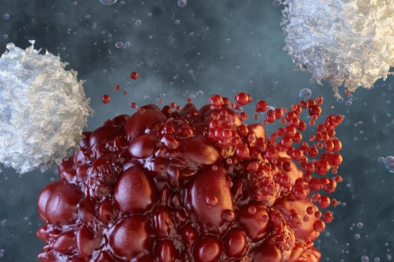 A protein called TRAILshort may act as a central immune brake that limits T-cell responses and CAR-T therapy. Cancer cells can weaken the immune system by producing a protein that prevents its most important defenders from attacking. Mayo Clinic researchers have identified how this protein works, revealing a possible target for treatments designed to improve [...]