 Login

National Academies Report Backs Climate Change Attribution Science
Attribution science is advancing quickly, researchers said. That could support lawsuits seeking damages for severe events worsened by global warming.


© Stephane Mahe/Reuters
Since riding Donald Trump’s coattails to power, Robert F. Kennedy Jr has taken his haphazard anti-vaccine views and enshrined them as a core tenet of US health policy.
According to 16 current and former health officials who spoke to Reuters, RFK Jr has taken much more drastic measures to push his anti-vaccine agenda than was previously known.
Speaking on condition of anonymity, two officials told the news agency that RFK approached National Institute of Health director Jay Bhattacharya earlier this year with a proposal to spend up to $5 billion in tax dollars to establish links between vaccines and autism.
As Reuters notes, that kind of spending would encompass ten percent of the NIH’s annual budget, a massive waste that would fly in the face of decades of established science showing vaccines definitively don’t cause autism.
“He’s an anti-vaccine activist. That’s who he is. That’s who he’s been for 20 years,” Paul Offit, director of the Vaccine Education Center at the Children’s Hospital of Philadelphia told Reuters. “To expect that as secretary of Health and Human Services he’d be anything other than that is wishful thinking.”
It certainly wouldn’t have been the first time RFK Jr attempted to twist the science to link vaccines with autism. Earlier this year, the US Department of Health and Human Services was caught spending $1.6 million funding a study on hepatitis B vaccines conducted by controversial Danish researchers Peter Aaby and Christine Stabell Benn.
Had the Africa Centres for Disease Control and Prevention — the public health arm of the Africa Union — not stepped in to stop the RFK-backed study, researchers would have withheld inoculations from thousands of newborn infants in Guinea-Bissau.
The situation drew notable comparisons to the Tuskegee Experiment, a highly unethical medical experiment that used hundreds of Black men as guinea pigs to study the long-term effects of syphilis. Despite the fact that effective treatment was available, nearly 400 men spent decades without treatment, suffering life-long consequences at the hands of manipulative researchers.
Though the Tuskegee Experiment may not have produced any medical breakthroughs or scientific discoveries, its cruel legacy seemingly lives on as inspiration for RFK and his HHS cronies.
More on RFK Jr: CDC Report on Vaccines and Autism Caught Citing Hallucinated Study That Does Not Exist
The post RFK Jr Wants to Waste $5 Billion in the Worst Way Imaginable appeared first on Futurism.


The next time you’re checking into a hospital, it’s possible that your nurse may actually be 8,000 miles away.
That’s the reality at a growing number of hospitals throughout the US and the world, which are turning to low-paid telehealth workers in the Philippines to help fill in the duty roster. As it is for remote driving and AI data labeling, the Philippines has become a wellspring of cheap labor for transnational healthcare companies, which are more than happy to exploit a country where millions of people live in poverty.
Stunning reporting by Rest of World detailed the lives of some of the 210,000 full-time Filipino workers employed in remote healthcare. This new class of workers makes up for a massive shortage of almost 80,000 registered nurses in the US.
One of the workers, Alice, is a licensed nurse working in Quezon City, a city of three million people in the Philippines. In 2019, she signed on with a telehealth company providing mental health and substance abuse treatment to US-based patients in California and New Mexico. Alice told RoW she earns $5 an hour, five times as much as the hospital job she left behind.
“We have this virtual clinic that functions like a lobby where patients check in,” Alice explained, “and then we traffic or triage them and send them to each of the providers’ personal Zoom room.”
To get a job communing with US patients through a screen, a Filipino worker simply needs to have a medical degree — it doesn’t matter what kind. While it’s likely that many telehealth recruits come from healthcare administration backgrounds, RoW notes that about 30 percent of the people employed in these jobs are trained nurses or other medical professionals.
The result is a scenario where tens of thousands of healthcare workers are physically present in their communities, but spend their days caring for patients half a world away in a desperate bid to pay the bills. Already facing a mounting shortage of trained healthcare professionals, the Filipino people have watched as telehealth corporations court the remaining hospital workers to oversee care for patients in the US.
“If they can’t go abroad, remote nursing is the next best thing because local wages are so low,” Nico Uba, secretary general of the group Filipino Nurses United told RoW. “Then local hospitals are left understaffed and overworked.”
The companies running this insidious scheme prey on two sets of healthcare workers on opposite ends of the world: the underpaid Filipino, for whom a wage of $5 an hour is much higher than the national average, and the overworked American, for whom a 50-hour work week is practically a vacation.
The remote healthcare industry is well aware of this tension, and is happy to take advantage. As JL Botor, president of the Healthcare Information Management Association of the Philippines told RoW, US hospitals can save up to 70 percent of their overhead costs by hiring Filipino workers instead.
In all, Botor notes that the country is a “clinical process outsourcing powerhouse,” as well as “a premier global hub for supporting overstressed international healthcare systems.”
More on healthcare: Lawsuit Claims the Mayo Clinic’s Use of AI Is Butchering Patient Care
The post US Hospitals Are Outsourcing In-Person Nursing to Remote Health Workers in the Philippines appeared first on Futurism.

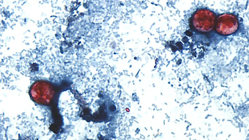
On Thursday, the New York Times reported that the Centers for Disease Control and Prevention had linked the outbreak of cyclosporiasis, an infection notorious for inducing explosive diarrhea, to iceberg lettuce from Taylor Farms that was supplied to Taco Bell.
Per the reporting, Taco Bell said it was voluntarily pulling the lettuce from select states where it received the contaminated produce: Indiana, Kentucky, Michigan, Ohio, and West Virginia. An announcement from the CDC warned the public not to eat food items with shredded lettuce from Taco Bell, but hasn’t identified Taylor Farms as the source.
But while Taco Bell may have served the tainted lettuce, it would be remiss to lay all of the blame on the fast food chain (although one aggrieved customer is already suing it after contracting the illness).
The contamination occurred at the farms or facilities where the food was processed, from a single supplier in Mexico, Food and Drug Administration officials said. The supplier, Taylor Farms, has been involved in previous outbreaks, including another outbreak of cyclosporiasis in a salad mix in 2013.
Taco Bell, to its credit, also immediately pulled its ingredients from some of its locations last week as soon as it learned about the situation, according to local reports.
And then there’s the broader political context that many experts are drawing attention to: how the Trump administration has slashed funding to the CDC and paralyzed the nation’s food safety bodies.
Last February, shortly after Trump took office, the US Department of Agriculture (USDA) dissolved its National Advisory Committee on Microbiological Criteria for Foods, which provided scientific guidance on food safety policy. Ryan Cooper at The Prospect notes that the committee was working on advice for foodborne illness outbreaks before being terminated. The FDA also shut down its National Advisory Committee on Meat and Poultry Inspection.
Not spared during the wave of cuts that wreaked the federal government, the USDA lost over 20,000 workers last year, with 913 of them coming from its Food Safety Inspection Service.
Last year, the CDC — which has lost some 3,000 employees under Trump — scaled back its food surveillance program called FoodNet that monitored eight common foodborne pathogens, changing it so that surveillance of the cyclospora parasite that causes cyclosporiasis was optional.
Dan Jernigan, a former director of CDC’s Center for Emerging and Zoonotic Infectious Diseases, told the Washington Post that the decision was made because funding for food safety had not been maintained over the past few years. That the US is now on track to have more cyclospora cases this year than any previous year in history is likely no coincidence.
Other incidents show how the administration has taken a recklessly laissez faire approach to public health. The US’s worst measles outbreak this century occurred after Trump had hollowed out the CDC — and after health secretary RFK had spent years heavily promoting anti-vaccine rhetoric. The CDC also reportedly fired its ship inspectors last year, a decision that came under scrutiny during a deadly hantavirus outbreak on a Dutch cruise ship. Some experts estimate that hundreds of thousands and possibly millions of children have died as the result of Trump dismantling USAID, which funded emergency food assistance programs and HIV response across the globe.
Jernigan left the agency last August in protest of Trump ousting its then director Susan Monarez and the policies of health secretary Robert F Kennedy Jr. But he said that the Trump cuts would have little impact on the ongoing outbreak, adding that FoodNet wasn’t designed for real-time outbreak detection or response.
As the WaPo reporting notes, federal and state health authorities have insisted that the cyclosporiasis investigation hasn’t been impeded because the parasite is still reportable to a separate national database. Still, Jernigan said that scaling back FoodNet wasn’t inconsequential.
“Routine outbreak investigators are the firefighters responding to a specific blaze. FoodNet is the citywide system that tracks how often fires occur and whether the overall risk is rising,” he told WaPo. “Removing cyclosporiasis from FoodNet does not take the firefighters away from the current outbreak, but it does remove part of the system that tells us how large the problem is and whether it is getting worse over time.”
Cyclosporiasis is a particularly tough illness to track. Symptoms can take weeks to present, making it difficult for health officials to trace back where sufferers were infected. A recent Wired piece drew attention to how many people may be wrongly misdiagnosing themselves as news of the diarrhea dilemma spread across social media.
Linda Yancey, an infectious disease specialist at Memorial Hermann Health System in Texas, warned that large outbreaks like this could become more common.
“Unfortunately with so many budget cuts, every federal agency is hurting for personnel and resources,” she told Time Magazine earlier this month. “We could be at greater risk of foodborne infections because the FDA is so understaffed and underfunded right now.”
So far, Michigan, the epicenter of the outbreak, has alone tallied over 4,300 cases of infection, while nationwide there could be 7,000 cases, though the Centers for Disease Control and Prevention has only confirmed 1,600 of them.
More on parasites: If You’ve Been Having Explosive Diarrhea, You May Want to Read This
The post Let’s Be Real, The Explosive Diarrhea Outbreak Is Trump’s Fault, Not Taco Bell’s appeared first on Futurism.

A new Bayesian system called ALADYNOULLI is designed to forecast how multiple diseases unfold over time from longitudinal electronic health record (EHR) histories. The work targets a central challenge in longitudinal modelling: evaluating prediction without “information leakage,” so that future diagnoses never influence training for past timepoints. By explicitly simulating real clinical follow-up, the researchers aim to make risk estimates more trustworthy for decision-making.
In the primary assessment, ALADYNOULLI produced dynamic 1-year forecasts at 10 fixed moments during follow-up, starting at enrollment and extending to years 1 through 9. At each timepoint, models were retrained using only data available up to that moment, then tested on 1-year outcomes. The key metric was the median area under the receiver operating characteristic curve (AUC) across these dynamic evaluations, summarizing how predictive accuracy changes as new diagnoses accumulate.
The study spans 28 diseases and reports robust discrimination for several clinically important endpoints. Performance was strong for atherosclerotic cardiovascular disease (ASCVD), breast cancer, atrial fibrillation, heart failure, and Parkinson’s disease, with AUC values of 0.879, 0.867, 0.801, 0.811, and 0.796, respectively. Importantly, the evaluation excluded people with prevalent disease at prediction time, aligning the comparisons with prospective risk.
ALADYNOULLI was also benchmarked against established clinical risk tools where feasible. At enrollment, static models trained only on baseline information were compared for both 1-year and 10-year horizons. While longer-term predictions naturally degraded, ALADYNOULLI retained meaningful power—for example, for ASCVD at 10 years—highlighting the advantage of learning from longitudinal EHR trajectories rather than relying solely on cross-sectional inputs.
Calibration analyses further supported reliability: predicted and observed event rates matched closely across millions of at-risk patient-time observations. The researchers report a mean squared error on calibration of 4.67 × 10⁻⁷, with mean predicted rates (5.55 × 10⁻⁴) closely tracking mean observed rates (5.45 × 10⁻⁴). This suggests not only ranking accuracy, but also quantitative risk realism.
To test whether results were driven by subtle temporal artifacts, the team ran sensitivity analyses that excluded events occurring shortly before prediction and varied washout windows. They also examined alternative horizons and a dynamic 10-year rolling interpolation strategy, which updates risk estimates as annual information becomes available. Together, these checks help confirm that temporal patterns are being learned in a causally aligned manner.
Overall, the findings position ALADYNOULLI as a clinically relevant framework for multi-disease risk forecasting, combining longitudinal EHR modelling with predictive discrimination and calibration. By rigorously preventing leakage and validating dynamic follow-up scenarios, the approach offers a pathway toward more actionable, time-aware risk prediction in real-world healthcare settings.
Subject of Research: Longitudinal EHR-based multi-disease risk prediction using Bayesian modelling and genetic/EHR integration
Article Title: A Bayesian framework for longitudinal EHR and genetic discovery.
Article References: Urbut, S.M., Ding, Y., Nakao, T. et al. A Bayesian framework for longitudinal EHR and genetic discovery. Nature (2026). https://doi.org/10.1038/s41586-026-10780-5
Image Credits: AI Generated
DOI: https://doi.org/10.1038/s41586-026-10780-5
Keywords: longitudinal EHR, Bayesian framework, disease risk prediction, dynamic forecasting, temporal validation, calibration, AUC, ASCVD
New blood vessels flood the developing brain, then must be sculpted into a functional network. In the cortex, this pruning depends on selective elimination of redundant connections, but the cellular command center behind vascular refinement has remained only partly understood. A new study now points to microglia—the brain’s immune sentinels—as active architects of cerebrovascular pruning during postnatal development.
Using live visualization in the frontal cortex of postnatal day 11 mice, researchers tracked how microglia behave around forming blood vessels. They observed that microglia do not merely associate with vasculature; instead, they progressively migrate toward vessels as development proceeds. As vessel remodeling advances, microglia become enriched at sites where pruning occurs, suggesting a causal role rather than passive surveillance.
To test function, the team manipulated microglia–vessel interactions and assessed vascular outcomes. Intervening in microglial activity altered the extent of vessel elimination, consistent with the idea that microglia mediate pruning to refine the emerging vascular map. The results connect microglial behavior directly to the efficiency and organization of the developing cortical circulation.
Central to the mechanism is an immune signaling pathway previously linked to neuronal synaptic refinement: PD-L1 and its receptor PD-1. The study identifies PD-L1–PD-1 signaling as a regulator of microglia-driven vascular pruning. When this axis was disrupted, pruning was impaired, indicating that immune checkpoint-like communication can shape vascular architecture in the developing brain.
The authors further implicate a specific molecular source from microglia. They report that microglial CD5L secretion promotes the PD-1–dependent pruning program. In other words, microglia release CD5L, engage PD-1 signaling through PD-L1 interactions, and thereby influence which vessels persist and which are eliminated.
Collectively, the work reframes cerebrovascular pruning as a neuroimmune-guided developmental process. By showing that microglia actively target redundant vasculature and by defining a PD-L1–PD-1/CD5L pathway, the study provides mechanistic evidence for how immune cells can tune vascular networks to match developing brain circuitry.
Beyond development, the findings raise broader questions about whether similar immune checkpoint signaling contributes to abnormal vascular remodeling in disease, where pruning can be either excessive or insufficient. For now, the study delivers a clear developmental role: microglia help determine what the brain keeps growing and what it discards.
DOI: https://doi.org/10.1038/s41593-026-02378-x
Article Title: Microglia prune developing cortical blood vessels through PD-1 signaling.
Article References: Zhang, M., Wang, Y., Yang, R. et al. Microglia prune developing cortical blood vessels through PD-1 signaling. Nat Neurosci (2026). https://doi.org/10.1038/s41593-026-02378-x
Image Credits: AI Generated
A newly published study in Nature Metabolism reports that brown fat can act as a hub of molecular messaging, using secreted microRNAs to coordinate physiology across the body. Researchers focused on brown adipose tissue—well known for its role in thermogenesis—and asked whether it also participates in long-distance communication via RNA signals.
To visualize and track these messages, the team employed brown-fat–specific RNA labeling in living systems. This strategy allowed them to connect RNA production within adipocytes to downstream effects elsewhere, moving beyond correlations toward a clearer map of inter-organ RNA traffic.
The central finding is the presence of a communication network mediated by microRNAs released from brown fat. These small regulatory RNAs appear to travel beyond the tissue of origin and engage gene-regulatory pathways in other organs, effectively relaying information about metabolic state.
Functionally, the work supports a model in which microRNA “dispatch” from thermogenic fat tunes cellular programs in remote targets. Such programs likely involve post-transcriptional regulation, a process microRNAs are known to influence by pairing with complementary sequences in messenger RNAs.
Importantly, the approach does not treat microRNAs as generic circulating molecules. Instead, the labeling confirms their brown-fat origin and strengthens the link between secretory RNA dynamics and systemic physiological outcomes.
The study also highlights the specificity of the messaging: not all RNAs are equally represented, and the labeled microRNA repertoire points to organized, tissue-defined signaling rather than random leakage.
Beyond mapping pathways, the results carry implications for metabolic disease research. If brown fat microRNAs can reshape gene expression in other organs, manipulating these signals could offer a route to modulate energy balance, insulin sensitivity, or inflammatory tone.
For now, the findings provide a blueprint for how RNA labelling combined with tissue specificity can uncover hidden layers of endocrine-like communication. As techniques improve, the field may move toward a broader “RNA connectome” linking organ behavior through secreted regulatory molecules.
Researchers describe their results as evidence that brown fat is not only a metabolic engine but also an information broadcaster. In the evolving landscape of viral-style science news, this discovery underscores how tiny RNA molecules can orchestrate large-scale biological conversations.
Subject of Research: Inter-organ communication via secreted microRNAs from brown adipose tissue.
Article Title: Brown fat-specific RNA labelling reveals a network of inter-organ communication by secreted microRNAs.
Article References: Lino, M., Palermo-Ruiz, G., Efthymiou, V. et al. Brown fat-specific RNA labelling reveals a network of inter-organ communication by secreted microRNAs. Nat Metab (2026). https://doi.org/10.1038/s42255-026-01558-0
Image Credits: AI Generated
Naked mole-rats have long puzzled biologists with their striking eusocial structure, where most adults remain reproductively suppressed while a queen controls the colony’s future. Now, a new study identifies a chemical “queen odour” that links social status to endocrine control, revealing a mammalian analogue to insect queen pheromones.
The molecule, termed IPM, is enriched by the queen and acts as the key mediator of reproductive suppression in non-breeding females. When IPM is present, circulating prolactin rises while progesterone is suppressed, locking the colony into a stable reproductive hierarchy characterized by reduced conflict and maintained group cohesion.
Crucially, the team connects this behavioural outcome to the brain. Using multiple approaches—including non-invasive functional ultrasound imaging (fUSI)—they show that IPM provokes strong activation of olfactory neurons and central neural circuits. This provides a neural substrate for how a chemical cue can trigger colony-wide physiological change, rather than relying on social learning or passive association.
The experiments also clarify sensory routing. Ablating olfaction abolishes aggression initiation and lowers prolactin levels, directly tying IPM detection to reproductive suppression. While the findings are consistent with sensing through the main olfactory epithelium, the authors note that the vomeronasal system may still contribute.
Queen removal normally destabilizes colonies, leading to aggression, reproductive competition, prolactin decline, and, in extreme cases, mortality. Yet when IPM is added to queenless bedding, the colony remains stable for months. During this period, fecal progesterone metabolite levels stay low, indicating sustained ovarian quiescence alongside elevated prolactin.
The study also addresses a longstanding paradox: transferring soiled bedding and urine from parent colonies fails to maintain suppression in isolated individuals. The authors propose that IPM’s chemistry helps explain this discrepancy. As a long-chain fatty acid ester with low volatility, IPM may persist within the colony environment but fail to travel effectively over distance or survive transfer pathways that preferentially carry other chemicals.
Comparative work extends beyond naked mole-rats. IPM is detected in breeding females of four additional Fukomys species. Although reproductive suppression mechanisms may differ across species, the presence of a similar queen-linked odorant suggests convergent chemical strategies for managing reproductive skew in eusocial mammals.
Overall, the research reframes reproduction control as a chemosensory-to-endocrine pathway: IPM activates olfactory–hypothalamic signalling, elevating prolactin and inhibiting GnRH release. This “social hormone” model offers a new principle for social evolution—chemical cues can synchronize behaviour and physiology at the scale of an entire mammal colony.
Subject of Research: Reproductive suppression and social hierarchy in eusocial mammals (naked mole-rats and related Fukomys species)
Article Title: A queen odour mediates reproductive suppression in a eusocial mammal
Article References:
Khallaf, M.A., Hart, D.W., Luo, W. et al. A queen odour mediates reproductive suppression in a eusocial mammal.
Nature (2026). https://doi.org/10.1038/s41586-026-10772-5
Image Credits: AI Generated
DOI: https://doi.org/10.1038/s41586-026-10772-5
Keywords: IPM; queen odour; reproductive suppression; prolactin; GnRH; olfaction; eusociality; functional ultrasound imaging; Fukomys

The first X-ray ever taken of a human body, in 1895, was of Wilhelm Röntgen’s wife’s hand, wedding ring and all. One hundred and thirty years later, four amateurs orbiting Earth at roughly 17,000 miles an hour recreated that portrait — a hand, a ring, the same pale architecture of bone — except this image came down from space.
It is the first diagnostic X-ray ever captured of a human being in orbit, and it is now confirmed. A study published in Radiology, the journal of the Radiological Society of North America, reports that the crew of SpaceX’s Fram2 mission acquired anatomic radiographs during their 2025 flight, and that three independent radiologists on the ground judged the orbital images statistically indistinguishable from scans taken before launch — good enough to spot a fracture, a foreign object, or the kind of injury a crew doctor would need to see fast.
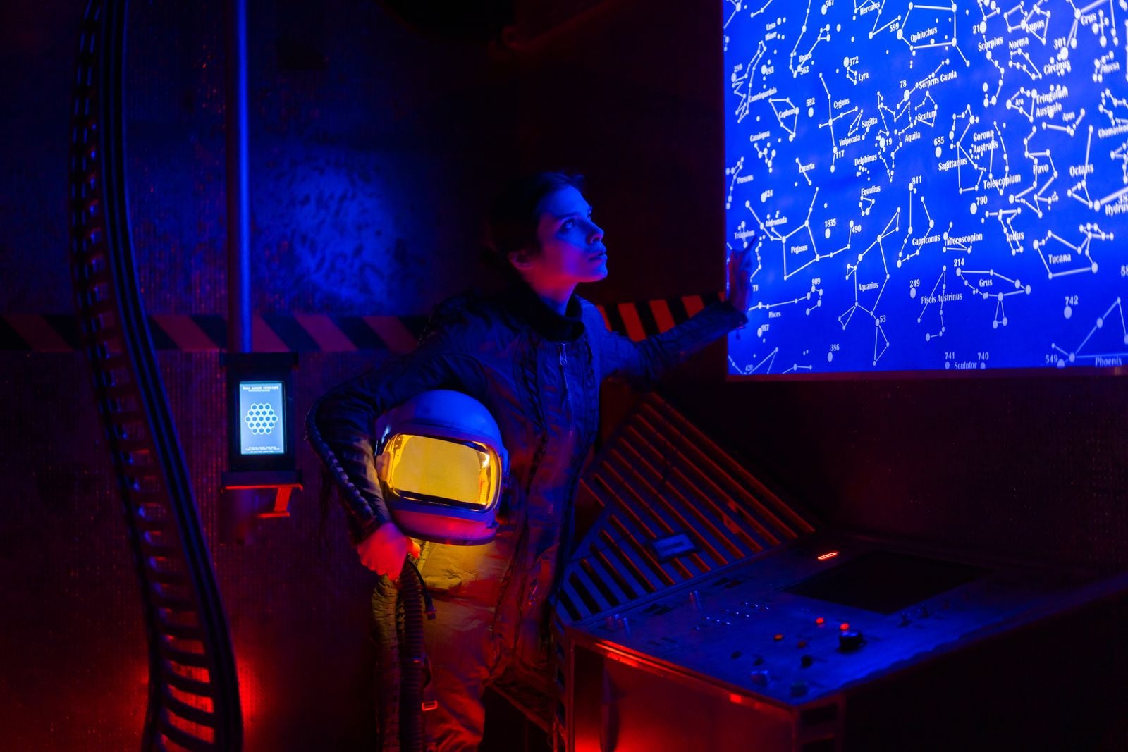
The device produced a full set of images across the multi-day flight: a phantom control object, a smartwatch, a hand, a forearm, a chest, an abdomen, and a pelvis. The scans were recorded digitally, so the crew could review them on the spot without developing film. The study was led by Dr. Sheyna Gifford, an aerospace medicine physician at Mayo Clinic, whose team had spent years working toward exactly this test.
Space medicine has a physics problem. Ultrasound, the workhorse of orbital diagnostics for more than four decades, transmits sound waves through gel-coupled tissue. It works fine inside a pressurized cabin. It struggles with bone, where the internal structure often stays a mystery on ultrasound, and it does nothing for the interior of a cracked component. X-rays, being electromagnetic radiation, pass straight through.
That distinction matters as crewed missions push farther from Earth than at any time in half a century. In April 2026, the Artemis II crew traveled about 252,700 miles from Earth, breaking the distance record Apollo 13 set by accident in 1970. Lunar surface stays under Artemis III and the missions that follow will place crews days away from any hospital. A broken tibia on the Moon is not a broken tibia in Houston.
The operational finding may matter as much as the clinical one. The Fram2 crew — cryptocurrency investor Chun Wang, filmmaker Jannicke Mikkelsen, engineer Rabea Rogge, and polar explorer Eric Philips — were all first-time flyers, and none were physicians. Three of them received roughly four hours of training on the portable system before launch, about the length of a morning workshop, then operated it themselves in microgravity.
That mirrors how portable X-ray already gets deployed on the ground. Gifford has pointed out that these units already run at the Kentucky Derby, on Super Bowl sidelines, and across low-resource regions worldwide, precisely because they can run on solar power and be operated by people with no medical training. Staying still enough to take a clean image in freefall was the open question, and the crew answered it.
The hardware also survived the ride. After launch vibration, re-entry heating, and a Pacific splashdown, the X-ray generator came back with only superficial structural damage. That is not a minor achievement. Most medical imaging equipment is designed on the assumption it will never leave a temperature-controlled room.
The mission itself was an unusual choice for a first-of-its-kind medical test. The privately funded flight launched from Kennedy Space Center and became the first crewed spaceflight to fly a 90-degree polar orbit, passing from pole to pole roughly every 46 minutes at an altitude of about 265 miles. Over three and a half days, the crew ran 22 research experiments, from growing mushrooms to photographing aurora, with the X-ray work among the headline objectives.
The polar trajectory was incidental to the radiography. What mattered was the profile: a short-duration flight with untrained operators willing to serve as both subjects and technicians. That is close to the exact operational shape of a lunar sortie crew, and the International Space Station rarely offers it.
Before the orbital attempt, Gifford’s group had already flown a portable unit on a parabolic flight in 2022, showing that viable diagnostic images were possible in simulated microgravity. The orbital flight was the graduation exercise, and it extended the capability beyond bone.
The smartwatch image is the tell. An X-ray that can image a fracture can also image a suspect weld, a cracked pressure vessel, or a foreign object lodged where it should not be. Gifford has framed this as the point: for a sustained presence in space, X-rays matter not only for crew but for electronics and spacesuits, which cannot be inspected inside without either taking them apart or seeing through them. On a lunar habitat where every spare part had to be launched from Earth, non-destructive inspection is itself a life-support function.
The terrestrial implications may be larger still. Diagnostic imaging is one of the widest gaps in global health infrastructure. A device rugged enough for spaceflight, cheap enough to deploy at scale, and simple enough to run after four hours of instruction is a plausible answer to that gap — the same argument that already puts these machines in rural clinics far from any hospital.
An X-ray is a diagnostic tool. It is not a treatment. A crew on the lunar surface with a confirmed compound fracture still faces the problem astronauts have always faced: the nearest orthopedic surgeon is a quarter-million miles away. The value of imaging in that scenario is triage — knowing whether to continue the mission, improvise a splint, or begin a medical evacuation that could take days.
Open questions remain. The images were taken in microgravity, not the one-sixth gravity of the lunar surface, where positioning a patient and holding source-detector geometry steady will behave differently again. Radiation exposure protocols inside a cabin already bathed in cosmic rays will need their own rulebook. And while the in-flight images matched the crew’s own preflight scans, the portable system’s ceiling against full hospital equipment is a separate question this flight did not settle.
The broader story is about who is now doing this kind of work. A mission funded by a private citizen, flown on a commercial capsule, carrying a commercial off-the-shelf X-ray unit, tested by an academic medical center, produced a peer-reviewed result in a mainstream journal. NASA was not the operator. Mayo Clinic was not the launch provider. SpaceX was not the principal investigator. Each played one part, and the parts fit.
That distributed model is what lunar-era space medicine will probably look like. The Apollo-era approach — one agency, one contractor stack, one flight surgeon in mission control talking to one crew — cannot scale to the cadence of missions that Artemis, Gateway, and commercial low-Earth-orbit stations imply. A portable X-ray a non-physician can operate is a small piece of hardware. It is also a preview of the operational philosophy that will have to keep future crews alive when Houston is a light-second and a half away.
The hand that came down from Fram2 sits in the same lineage as the one Röntgen made in 1895. Same bones, same rings, the same faint gray shadow of tissue around them. One was taken in a laboratory in Würzburg. The other was taken by an amateur, hurtling over the poles, 265 miles above the planet, and it came home clear enough to read.
The post Four first-time astronauts with only four hours of training took the first diagnostic X-rays ever captured on human beings in orbit, and radiologists on the ground judged the images clinically usable enough to catch a fracture a quarter-million miles from the nearest hospital appeared first on Space Daily.


Read more of this story at Slashdot.
Apocryphal tales of its meals causing the runs have plagued Taco Bell since time immemorial. But this latest parasitic outbreak is something else entirely.
As the Washington Post reports, federal and state authorities are investigating whether the fast food chain is behind the outbreak of explosive diarrhea cases across the country — further cementing its scatological legacy.
The illness, caused by the cyclosporiasis parasite, has sickened as many as 5,100 people across the country, according to the Centers for Disease Control and Prevention. In Michigan, which appears to be the epicenter of the outbreak, over 3,700 people have been infected, state authorities said.
Taco Bell’s suspected role in the outbreak comes after reports that the chain’s Michigan locations began pulling some of its ingredients. Signs in front of the restaurants stated that they were no longer serving lettuce, cilantro, onion, pico de gallo, and guacamole due to a “nationwide recall,” though what recall they were referring to is unclear.
Health officials have so far struggled to narrow down how the parasite is spreading. While usually found on fresh produce, symptoms can take weeks to manifest, making it difficult to trace back to the source.
Lettuce has emerged as one of the likeliest culprits. In an update this week, Michigan’s chief medical executive Natasha Bagdasarian said that “early information has shown lettuce as a common product that regularly comes up during the investigation.”
“Although we do not have a definite product identified as the source of the outbreak, we want to let Michiganders know what we have learned so far so they can take steps to protect their families,” Bagdasarian added.
Federal authorities haven’t singled out any veggies yet, though acting Food and Drug Administration director Don Prater acknowledged Michigan’s fixation on lettuce.
“What I can say at this point is that we’re continuing our traceback investigation on multiple produce items, including the lettuce,” he said, per WaPo.
So far, no nationwide recalls have been made related to the outbreak. One person familiar with the federal investigation, however, praised Taco Bell’s response.
“It’s good the chain has already voluntarily stopped serving a number of items,” they told the newspaper. They added that some who became sick said they didn’t eat at the chain, suggesting that it’s not the only one responsible.
More on parasites: If You’ve Been Having Explosive Diarrhea, You May Want to Read This
The post Evidence Grows That Taco Bell Infected Massive Number of Customers With Explosive Diarrhea appeared first on Futurism.


New research from Columbia University found that adults who cut their sleep by about 80 minutes for six weeks packed on a pound (0.45 kg) and moved less during the day.
The post Losing Even 80 Minutes of Sleep a Night Linked to Weight Gain appeared first on Sci.News: Breaking Science News.
Read more of this story at Slashdot.
Last February, Alice Gilman, the cofounder and chief operating officer of a secretive Silicon Valley longevity startup called R3 Bio, sat for an interview with podcast Skyline Drive. In the interview, she discussed R3 Bio’s aspirations to grow nonsentient humanoid life-forms — from which, in theory, they can grow and harvest organs for implantation into human bodies, or to test future drugs and other treatments without the need for live animal testing.
“It’s just pretty much a heart, lungs, and everything that you would find in a body,” Gilman tells host and journalist Mangesh Hattikudur. “But it’s not technically alive because it’s exclusively the organs. And they’re in this little biological platform that you can test on and you can map out the cross-organ interactions.”
Throughout their conversation, as Hattikudur notes, Gilman refers to these imagined unfeeling meat suits — which will ideally be born without brains — as “organ sacks.” Needless to say, it’s an ambitious (and to many, fundamentally unsettling) concept that raises plenty of nagging bioethical questions — a reality that the company is clearly wrestling with as it entertains the idea in public.
“I try to not be scary to an average person,” Gilman told Hattikudur.
Months after this interview took place, however, Gilman fought to keep it under wraps, as Skyline Drive told Futurism. Why? It’s not exactly clear.
About a week before the podcast episode was set to air, producers for the Skyline Drive reached out to Gilman to fact-check the conversation. To their surprise, Gilman requested that the episode be postponed. Despite describing imagined “organ sacks” in detail, she denied that R3 Bio was working on the concept “for now, period,” and declined to say what it was working on instead.
Gilman didn’t argue that the interview was factually incorrect. Instead, the podcast team said, she vaguely referred to R3 Bio as a “federal asset,” seemingly implying that she had to tread lightly due to government involvement. To be certain, “federal asset” usually implies some kind of ownership or domain over something held by the federal government. Which would imply that the federal government is investing in a company that’s claimed to have worked on “organ sacks.” She also insisted that what she said in her nearly two-hour-long interview was all theory and opinion.
R3 Bio only recently burst into the public sphere. In March, it announced its existence in a buzzy Wired article, in which R3 Bio told the magazine that it was specifically working to create “organ sacks.” Company investor Boyang Wang elaborated on the idea, telling Wired that “replacement is probably better than repair when it comes to treating diseases or regulating the aging process in the human body,” and that “if we can create a nonsentient, headless bodyoid for a human being, that will be a great source of organs.”
But R3 Bio’s ambitions seemingly go far beyond “organ sacks.” A few days after Wired’s piece ran, MIT Technology Review published a deep dive about R3 Bio’s apparent aspirations and its cofounder John Schloendorn — a self-described “biological designer” who has given confidential talks about “full body replacement” and “recent lab progress towards making replacement bodies.” Generally speaking, replacement bodies refer to physical clones that will allow for humans to transplant their heads onto a whole new human body, a still-theoretical idea that’s attracted the fascination of several scientists. (In response to the MIT Tech piece, R3 Bio issued what the outlet described as a “sweeping disavowal” of its findings, and said that “any allegations of intent or conspiracy to create human clones or human beings with brain damage are categorically false.”)
In her interview with Skyline Drive, which took place before Wired or MIT Tech published their respective pieces, Gilman says that the company has “a few milestones that we’re hoping to achieve. The first milestone being we are working on replacing animal testing by using skin stem cells.”
“It’s mostly a stealth project, but the essence of the surface level of what we do is we take skin cells from monkeys and we reprogram them and do some Yamanaka miracles,” she said, referring to the Nobel Prize-winning scientist Shinya Yamanaka’s transformational stem cell research, “that result in fully grown organ systems.”
In its March reporting, Wired found that the company had put out a job listing for a veterinarian in Puerto Rico who could “implant embryos, monitor pregnancies and help deliver healthy births” in nonhuman primates. On its website, however, R3 Bio states that it does “not work with live primates.”
Gilman gave the Skyline Drive interview from an island in Puerto Rico. R3 Bio wanted “monkey access,” Gilman told Hattikudur, but didn’t want to put monkeys in cages, a practice that the cofounder believes is unethical; after all, one goal of her company is to eliminate the need for animal testing.
So instead, they picked an island with a lot of monkeys.
“I’m a big animal rights advocate… I don’t want to keep monkeys in cages,” she told Hattikudur. “And there’s an option to have them run around the island with bracelets on.”
“We wanted monkey access and there’s a lot of monkeys here,” she added. “After negotiating with local companies that are supervising them, we can walk up to them, get some of their skin cells.”
During the conversation with Skyline Drive, Gilman also discussed one very big scientific problem the company is facing: that the less sentient an “organ sack” is, the less likely it is to function effectively.
“So do these organ sacks have the stem of a brain?” Hattikudur asked.
“So the mice models can exist with a very limited set of nervous systems… but then there are other constraints on the ability of the organs to preserve themselves,” Gilman responded. “So there’s a way in which you can skew towards social acceptance and make less nervous system, and then when you scoot towards social acceptance, it looks better, but it’s less scientifically feasible.”
“So we are still making observations about how that would work,” she added.
How far along R3 Bio is right now is murky at best. Gilman cryptically noted that she lives in a “contextual world,” and in that world, “just because we don’t talk about a lot of things, that doesn’t mean they’re not happening.”
On its website, R3 Bio says that while it “would be excited by the future prospect of full-scale human organ fabrication,” it views “the science as currently not ready.”
“We have no ongoing work in this area,” the website notes.
During the interview, Gilman remained confident about the positive impact she believes her company will have. She told Hattikudur that her “worst case scenario” is that she saves “millions of lives,” and that’s a “bet worth taking, regardless of any kinds of blows that are gonna come our way.”
We reached out to R3 Bio to ask why it pushed to postpone the podcast, if it’s currently or planning to obtain skin cells from cage-free island monkeys — and if so, how — and if it has indeed stalled its efforts to create the “organ sacks” it’s now repeatedly discussed in the press. We also asked the company how it defines having “no ongoing work” in the realm of “full-scale human organ fabrication.” We did not hear back at the time of publishing.
In an interview with Futurism, Hattikudur said he was “surprised” by Gilman’s push to suppress the interview.
“The company hasn’t been exactly transparent about what they’re doing,” said Hattikudur. “There’s a lot of mix between the hype they’re telling people and what they’ve actually achieved in the lab, which is not very clear.”
“If they were honest that this is what they believe in, and this is what they’re working on, it would actually be less of a big deal,” he added. “It’s more that they awkwardly seem to be hiding something which everyone kind of knows about.”
More on R3 Bio: A Startup Has Been Quietly Pitching Cloned Human Bodies to Transfer Your Brain Into
The post Secretive Silicon Valley Startup Tries to Quash Interview, Says It’s Definitely Not Working on Growing Brain-Free “Organ Sacks” appeared first on Futurism.
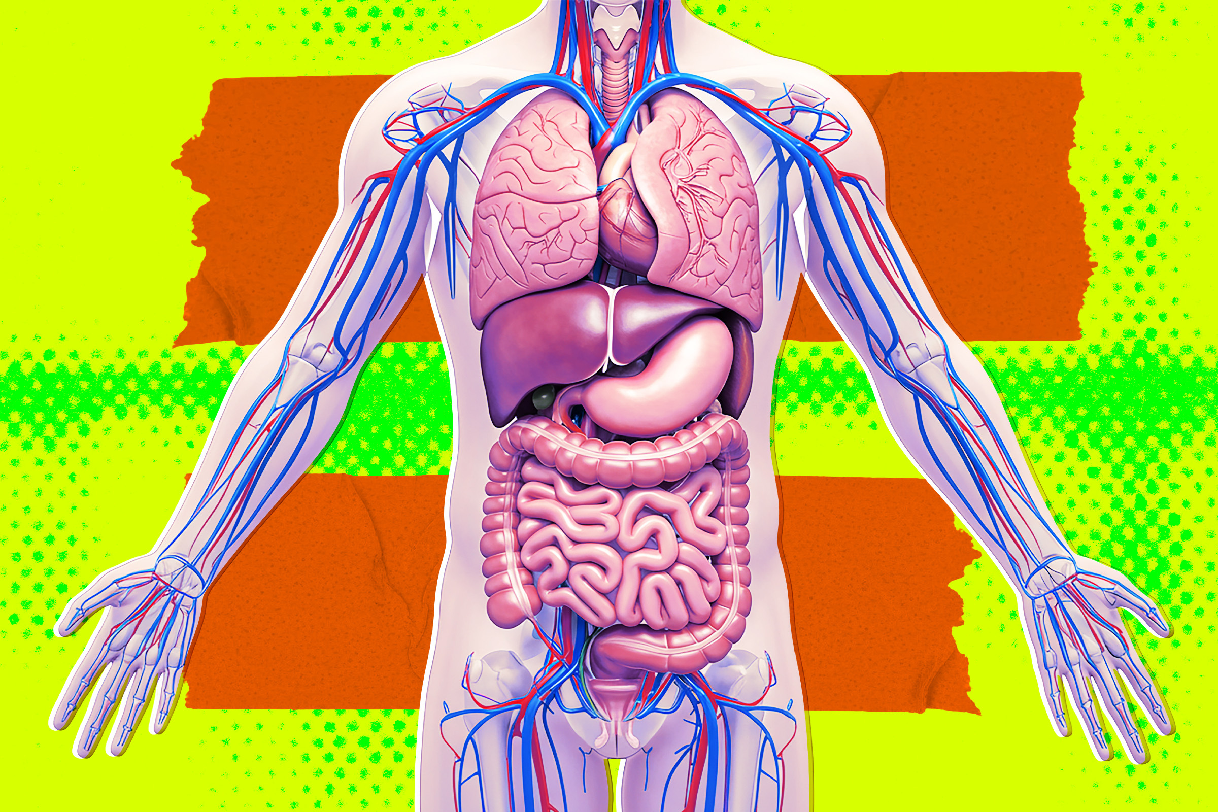
 A newly identified inflammatory signaling target may help weaken the protective environment surrounding pancreatic tumors and make existing treatments more effective. Pancreatic tumors are not made up of cancer cells alone. They are surrounded by scar-like tissue, immune cells, and support cells that can shield the disease from treatment. A new study suggests that disrupting [...]
One of the largest health networks integrating AI into the medical space is facing allegations that its high-tech tools are ruining patients’ lives.
In a new civil suit, former Mayo Clinic research director and AI compliance lead Traci Tamiko Eto alleges that the globally recognized hospital retaliated against her for blowing the whistle on its disastrous implementations of AI health tools.
According to reporting by Minnesota Public Radio, Eto first joined the Mayo Clinic in 2023. After flagging potential privacy problems linked with the hospital’s Mayo Clinic Platform, an AI-integrated data system, Eto says her supervisor ignored her concerns. Instead, she says her boss “insisted” that fixing the issue would “jeopardize the pace on ongoing research projects, which in turn would compromise Mayo’s competitive advantage.”
Eto continued, MPR reports, flagging multiple failures of Mayo Clinic higher-ups to follow federal regulations on review processes of new tech, like MAYA, the clinic’s AI-integrated digital assistant. According to the lawsuit, the team working on MAYA deleted unflattering test results, mischaracterized the tool’s abilities, and made decisions that put data security in jeopardy.
Tucked inside her ten separate whistleblower complaints, MPR notes, is the allegation that the team working on MAYA knew the tool had an error rate as high as 67 percent. Eto alleges that instead of flagging that horrifying number, MAYA staffers tried to hide it.
As a result, Eto says she began to be excluded from executive meetings in early 2025, before being told she was a “poor cultural fit.” She was offered a choice, the lawsuit alleges: either resign, or face alterations to her personnel file “that would render her unemployable at Mayo and would impede her career outside the institution.”
As the major lawsuit moves forward, the threat of career-ending backlash remains.
“When an individual decides that they are willing to file a lawsuit against a public goliath on matters that are hot button issues in our country, she is literally risking her career,” Eto’s attorney, Artur Davis told MPR. “She is risking being attacked professionally. She is risking her reputation, and for her to do that tells you something about her level of confidence that she’s right.”
Mayo was pretty tight-lipped in response to the suit.
“Our research and clinical innovation are conducted in accordance with applicable laws and regulations and we remain steadfast in upholding the trust patients place in us and respecting their privacy,” the organization’s communications director told MPR in a statement. “Mayo Clinic does not comment on pending or active litigation.”
While the suit revolves around Eto’s wrongful termination, it represents a significant referendum on the state of AI tools in the medical space. If a healthcare titan like Mayo Clinic is lying about the efficacy of its AI-integrated tools, what does that mean for the rest of the industry?
“If [people] care about the notion that AI has to be handled in a responsible manner,” Davis said, “with integrity, and there have to be rules and guidelines, this is a case that should matter to you.”
The post Lawsuit Claims the Mayo Clinic’s Use of AI Is Butchering Patient Care appeared first on Futurism.

