❌

Reading view

Hidden Fat Around Blood Vessels Could Hold the Key to Preventing Heart Disease

When doctors talk about obesity and type 2 diabetes, they often focus on blood sugar levels, body weight, and cholesterol. Yet one of the greatest dangers of these conditions is the damage they cause to blood vessels. This damage can quietly develop over many years and eventually lead to heart attacks, strokes, and other serious […]

The post Hidden Fat Around Blood Vessels Could Hold the Key to Preventing Heart Disease appeared first on Knowridge Science Report.

  •  

CCRT Boosts Cognition and Biomarkers in Older Adults

In the rapidly aging global population, mild cognitive impairment (MCI) has emerged as a critical area of focus for researchers dedicated to understanding and mitigating the progression of cognitive decline in older adults. Recent groundbreaking research has illuminated promising interventions, and among the most compelling is the application of Cognitive Control and Reasoning Training (CCRT). The latest study, conducted by Tan, Ping, Zhang, and colleagues, presents robust evidence that CCRT can notably improve cognitive function and modulate blood biomarkers in community-dwelling older adults experiencing MCI, offering a beacon of hope for preventative strategies in geriatric cognitive health.

At its core, Cognitive Control and Reasoning Training is a targeted, intensive cognitive exercise regimen designed to enhance executive functions—the brain’s system for managing attention, problem-solving, and flexible thinking. The new study rigorously quantifies the impact of CCRT through a multidimensional approach, combining neuropsychological assessments with advanced biomarker profiling from blood samples. This integrative methodology provides unprecedented insight into how cognitive training not only improves mental agility but also induces measurable physiological changes associated with brain health and neuroplasticity.

One of the most compelling aspects of this research is its focus on community-dwelling older adults rather than institutionalized populations, emphasizing real-world applicability and accessibility of CCRT interventions. By centering on MCI, a condition characterized by noticeable but not debilitating cognitive decline, the study targets a critical window of opportunity where intervention can potentially halt or even reverse the trajectory toward dementia. This shift in clinical focus is crucial, as it aligns with precision medicine’s broader goal of early detection and personalized intervention.

Methodologically, the study implemented a longitudinal design encompassing a sizeable cohort subjected to a structured CCRT protocol over several months. Participants underwent repeated evaluations using standardized cognitive batteries that assess memory, executive function, attention, and processing speed. Concurrently, blood samples were meticulously analyzed to detect variations in neuroinflammatory markers, neurotrophic factors such as brain-derived neurotrophic factor (BDNF), and markers indicative of oxidative stress. The dual assessment strategy enabled a holistic evaluation of both cognitive and systemic effects induced by the training.

The findings reveal statistically significant improvements in global cognitive scores among those exposed to CCRT, notably in domains linked to working memory and executive control—areas crucial for everyday function and independence. More intriguing, however, were the concomitant changes in blood biomarker profiles, suggesting that cognitive training may exert its benefits through systemic modulation of neurobiological processes. For example, elevated levels of BDNF, a key molecule involved in synaptic plasticity and neuronal survival, were observed post-intervention, signaling enhanced neuroadaptive capacity.

Another striking discovery was the reduction of pro-inflammatory cytokines in the participants’ blood samples following CCRT, underscoring the interplay between cognitive health and immune regulation. Chronic low-grade inflammation has been implicated in the pathogenesis of neurodegenerative diseases, and the ability of targeted cognitive training to attenuate these inflammatory pathways positions CCRT as a multidisciplinary therapeutic tool. Such biomolecular insights offer mechanistic explanations bridging behavioral improvements with underlying neurophysiology.

The implications of this study extend well beyond individual cognitive enhancement, challenging long-held notions about the irreversibility of age-related cognitive decline. The bi-directional relationship between brain function and peripheral biomarkers captured herein supports an emerging paradigm in neuroscience: that cognitive training is not merely a psychological exercise but a biologically rooted intervention capable of reshaping brain-body interactions. This realization paves the way for future integrated treatment strategies combining behavioral therapies with pharmacological approaches targeting neuroinflammation and neurotrophic support.

Importantly, the research team also addressed the sustainability of the cognitive and biological gains induced by CCRT. Follow-up assessments conducted several months post-intervention demonstrated that while some decline in measured benefits occurred, trained individuals retained significantly better cognitive functioning and biomarker profiles compared to control groups. This longevity suggests that CCRT programs may induce lasting neuroplastic changes, albeit emphasizing the need for continuous or booster sessions to maintain optimally enhanced cognitive states.

In parallel, the study highlights the feasibility of implementing CCRT in community settings, utilizing digital platforms and remotely supervised protocols, thereby overcoming traditional barriers such as mobility constraints and access to specialized care. By democratizing cognitive health interventions, this research heralds a new era where preventative cognitive maintenance can be integrated into routine elderly care, dramatically shifting public health strategies aimed at aging populations.

Moreover, the data illuminate potential predictive biomarkers that could be employed for precision targeting of cognitive interventions. Individuals who exhibited greater baseline neuroinflammatory markers and lower BDNF benefitted disproportionately from CCRT, suggesting that biomarker screening might inform patient selection and customization of training programs. Future clinical trials exploring dose-response relationships and personalized adjustments of training complexity are underway to optimize efficacy.

From a broader societal perspective, these findings carry substantial implications for reducing the global dementia burden. With populations aging rapidly worldwide, scalable interventions like CCRT could alleviate healthcare pressures by preserving functional independence and delaying institutionalization. Furthermore, the intersection of cognitive neuroscience and immunology as demonstrated here invites cross-disciplinary collaborations aimed at holistically addressing aging-related cognitive challenges.

While the study’s innovations are groundbreaking, the authors acknowledge several limitations requiring further exploration. The relatively short trial duration and modest sample size warrant larger, multicenter studies to validate and expand upon these results. Additionally, disentangling the specific cognitive components or mechanisms of CCRT responsible for biomarker modulation remains an open question, inviting sophisticated neuroimaging and molecular studies.

Nevertheless, this pioneering research reinforces the hopeful narrative that cognitive decline is not an inevitable consequence of aging but a modifiable condition amenable to targeted, science-backed interventions. The synthesis of behavioral training and biomarker tracking sets a new gold standard for developing interventions grounded in mechanistic understanding, moving cognitive health management toward a future where personalized, preventative care is the norm rather than the aspiration.

As public health systems grapple with the mounting challenges of neurodegenerative diseases, findings like these imbue the scientific community, clinicians, and caregivers with renewed optimism. By harnessing the brain’s inherent plasticity through methods like CCRT, we can envision a future in which older adults maintain cognitive vitality longer, enriching quality of life and sustaining autonomy well into advanced age.

This study by Tan et al. will undoubtedly spark extensive dialogue and inspire subsequent research into multifaceted cognitive therapies. As the field evolves, integrating cognitive training with nutritional, pharmacological, and lifestyle interventions could forge comprehensive, multimodal treatments. Such innovations promise to transform the landscape of brain aging, shifting the narrative from inevitability to empowerment.

In summary, the intersection of cognitive training and biomarker science elucidated by this study marks a monumental step in geriatric cognitive health. The compelling evidence for CCRT’s efficacy positions it as a cornerstone in emerging strategies designed to combat cognitive decline, offering scientific validation to a therapeutic approach that is accessible, scalable, and deeply rooted in our understanding of brain and body biology. The potential societal benefits underscore the urgency of expanding this research frontier to realize the promise of healthier cognitive aging for millions worldwide.


Subject of Research: Effects of Cognitive Control and Reasoning Training (CCRT) on cognitive function and blood biomarkers in community-dwelling older adults with Mild Cognitive Impairment (MCI).

Article Title: A study on the effects of CCRT on cognitive function and blood biomarkers in community-dwelling older adults with MCI.

Article References:
Tan, Z., Ping, J., Zhang, H. et al. A study on the effects of CCRT on cognitive function and blood biomarkers in community-dwelling older adults with MCI. BMC Geriatr (2026). https://doi.org/10.1186/s12877-026-07730-1

Image Credits: AI Generated

  •  

Unraveling Childhood Trauma and Suicidality in Bipolar Disorder

In a groundbreaking study published in Translational Psychiatry, researchers have unveiled intricate behavioral mechanisms that interconnect childhood maltreatment, cognitive function, impulsivity, and suicidality in individuals with bipolar disorder. This revelation stems from applying a sophisticated network analysis approach that sheds new light on the pathways through which early adverse experiences shape mental health trajectories and suicide risk in this vulnerable population. The study’s innovative framework marks a significant advance in psychiatric research, offering fresh perspectives on targeted interventions and prevention strategies for suicidality driven by complex biopsychosocial interactions.

Bipolar disorder, characterized by extreme mood fluctuations between mania and depression, is already known to confer a heightened risk for suicide. However, identifying the underlying behavioral and cognitive pathways that mediate this risk remains a challenging scientific endeavor. The recent study navigates these complexities by mapping the nuanced relationships among childhood maltreatment, cognitive deficits, impulsivity traits, and suicidal tendencies using network modeling techniques, which excel at capturing interconnected variables and their mutual influences. This multidimensional lens promises a more holistic understanding than traditional analytical methods.

The research team collected comprehensive behavioral and clinical data from individuals diagnosed with bipolar disorder, encompassing measures of childhood adversity, cognitive performance, impulsivity scales, and historical records of suicidal behavior. Instead of isolating each factor independently, the scientists utilized network analysis algorithms to visualize and quantify the dynamic interplay between these variables. This approach illuminated distinct behavioral pathways whereby adverse childhood experiences indirectly exacerbate suicidality through their impact on cognitive impairment and impulsivity, rather than through straightforward direct effects.

One of the most compelling outcomes of the investigation is the identification of key cognitive domains acting as mediators between early maltreatment and suicidality. Specifically, deficits in executive functions—the set of cognitive processes involved in planning, decision-making, and inhibitory control—appear central to the cascade linking childhood trauma with elevated suicide risk. Executive dysfunctions increase susceptibility to impulsive behaviors, which are well-documented precipitants of suicidal acts, especially in bipolar populations. These findings suggest that targeting cognitive remediation might attenuate impulsivity and, consequently, suicidality.

Impulsivity, a multifaceted construct encompassing rash decision-making, inability to delay gratification, and heightened risk-taking, emerged as a critical behavioral mechanism connecting cognitive deficits and suicidal tendencies. The network model reveals that impulsivity serves as a conduit translating impaired executive functioning into concrete suicide-related behaviors. Importantly, the data indicate that impulsivity is more than a standalone risk factor; it functions within a broader psychosocial context shaped by preceding cognitive disruptions and trauma exposure. This layered insight has profound therapeutic implications.

Furthermore, the study underscores the pervasive influence of childhood maltreatment on adult psychiatric outcomes, particularly within bipolar disorder cohorts. Maltreatment experiences—including emotional neglect, physical abuse, and sexual abuse—were shown to exert both direct and indirect effects on suicidality, mediated through cognitive and behavioral dimensions. These revelations reaffirm the critical need for early identification and intervention in maltreated children to mitigate long-term psychiatric sequelae and suicidality risk.

The application of a network-based analytical framework advances the conceptualization of psychiatric morbidity from linear cause-effect models toward dynamic, interconnected systems. By embracing complexity rather than oversimplifying, this paradigm facilitates the discovery of novel intervention targets and more precise risk stratifications. For example, network centrality measures highlight executive cognitive functions and impulsivity as hubs within the behavioral network, pinpointing potential fulcrums for clinical efforts.

The study also prompts reconsideration of suicide prevention strategies in bipolar disorder. Traditional approaches have focused on symptomatic control and mood stabilization; however, the present results propose that cognitive enhancers and behavioral therapies tailored to impulsivity reduction may prove equally pivotal. Cognitive remediation therapies aiming to bolster executive capabilities and impulse-control training could form integral components of comprehensive care frameworks.

In light of these findings, future research is encouraged to explore neurobiological correlates of the identified behavioral pathways. Elucidating the neural substrates underpinning executive dysfunction and impulsivity—particularly how childhood maltreatment may epigenetically or neurodevelopmentally alter these circuits—could potentiate biomarker discovery and personalized medicine approaches. Multimodal imaging and longitudinal designs might be especially informative to capture trajectory changes.

Additionally, replication across diverse populations, including mood disorder subtypes and varying socio-demographic backgrounds, will be vital to establish the generalizability and robustness of this network model. The researchers advocate for integration of environmental, genetic, and epigenetic variables in subsequent modeling efforts to achieve even more comprehensive representations of suicide risk networks.

The research embodies the contemporary shift toward systems psychiatry, which acknowledges the intricacies of mental disorders as products of interacting biological, psychological, and social factors. This holistic vision aligns with global mental health priorities emphasizing precision approaches to diagnosis and treatment that transcend symptomatic criteria alone.

Moreover, this study’s insights into behavioral pathways offer tangible opportunities to inform clinical practice and public health policies. Screening tools that combine assessments of childhood maltreatment histories, cognitive performance metrics, and impulsivity profiles could enhance early detection of high-risk individuals with bipolar disorder. This proactive stance may facilitate timely psychosocial support and crisis intervention.

Beyond clinical settings, the work reinforces ongoing societal initiatives to prevent childhood maltreatment through education, family support, and social services. By illustrating the long-term mental health consequences that reverberate decades after maltreatment, the study advocates for sustained investments in early trauma prevention as foundational suicide prevention.

As suicide remains a leading cause of mortality worldwide, particularly among those living with mood disorders, unraveling the complex behavioral networks underlying risk is paramount. This research delineates a nuanced roadmap linking early adversity, cognitive vulnerabilities, emergent impulsivity, and fatal outcomes, thus enriching scientific understanding while offering practical hope for reducing suicidality.

In summary, this pioneering investigation leverages a network approach to dissect the convoluted interactions among childhood maltreatment, cognitive dysfunction, impulsivity, and suicidal behavior within bipolar disorder. It highlights executive functioning and impulsivity as pivotal intermediates influenced by early trauma, which collectively escalate suicide risk. The study champions integrated, system-aware models as essential for advancing both theory and treatment of suicide in this challenging clinical population.

The fusion of behavioral science, computational modeling, and psychiatric epidemiology exemplified here sets a new standard for the field. It invites stakeholders across disciplines—from clinicians and neuroscientists to policymakers and community advocates—to embrace complexity and develop multifaceted interventions that address the roots of suicidality. By doing so, it promises to transform lives and redefine hope for millions grappling with bipolar disorder worldwide.


Subject of Research: Behavioral mechanisms linking childhood maltreatment, cognition, impulsivity, and suicidality in bipolar disorder.

Article Title: Mapping behavioural mechanisms linking childhood maltreatment, cognition, impulsivity, and suicidality in bipolar disorder: A network approach.

Article References:
Fares-Otero, N.E., Iversen, A.E., Gutiérrez-Zotes, A. et al. Mapping behavioural mechanisms linking childhood maltreatment, cognition, impulsivity, and suicidality in bipolar disorder: A network approach. Transl Psychiatry 16, 317 (2026). https://doi.org/10.1038/s41398-026-04177-1

Image Credits: AI Generated

DOI: 20 June 2026

  •  

Desflurane vs. Propofol: Impact on Elderly Brain Health

In the evolving landscape of anesthetic management for elderly patients, the impact of anesthetic agents on cognitive outcomes remains a critical area of investigation. Recent research published in BMC Geriatrics in 2026 has provided new insights into the differential effects of desflurane and propofol on perioperative neurocognitive disorders (PND) in older adults undergoing major urological surgeries. This randomized, controlled trial extends our understanding of how anesthetic choices may influence postoperative cognitive trajectories, a subject of immense importance given the increasing aging population and prevalence of complex surgeries in this demographic.

Perioperative neurocognitive disorders encompass a spectrum of cognitive impairments that arise in the perioperative period, including delirium and longer-lasting cognitive decline. Older adults are particularly susceptible due to age-related neurodegenerative changes, comorbidities, and the physiological stresses induced by anesthesia and surgery. Establishing whether specific anesthetics can exacerbate or mitigate these neurocognitive phenomena has significant implications for optimizing perioperative care and improving long-term functional outcomes for elderly patients.

The study under discussion rigorously compared desflurane, a volatile inhaled anesthetic known for its rapid onset and recovery, with propofol, an intravenous sedative-hypnotic agent valued for its favorable safety profile and neuroprotective properties observed in preclinical studies. The trial enrolled a robust cohort of older individuals scheduled for extensive urological procedures, randomizing them to receive either desflurane or propofol-based anesthesia. Cognitive function was meticulously assessed preoperatively and at multiple postoperative intervals using standardized neuropsychological tests sensitive to subtle cognitive changes, allowing for detailed temporal mapping of cognitive trajectories.

Findings from this investigation demonstrated notable differences between the two anesthetic regimens regarding the incidence and severity of perioperative neurocognitive disorders. Patients anesthetized with desflurane exhibited a higher frequency of early postoperative delirium and more pronounced cognitive decline at follow-ups compared to those who received propofol. These results resonate with emerging evidence suggesting that volatile anesthetics may contribute to neuroinflammation and neuronal stress, mechanisms hypothesized to underlie PND, whereas propofol’s antioxidative and anti-inflammatory effects potentially confer neuroprotection.

Critically, the study employed strict methodological standards to minimize bias and confounding, including blinding of outcome assessors and adjustment for variables such as baseline cognitive status, comorbidity burden, and surgical complexity. This rigorous approach increases confidence that the observed differences in cognitive outcomes stem from the anesthetic agents themselves rather than extraneous factors. Furthermore, the trial’s focus on major urological surgeries — procedures that are often lengthy and physiologically demanding — adds clinical relevance, as these patients typically face heightened risks of adverse neurocognitive sequelae.

The implications of these findings extend beyond a mere comparison of anesthetic efficacy and safety. They invite a reevaluation of perioperative protocols for elderly patients, advocating for anesthetic strategies that prioritize cognitive preservation alongside hemodynamic stability and analgesia. Propofol’s apparent advantage in attenuating PND risk may encourage clinicians to tailor anesthesia plans more judiciously, potentially integrating adjunctive measures such as enhanced monitoring of cerebral oxygenation and inflammation markers.

Mechanistically, the differential impact of desflurane and propofol on cognitive outcomes may relate to their interactions with neuronal glutamate receptors, modulation of synaptic plasticity, and influence on the blood-brain barrier permeability. Desflurane, despite its rapid elimination, has been associated with increased production of reactive oxygen species and microglial activation in animal models, processes that can trigger neuroinflammation and synaptic dysfunction. In contrast, propofol’s chemical structure facilitates scavenging of free radicals and suppression of pro-inflammatory cytokines, suggesting a neuroprotective milieu during the vulnerable perioperative period.

Additionally, this study sheds light on the critical period immediately after surgery, where interventions targeting delirium prevention could be best leveraged. The higher delirium rates observed with desflurane highlight the necessity for vigilant postoperative cognitive assessments and early rehabilitative efforts in patients exposed to volatile anesthetics. Conversely, the more favorable cognitive profile seen with propofol might translate into shorter hospital stays, reduced postoperative complications, and better quality of life trajectories, outcomes that remain to be explored in subsequent longitudinal research.

Beyond the direct effects on cognition, the choice of anesthetic also influences systemic physiological responses including hemodynamic fluctuations and respiratory function, which can indirectly affect brain perfusion and oxygenation. This study’s comprehensive monitoring protocols provide a nuanced framework for balancing these considerations while still focusing on optimizing neurological outcomes in frail and complex surgical populations.

The authors acknowledge certain limitations such as the single-center design and the need for larger-scale multicenter studies to validate these results across diverse patient populations and surgical contexts. Nonetheless, this investigation constitutes a significant advancement in the personalized management of anesthesia in elderly patients, framing future research priorities that integrate neurocognitive outcomes as central endpoints in anesthesiology trials.

In conclusion, this landmark trial delineates a clear divergence in neurocognitive outcomes based on the anesthetic agent used, favoring propofol over desflurane in terms of mitigating perioperative neurocognitive disorders in older adults undergoing major urological surgery. These insights compel a paradigm shift towards individualized anesthetic regimens that prioritize cognitive health, thereby enhancing recovery trajectories and long-term well-being in the vulnerable aging population. As the global burden of neurocognitive disorders grows, such targeted perioperative strategies stand poised to transform clinical practice and elevate standards of geriatric surgical care profoundly.


Subject of Research: Effects of anesthetic agents on perioperative neurocognitive disorders in elderly patients undergoing major urological surgery.

Article Title: Effect of desflurane versus propofol on perioperative neurocognitive disorders in older adults undergoing major urological surgery: a randomized trial.

Article References:
Somnuke, P., Siriussawakul, A., Pongraweewan, O. et al. Effect of desflurane versus propofol on perioperative neurocognitive disorders in older adults undergoing major urological surgery: a randomized trial. BMC Geriatr (2026). https://doi.org/10.1186/s12877-026-07823-x

Image Credits: AI Generated

  •  

Emotion Regulation Drives VR Therapy for Auditory Hallucinations

In recent years, the use of virtual reality (VR) in clinical psychology has surged, offering innovative approaches to treat some of the most challenging mental health conditions. A groundbreaking study published in 2026 by Glenthøj, Vernal, Mariegaard, and colleagues in Schizophrenia examines an essential question: can emotion regulation be identified as a primary mechanism driving the efficacy of virtual reality-assisted therapy for auditory hallucinations? This investigation, a secondary analysis stemming from the CHALLENGE randomized clinical trial, shines a new light on how immersive technologies might not just help manage symptoms but actively transform the neuropsychological processes underlying psychosis.

Auditory hallucinations, or hearing voices that are not present, are among the most debilitating symptoms experienced by individuals with schizophrenia and related disorders. Conventional treatments, while somewhat effective, often leave patients with residual symptoms, significantly impairing their quality of life. The advent of VR-assisted therapy offers a promising frontier by immersing patients in controlled, yet highly realistic environments where they can confront and modulate their hallucinatory experiences under therapeutic guidance. The CHALLENGE trial represents one of the largest and most comprehensive efforts to rigorously assess these interventions and tease apart the psychological factors mediating their success.

This study’s unique contribution lies in its focus on emotion regulation as the potential key mechanism of therapeutic change. Emotion regulation refers to the complex set of cognitive and behavioral processes through which individuals influence their emotional experiences, expressions, and responses. In psychosis, dysregulated emotional reactions to hallucinations can exacerbate distress and dysfunction, creating a vicious cycle of increased symptom severity. By using VR to simulate hallucinatory voices within a safe environment, patients may practice adaptive emotion regulation strategies that reduce the impact of their symptoms in daily life.

The methodology underpinning this secondary analysis involved sophisticated statistical modeling to track changes in patients’ emotion regulation capabilities alongside reductions in hallucination severity and distress over the course of VR therapy. Participants underwent multiple VR sessions designed to confront hallucinations through interactive avatars representing the voices, coupled with real-time coaching in cognitive reappraisal, mindfulness, and distress tolerance techniques. This allowed researchers to dynamically examine how improvements in managing emotions corresponded with clinical outcomes, offering evidence for causality rather than mere correlation.

Findings suggest that emotion regulation improvements significantly mediated the reduction of hallucination-related distress and overall symptom burden. Patients who demonstrated greater gains in their ability to reinterpret and modulate negative emotional responses to the voices subsequently reported notably diminished auditory hallucination severity. Such results provide compelling experimental support for emotion regulation as a central therapeutic mechanism in VR-assisted interventions, marking a pivotal advancement in psychosis treatment models.

From a neuroscientific perspective, these results align well with emerging understandings of the neural substrates involved in psychosis. Dysfunction within prefrontal-limbic circuits, which govern emotional control and executive function, is thought to underpin many psychotic symptoms. VR therapy may promote neuroplasticity within these networks by encouraging patients to engage in adaptive emotional strategies repeatedly in immersive, salient contexts, effectively rewiring maladaptive patterns that perpetuate hallucinations and distress.

Moreover, the immersive and interactive attributes of VR make it uniquely suited to facilitate experiential learning in emotion regulation. Unlike traditional talk therapy, VR creates visceral, multi-sensory exposures that can evoke authentic emotional responses. This experiential intensity may enhance the encoding and generalization of therapeutic gains, supporting transfer of emotion regulation skills into everyday challenging encounters with hallucinations. The personalized, graded nature of VR exposures further tailors treatment to individual patients’ needs and tolerance levels, optimizing clinical impact.

Importantly, this study also addresses critical questions about the durability and specificity of these effects. Secondary analyses revealed that emotion regulation gains were maintained at follow-up assessments several months post-therapy, suggesting lasting positive neuropsychological change rather than transient symptom suppression. Additionally, these improvements were specific to emotion regulation constructs rather than generalized cognitive or attentional abilities, underscoring the targeted nature of VR’s therapeutic impact.

These insights offer profound clinical implications. By pinpointing emotion regulation as a core mechanism, future interventions can increasingly focus on honing these skills within VR platforms, potentially integrating neurofeedback, bio-sensing, or adaptive difficulty adjustments to accelerate recovery. Furthermore, emotion regulation could serve as a predictive biomarker to identify patients most likely to benefit from VR-assisted therapy, enabling precision psychiatry approaches tailoring treatment plans according to individual emotional processing profiles.

The study’s implications extend beyond psychosis treatment per se, offering a conceptual framework for utilizing VR in other psychiatric domains where emotional dysregulation plays a pivotal role, such as anxiety disorders, PTSD, and depression. By demonstrating how immersive technologies can catalyze emotion regulation neural circuits, this research paves the way for wide-reaching applications of VR as a transformative therapeutic tool in mental health care.

Nevertheless, the authors acknowledge ongoing challenges in scaling VR therapies, including technological accessibility, patient engagement variability, and the need for standardized protocols to ensure intervention fidelity. Ethical considerations surrounding the intensity of VR experiences and the safeguarding of vulnerable patients also merit ongoing scrutiny as this field evolves. Continued randomized trials with larger samples and diverse populations will be essential to consolidate evidence and inform best practice guidelines.

In summary, the CHALLENGE trial secondary analyses present a landmark investigation revealing that emotion regulation improvements are not merely outcomes but active mechanisms driving symptom improvement in VR-assisted therapy for auditory hallucinations. This discovery heralds a new era where immersive therapeutics grounded in robust neuropsychological theory have the potential to reshape mental health treatment paradigms fundamentally.

As virtual reality continues to infiltrate clinical settings, the intersection of technology and psychology promises unprecedented opportunities to unlock healing mechanisms once obscured by conventional approaches. This pioneering research by Glenthøj et al. charts a visionary pathway—one where harnessing the brain’s intricate emotional machinery through immersive digital environments can rewrite the narratives of suffering for individuals grappling with psychosis worldwide.

The convergence of virtual environments, cognitive science, and clinical innovation showcased in this work highlights a future where mental illness is not merely managed but dynamically transformed through mechanistic precision in therapy design. For patients burdened by the relentless voices of auditory hallucinations, the promise of VR-assisted emotion regulation offers a beacon of hope toward reclaiming agency, resilience, and meaning in their lives.


Subject of Research: Emotion regulation as a mechanism of change in virtual reality-assisted therapy for auditory hallucinations.

Article Title: Is emotion regulation a mechanism of change in virtual reality-assisted therapy for auditory hallucinations? Secondary analyses of the CHALLENGE randomized clinical trial.

Article References:
Glenthøj, L.B., Vernal, D.L., Mariegaard, L.S. et al. (2026). Is emotion regulation a mechanism of change in virtual reality-assisted therapy for auditory hallucinations? Secondary analyses of the CHALLENGE randomized clinical trial. Schizophrenia. https://doi.org/10.1038/s41537-026-00779-2

Image Credits: AI Generated

  •  

Ineupatorolide B Eases Depression via Vagus Nerve

In an era where depression remains one of the most pervasive mental health disorders worldwide, emerging research continues to unravel the complex interactions within our bodies that may offer new treatment avenues. A groundbreaking study recently published in Translational Psychiatry has illuminated a remarkable connection between the vagus nerve, gut microbiota, and the antidepressant effects of a novel compound called ineupatorolide B. This discovery not only broadens the scientific understanding of depression but also signals new directions for therapeutic interventions targeting the gut-brain axis.

Depression’s multifaceted nature has long challenged researchers and clinicians alike. Traditionally, treatments have focused on modulating neurotransmitters in the brain, such as serotonin and dopamine. However, increasing evidence reveals that the gut-brain axis — a bidirectional communication pathway linking the central nervous system and the gastrointestinal tract — plays a critical role in regulating mood and behavior. The vagus nerve acts as a central highway in this communication chain, transmitting signals between the gut microbiota and the brain. New data suggests that manipulating this neural conduit might be key to unlocking novel antidepressant mechanisms.

At the core of the study by Wang and colleagues lies ineupatorolide B, a bioactive compound isolated from natural sources with previously unexplored neuropsychiatric potential. Using a well-validated mouse model of depression, the researchers meticulously demonstrated that administration of ineupatorolide B amends depressive-like behaviors, an effect intricately linked to alterations in the gut microbial community. The compound’s antidepressant actions were shown to be dependent on an intact vagus nerve, underscoring the impossibility of fully dissociating gut-brain interactions from mood regulation.

To induce depressive symptoms in mice, the team employed chronic stress paradigms mimicking real-world psychological stressors. Behavioral assessments post-treatment revealed notable improvements in exploratory behavior and reduced signs of despair, pointing to the efficacy of ineupatorolide B. Subsequent gut microbiota analyses uncovered a significant shift in microbial diversity and composition following treatment, characterized by an enrichment of beneficial bacterial taxa previously associated with anti-inflammatory and neuroprotective functions.

The vagus nerve, often described as the “information superhighway” of the parasympathetic nervous system, emerged as the linchpin in this neuro-gastrointestinal axis. When the researchers surgically severed or pharmacologically inhibited the vagus nerve, the antidepressant benefits of ineupatorolide B were abolished. This pivotal finding confirms that gut microbiota alterations alone are insufficient and must be coupled with vagus-mediated signaling to exert mood-enhancing effects, highlighting a complex neuroimmune dialogue.

Delving deeper, molecular and immunohistochemical analyses revealed that ineupatorolide B impacts systemic and neural inflammatory pathways. The compound reduced expression of pro-inflammatory cytokines within the hippocampus, a brain region critically implicated in mood disorders, while simultaneously fostering neurogenesis and synaptic plasticity. These neurobiological enhancements offer plausible mechanistic explanations for the behavioral improvements observed and reinforce the notion that inflammation is intricately tied to depression’s pathophysiology.

Perhaps most intriguingly, the study unveiled a specific pattern of microbial metabolites altered by ineupatorolide B treatment. These metabolites are hypothesized to cross the gut-blood barrier and act on vagal afferent fibers, modulating neuronal excitability and neurotransmitter release. Such findings cultivate an emerging paradigm where microbial metabolites are not mere byproducts but active signaling molecules intricately shaping the neurochemical milieu of the brain.

The implications of these findings ripple far beyond basic science. In clinical settings, harnessing vagus-dependent pathways could revolutionize antidepressant therapies by focusing on microbiota modulation rather than conventional neurotransmitter-targeted drugs, many of which suffer from delayed onset and limited efficacy. This research provides a compelling preclinical rationale for developing compounds that modulate gut-brain interactions more precisely and with fewer adverse effects.

Moreover, the identification of ineupatorolide B as a bioactive agent extends the possibilities of phytochemical and natural product libraries as untapped reservoirs for neuropsychiatric drug discovery. While still in early stages, translational efforts can now investigate whether such compounds, alone or in synergy with probiotic treatments, can elicit robust therapeutic outcomes in humans suffering from depression and related disorders.

Future inquiries will need to delineate the precise molecular receptors on vagal afferents activated by microbial metabolites induced by ineupatorolide B, a vital step toward targeted drug design. Additionally, unraveling the longitudinal effects and safety profiles of modulating the gut-brain axis via this compound will be essential in moving toward clinical applications.

Beyond depression, these insights may have broader implications for other neuropsychiatric and neurodegenerative diseases also characterized by gut dysbiosis and neuroinflammation, such as anxiety, Parkinson’s disease, and multiple sclerosis. As the field of psychobiotics burgeons, the vagus nerve stands out as a promising therapeutic target whose modulation could recalibrate the neural circuits governing emotion, cognition, and behavior.

The integration of microbiology, neurobiology, and pharmacology showcased in this study epitomizes the power of interdisciplinary science to unlock novel therapeutic pathways. Wang and colleagues’ work thus represents a beacon in personalized medicine strategies aimed at mental health, a field urgently craving innovation as global depression rates continue to climb amid societal challenges.

In summary, the discovery that ineupatorolide B’s antidepressant efficacy is mediated by vagus nerve-dependent modulation of gut microbiota ushers in a transformative perspective on depression treatment. By bridging peripheral microbial ecosystems with central nervous system function via neural pathways, this research redefines our understanding of depression’s origins and therapeutic targets, inspiring the next generation of holistic and targeted interventions for mental health.

As the scientific community rallies around the gut-brain axis, studies such as this underscore the necessity of viewing depression through a systemic lens, acknowledging the profound interplay between the body and mind. This conceptual shift stands poised to not only improve treatment outcomes but also destigmatize mental illness by emphasizing its biological complexity.

Ultimately, as neuroscience continues to unravel the enigmatic connections between microbiota, neural circuits, and behavior, hopes rise for innovative treatments grounded in nature’s own chemical repertoire. Ineupatorolide B shines as a fascinating candidate at this frontier, exemplifying the promise held by gut-mediated neuromodulation in conquering depression’s global burden.


Subject of Research:
Vagus nerve-mediated antidepressant effects of ineupatorolide B through modulation of gut microbiota in a mouse depression model.

Article Title:
Vagus nerve–dependent antidepressant effects of ineupatorolide B via gut microbiota modulation in a mouse model of depression.

Article References:
Wang, S., Zhang, Y., Wu, N. et al. Vagus nerve–dependent antidepressant effects of ineupatorolide B via gut microbiota modulation in a mouse model of depression. Transl Psychiatry (2026). https://doi.org/10.1038/s41398-026-04127-x

Image Credits: AI Generated

DOI: https://doi.org/10.1038/s41398-026-04127-x

  •  

Rice Roots Attract Bacillus Using Heptadecanoic Acid

In a groundbreaking revelation that could reshape our understanding of plant-microbe interactions and agricultural biotechnology, researchers have unveiled a novel chemical communication mechanism between rice roots and beneficial soil bacteria. This newly discovered pathway centers on the secretion of heptadecanoic acid, a specific fatty acid, which effectively recruits Bacillus species to the rhizosphere, the densely populated soil region surrounding plant roots. Such a mechanism not only highlights the intricate biochemical dialogues that underpin plant-microbe symbioses but also opens promising avenues for sustainable crop management through microbiome engineering.

Rice, a staple food that sustains more than half of the world’s population, has long been studied for its interactions with the soil ecosystem. The rhizosphere harbors myriad microbial communities, some of which promote plant health by enhancing nutrient acquisition, defending against pathogens, or improving stress tolerance. Identifying the exact signals that facilitate recruitment of beneficial bacteria has presented a considerable scientific challenge. The recent study by Zeng, Wen, Zhao, and colleagues provides essential insights and fills a critical gap by pinpointing heptadecanoic acid as a chemical attractant secreted by rice roots, which acts as a molecular beacon for Bacillus.

Heptadecanoic acid, also known as margaric acid, is a saturated fatty acid containing 17 carbon atoms. Traditionally, plant root exudates—complex mixtures of organic acids, sugars, amino acids, and secondary metabolites—have been observed to influence microbial colonization, but the specific involvement of fatty acids as signals was less appreciated. The team’s meticulous biochemical analyses have now elucidated that rice roots not only exude heptadecanoic acid but do so strategically when recruiting beneficial Bacillus species. This revelation signifies a refined understanding of the selective microbial recruitment strategies employed by plants.

The team’s research employed an array of advanced techniques—ranging from root exudate profiling through gas chromatography-mass spectrometry (GC-MS) to microbial chemotaxis assays—to validate that Bacillus species detect and are attracted to gradients of heptadecanoic acid in the rhizosphere. Importantly, not all soil microbes respond equivalently, underscoring the specificity of this fatty acid signal. Functional studies further demonstrated that disrupting rice root production of heptadecanoic acid diminished Bacillus colonization, which correspondingly weakened the plant’s resistance to common soil-borne pathogens.

Understanding the biochemical basis for such recruitment enables scientists to rethink approaches to crop protection and enhancement. Bacillus species are well-known for their plant growth-promoting traits, including production of antimicrobial compounds, enhancement of nutrient solubilization, and induction of systemic resistance in plants. By harnessing the heptadecanoic acid signaling pathway, it is conceivable that agricultural inoculants could be optimized to improve colonization efficiency and consistency in field conditions, potentially reducing the need for chemical pesticides and fertilizers.

This molecular dialogue highlights a sophisticated communication network where plants actively shape their microbial environment. The discovery that a fatty acid functions not merely as a structural or energy-storage molecule but as a signaling compound redefines our conception of root exudate complexity. Moreover, heptadecanoic acid’s hydrophobic nature prompts questions about its diffusion dynamics in soil and the mechanisms by which Bacillus receptors detect it. Future exploration into bacterial chemoreceptors and downstream signaling pathways activated by this molecule could illuminate novel targets for microbiome manipulation.

Beyond direct agricultural implications, this finding resonates with broader ecological principles. The soil microbiome’s composition significantly influences plant community dynamics, ecosystem productivity, and resilience to environmental stresses. By uncovering a precise mediator of microbe recruitment, the work by Zeng and colleagues contributes to the fundamental ecological theory of plant-soil feedback loops, suggesting that plants possess active biochemical agency in curating their microbial consortia.

Interestingly, the secretion of heptadecanoic acid is not constant but modulated by environmental cues and plant developmental stages, emphasizing the dynamic nature of root exudation profiles. This temporal regulation indicates that plants might selectively recruit beneficial microbes in response to specific physiological needs or external threats, adding an adaptive dimension to this interaction. Understanding these controls could inform timing strategies for microbial inoculant application in agriculture to coincide with natural recruitment periods.

The authors also raise intriguing evolutionary considerations. The conserved ability among rice varieties to secrete heptadecanoic acid suggests selective pressure favoring plants capable of enlisting Bacillus partners. Parallel studies on other cereal crops could reveal whether this fatty acid signaling is a widespread mechanism or unique to rice. Such comparative analyses across plant taxa may unearth a broader family of fatty acid signals mediating symbioses and expand the toolkit for synthetic microbiome engineering.

This pioneering investigation underscores the necessity of interdisciplinary approaches combining plant biology, microbiology, analytical chemistry, and ecology to fully decipher plant-microbe interactions. It leverages cutting-edge omics technologies and bioinformatics pipelines to characterize metabolite profiles and microbial responses, illustrating the power of integrative science. The robust experimental design and reproducible methodologies set a new benchmark for future studies aiming to parse root exudate signaling.

As the global agricultural sector grapples with the challenges of increasing food production sustainably on finite arable land under climate change pressures, the discovery of a natural plant mechanism to recruit growth-promoting and protective microbes represents a potentially transformative tool. Strategies informed by such foundational knowledge could reduce reliance on synthetic inputs, lower environmental footprints, and enhance crop resilience, aligning with ambitious global sustainability goals.

In conclusion, the identification of heptadecanoic acid as a pivotal signaling molecule secreted by rice roots to attract beneficial Bacillus species marks an exciting advance in the field of plant-microbe research. It unravels a previously unrecognized layer of biochemical communication with profound implications for agriculture and ecology. Continued investigation into the molecular details, environmental regulation, and cross-species relevance of this signaling mechanism promises to unlock innovative avenues for crop improvement and sustainable farming practices worldwide.


Subject of Research: Plant-microbe interactions focusing on the biochemical signaling between rice roots and Bacillus species mediated by heptadecanoic acid secretion.

Article Title: Author Correction: Rice roots recruit Bacillus via the secretion of heptadecanoic acid.

Article References: Zeng, J., Wen, T., Zhao, J. et al. Author Correction: Rice roots recruit Bacillus via the secretion of heptadecanoic acid. Nat. Plants (2026). https://doi.org/10.1038/s41477-026-02341-5

Image Credits: AI Generated

  •  

Scientists Discover the “Achilles’ Heel” of Two of the World’s Deadliest Diarrhea Bacteria

Fleckenstein ETECScientists have uncovered a shared target used by multiple diarrhea-causing bacteria to invade the gut. Despite decades of research, scientists still do not have vaccines against two of the world’s most common causes of severe bacterial diarrhea: enterotoxigenic E. coli (ETEC) and Shigella. Together, these pathogens infect hundreds of millions of people each year and [...]
  •  

'You kill the bacteria and heal the wound at the same time': Emerging nanotech could be the future of wound healing

For most people, a minor cut or scrape is no big deal — the body heals itself quickly, and antibiotics can deal with any infections. But some wounds, such as severe burns and diabetic ulcers, are prone to bacterial infections that can become resistant to antibiotics.

"Diabetic wounds are very difficult to heal and people live with these wounds for pretty much the rest of their life," says Vitaliy Khutoryanskiy, a materials scientist at the University of Reading in the United Kingdom.

To address this problem, scientists are developing new ways to treat infected wounds using specially designed nanomaterials that are activated with light and deliver precise antimicrobial action. The approach has shown promise in reducing infection and accelerating wound healing in experiments on mice and pigs but has not yet been tested in people.

Chronic, non-healing wounds offer ideal conditions for the formation of resilient biofilms, which delay healing and significantly raise the risk of amputation. The vast majority of such wounds — over 78 percent — have these stubborn layers of bacteria, which are often antibiotic-resistant.

The new light-activated nanomaterials offer a different way to eradicate bacterial infections, by converting light into localized heat, or by reacting with oxygen present in the tissues to produce toxic molecules that kill bacteria with minimal damage to the surrounding tissue.

Our skin can naturally absorb tiny amounts of radiation but with the help of specially designed nanomaterials, says Zhenpeng Qin, a materials scientist at the University of Texas at Dallas, "you can heat the tissue to a higher temperature." The heat weakens the bacteria and helps with tissue repair. Qin, who coauthored an exploration of the technique in the 2024 Annual Review of Biomedical Engineering, notes that similar, light-triggered therapies have been used to deliver toxins to target certain skin and esophageal cancers, but they have not been applied extensively to wound care.

In one promising study with wounds, Raffaele Mezzenga, a materials scientist from ETH Zurich, and his colleagues began with a naturally occurring antimicrobial protein called lysozyme, which was extracted from egg whites. They engineered the protein into a gel mixed with a light-absorbing dye. In the presence of near-infrared light, the dye heats up, melting the gel and releasing active lysozyme. When the light is turned off and the material cools, the lysozyme reverts to its inactive form.

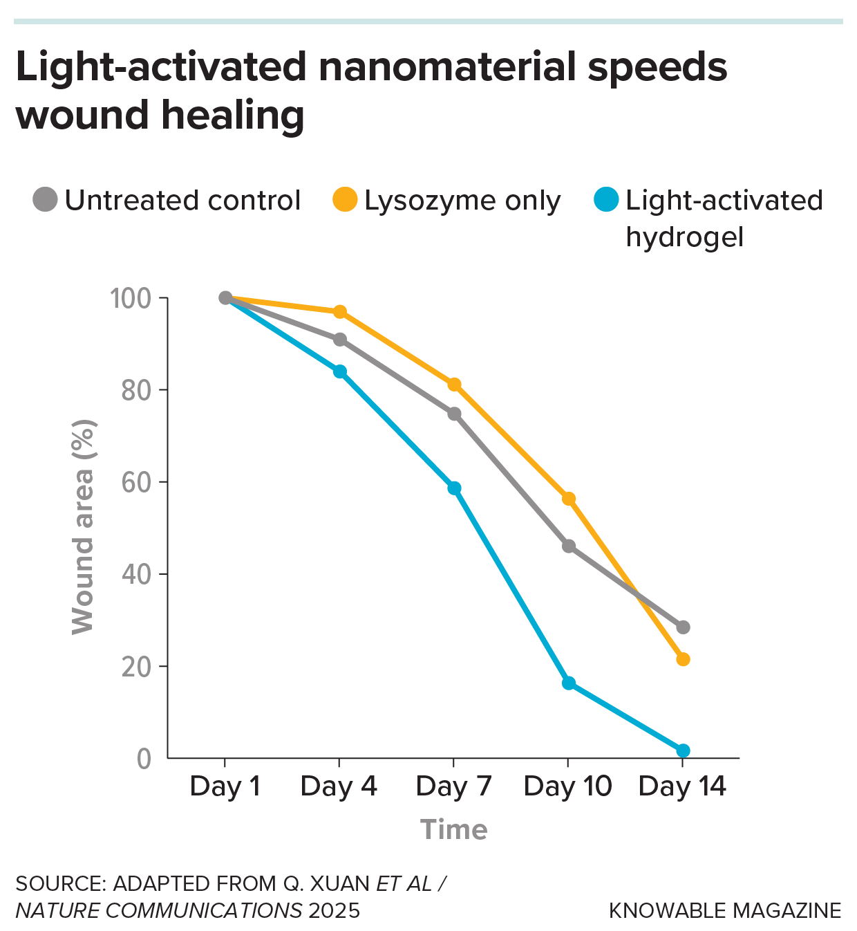
When the team applied the gel to wounds in mice and pigs, they found it eradicated more than 95 percent of the bacteria present. The wounds also healed more quickly, because the lysozyme — which is toxic for healthy cells, too — was activated in the wound only when irradiated with light, saving the skin from overexposure. To boost healing still further, the team added magnesium ions to the gel, which prime immune cells called macrophages to shift from an inflammatory state to one that promotes healing. "The healing will be much faster because you kill the bacteria and heal the wound at the same time," says Mezzenga.

A chart showing percentage of wound healing over time

Light-activated nanomaterials that release noxious compounds only when and where they are needed can help eradicate wound infections while preventing damage to unaffected tissues. Here, mice with antibiotic-resistant wound infections were treated with a hydrogel that releases lysozyme, an antibacterial protein, only when activated by light. Their wounds healed more quickly than those of mice left untreated or treated with lysozyme alone. (Image credit: Adapted from Q. Xuan et al/Nature Communications and Knowable Magazine)

Since bacterial biofilms are especially persistent on the surfaces of medical implants — where they can cause recurring infections and sometimes require repeated surgeries or even amputations — the team also tested their gel on infected prosthetic joints in mice. They injected the gel around an infected implanted needle and shone near-infrared light through the skin. The treatment cleared biofilms and eradicated about 99 percent of bacteria around the implant, while preserving bone tissue.

In another recent study, scientists from Gannan Medical University and Shanghai University in China treated wounds using a nanomaterial made of gold nanoparticles and graphene-oxide "quantum dots," which are tiny, carbon-based semiconducting particles. When irradiated with blue light, the gold particles absorb the light energy and convert it into heat, while graphene oxide helps to transfer electrons across the material. This boosts reactions that produce toxic, unstable molecules called reactive oxygen species that react with structures on bacterial membranes and destroy them.

When the scientists added this material to a bacterial solution and shone blue light on it for 10 minutes, the mild heat and reactive oxygen species worked together to cause bacterial membranes to disintegrate. Using a stain that distinguished dead from living bacteria, the researchers confirmed that the treatment had killed 97 percent of the bacteria.

Testing the nanomaterial in mice revealed that after nine days, the wounds on treated mice showed 99 percent healing, while those of untreated mice showed only about 70 percent healing.

While these techniques have shown promise in the lab, further work will be needed before they can be applied to people. "There is still some way to go," says Lars Kaestner, a biologist at Saarland University in Germany. To be useful in a clinical setting, he notes, researchers would need to do extensive safety testing and lower the cost of the nanomaterials.

Nevertheless, the idea provides hope for patients with chronic wounds that fail to heal with conventional antibiotics, particularly as drug-resistant infections become more common in hospitals and diabetic care.

"It's a good concept," says Qin. "Wound healing and antibacterial resistance are very big challenges. And I think any advance that we can make in these areas would be welcome."

This article originally appeared in Knowable Magazine, a nonprofit publication dedicated to making scientific knowledge accessible to all. Sign up for Knowable Magazine's newsletter.

  •  

Why Older Adults Need To Pay Closer Attention to Vitamin B12

Vitamin B12 NewspaperVitamin B12 is needed in only trace amounts, yet its absence can have far-reaching effects. Two micrograms is an almost unimaginably small amount. It weighs less than a tiny fragment of a grain of table salt. Yet adults need only around this amount of vitamin B12 each day, depending on the guideline used, to support [...]
  •  

Scientists Say a Daily Probiotic May Help Fight Depression in Older Adults

Stressed Tired Woman Taking Vitamin Painkiller Antidepressant PillA new study suggests that altering the gut microbiome with probiotics may provide additional benefits for older adults being treated for depression. Could a daily probiotic help ease depression in older adults? A new clinical trial suggests the answer may be yes, adding to growing evidence that the gut and brain are more connected than [...]
  •  

Metaplasticity Enhances Life-Course Brain Health Strategies

In a groundbreaking advance poised to reshape how we approach brain health across the human lifespan, recent research reveals that integrating the principles of metaplasticity into therapeutic interventions can dramatically enhance their effectiveness. Metaplasticity, often described as the “plasticity of plasticity,” refers to the brain’s dynamic ability to regulate its own synaptic plasticity based on prior activity and environmental inputs. This emerging understanding promises to revolutionize strategies for cognitive enhancement and neurological rehabilitation, offering personalized and adaptive therapies that evolve with the individual’s brain state.

The concept of metaplasticity extends the traditional notion of neuroplasticity, emphasizing that synaptic changes are not static but modulated by previous experiences. This regulatory mechanism ensures that synaptic modifications occur within optimal ranges, preventing the neural circuits from becoming either too rigid or overly malleable. Recognizing this, scientists are now exploring interventions that do more than promote plasticity—they strategically prime the brain’s plastic potential to achieve sustained and balanced neural adaptability.

Life-course brain health is a complex interplay of genetic, environmental, and experiential factors that cumulatively influence neural function. Traditional interventions often apply a one-size-fits-all model, ignoring the fluctuating states of neural readiness and adaptation across developmental stages. By contrast, therapies informed by metaplasticity tailor their application according to the individual’s neurobiological context, potentially enhancing outcomes in cognitive development, neurorehabilitation after injury, and age-related cognitive decline.

The research underscores the importance of timing and context in brain health interventions. Synaptic plasticity is not merely about stimulating neurons but about doing so when they are optimally susceptible to change. This temporal precision is critical during sensitive periods in childhood and adolescence when the brain’s structure and function undergo rapid transformation. Incorporating metaplasticity principles means recognizing these windows of opportunity and adapting interventions to harness the brain’s highest potential for growth and recovery.

Moreover, metaplasticity offers intriguing insights into why certain neurological and psychiatric conditions manifest, persist, or respond variably to treatment. In disorders such as autism spectrum disorder, schizophrenia, and depression, dysregulation of plasticity mechanisms is increasingly implicated. Understanding how metaplasticity governs synaptic strength and flexibility not only facilitates more refined diagnostic frameworks but also guides innovative therapeutic designs that can recalibrate neural networks more effectively.

Neurorehabilitation strategies following traumatic brain injury or stroke stand to benefit immensely from a metaplasticity-informed approach. Current protocols focus heavily on repetitive stimulation or training, but without considering the brain’s shifting landscape of plasticity thresholds. By integrating metaplastic principles, clinicians can optimize the intensity and nature of rehabilitation exercises to coincide with periods when neuronal circuits are most adaptable, potentially accelerating recovery and improving functional outcomes.

This paradigm shift also extends into the realm of aging and neurodegenerative diseases. As brain plasticity declines with age, interventions designed to restore or boost neuroplastic capacity need to be sensitive to the evolving metaplastic state of neural circuits. Targeting these mechanisms might slow cognitive decline, improve memory retention, and enhance quality of life for older adults. Such interventions could include tailored cognitive training paradigms, pharmacological agents, or even non-invasive brain stimulation techniques employed with metaplastic timing considerations.

Critically, the application of metaplasticity in clinical settings demands a robust biomarker framework to assess brain state and plastic potential dynamically. Advances in neuroimaging and electrophysiological monitoring are instrumental in this regard, enabling real-time tracking of circuit responsiveness and guiding intervention adjustments. This precision medicine approach promises not only better therapeutic efficacy but also minimizes adverse effects associated with inappropriate stimulation.

As the scientific community deepens its understanding of metaplasticity, ethical considerations around intervention timing and intensity come into sharper focus. The prospect of manipulating brain plasticity across the lifespan raises questions about long-term consequences, individual variability, and the balance between enhancement and harm. Ongoing research must address these concerns through rigorous longitudinal studies and multidisciplinary dialogues involving neuroscience, bioethics, and patient advocacy.

Education systems may also harness knowledge on metaplasticity to optimize learning processes. By structuring curricula and educational environments that align with periods of heightened neural receptivity, educators can facilitate more effective skill acquisition and cognitive development. This integration of neuroscience and pedagogy represents a transformative step towards personalized learning experiences tailored to the brain’s natural rhythms of plasticity.

The public health implications are profound, as metaplasticity-guided interventions could democratize brain health strategies, making them more accessible and tailored beyond clinical populations. Community programs aimed at mental wellness, resilience building, and cognitive maintenance might incorporate metaplastic principles to maximize their impact, fostering healthier neural aging trajectories and buffering against stress-related neural degradation.

Future research efforts are focused on unraveling the molecular substrates that orchestrate metaplastic adjustments, including intracellular signaling pathways, neurotransmitter systems, and gene expression dynamics. These investigations hold promise for the development of novel pharmacotherapies that target metaplastic regulators, offering precise modulation of neural plasticity in disease states and healthy brains alike.

In conclusion, applying the burgeoning knowledge of metaplasticity marks a profound evolution in brain health interventions. This approach compels a reexamination of how neuroscientists and clinicians conceptualize brain adaptability, emphasizing a lifelong, context-dependent modulation of neuroplastic potential. As this paradigm gains traction, it sets the stage for more refined, effective, and ethically conscious strategies to optimize brain function from infancy through advanced age.


Subject of Research: Life-course brain health interventions and the role of metaplasticity in optimizing therapeutic outcomes.

Article Title: Life-course brain health intervention choices are improved by applying knowledge of metaplasticity.

Article References:
Scher, M.S. Life-course brain health intervention choices are improved by applying knowledge of metaplasticity. Pediatr Res (2026). https://doi.org/10.1038/s41390-026-05183-1

Image Credits: AI Generated

DOI: 10.1038/s41390-026-05183-1

  •  

Sensory Hyposensitivity’s Role in Depression: A Review

In a groundbreaking systematic narrative review recently published in Translational Psychiatry, researchers Sharpley, Odierna, Bitsika, and colleagues have unveiled new insights into the underexplored dimension of sensory hyposensitivity in depression. This comprehensive analysis not only delineates the form and process of sensory hyposensitivity but also delves into its multifaceted functions within depressive pathology, potentially revolutionizing how the scientific community understands and approaches depressive disorders.

Depression, a pervasive and multifactorial mental health condition, has traditionally been studied through emotional, cognitive, and neurochemical lenses. However, this novel review places sensory processing anomalies—specifically sensory hyposensitivity—at the forefront of depression research. Sensory hyposensitivity refers to a reduced responsiveness to sensory stimuli, encompassing diminished perception or processing of tactile, auditory, visual, or olfactory inputs. This investigation critically examines how such sensory deficits may contribute to the manifestation and maintenance of depression.

The authors begin by methodically synthesizing available literature, tracing the contours of sensory hyposensitivity as a discrete but interconnected phenomenon within depressive symptomatology. Their narrative review compiles evidence from neuroimaging studies, psychophysical assessments, and clinical observations, underscoring consistent findings of blunted sensory responsiveness across various sensory modalities in depressed individuals. These findings suggest that sensory hyposensitivity is not an epiphenomenon but an integral facet of the disorder.

Mechanistically, the review advances the hypothesis that sensory hyposensitivity stems from dysfunctional neural circuitry involving sensory cortices and integrative regions such as the thalamus and limbic system. The authors highlight aberrations in neurotransmitter systems, including serotonergic and glutamatergic pathways, which may dampen sensory signal processing. These neurobiological underpinnings align well with the clinical profiles of patients exhibiting attenuated sensory experiences.

Importantly, the review moves beyond description to investigate the process through which sensory hyposensitivity unfolds in depression. It posits a bidirectional relationship whereby impaired sensory processing exacerbates affective symptoms, which in turn further blunts sensory responsiveness, fostering a vicious cycle. This iterative dynamic may partly explain why some depressive episodes are resistant to traditional treatments aimed primarily at mood symptoms rather than sensory deficits.

The functional consequences of sensory hyposensitivity also receive detailed focus. The authors discuss how diminished sensory input may impair environmental engagement and reduce hedonic experience, thus contributing to anhedonia—a core symptom of depression. Additionally, sensory blunting may interfere with social interactions, as attenuated perception of social cues can hinder effective communication and emotional reciprocity.

One of the most compelling arguments articulated in the review is the potential role of sensory hyposensitivity as a biomarker for specific depression subtypes. By stratifying patients based on sensory processing profiles, clinicians could tailor interventions more precisely. For example, sensory retraining therapies or neuromodulation targeting sensory circuits might emerge as adjuncts to conventional pharmacotherapy and psychotherapy.

Furthermore, the review evaluates methodological challenges in studying sensory hyposensitivity, such as heterogeneity in assessment tools and variability in patient populations. The authors call for refined, standardized protocols employing multimodal sensory testing and integration of objective biomarkers, including electrophysiological and neuroimaging indices, to enhance reliability and validity.

The translational implications of these findings are profound. Integrating sensory hyposensitivity into diagnostic frameworks could reshape clinical evaluation practices, leading to earlier detection and more nuanced treatment planning. Moreover, raising awareness about sensory deficits among healthcare providers may improve patient outcomes by addressing often overlooked but debilitating symptoms.

The authors also speculate on the evolutionary context of sensory hyposensitivity in depression. They suggest it may represent a maladaptive remnant of an ancient conservation-withdrawal response, wherein decreased sensory responsiveness served to minimize energy expenditure during periods of adversity. Understanding this evolutionary dimension may inform novel treatment paradigms that seek to recalibrate sensory processing rather than simply alleviate mood disturbances.

Importantly, this review sheds light on distinct sensory modalities affected in depression, showing that tactile and auditory hypo-responsiveness are particularly pronounced, with visual and olfactory deficits also noted but less consistently. This differential pattern invites further research into modality-specific interventions and highlights the complexity of sensory system involvement.

Another area of focus is the developmental trajectory of sensory hyposensitivity in depressive disorders. The authors discuss evidence indicating that sensory processing impairments may precede or coincide with the onset of depressive symptoms, suggesting a potential predictive role in at-risk populations. Early identification of sensory abnormalities could thus serve as a preventive strategy.

The review also underscores the necessity of interdisciplinary collaboration in advancing research on sensory hyposensitivity. Integrative approaches bridging neuroscience, psychology, psychiatry, and sensory science are poised to unravel the nuanced relationships between sensory processing and affective disorders, fostering innovation in therapeutic approaches.

In conclusion, Sharpley and colleagues’ seminal review elucidates sensory hyposensitivity as a critical, yet underappreciated, dimension of depression. By charting its form, process, and function with remarkable depth, the authors pave the way for novel conceptualizations, diagnostic refinements, and treatment interventions that promise to transform depression care. This paradigm shift beckons a new era in mental health research, where sensory experiences are recognized not just as peripheral but as central to understanding and healing depression.

Subject of Research: Sensory hyposensitivity in depression

Article Title: A systematic narrative review of the form, process, and function of sensory hyposensitivity in depression

Article References:
Sharpley, C.F., Odierna, G.L., Bitsika, V. et al. A systematic narrative review of the form, process, and function of sensory hyposensitivity in depression. Transl Psychiatry (2026). https://doi.org/10.1038/s41398-026-04165-5

Image Credits: AI Generated

DOI: https://doi.org/10.1038/s41398-026-04165-5

  •  

Diagnostic dilemma: A fish stabs a man through the throat and the base of the skull

The patient: A 31-year old man in Louisiana

The symptoms: When the patient reached the hospital, he was bleeding from the mouth and had severe neck pain and stiffness. Upon examining the man, his doctors noted a tear in the back of his throat on the right side.

What happened next: The man, a sports fisherman, had been brought to the hospital by boat and helicopter after an accident during an ocean fishing trip. He had caught a white marlin (Kajikia albida), a type of large fish with a long pointy "bill," weighing approximately 60 pounds (27 kilograms). This species can grow up to 180 pounds (82 kg).

As the man leaned over the side of the boat to release the hook from the fish, it jumped and struck him in the mouth with its bill, sending the man tumbling backward into the boat. He felt pain in his neck, which then radiated into his spine.

At the hospital, an X-ray of the man's upper spine did not reveal any abnormalities. However, his intensifying spinal pain and neck stiffness prompted one of the doctors to order a computed tomography (CT) scan of the same area. In the CT scan, physicians noted "a wedge-shaped, hyperdense object," they wrote in a report of the case. The object had penetrated the back of the man's throat and entered his spinal canal. It stuck there, piercing the foramen magnum — a large hole at the base of the skull that the spinal cord passes through.

The diagnosis: The doctors immediately performed surgery on the man to remove the penetrating object. They administered general anesthesia, and the surgeon used retractors — instruments that hold wounds open — to expose the object, which turned out to be the broken tip of a fish bill.

The treatment: The sharp bill fragment was so firmly wedged in the man's skull that the surgeon had to make an additional incision above the man's topmost vertebra in order to remove it, pulling it out along the path of entry. It measured about 1.4 inches (3.5 centimeters) long.

After the fragment was removed, the patient was given five different types of antibiotics both to prevent infection from microbes found in the throat and from bacteria that are unique to marine environments. He was discharged eight days later and continued to take multiple antibiotics for the next two weeks.

At his final follow-up visit, the man had fully recovered and showed no lingering neurological symptoms.

What makes the case unique: In 1848, doctors published the first medical case study of a skull injury from a foreign object: a gunpowder explosion had sent an iron bar through the skull of railroad worker Phineas Gage. Gage recovered, but he exhibited personality changes that doctors attributed to his significant brain injuries.

In another notable case, published in 1895, a man had tripped and fallen face-first onto an oilcan. The spout penetrated his cheek and became stuck at the base of his skull, causing temporary incontinence and permanent memory loss.

Since those 19th-century cases, physicians have reported instances of cranial trauma in different parts of the skull and from a variety of objects, including a crochet hook, a pitchfork, a bike key and a harpoon.

This man's case is the first reported instance of an injury to the foramen magnum, as well as the first injury caused by a fish bill, according to the report.

This article is for informational purposes only and is not meant to offer medical advice.

  •  

Tiny Plastic Particles Could Strongly Harm Liver Health

Microplastics are everywhere. They are tiny pieces of plastic that form when larger plastic items, such as bottles, food containers, plastic bags, and packaging materials, slowly break down over time. These particles are so small that people often cannot see them with the naked eye. Scientists have found microplastics in rivers, oceans, soil, drinking water, […]

The post Tiny Plastic Particles Could Strongly Harm Liver Health appeared first on Knowridge Science Report.

  •  

Hidden Nutrient in These Fruits May Strongly Protect Your Heart

People are often told to eat more fruits and vegetables if they want to stay healthy. The advice sounds simple and easy to follow. But scientists now say that not all fruits and vegetables provide the same health benefits. A new study suggests that many people may be missing an important group of natural compounds […]

The post Hidden Nutrient in These Fruits May Strongly Protect Your Heart appeared first on Knowridge Science Report.

  •  
❌