❌

Reading view

Bumble Bees Solve an Insect Version of a Famous Primate Intelligence Test

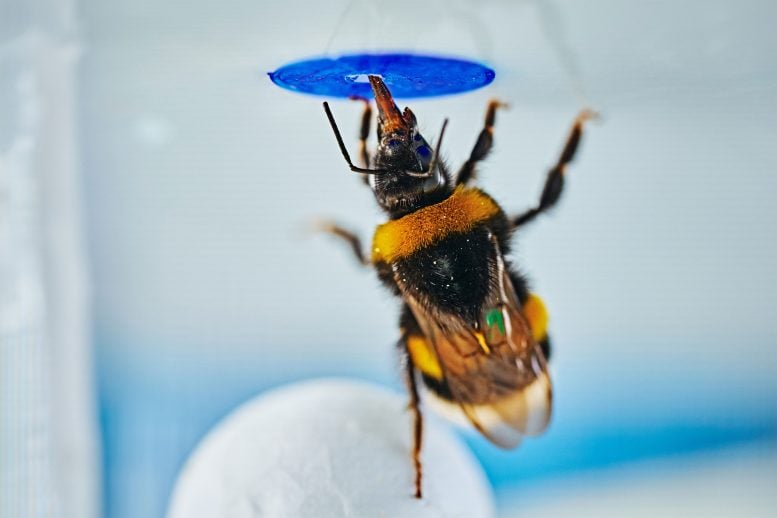
Bumble Bee Standing on a BallBumble bees demonstrated an unexpected ability to solve a novel object-based challenge without training. A bee’s brain is smaller than a sesame seed, yet in a new experiment, some bumble bees appeared to solve a problem that required more than simple instinct. They had to move an object into position, climb onto it, and use [...]
  •  

UCLA Scientists Uncover Potential Method to Repair Damaged Kidneys

A groundbreaking discovery at the University of California, Los Angeles (UCLA) has unveiled a promising new therapeutic avenue for kidney regeneration, leveraging a drug initially designed to repair heart tissue following myocardial infarction. This drug, known as AD-NP1, targets a specific protein—ENPP1—that plays a critical role in impeding the body’s natural healing processes in injured tissues. The revelation that AD-NP1 can also accelerate kidney repair heralds a significant advancement in regenerative medicine, especially for patients suffering from chronic kidney disease (CKD), a condition that affects millions globally.

The heart of this research revolves around a protein named ectonucleotide pyrophosphatase/phosphodiesterase 1 (ENPP1). When tissues such as the heart or kidney sustain injury, ENPP1 production escalates, initiating a cascade of metabolic disturbances that hinder energy synthesis within cells. This disruption compromises cellular function and ultimately stalls effective tissue regeneration. In the context of the kidney, which is vital for maintaining systemic homeostasis, such impediments can lead to progressive scarring and functional decline, culminating in kidney failure.

Through years of meticulous experiments, the UCLA team delineated the detrimental impact of ENPP1 on the kidney’s reparative capacity. They observed that injured renal tissue upregulates ENPP1 expression, which in turn resets the metabolic landscape of cells in the damaged microenvironment. This altered metabolic signaling disrupts mitochondrial function, thereby curtailing ATP production essential for cell proliferation and repair. By employing genetic knockout models in mice, where ENPP1 expression was inhibited, the researchers documented a striking enhancement in renal recovery post-injury. These mice demonstrated reduced fibrotic scarring and improved renal biomarkers, signaling restored kidney function.

Central to translating these findings into potential clinical interventions is AD-NP1, a sophisticated monoclonal antibody engineered to specifically neutralize ENPP1. Unlike broad-spectrum immunosuppressants or anti-inflammatory agents, AD-NP1 offers targeted therapeutic precision by binding solely to human ENPP1, circumventing off-target effects. Initially conceptualized to mitigate cardiac fibrosis and promote myocardial regeneration following heart attacks, AD-NP1’s mechanism exerts a similar rejuvenating influence on kidney tissues. The antibody effectively halts the ENPP1-induced metabolic sabotage, facilitating an environment conducive to cellular proliferation and tissue repair.

In experimental paradigms, the administration of AD-NP1 in mice subjected to nephrotoxic diets and chemically induced renal injuries yielded compelling outcomes. Within merely seven days of treatment, subjects exhibited marked improvements in kidney function tests and histological assessments showed diminished collagen deposition and scarring. These findings underscore the drug’s potential to transform the management of acute and chronic kidney injuries by reinstating the organ’s intrinsic regenerative programs, which are often stifled in disease states.

Moreover, the team’s translational approach integrated analyses of human kidney biopsies acquired from individuals with chronic kidney disease. These samples consistently manifested elevated ENPP1 levels relative to healthy controls, substantiating the protein’s pathological role in human renal disorders. This correlation not only reinforces the relevance of the preclinical models used but also paves the way for clinical evaluation of AD-NP1’s efficacy in human subjects suffering from renal ailments.

The broader implications of this discovery extend into the metabolic regulation of tissue repair. ENPP1’s role appears multifaceted, orchestrating cellular energy dynamics and intercellular communication networks that dictate regenerative outcomes. Inhibiting this protein intervenes in maladaptive signaling cascades, thereby restoring bioenergetic balance and enabling clonal expansion of healthy cells adjacent to injury sites. This paradigm shift in understanding organ regeneration emphasizes metabolic modulation as a cornerstone of effective healing strategies.

Prior to this research, the therapeutic targeting of ENPP1 was primarily confined to cardiac medicine. The successful repurposing of AD-NP1 for kidney injury illustrates the concept of cross-organ regenerative mechanisms, where molecular pathways governing tissue healing are conserved albeit contextually modulated. This discovery propels the field into a new frontier, advocating for the exploration of shared regenerative targets that can be manipulated across diverse organ systems.

AD-NP1’s journey into clinical trials commenced recently with FDA approval of a Phase 1 safety study in human patients recovering from heart attacks. This milestone reflects the rigorous preclinical validation of the drug’s pharmacodynamics and safety profile. Building on these milestones, ongoing efforts at UCLA aim to extend these trials to include patients with progressive kidney disease, setting the stage for comprehensive evaluation and potential future clinical application.

Underlying this innovative research are substantial investments from the National Institutes of Health, California Institute of Regenerative Medicine, and the U.S. Department of Defense. This underscores the strategic importance and translational potential of regenerative medicine therapies in addressing chronic diseases that impose heavy societal burdens. The interdisciplinary collaboration among cardiovascular scientists, nephrologists, and molecular biologists at UCLA exemplifies the synergy needed to unlock complex biological processes for therapeutic gain.

In summation, the identification of ENPP1 as a metabolic gatekeeper that impedes tissue repair, combined with the development of AD-NP1 to neutralize its effects, represents a significant leap forward in regenerative medicine. The drug’s ability to foster organ regeneration by restoring metabolic and cellular homeostasis offers hope for millions afflicted by kidney disease and opens novel avenues for organ repair strategies. As clinical trials progress, the scientific community eagerly anticipates further validation of these findings, potentially heralding a new era where molecularly targeted therapies can substantially reverse organ damage and restore quality of life.

Subject of Research: Kidney regeneration, metabolic regulation of tissue repair, ENPP1 protein inhibition, monoclonal antibody therapy

Article Title: UCLA Researchers Discover Novel Role for ENPP1 Inhibition in Enhancing Kidney Repair via AD-NP1

News Publication Date: Not specified (based on provided content)

Web References: Cell Stem Cell Journal Article

Keywords: ENPP1, kidney injury, chronic kidney disease, tissue regeneration, monoclonal antibody, AD-NP1, metabolic signaling, renal repair, fibrosis, UCLA, regenerative medicine, Phase 1 clinical trial

  •  

Scientists Reveal New Discoveries About Cells Forming Brain’s Protective Barriers

In the complex landscape of brain development, the formation of the brain’s protective barriers—the blood-brain barrier (BBB) and the blood-cerebrospinal fluid (CSF) barrier—has long fascinated neuroscientists. These barriers are composed of highly specialized cells that serve a critical function: permitting necessary nutrients to enter the brain while preventing harmful substances, pathogens, and toxins circulating in the bloodstream from crossing into the delicate neural environment. Despite a deep understanding of their function, the cellular mechanisms orchestrating their assembly during embryogenesis have remained elusive. A groundbreaking study led by researchers at the University of California San Diego, recently published in Cell, unveils a surprising and paradigm-shifting contributor to brain barrier development: cellular senescence.

Cellular senescence is traditionally characterized as a permanent cessation of cell division, commonly associated with aging and pathology. These so-called “zombie” cells, which remain metabolically active but fail to proliferate, accumulate in aging tissues, contributing to chronic inflammation, tissue dysfunction, and cognitive decline. As a consequence, senescent cells have become key targets in developing therapeutic strategies to mitigate age-related diseases. However, recent evidence challenges the simplistic association of senescence solely with aging and damage. Instead, senescence appears highly context-dependent, playing complex and sometimes beneficial roles in development and healing.

Emerging studies have revealed transient senescent cell populations in mouse embryos during the formation of limbs and kidneys, as well as in wound healing contexts, where senescence promotes tissue repair. These discoveries suggest that senescence may be an evolutionarily conserved mechanism to guide tissue remodeling and regeneration when temporally controlled. The UC San Diego research team, led by Assistant Professor Hiruy Meharena, has extended this concept into the realm of brain development, uncovering diverse senescence-associated cell states that actively contribute to constructing the brain’s vital protective barriers.

Through a multifaceted approach combining single-cell RNA sequencing, advanced imaging, and precise genetic lineage tracing in developing mouse brains, the investigators identified three distinct cell types that transiently or persistently enter senescence during the formation of the BBB and blood-CSF barrier. These cell types include vascular endothelial cells, brain-resident macrophages, and choroid plexus epithelial cells, each exhibiting unique senescence profiles and functional roles in the brain’s developmental timeline.

Vascular endothelial cells, which line the embryonic blood vessels, and brain-resident macrophages engage senescence transiently during critical windows of brain vascular remodeling and patterning. These senescent states appear to fine-tune the formation of the intricate vascular network essential for establishing the highly selective BBB, coordinating cellular interactions and molecular signaling pathways that regulate tight junction formation and barrier integrity. This temporary senescent phase helps orchestrate the vascular architecture that prevents harmful molecules in the blood from infiltrating brain tissue.

In contrast, choroid plexus epithelial cells—responsible for generating cerebrospinal fluid and forming the blood-CSF barrier—exhibit a persistent senescence-like state that endures beyond embryonic development into adulthood. This finding defies the prevailing dogma that developmental senescence is exclusively transient and suggests a novel dimension of senescence’s role in maintaining barrier function over the lifespan. The sustained senescent phenotype in these epithelial cells likely supports the continuous protective and homeostatic functions of the blood-CSF barrier, which plays a critical role in brain fluid regulation and immune surveillance.

One of the study’s most unexpected revelations emerged when the researchers experimentally ablated senescent cells during embryogenesis. The resulting mice exhibited marked abnormalities in the structural and functional development of both brain barriers, accompanied by disrupted fluid balance within the central nervous system. These defects underscore the indispensability of senescent cells in normal brain barrier formation and highlight the complex interplay between senescence and tissue morphogenesis.

This nuanced view of senescence challenges the traditional perception of a monolithic, detrimental “senescence program,” revealing instead a spectrum of senescence-associated cell states tailored to specific developmental contexts and cell types. Senescence acts not as a simple arrest signal but as a dynamic, multifaceted process facilitating intercellular cooperation during neurovascular development. It integrates molecular cues that modulate cell behavior, extracellular matrix remodeling, and immune cell function to sculpt the brain’s defensive frontiers.

The implications of these findings extend beyond developmental biology into the pathophysiology of brain disorders. Since the integrity of the BBB and blood-CSF barrier is compromised in neurodegenerative diseases, stroke, and neuroinflammation, understanding the diverse roles of senescent cells in barrier biology may herald new therapeutic avenues for restoring barrier function and mitigating neurological deficits. The UC San Diego team is now probing how senescence-related mechanisms unfold in disease contexts, aiming to decipher whether modulating senescent cell populations can ameliorate or prevent barrier dysfunction.

Moreover, the identification of persistent senescence in adult choroid plexus cells prompts intriguing questions about the balance between beneficial senescence and potential chronic senescent cell accumulation contributing to pathology. Future research will explore how this persistent senescence is regulated and whether it participates in age-related changes in brain fluid dynamics or susceptibility to CNS infections.

This research challenges the classical narrative that equates senescence with detrimental aging by elucidating its fundamental, constructive roles in brain development. It elevates the concept of senescence from a pathological endpoint to an essential, adaptive cellular state finely tuned in time and space. By redefining senescence as a developmental and homeostatic facilitator, the study sets a new trajectory for investigating cellular aging, brain barrier biology, and neurovascular health.

As neuroscience embraces the complexity of cellular states within the brain’s microenvironment, these insights offer a richer, more integrative framework. The study’s comprehensive use of cutting-edge genomic, imaging, and genetic tools exemplifies the power of modern neurobiology to unravel the elusive choreography underlying brain structure and function. Continued exploration of developmental senescence promises to deepen our understanding of brain physiology and opens transformative possibilities for innovative treatments targeting the blood-brain and blood-CSF barriers across the lifespan.


Subject of Research: Animals

Article Title: Persistent and transient senescent cells contribute to brain barrier development

News Publication Date: 10-Jun-2026

Web References: https://www.cell.com/cell/fulltext/S0092-8674(26)00581-7

References: DOI 10.1016/j.cell.2026.05.022

Image Credits: Ella Maru Studio, conceptualized by Ashley Watson and Hiruy Meharena

Keywords: Cellular neuroscience, cellular senescence, developmental neuroscience, molecular neuroscience, cellular physiology, cell development, cellular processes

  •  

New Research Reveals Rays Use Chemical Signals to Warn Others of Danger

In a groundbreaking discovery that broadens our understanding of marine life communication, researchers at Oregon State University have identified a novel chemical signaling mechanism among bat rays (Myliobatis californica) that warns conspecifics of looming threats. This chemical disturbance cue, previously documented only in bony fish, now emerges as a key anti-predator strategy in cartilaginous fishes—specifically rays—revealing a layer of behavioral complexity that challenges long-held assumptions about elasmobranch social interactions.

Dr. Joshua Bowman, leading the study from OSU’s Big Fish Lab, devised an elegant experimental setup employing three isolated tanks interconnected through water flow but segregated visually and acoustically to eliminate confounding sensory influences. Each tank housed an individual bat ray, with one designated as the “signaler” and the other two as “receivers.” By simulating predatory pressure through a gentle chase of the signaler ray, the experimenters induced a natural defensive response while ensuring no physical harm.

Strikingly, within mere seconds of the signaler tank’s water being directed towards the receiver tanks, the bat rays in these isolated environments exhibited marked behavioral shifts characterized by increased swimming speed and evasive maneuvers. These responses mimic a flight reaction, confirming the transfer of a chemical alarm cue through the aquatic medium—a sensory pathway previously undocumented in cartilaginous species.

This discovery bears profound implications for understanding elasmobranch ecology and survival strategies. Bat rays, along with sharks and skates, belong to a class of cartilaginous fishes characterized by their distinct evolutionary lineage and physiology. These findings suggest sophisticated intraspecies chemical communication channels functioning as early-warning systems against predation risks, potentially reshaping our perception of their behavioral ecology.

Moreover, the research carries significance beyond bat rays. Whites sharks, apex predators renowned for their fearsome reputation, are known to exhibit avoidance behaviors when orcas, their potential predators, are nearby. Yet, these sharks often do not have direct visual contact with orcas. This study’s insights into chemical alarm signaling hint at unseen communication mechanisms that might underlie such evasive behavior, opening new avenues for exploration into shark sensory biology and their interactions within the marine predator-prey network.

The experiment’s methodological rigor enhances the credibility of these findings. By isolating variables—eliminating acoustic and visual cues—the team conclusively linked behavioral changes to chemical signals. High-resolution overhead video recordings captured nuanced shifts in locomotion dynamics, quantitatively analyzing the rays’ rapid swimming velocity increases immediately following exposure to the alarm chemicals. These data underscore a robust flight response triggered solely by chemical disturbance cues.

While the precise chemical compound or combination responsible for the alarm signaling remains unidentified, the study establishes a foundation for future biochemical and molecular investigations. Identifying the molecular structure of the alarm cue could unlock transformative understandings of elasmobranch chemical ecology, potentially informing conservation strategies and offering biomarkers for monitoring stress responses in marine populations.

Importantly, the research foregrounds broader ecological and ethical considerations. The evolutionary origin of these chemical disturbance cues underscores their critical role in survival and natural behavior modulation. Human interactions—whether through scientific research, fisheries, or recreational activities—may inadvertently trigger stress dissemination across populations via such chemical signals, amplifying disturbance impacts beyond the initially affected individual.

The OSU Big Fish Lab, recognized as a leading shark research facility on the U.S. West Coast, continues to spearhead innovative studies uncovering the intricate lives of marine cartilaginous fishes. This collaboration, involving co-authors Jamie Cornelius and Mauricio Cantor, integrates expertise spanning marine ecology, physiology, and animal behavior, thereby enhancing multidisciplinary perspectives crucial to advancing marine science frontiers.

By employing bat rays as a proxy for studying elusive white shark behavior, the researchers circumvent logistical challenges inherent to large predator studies. Bat rays’ accessibility and manageable size provide an ethical and practical model to dissect communication modalities applicable across related species, ensuring experimental feasibility while preserving ecological validity.

This study, recently published in the Journal of Experimental Zoology Part A: Ecological and Integrative Physiology, represents a significant milestone in marine biology. It not only documents, for the first time, chemically mediated disturbance cues in cartilaginous fishes but also stimulates critical reflection on the sensory worlds of sharks and rays, organisms whose behavioral sophistication may be far richer than previously appreciated.

Collectively, these findings herald a paradigm shift in our understanding of underwater communication networks. They remind us that aquatic ecosystems host complex, often hidden, language systems integral to species survival. As we deepen our inquiry into these chemical dialogues, we unlock new insights essential for protecting marine biodiversity in a rapidly changing ocean environment.

Subject of Research: Animals
Article Title: Behavioral Evidence for a Chemical Disturbance Cue in Bat Rays (Myliobatis californica)
News Publication Date: 10-Jun-2026
Web References: http://dx.doi.org/10.1002/jez.70102
References: Journal of Experimental Zoology Part A: Ecological and Integrative Physiology
Keywords: chemical disturbance cue, bat rays, cartilaginous fish, elasmobranchs, shark behavior, marine communication, anti-predator strategy, predator-prey interactions, aquatic chemical signaling, flight response, marine ecology, behavioral study

  •  

Innovative Heat Sensor Developed to Monitor Living Cells

In a groundbreaking advance at the intersection of bioengineering and microbiology, researchers at Harvard’s John A. Paulson School of Engineering and Applied Sciences have unveiled a pioneering device known as a pico-calorimeter. This remarkable instrument is capable of directly detecting heat signals emanating from minute populations of living cells, a feat that until now has remained technologically elusive due to the vanishingly small scale of these thermal signatures. By enabling real-time metabolic tracking at unprecedented sensitivity, the pico-calorimeter promises to revolutionize our understanding and monitoring of cellular activity, bacterial growth, and antibiotic response.

At the core of this innovation is the principle that living cells, as they metabolize nutrients, grow, divide, and react to external stimuli such as drugs, intrinsically emit heat. This metabolic heat, although incredibly subtle, offers a direct, unmediated insight into the physiological state of cells. Unlike conventional methods that infer cellular metabolism indirectly through oxygen consumption or chemical byproducts, the pico-calorimeter provides a direct calorimetric measurement, offering a more immediate and accurate assay of cellular energetics.

The technical architecture of the device is a marvel of microengineering. It consists of three ultrafine glass capillaries aligned on a delicately micromachined membrane with near-atomic precision. One of these capillaries harbors the biological sample suspended in a liquid growth medium, while the remaining two serve as thermodynamic reference points. As the bacteria or other cells within the sample metabolize and proliferate, they release minute quantities of heat. This thermal flux generates infinitesimal temperature gradients relative to the reference capillaries, which are then transduced into electrical signals by a nearby thermopile—an exquisitely sensitive heat-to-electricity converter capable of resolving fluctuations on the order of picowatts.

A critical enhancement in this iteration of the pico-calorimeter lies in its housing within a vacuum chamber. This vacuum environment thermally isolates the sensor assembly from ambient thermal noise, dramatically amplifying sensitivity by nearly an order of magnitude compared to antecedent designs. The prior versions employed liquid droplets on suspended membranes, which, while innovative, presented operational challenges and limited sensitivity. The current vacuum-sealed, microfluidic integration advances robustness, usability, and measurement fidelity, marking a milestone in the delicate art of thermal detection at the cellular scale.

Demonstrating the device’s prowess, the research team employed the pico-calorimeter to monitor Escherichia coli growth dynamics, beginning with an initial seed population as modest as thirty to forty individual bacterial cells. This capability to detect and analyze such small biological communities unveils immense potential for applications ranging from fundamental microbiology to clinical diagnostics. Notably, the device was employed to examine bacterial metabolic responses upon introduction of antibiotics with varying mechanisms of action—chloramphenicol, rifampicin, and ampicillin—capturing differential heat signatures indicative of altered cellular viability and activity.

These direct calorimetric measurements have revealed metabolic perturbations well in advance of what traditional culture-based microbiological assays could discern. This capability is particularly transformative in clinical scenarios such as sepsis, where detecting and characterizing bacterial populations at extremely low concentrations in blood is vital but presently time-consuming. The pico-calorimeter’s sensitivity could drastically truncate diagnostic timelines from days to hours, allowing more timely and targeted antimicrobial interventions.

Beyond measuring growth and antibiotic susceptibility, the pico-calorimeter enables continuous, real-time monitoring of fundamental cellular parameters including viability, proliferation rate, and stress responses. This temporal resolution offers researchers the invaluable ability to dynamically track cellular states and transitions, capturing metabolic shifts as cells respond to environmental perturbations or pharmacological treatments. Such insights may open new frontiers in cell biology, pharmacodynamics, and personalized medicine.

This breakthrough device represents the culmination of roughly two decades of meticulous research in the Vlassak laboratory, with contributions building from initial exploration of nano-calorimetry techniques aimed at materials science phenomena such as phase transitions in shape memory alloys and metallic glasses. The interdisciplinary bridge extended to biological systems has ushered in ultrasensitive calorimetry methods capable of assessing metabolic heat production in large cells and complex embryonic development processes, laying foundational expertise for this current bio-calorimetric leap.

Technologically, the fabrication of the pico-calorimeter draws upon state-of-the-art micro- and nanofabrication facilities, including the Harvard Center for Nanoscale Systems. The integration of microfluidics and micromachined sensing elements within thermally isolated environments exemplifies the cutting-edge convergence of materials science, electrical engineering, and biological instrumentation. The multidisciplinary synergy underscores the technological sophistication essential for detecting thermal signals billions of times smaller than everyday heat fluxes.

Commercially, the Harvard Technology Development office has filed multiple patents covering the pico-calorimeter and its applications in antimicrobial susceptibility testing. Juanjuan Zheng, a pivotal figure in this research and founder of an innovative startup, is leveraging the technology to deliver practical, label-free, real-time metabolic readouts. These tools hold promise for broad adoption in life sciences research, pharmaceutical development, and clinical diagnostics, harnessing the power of heat measurement to glean functional insights from minute biological samples.

As the research community embraces this transformative platform, interest has surged from a diverse array of scientific domains. The ability to capture direct metabolic information from tiny, dynamic living systems resolves a long-standing challenge in single-cell and small-population analysis. This heralds a new era where functional metabolic data provides early and actionable information on biological states, ranging from cellular growth and stress response to drug efficacy and resistance patterns.

In essence, the Harvard pico-calorimeter transcends conventional bioanalytical tools by transforming infinitesimal heat emissions into a vivid metabolic narrative. This technological breakthrough not only augments our scientific toolkit but also possesses the potential to accelerate biomedical discovery, enhance therapeutic precision, and ultimately improve patient outcomes by enabling rapid, noninvasive interrogation of cellular function. The implications for healthcare, research, and biotechnology are profound, heralding an exciting frontier where the subtle warmth of life becomes an unequivocal signal of cellular health and response.


Subject of Research: Cells
Article Title: A pico-calorimeter for cellular metabolism and antimicrobial susceptibility testing
References: Proceedings of the National Academy of Sciences
Image Credits: Vlassak Group / Harvard SEAS
Keywords: Calorimetry, Chemical analysis, Life sciences, Biophysics, Single cell profiling, Single molecule analysis, Materials science, Physics, Applied sciences and engineering, Applied physics, Engineering, Electrical engineering, Chemical engineering, Mechanical engineering, Health and medicine, Biomedical engineering

  •  

This Surprising Hair Type Could Hold the Key to Chronic Itch Relief

Young Man Scratching Itchy Skin Allergic RashA newly identified sensory pathway links fine hairs to itch sensations, revealing an unexpected biological system that may contribute to chronic itching disorders and provide new targets for treatment. An itch can seem simple, but scientists are finding that the sensation is far more complex than it appears. Researchers at the University of Michigan have [...]
  •  

New Species of Walking Shark Discovered off Papua New Guinea

Hemiscyllium dudgeonae, male paratype, Nubwageta, Milne Bay province, Papua New Guinea. Image credit: M.V. Erdmann.

Marine biologists have identified a new species of the shark genus Hemiscyllium in the waters of eastern Papua New Guinea, expanding a remarkable group of reef-dwelling sharks known for using their four fins to ‘walk’ across the seafloor.

The post New Species of Walking Shark Discovered off Papua New Guinea appeared first on Sci.News: Breaking Science News.

  •  

“Geriatric” Butterfly Species Lives Almost Three Times Longer Than Its Relatives, Study Finds

In the dense, vibrant rainforests stretching across South and Central America, a remarkable evolutionary marvel is unfolding in the form of a tropical butterfly genus that defies traditional biological limits on lifespan. According to a groundbreaking study led by researchers at the University of Bristol, as published recently in Nature Communications, Heliconius butterflies have evolved a sophisticated anti-aging mechanism that dramatically extends their lifespans compared to their close relatives. This discovery not only challenges existing paradigms surrounding insect longevity but also opens a new frontier in understanding the molecular and physiological bases of aging in natural populations.

Heliconius butterflies, widely distributed throughout neotropical ecosystems, have long intrigued biologists due to their exceptional longevity. Unlike most butterfly species, which complete their adult phase within a few weeks, Heliconius butterflies exhibit lifespans that soar to nearly a full year, with median lifespans averaging around three times those of closely related species. Specifically, Heliconius hewitsoni can live up to 348 days in the wild, a staggering contrast to the mere 14 days observed in its relative Dione juno. This magnified difference—in terms of maximum lifespan—exceeds a 25-fold disparity, presenting a natural laboratory to investigate the factors that contribute to longevity.

Delving into the evolutionary dynamics behind this phenomenon, the research team employed an integrative methodology that combined field observations, controlled insectary experiments, and mark-release-recapture studies across multiple Heliconiini tribe species. This multifaceted approach allowed the scientists to quantify not only lifespan but also the patterns of physiological aging, including metrics such as physical performance decline via grip-strength assessments. Intriguingly, in some Heliconius species like Heliconius hecale, the expected decline in physical capabilities that accompanies aging in most organisms was virtually absent, underscoring an evolved resistance to senescence.

The research highlights a unique ecological adaptation: adult pollen feeding. Heliconius butterflies are one of the few lepidopteran groups capable of digesting pollen throughout their adult lives, contrasting with the nectar-reliant feeding habits of most butterflies. This dietary specialization appears intricately linked to their extended healthspan and longevity. Pollen is a rich source of proteins and amino acids, which are hypothesized to support enhanced somatic maintenance and cellular repair mechanisms, potentially underpinning slower rates of aging and sustained physiological function.

To rigorously test the relationship between diet and aging, the study contrasted Heliconius hecale with its non-pollen-feeding relative Dryas iulia under controlled conditions that varied access to pollen. Remarkably, while Heliconius hecale individuals sustained their muscular function and body mass longer and evaded typical age-related deterioration, even those deprived of pollen retained a longevity advantage relative to Dryas. This finding implies that while pollen feeding contributes nutritionally, intrinsic genetic or epigenetic adaptations also play a critical role in extending lifespan, suggesting a complex interplay between evolved traits and ecological factors.

From an evolutionary biology perspective, the divergence in longevity and aging rates within the Heliconiini tribe exemplifies natural selection’s power in shaping life-history traits in response to ecological niches and resource utilization. The accelerated lifespan and delayed senescence in Heliconius suggest that adaptive shifts—such as adult pollen feeding—can drive profound modifications in physiology and perhaps modulate molecular pathways linked to aging, including oxidative stress resistance, protein homeostasis, and metabolic regulation.

The implications of this research extend beyond the entomological sphere, offering a promising model for the biology of aging and lifespan extension. Insects have already revealed fundamental insights into genetic regulation and cellular processes, and the Heliconius genus introduces a new dimension by combining extraordinary longevity with ecological and evolutionary relevance. Decoding the molecular mechanisms enabling Heliconius butterflies to circumvent typical aging processes could illuminate targets for biomedical research into human healthspan and age-related diseases.

Jessica Foley, the study’s lead author, emphasized the significance of these findings within the broader context of longevity research. Highlighting the vast interspecies disparity in lifespan—from ephemeral mayflies surviving mere days to ant and termite castes living decades—she notes that Heliconius butterflies offer a naturally evolved system to decode the underlying biology that governs such diversity. Their relatively recent evolutionary divergence from short-lived relatives strengthens the value of this natural comparative experiment.

Furthermore, the study underscores the utility of integrating ecological, physiological, and evolutionary data to unravel complex biological phenomena. By blending fieldwork with laboratory precision, the researchers were able to identify robust patterns of aging and performance that are rarely accessible in wild insect populations. This holistic approach models a pathway forward for future studies aimed at dissecting the multifactorial influences on longevity and healthspan.

In conclusion, the revelation that Heliconius butterflies not only live exceptionally long lives but also maintain functional integrity well into old age represents a profound breakthrough in understanding natural aging mechanisms. Their case underscores how dietary innovations and evolutionary pressures can collectively sculpt lifespan, challenging the limits traditionally imposed by physiology. As research progresses, the Heliconius genus is poised to emerge as a vital model system, shedding light on the deep evolutionary roots of ageing and longevity and potentially inspiring novel therapeutic approaches for age-associated conditions in humans.


Subject of Research: Animals

Article Title: Evolution of increased longevity and slowed ageing in a genus of tropical butterfly

News Publication Date: 16-Jun-2026

Web References: 10.1038/s41467-026-73635-7

Image Credits: Louise Bestea

Keywords: Insects, Lepidoptera, Rainforests, Tropical forests, Evolutionary biology

  •  

Young Frogs Adopt Cautious Behavior in Response to Disease Outbreak

For a diminutive amphibian measuring no larger than a single fingernail, the allocation of energy can mark the fine line between survival and demise. Recent groundbreaking research from the University of Florida delves into the intricate decisions juvenile frogs make regarding energy distribution, especially when confronted with lethal pathogens. By leveraging a blend of empirical data and computational modeling, the study uncovers nuanced strategies these tiny creatures adopt, balancing growth and immune defense — insights that not only illuminate amphibian biology but also shed light on broader ecological and evolutionary dynamics.

Central to this study is the common coqui frog (Eleutherodactylus coqui), an emblematic species native to Puerto Rico known for its direct development, bypassing the traditional tadpole stage. This biological peculiarity means these frogs hatch as fully formed miniature adults, profoundly influencing how energy constraints impact their early life stages. Given their extraordinarily small size, approximately equivalent to a human pinky nail, tracking individual frogs in natural environments is virtually impossible. Consequently, the research team integrated robust field and experimental observations into sophisticated computer models, simulating how these frogs allocate scarce resources across growth and immunity throughout a typical year.

The research focuses poignantly on the interplay between the coqui frogs and a devastating fungal pathogen, Batrachochytrium dendrobatidis (Bd), commonly known as chytrid fungus. This pathogen has wreaked havoc on amphibian populations globally, making it a critical subject for understanding disease ecology. Leveraging data encompassing seasonal fluctuations in food availability and infection risk, the models capture the dynamic energy budgeting decisions these amphibians undertake. Intriguingly, findings reveal a compelling survival strategy: juvenile frogs prioritize rapid growth during early life phases, deferring costly immune investments until infection intensity escalates to dangerous levels.

This strategic delay in immune response is hypothesized to optimize survival probabilities. Small, slow-growing individuals face heightened predation risks, making early growth paramount. By investing in immunity only when faced with a significant pathogenic threat, frogs effectively balance the trade-offs between immediate survival pressures and long-term health. The models demonstrate that this energy allocation strategy maximizes fitness, highlighting the sophistication of amphibian physiological responses to environmental and pathogenic stressors.

Temporal variation emerges as a decisive factor modulating these energy allocation decisions. Seasonal oscillations in temperature and precipitation orchestrate changes in food resources and pathogen prevalence. Frogs hatching during favorable summer conditions, when prey is abundant, can allocate energy more flexibly across growth and immune defense, enhancing their chances for survival and successful maturation. Conversely, cohorts born during cooler and drier periods encounter constrained energy budgets, compelling a more frugal distribution that compromises both growth rates and immune competence, with dire consequences for survival.

This research also illuminates the broader implications of climate change for amphibian populations. Extending or intensifying cool, dry periods could exacerbate energetic limitations faced by juvenile frogs, impeding rapid growth and compromising immune defenses. Such shifts threaten to undermine population resilience against pathogens like chytrid fungus, potentially precipitating more severe declines. Understanding these vulnerabilities is crucial for conservation efforts aiming to buffer amphibian species against accelerating environmental perturbations.

Methodologically, the study exemplifies the power of integrating mechanistic disease models with empirical data sets. By simulating individual-level energy trade-offs in relation to pathogen exposure within fluctuating environments, the researchers transcend traditional population-level disease modeling. This approach unveils heterogeneity in individual responses, a key dimension often masked in aggregate analyses, enabling a more precise understanding of disease dynamics and host resilience.

The study’s lead author, Zuania Colón-Piñeiro Ph.D., emphasizes the importance of dissecting individual heterogeneity in disease ecology: “Not all individuals respond identically to pathogens, and understanding this heterogeneity is vital for predicting population outcomes.” Her collaboration with senior author Ana V. Longo Ph.D. underscores the synergy between empirical natural history and theoretical modeling, with Longo highlighting the unique biology of coqui frogs that facilitates this research and provides broader insights into amphibian developmental strategies.

The implications of these findings extend beyond the realm of wild amphibians. The computational framework developed by the team offers a valuable tool for conservationists managing captive breeding programs. By modeling energy allocation strategies under varying environmental conditions and pathogen pressures, conservationists can optimize rearing protocols to boost survival rates post-release, enhancing the efficacy of reintroduction programs. This translational potential exemplifies the study’s impact, providing a bridge between fundamental ecological theory and practical wildlife management.

Importantly, the research team advocates for the continued integration of fieldwork and modeling. Models, while powerful, rely on accurate and extensive biological data to yield meaningful predictions. Field studies that track environmental variables, pathogen prevalence, and individual growth trajectories remain indispensable. This dual approach ensures that theoretical insights remain grounded in biological reality, ultimately guiding more effective interventions to safeguard vulnerable species.

The study, entitled “Playing it safe at early life stages: Balancing energy allocations to maximize fitness under seasonal pathogen dynamics,” stands as a testament to interdisciplinary collaboration, involving expertise across multiple departments at the University of Florida, the University of Puerto Rico, and partner institutions. By illuminating how minute amphibians navigate the energetic tightrope of survival amidst disease threats and fluctuating environments, it adds a significant chapter to our understanding of life history evolution and disease ecology.

As amphibian declines continue to raise alarms on global biodiversity fronts, insights from such detailed mechanistic studies are invaluable. They remind us that survival strategies are seldom straightforward and that even the tiniest creatures deploy sophisticated physiological tactics honed by evolution. Understanding these strategies underpins efforts to stem biodiversity loss, highlighting the intricate tapestry connecting organismal biology, environmental dynamics, and disease.


Subject of Research: Animals

Article Title: Playing it safe at early life stages: Balancing energy allocations to maximize fitness under seasonal pathogen dynamics

News Publication Date: 15-Jun-2026

Web References: http://dx.doi.org/10.1111/1365-2656.70282

Image Credits: Alberto López

Keywords: Frogs, Amphibians, Developmental biology, Evolutionary developmental biology, Immune system, Adaptive immune system, Immune regulation

  •  

How Scientific Advances Are Transforming Our Understanding of the Biology of Ageing

In the pursuit of understanding the enigmatic phenomenon of ageing, modern biology finds itself at a crossroads more complex than ever before. Once described as “an unsolved problem in biology” by Nobel laureate Peter Medawar in the mid-20th century, ageing has since evolved into a multifaceted enigma that transcends simple explanations or singular mechanisms. Recent advances in the field have empowered scientists to probe the molecular and genetic underpinnings of ageing at unprecedented depth, studying thousands of genes within individual cells and dissecting the interplay of diverse biological pathways. Yet this surge in knowledge reveals not clarity but the intricate complexity inherent to the ageing process.

Dr. Piotr Chmielewski from Wroclaw Medical University offers a timely and critical review that reshapes our understanding of ageing biology. His article published in the journal Biogerontology challenges the long-held quest for a universal cause of ageing and posits instead that ageing is a product of dynamic, overlapping biological processes operating concurrently across various organizational levels. In this paradigm shift, the once singular biological quandary becomes a tapestry of interconnected cellular and molecular systems whose interactions collectively propel the ageing process.

The leaps in technology over recent decades have drastically expanded the horizons of ageing research. Signaling networks such as mTOR, AMPK, FOXO, and IGF-1 have been identified as crucial regulators of growth, metabolism, and longevity, illuminating intracellular pathways that influence lifespan. The development of epigenetic clocks—biological age estimators derived from patterns of DNA methylation—has enabled researchers to quantify biological age beyond chronological markers. Furthermore, advancements in single-cell sequencing technologies now allow unprecedented resolution in observing cellular changes during ageing. Despite these breakthroughs, Dr. Chmielewski emphasizes the paradox that greater knowledge reveals deeper uncertainties concerning the fundamental nature of ageing.

Historically, the pursuit of a singular ageing mechanism has dominated the field, with hypotheses including oxidative stress, accumulation of DNA damage, telomere attrition, mitochondrial decline, epigenetic alterations, and chronic low-level inflammation each proposed as primary drivers. Over time, however, it has become apparent that none of these factors alone account for the totality of ageing-related changes. Instead, ageing appears akin to a gradual loss of systemic organization, a complex biological network gradually unraveling through cumulative and interacting defects rather than a discrete failure attributable to any one cause.

This multidimensional view underpins the widely accepted concept of the hallmarks of ageing, encompassing biological features such as genomic instability, telomere shortening, epigenetic alterations, proteostasis loss, mitochondrial dysfunction, cellular senescence, stem cell exhaustion, and altered intercellular communication. These hallmarks do not function in isolation but in intricate crosstalk, amplifying each other’s effects and contributing to the systemic degeneration observed in ageing organisms.

A pivotal advance driving contemporary age biology research lies in the use of epigenetic clocks, tools that estimate biological age by profiling DNA methylation patterns at select genomic loci. While these clocks correlate strongly with chronological age and predict disease risk and mortality, their exact biological significance remains uncertain. Dr. Chmielewski articulates a critical question facing the field: do epigenetic clocks capture core ageing mechanisms, or are they merely biomarkers reflecting downstream effects of other processes? The distinction is monumental because it determines whether interventions targeting epigenetic marks will achieve true rejuvenation or merely modulate surrogate ageing indicators.

Experimental therapies such as senolytics have shown promise by selectively eliminating senescent cells, which accumulate with age and contribute to tissue dysfunction and inflammation. Yet the complexity of ageing biology calls for caution—what works in rodent models may not directly translate to humans. Fundamental disparities in lifespan, metabolic rate, cellular turnover, tissue architecture, immune function, and repair capacity separate rodents from humans, limiting the applicability of animal data to human therapeutics. This gap underscores the critical need for human-centric clinical research in ageing interventions.

A profound insight emerging in recent years is the recognition that youthfulness is not the mere absence of cellular damage but rather a robust capacity for adaptation and repair. Living organisms continually face metabolic errors, DNA mutations, and protein misfolding; however, their resilience lies in effective stress response systems, regenerative potential, and metabolic plasticity. Interventions such as caloric restriction, intermittent fasting, and physical activity illustrate this principle: they do not halt damage accumulation but enhance the body’s adaptive responses, maintaining functionality and delaying age-related decline.

Shifting research focus accordingly, Dr. Chmielewski suggests a new narrative framing ageing research. Instead of solely asking “What causes ageing?”, the field is increasingly exploring “How do some organisms maintain function, resilience, and adaptive capacity despite ongoing damage?” This reframed question moves the scientific community towards identifying factors that preserve healthspan and mitigate functional loss rather than searching for elusive “anti-ageing” magic bullets.

Such an outlook heralds a future where ageing interventions emphasize support for systemic maintenance, resilience, and repair mechanisms. This direction could translate into therapeutic strategies that bolster cellular quality control, metabolic regulation, and immune competence, collectively sustaining organismal health beyond current expectations. In refocusing ageing as an emergent property of complex systems rather than a singular failure, biogerontology embraces a more holistic understanding that aligns with the observed heterogeneity and dynamism of ageing biology.

Ultimately, Dr. Chmielewski’s review challenges simplistic narratives and advocates for embracing complexity as the path forward. The realization that ageing is a multi-layered phenomenon demanding integrative study promises to inspire innovation in research approaches and therapeutic design. By charting this nuanced course, the scientific community stands poised to unlock the mysteries of longevity, delay the onset of age-related diseases, and fundamentally improve human health across the lifespan.


Subject of Research: Not applicable

Article Title: Ageing was never a singular problem in biology: implications for mechanisms, measurements and interventions

News Publication Date: 20-Apr-2026

Web References: http://dx.doi.org/10.1007/s10522-026-10436-x

References: Chmielewski, Piotr Paweł. “Ageing was never a singular problem in biology: implications for mechanisms, measurements and interventions.” Biogerontology (2026).

Image Credits: Wroclaw Medical University

Keywords: Gerontology, Ageing populations, DNA, Life sciences, Molecular biology

  •  

Female Chickadees Select Male Partners Based on Cognitive Abilities

In a groundbreaking study recently published in eLife, researchers have unveiled compelling evidence that female North American mountain chickadees, a species traditionally characterized by socially monogamous mating systems, selectively seek out males exhibiting superior spatial cognitive abilities for extra-pair copulations. This revelation adds a fascinating dimension to our understanding of mating behavior and cognitive evolution in wild avian species, highlighting the intricate interplay between cognitive traits and reproductive strategies.

The study centers on mountain chickadees, non-migratory birds inhabiting temperate environments, renowned for their reliance on sophisticated spatial navigation to store and retrieve food caches. These birds’ survival depends heavily on their capacity for spatial learning and memory, making them an ideal model to investigate the role of cognitive function in mating preferences. By focusing on this species, the research addresses a critical question: do female birds prioritize cognitive skills of potential mates, beyond social pair bonds, to maximize reproductive success?

To quantify spatial cognition, the researchers engineered an innovative experimental setup involving ‘smart’ feeder arrays configured to dispense sunflower seeds at unique locations, individualized for each tagged chickadee. By systematically tracking how many errors each male made in locating their designated food source, the team accurately gauged spatial learning efficiency and memory retention. This mechanistic approach enabled an objective assessment of cognitive performance under naturalistic conditions, bypassing subjective behavioral observations.

Over the course of three breeding seasons, the research team meticulously documented parentage using genetic analyses to detect the prevalence and paternal identity of extra-pair offspring. Their findings revealed that nearly 70% of nests contained offspring sired by males other than the social partner, with approximately one-third of sampled chicks resulting from such extra-pair copulations. Notably, males demonstrating superior spatial cognition sired significantly more extra-pair young, suggesting these cognitive traits confer a conspicuous reproductive advantage.

Importantly, the data showed that males excelling in spatial tasks could sire between six and seven extra-pair offspring annually, dwarfed not by age but by cognitive prowess. This finding disrupts conventional assumptions that reproductive success correlates linearly with age or physical dominance, positioning advanced cognitive abilities as a pivotal determinant in extra-pair mating dynamics. Females appear to leverage these abilities when selecting mates beyond their social partner, potentially securing superior genetic quality for their progeny.

Further analyses indicated that males with higher cognitive scores did not suffer reproductive deficits within their social nests, maintaining comparable clutch sizes and chick mass relative to cuckolded males. Intriguingly, cognitive superiority was associated with heavier offspring overall, which likely translates to increased survival probabilities and long-term fitness benefits. Thus, spatial cognition appears to enhance male reproductive success both directly and indirectly.

Exploring female factors, the study unveiled a compelling pattern: females exhibiting lower spatial cognitive capabilities were more prone to have extra-pair young. This suggests a strategic response where cognitively limited females seek extra-pair copulations as an adaptive mechanism to compensate for their own deficiencies, thereby securing mates with enhanced spatial abilities. This bidirectional cognitive dynamic underscores complex sexual selection pressures governing mate choice.

Crucially, the likelihood of social males being cuckolded did not directly relate to their cognitive performance. Instead, female choice driven by assessing alternative males’ spatial skills determined extra-pair mating outcomes. This decouples social pair bond stability from cognitive prowess, signaling selective pressures favoring cognitive traits beyond social fidelity. It also emphasizes the nuanced cognitive evaluation females employ during mate selection processes.

These findings have profound implications for evolutionary biology, particularly in understanding how cognitive characteristics influence sexual selection. By demonstrating that spatial cognitive abilities contribute to reproductive success and offspring viability, the study offers robust empirical evidence supporting natural selection acting on cognitive traits in natural populations, reinforcing the theory that cognitive evolution is intertwined with reproductive strategies.

Moreover, the research methodology blending fieldwork, engineered cognitive assessments, and genetic parentage analysis exemplifies a cutting-edge interdisciplinary approach. This allows researchers to unravel the subtle yet consequential behavioral and genetic factors shaping population dynamics in wild animals, paving the way for future studies investigating cognition’s role across diverse taxa.

Senior author Vladimir Pravosudov summarizes the significance: “Our results demonstrate that sexual selection extends into cognitive domains, with females preferring males whose spatial skills enhance offspring survival. This contributes to shaping cognitive evolution in species where spatial memory directly influences fitness.” The study thereby enriches our comprehension of how natural and sexual selection jointly sculpt complex traits like cognition.

In conclusion, the evidence presented calls for a paradigm shift in considering cognitive traits as central to mating strategies and reproductive fitness. This research not only deepens insights into mountain chickadee behavior but also broadens the evolutionary narrative, spotlighting cognition’s integral role in survival and reproductive success. Future explorations may unravel similar patterns in other species, expanding our grasp of the cognitive dimensions of sexual selection.

Subject of Research: Animals

Article Title: Male chickadees with better spatial cognition sire more extra-pair young

News Publication Date: 16-Jun-2026

Web References: http://dx.doi.org/10.7554/eLife.110905.1

Image Credits: Vladimir Pravosudov (CC BY 4.0)

Keywords: Ecology, Animals, Birds, Cognition, Spatial memory, Evolutionary biology, Natural selection

  •  

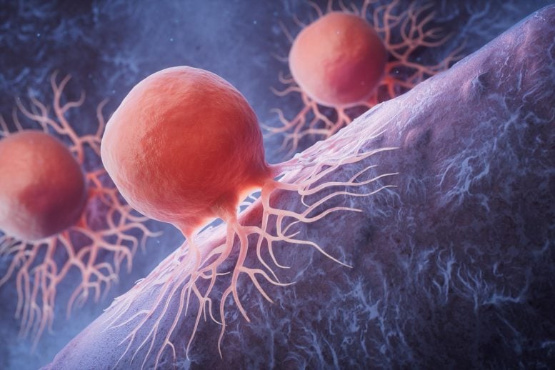
New Study Reveals Unexpected Way To Destroy Pancreatic Cancer Cells

Biological Cancer Cells RenderingA new study suggests that overstimulating key cancer-growth pathways, rather than shutting them down, could offer a novel way to target pancreatic tumors driven by KRAS mutations. New research points to a possible way to fight pancreatic cancer by turning the disease’s own growth signals against it. The research, published in the journal Aging, focused [...]
  •  

“We’ve Never Seen Anything Like This”: Discovery of a Hidden Biological Clock Reveals the “Master Key” to Growth and Development


A hidden biological clock has been discovered by genetic researchers, revealing how proteins act as a timing mechanism that controls living organisms’ growth into adulthood.

Cold Spring Harbor Laboratory (CSHL) researchers in New York revealed their findings in a recent paper published in the Proceedings of the National Academy of Sciences, identifying how a feedback circuit between the proteins MYRF-1 and LIN-42 functions as the worm genome’s master developmental clock.

Their research focused on the worm species Caenorhabditis elegans, revealing how these proteins set the timing of pulses that guide the worm through its development.

A Missing Biological Clock

“Development requires that cells change identity in the correct order while the organism continues to grow. How multicellular animals generate and synchronize this timing information across tissues has remained unclear,” the authors write.

The search for this missing biological clock was the core of the CSHL team’s research. Genetic and developmental problems are often expressed as a failure of developmental processes to initiate, which could be tied to problems with internal timing sequences that organize such behaviors. This marks the first time researchers have discovered a non-repeating biological clock.

“This is the central clock for all cells in the worm,” explained co-author Christopher Hammell. “It’s responsible for coordinating a finite series of sequential pulses of gene expression that must occur only once, and in order, for proper developmental progression. It’s like a ratchet. It turns genes on and off multiple times during development, but ultimately, it’s only going in one direction.”

Exploring the Genetic Issues

CSHL researchers paired the modern AI tool AlphaFold with more traditional genetic research techniques, such as protein sequencing, molecular experiments, and DNA sequencing, to identify MYRF-1’s role in the worm’s development.

The team found that protein signals both initiate and terminate developmental stages, keeping growth on track. MYRF-1 pulses fire first, then activate LIN-42, which determines the strength and duration of the timing pulses. This biological clock activates stage-specific regulatory RNAs and synchronizes developmental checkpoints, creating a rhythmic gene-regulation metronome that enforces the irreversible nature of growth and molting. 

In tests to confirm how this biological clock operates, researchers discovered that blocking MYRF-1 expression disrupts the worms’ development cycle.

Understanding the Rhythm of Growth

“We’ve never seen anything like this before,” Hammell says. “MYRF-1 is part of this master regulatory clock for all cells, but it’s also acting as a key maker and the master key for each stage of growth. Without the right key for each stage, development hits a wall and can’t progress.”

The researchers say that understanding how these proteins interact to create cellular clocks that guide development will help researchers better understand cell growth, progression, and differentiation.

“The MYRF-1/LIN-42 circuit runs in all cells,” Hammell says. “And every one of these independent cellular clocks appears to be in sync when you watch normal development. But are they communicating with each other? We’ve never thought deeply about that question before.”

A better understanding of these clocks could offer potential routes to cures for genetic and developmental issues by allowing medical science to jump-start processes that fail to initiate naturally.

Fundamentally, by restoring synchronization of these growth, development, and differentiation processes, scientists and medical practitioners could see better future success in alleviating certain genetic conditions.

The team’s recent paper, “A Molecular Timer Couples Organism-Wide Temporal Identity to Developmental Checkpoints,” appeared in Proceedings of the National Academy of Sciences on May 6, 2026.

Ryan Whalen covers science and technology for The Debrief. He holds an MA in History and a Master of Library and Information Science with a certificate in Data Science. He can be contacted at ryan@thedebrief.org, and follow him on Twitter @mdntwvlf.

  •  

A 5.3-million‑year‑old whale graveyard has been found on the floor of the Indian Ocean

When a whale dies, a very special natural phenomenon can come alive. The carcass might float at the surface for some time, attracting sharks and other predators. As it becomes weathered, it may start to sink, falling through the water until it eventually settles on the seafloor, where deep-sea scavengers feast upon it. The scientific […]

The post A 5.3-million‑year‑old whale graveyard has been found on the floor of the Indian Ocean appeared first on Knowridge Science Report.

  •  

New light-powered fiber detects tiny amounts of bacteria in just one minute

Researchers in Japan have developed a new light-based technology that can rapidly gather thousands of bacteria into a tiny area, making them much easier to detect. The breakthrough could help doctors diagnose infections earlier and may also support environmental monitoring and other forms of biological testing. The research team, led by Professor Takuya Iida from […]

The post New light-powered fiber detects tiny amounts of bacteria in just one minute appeared first on Knowridge Science Report.

  •  

Good news—we have extra time before the Sun ends life on Earth

It’s a bit worrying when a scientific paper begins, “How long will life on Earth survive?” But in this case—a study by Jacob Haqq‐Misra of Blue Marble Space and Eric Wolf at the University of Colorado Boulder—the billion-plus-year timeline under consideration shouldn’t cause you too much existential panic.

The context for this question is that we understand the Sun will brighten as it eventually matures into a red giant that swallows the Earth in a solar furnace. So, where along that 5 billion-year path will life on Earth, in fact, be cooked?

Weathering and the weather

This isn’t just a question of incoming radiation. Among the thermostat-like stabilizing feedback loops in Earth’s climate, the cycling of CO2 through the solid Earth is a major factor over timescales this long. The weathering of silicate rocks at the surface converts atmospheric CO2 into carbonate that ends up on the seafloor, where it can be subducted into the mantle with tectonic plates. (And eventually, it can cycle back out to the atmosphere through volcanoes.)

Read full article

Comments

© NASA/GSFC/SDO

  •  

A Venus Flytrap Mystery That Has Perplexed Scientists Since Darwin Has Finally Been Solved

One of nature’s most mysterious predators isn’t a mammal stalking remote forests or jungles, nor any large, finned creature lurking in the ocean depths: it is a bizarre little plant that grows only in certain wetland areas along the East Coast of the United States.

With the distinctive hinged leaves that it uses to capture insects, the Venus flytrap is one of nature’s most curious offerings. The small bog-dwelling plant owes its success to the way it can generate an inviting aroma with the nectar it produces, which lures insects. Then, with the help of tiny follicles that act as triggers, the insects become trapped within the flytrap’s leaves as they close like jaws around its prey.

Scientists have long been perplexed by exactly how these plants can react quickly enough to capture even some of the fastest flying insects. Even Charles Darwin seemed baffled by these curious examples of carnivorous flora, describing them as “a plant [that] should secrete, when properly excited, a fluid containing an acid and ferment, closely analogous to the digestive fluid of an animal.”

While the exact mechanism that allows the mysterious Venus flytrap to carry out its distinctive method of feeding has long remained unknown, new research is finally shedding light on the mystery.

The Venus Flytrap’s Secret is Finally Revealed

According to recent experiments detailed in a new study, when the hair-trigger mechanism is activated on these plants, cells on the jaw-like leaves soften, which initiates the closing movement within fractions of a second, trapping any insects unlucky enough to have landed searching for nectar.

Venus flytrap
(Image Credit: S. Schuster/Unsplash)

Dr Yoël Forterre, a physicist at the French National Centre for Scientific Research (CNRS) and Aix-Marseille University, says that this movement had been so mysterious to Darwin that “he was convinced that the plant had a muscle inside, but plants do not have muscles and they do not have nerves.”

“For more than a century there have been many hypotheses,” Forterre, senior author of the recent study, said. “It’s very surprising that plant cell walls can tune their mechanical properties so fast.”

The Challenge of Measurement

One of the primary issues with resolving the plant’s curious mechanism involved overcoming challenges associated with measuring such a remarkably fast system of movement, let alone one in a plant, which possesses none of the usual mechanisms nature relies on for such activities.

Equally perplexing is that the Venus flytrap seemingly recognizes when an accidental trigger has been initiated, rather than a successful capture of an insect.

“If you close it accidentally with a drop of water, it will close and then reopen the next day,” Forterre says. “If it catches an insect, it has to digest it and dissolve the skeleton, which will take several weeks.”

In experiments Forterre and his colleagues performed, dental glue was used to secure the plant’s leaves, which allowed for the normal trigger mechanisms to be engaged, but left the plant’s leaves open throughout the process.

Past research had already shown that electrical signals had been detected coinciding with the manipulation of the trigger hairs present on the plant’s leaves, in which this sudden surge of electricity “activates” the plant’s closing mechanism within a fraction of a second.

Solving the Flytrap’s Mystery

Applying a new approach, Forterre and colleagues used the metallic tip of a nanoindenter, devices used to make very small mechanical measurements. Applying the device to the outer leaves revealed that its normal stiffness appeared to soften significantly once the hair mechanisms were triggered.

The reason this occurs, according to Forterre, is that the cells themselves temporarily become more flexible. Past theories suggested that the movement was initiated by the movement of water contained within the leaf.

The newly-identified mechanism reveals a process very similar to the function of a plunger, which, if inverted, will flip back to its normal position when pressure is applied.

Forterre says he is unaware of any similar plant in nature that displays such unusual mechanical properties, which he says allows the Venus flytrap to initiate a “very rapid change of mechanical properties of the cells.”

“I’ve been obsessed by this for 20 years,” Forterre recently told The Guardian, noting that “as a physicist, I thought we should understand the motor, the forces.”

Now, thanks to his team’s findings, one of the plant kingdom’s strangest inhabitants has finally revealed its secrets.

The team’s new study, “Fast cell wall softening causes Venus flytrap closure,” appeared in Science on June 11, 2026.

Micah Hanks is the Editor-in-Chief and Co-Founder of The Debrief. A longtime reporter on science, defense, and technology with a focus on space and astronomy, he can be reached at micah@thedebrief.org. Follow him on X @MicahHanks, and at micahhanks.com.

  •  
❌